

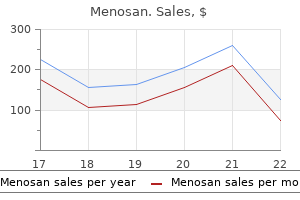
60caps menosan with visa
It is important to give the child a sense that she will be in control of the examination process symptoms precede an illness buy menosan on line amex. Young girls should feel that they are participating in their examination, not that they are being coerced or forced to have a gynecologic exam. If the interaction is poor during the first visit, the negative experience will detract from future physician-patient interactions (Lara-Torre, 2008). Other commonly seen diagnoses at a pediatric gynecology visit include labial adhesions, vulvar lesions, suspicion of sexual abuse, and genital trauma. Emphasize that the most important part of the examination is just "looking" and there will be conversation during the entire process. To successfully examine a child, one needs the cooperation of the patient and a medical assistant. Physicians may elect to treat the primary symptoms of vulvovaginitis for 2 to 3 weeks, realizing that on rare occasions they could be missing something more serious. It is recommended that the examination start with the nongenital areas, such as listening to the heart and lungs; an abdominal exam and inspection of the skin should be performed. This allows one to establish a rapport and mimics the traditional visits the child has with the pediatrician. In rare circumstances, it may be necessary to use continuous intravenous conscious sedation or general anesthesia to complete an essential examination. The most important technique to ensure cooperation is to involve the child as a partner. Children should ideally feel they are part of the exam rather than having an "exam done to them. A handheld mirror may help in some instances when discussing specifics of genital anatomy. It is critical to have all tools, culture tubes, and equipment within easy reach during a pediatric genital examination. Children often cannot hold still for long intervals while instruments are being located. The first aspect of the pelvic examination is evaluation of the external genitalia. Young children may be examined in the frog leg position, and children as young as 2 to 3 years of age may be examined in lithotomy with use of stirrups. The introitus will gape open with gentle pressure downward and outward on the lower thigh or undeveloped thigh or labia majora area (traction). Asking the child to pretend to blow out candles on a birthday cake may facilitate the process. Visualization of the introitus is better achieved using the above described traction and Valsalva than separation, as it gives a deeper view of the structures and partial visualization of the vagina. An assistant pulls upward and outward on the labia majora on one side while the examiner does the same with the nondominant hand on the contralateral labia. Then an oto/ophthalmoscope is used as a magnifying instrument and light source but is not inserted into the vagina. While the light from the oto/ophthalmoscope is shone into the vagina, the examiner can evaluate the vaginal walls and visualize the cervix as a transverse ridge, or flat button, that is redder than the vagina. This technique is generally successful in cooperative children unless there is a very high crescent-shaped hymen, in which case it is too difficult to shine the light into the small aperture of the vaginal introitus. Following inspection of the vagina and cervix, vaginal secretions may be obtained for microscopic examination and culture (the technique is described later). They may have septums, microperforations, finger-like extensions, or be completely imperforate. Bumps are usually a normal variant and are often attached to longitudinal ridges within the vagina. Hymens in newborns are estrogenized, resulting in a pink thick elastic redundancy. Older unestrogenized girls will have thin nonelastic hymens with significant signs of vascularity. The vaginal epithelium of the prepubertal child appears redder and thinner than the vagina of a woman in her reproductive years. The vagina is 4 to 6 cm long, and the secretions in a prepubertal child have a neutral or slightly alkaline pH. Recurrent vulvovaginitis, persistent bleeding, suspicion of a foreign body or neoplasm, and congenital anomalies may be indications to perform vaginoscopy and examine the inside of the vagina. Vaginoscopy in a prepubertal child most often requires sedation with a brief inhalation or intravenous anesthetic, but it can also be performed in the office with older, cooperative children in select circumstances. The introduction of any instrument into the vagina of a young child takes skillful patience. The prepubertal vagina is narrower, thinner, and lacks the distensibility of the vagina of a woman in her reproductive years. There are many narrow-diameter endoscopes that will suffice, including the Kelly air cystoscope, contact hysteroscopes, pediatric cystoscopes, smalldiameter laparoscopes, plastic vaginoscopes, and special virginal speculums designed by Huffman and Pederson. The ideal pediatric endoscope is a cystoscope or hysteroscope because the accessory channel facilitates the retrieval of foreign bodies as well as vaginal lavage. Local anesthesia of the vestibule may be obtained with 2% topical viscous lidocaine (Xylocaine) or longer-acting products such as lidocaine/prilocaine. A complete vaginal evaluation should never be performed under duress or by force, frequently the reason to use sedation when performing this examination on children. Common reasons to perform a rectal examination include genital tract bleeding, pelvic pain, and suspicion of a foreign body or pelvic mass. The child should be warned that the rectal examination will feel similar to the pressure of a bowel movement. The critical factors surrounding the pelvic examination of a female adolescent are different from those of examinations of children 2 to 8 years old. Many female adolescents do not want their mother, guardian, or other observers in the examining room. In many adolescent gynecology visits, a full pelvic exam is unnecessary (Lara-Torre, 2008). Each adolescent is at a different stage of development, and the approach to the exam may require variations that fit her developmental stage. A patient in early adolescence (12 to 14 years of age) may behave and need similar support as those in the prepubertal stages. They may ask for their mothers to be there, be fearful of the examination concept, and need more than one visit to achieve the goals of the visit. Adolescents often come for examinations with preconceived ideas that it will be very painful. Slang terminology for speculums among teens includes the threatening label "the clamp. Providers can counsel patients that they will inform them of each step in the process and then ask the teen if she is ready before performing each step. This places the teen in control of the tempo and allows her to anticipate the next element of the examination. Allowing them to see and touch the instruments also may assist in demystifying the exam and allow for it to flow more smoothly. The examiner provides pressure lateral to the introitus on the perineum prior to insertion of the speculum. It is estimated that 80% to 90% of outpatient visits of children to gynecologists involve the classic symptoms of vulvovaginitis: introital irritation (discomfort/pruritus) or discharge (Table 12. With puberty the prepubertal vagina becomes acidic under the influence of bacilli dependent on a glycogenated estrogen-dependent vagina. Breast budding is a reliable sign that the vaginal pH is shifting to an acidic environment. The pathophysiology of the majority of instances of vulvovaginitis in children involves a primary irritation of the vulva, which may be accompanied by secondary involvement of the lower one third of the vagina. Most cases involve an irritation of the vulvar epithelium by normal rectal flora or chemical irritants. There often are predisposing factors that lead to vulvar irritations such as the use of perfumed soaps, the pressure from tight seams of jeans or tights, and the like, which create denudation, allowing the rectal flora to easily infect the irritated epithelium. Except for the cervix, any mass discovered on rectal examination in a prepubertal exam should be considered abnormal. In this age of reliable access to ultrasonography, the internal genital exam to evaluate for the uterus and ovaries can be performed with the assistance of sonography, sparing the child from a rectopelvic or pelvic examination.
Order menosan canada
A mild interface or perivascular lymphocytic infiltrate with overlying parakeratosis may be present symptoms zoning out purchase menosan 60 caps on-line. T-cell gene rearrangement studies may demonstrate monoclonality; however, the meaning of this finding is unclear at this time. Ersoy-Evans S, et al: Narrowband ultraviolet-B phototherapy in pityriasis lichenoides chronica. Meziane L, et al: Febrile ulceronecrotic Mucha-Habermann disease: treatment with infliximab and intravenous immunoglobulins and review of the literature. Nanda A, et al: Febrile ulceronecrotic Mucha-Habermann disease (pityriasis lichenoides et varioliformis acuta fulminans) associated with parvovirus infection. Etanercept has been reported as effective, but infliximab has been reported to cause pityriasis lichenoides. Febrile ulceronecrotic MuchaHabermann disease has been treated successfully with methotrexate as well as with infliximab and intravenous immune globulins. Individual lesions respond to irradiation, and the relapsing course may remit with low-dose methotrexate. Large cell lymphomas of the skin have similar histologic and clinical features, so immunophenotyping is essential for prognosis. Lesions are erythematous, scaly macules and flat papules with very slow evolution. Resolution may leave persistent areas of hypopigmentation, which last for months to years. In many patients, the hypopigmented macules are the most prominent clinical finding. Skin lesions of pyogenic lymphoma may be seen secondary to a pyogenic lymphoma of other organs. The prognosis is poor in patients who have extracutaneous disease preceding or near the time of cutaneous involvement. The malignant cells are pleomorphic, large or medium cell types or are immunoblastic. Onset has been reported during glatiramer acetate treatment of multiple sclerosis. Relapses in the skin are common, but the development of extracutaneous, bone marrow, or lymph node involvement is uncommon. Clonal populations may occasionally be demonstrated in peripheral blood, but differ from those in the skin. Histologically, there is a dense dermal nonepidermotropic infiltrate with atypical tumor cells whose large nuclei have one or several prominent nucleoli and abundant cytoplasm. These primary cutaneous lymphomas usually present with one or several red-purple papules, nodules, or tumors 5 mm to 15 cm in size. The presence of a mixed population of suppressor cells, B cells, and histiocytes usually favors the diagnosis of reactive lymphoid hyperplasia. Other than low-dose methotrexate, chemotherapy generally has little role in the treatment of this disease. Local hyperthermia has been used successfully, as has inhibition of the mammalian target of rapamycin. It is more common in Asia, where it affects primarily women with a mean age of 40. In Korea, it is reported to be the most common form of cutaneous lymphoma after mycosis fungoides. A hydroa vacciniforme-type has been described in children in Mexico and in adults and children in Japan and Korea. Skin lesions are facial, and extremity papulovesicles ulcerate and heal with scarring. Histologically, the dermis and subcutaneous fat are infiltrated with intermediate-sized, atypical lymphocytes, within and around the walls of small and medium-sized vessels. Fujita H, et al: Primary cutaneous anaplastic large cell lymphoma successfully treated with low-dose oral methotrexate. Guitart J, et al: Cutaneous T-cell lymphomas: a spectrum of presentations with overlap with other cytotoxic lymphomas. Massone C, et al: the morphologic spectrum of primary cutaneous anaplastic large T-cell lymphoma: a histopathologic study on 66 biopsy specimens from 47 patients with report of rare variants. Cutaneous lesions occur in less than 10% of patients and present as papules, plaques, or nodules. The skin lesions may not represent lymphoma cutis because palisaded granulomatous and nonspecific dermal infiltrates may occur. The course is low-grade until the lymphoma transforms to a high-grade, large cell lymphoma. Subcutaneous (panniculitis-like) T-cell lymphoma Clinically, patients are usually young adults who present with subcutaneous nodules. Weight loss, fever, and fatigue are common and may herald the onset of a rapidly progressive hemophagocytic syndrome. Histologically, there is a lacelike infiltration of the lobules of adipocytes, mimicking panniculitis, especially lupus profundus. A characteristic feature is rimming of neoplastic cells around individual adipocytes with nuclear molding and atypia. Karyorrhectic debris, dermal involvement, and epidermotropism are clues to the diagnosis of / T-cell lymphoma. This virus is endemic in Japan, Southeast Asia, the Caribbean region, Latin America, and equatorial Africa. Lesions resemble mycosis fungoides, except that patches are uncommon, and plaques and nodules predominate. Histologically, the skin lesions contain lichenoid infiltrates of medium-sized lymphocytes with convoluted nuclei. Although the morphologic appearance of the malignant lymphocytes composing these primary cutaneous lymphomas is identical to lymphomas based in lymph nodes, they have a distinctly different clinical behavior and immunophenotypic profiles. This renders the classification systems based on lymph node histology of limited benefit in the diagnosis of primary cutaneous B-cell lymphomas. More simplified schemes have thus been proposed that apply to primary cutaneous lymphomas only. The great majority of primary cutaneous B-cell lymphomas are composed of cells with the morphologic characteristics of the B cells normally found in the marginal zone or germinal centers of lymph nodes. Secondary cutaneous involvement can occur with all forms of B-cell lymphoma based primarily in lymph node or other sites. The clinical features are similar to those of primary cutaneous lymphoma, with violaceous papules or nodules. Typically, the histologic structure of secondary lesions in the skin is similar to that of the lymphoma at the site of origin, usually the lymph nodes. The pattern in the skin, however, may not be sufficient to classify the lymphoma, making lymph node biopsy necessary in most patients. It is therefore critical to evaluate any patient suspected of having primary cutaneous B-cell lymphoma to exclude involvement at another site. Radiation is typically used for indolent forms of cutaneous B-cell lymphoma, but excision, rituximab, intralesional corticosteroids, and systemic chemotherapy have also been used in select cases. Higher-grade lymphomas, such as leg-type lymphoma, primary cutaneous follicle center lymphoma occurring on the leg, and precursor B-cell lymphoblastic lymphoma, are typically treated with systemic chemotherapy regimens, including combinations of anthracycline-containing chemotherapies and rituximab. Immunocytomas are associated with European Borrelia and occur as tense, shiny, pink to red nodules on the legs of older patients. The neoplastic cells are medium-sized gray cells with predominantly cleaved nuclei that proliferate within the space surrounding and between benign germinal centers. Light-chain restriction is easiest to identify in the plasma cell population by means of in situ hybridization. Clonal immunoglobulin gene rearrangements can usually be demonstrated, and lightchain restriction can typically be demonstrated in plasma cells at the periphery of the lymphoid aggregates. They present on the legs of older men and are characterized by sheets of plasmacytoid B cells with Dutcher bodies.
Cheap menosan master card
Other specific causes of vulvovaginitis may include systemic diseases medications you cant drink alcohol purchase menosan australia, chicken pox, and herpes simplex infection. There is nothing specific about the symptoms or signs of childhood vulvovaginitis. A discharge that is both bloody and purulent is likely not from vulvovaginitis but from a foreign body (see "Vaginoscopy for Prepubertal Bleeding without Signs of Puberty," presented later in this chapter), although patients infected with some pathogens, particularly Shigella boydii, often present with a bloody or blood-tinged discharge. The signs of vulvovaginitis are variable and not diagnostic, but they include vulvar erythema, edema, and excoriation. The differential diagnosis of persistent or recurrent vulvovaginitis not responsive to treatment should include considerations of a foreign body, primary vulvar skin disease (allergic or contact dermatitis), ectopic ureter, and child abuse. If the predominant symptom is pruritus, then pinworms or an irritant/nonspecific vulvitis is the most likely diagnosis. The vulvar skin of children may also be affected by systemic skin diseases, including lichen sclerosus, seborrheic dermatitis, psoriasis, and atopic dermatitis. The classic perianal "figure-8" or "hourglass" rash is indicative of lichens scleroses with white patches and in some cases local trauma. An ectopic ureter emptying into the vagina may only intermittently release a small amount of urine; thus this rare congenital anomaly should be considered in the differential diagnosis in young children. Treatment of Vulvovaginitis the foundation of treating childhood vulvovaginitis is the improvement of local perineal hygiene. Both parent and child should be instructed that the vulvar skin should be kept clean, dry, and cool, and irritants should be avoided. The child should be instructed to void with her knees spread wide apart (even while facing the toilet to improve urine draining) and taught to wipe from front to back after defecation. Chemicals that may be allergens or irritants, such as bubble bath, must be discontinued. Instructing patients to use nonmedicated, nonscented wipes rather than toilet paper may prevent the self-inoculation of the vagina with small pieces that can initiate a chronic discharge. Most episodes of childhood vulvovaginitis are cured solely by improved local hygiene. The majority of symptoms improve with hygienic changes and sitz baths (warm water, no soaps or chemicals). Utilizing this approach for a 2-week period should resolve most symptoms in patients with nonspecific vulvovaginitis. When this intervention fails, the suspicion for bacterial colonization is greater and a reasonable approach is the use of broad-spectrum oral antibiotics such as amoxicillin or trimethoprim sulfamethoxazole given for 10 to 14 days. Without continuation of the hygiene measures, then broad-spectrum antibiotics will only offer temporary relief, and the problem is likely to recur (Bercaw-Pratt, 2014). If patients are going to be treated with antibiotics, one should attempt to collect a culture of the vulvovaginal discharge prior to initiation of the antibiotics. In noncooperative children, treatment should not be withheld if unable to collect a specimen and empiric treatment may be started. For attempting to collect a specimen, many techniques have been described including the use of a very slim urethral Dacron swab moistened with nonbacteriostatic saline (used for collection of male urethral cultures). The outer catheter serves as an insulator, and the inner catheter is used to instill a small amount of saline and aspirate into the vaginal fluid. The results of the vaginal culture may demonstrate a single organism that is a respiratory, intestinal, or sexually transmitted disease pathogen. The presence of sexually transmitted organisms in a child is usually a strong indication that sexual abuse may have taken place and appropriate referral and follow-up is necessary (see Chapter 9). Denuded epithelium of adjacent labia minora agglutinates and fuses the two labia together, creating a "flat appearance" of the vulvar surface. A telltale somewhat translucent vertical midline line is visible on physical exam at the site agglutination. This thin, narrow line in a vertical direction is pathognomonic for labial adhesions. Labial adhesions are often partial and only involve the upper or lower aspects of the labia. Associated urinary infections have also been reported and may be the presenting symptom leading to the diagnosis. Most of the time treatment requests are driven by the parental concern of a closed vagina and their interpretation that this may lead to an inability to have children in the future or engage in intercourse. Although they do not explicitly say this, upon further questioning, many parents disclose this kind of concern. With appropriate counseling and reassurance of the benign and common nature of this condition, as well as the likely resolution during puberty, most parents are reassured and follow advice. The majority of the patients will fall into this category and can be reassured and followed over time to spontaneous resolution when they produce their own endogenous estrogen. If spontaneous separation does not occur at puberty, and manual separation is required, the presence of a better estrogenized skin will decrease the chances of recurrence which in children can range from 25% to 65%. Some children will present with symptoms and may include voiding difficulties, dysuria, frequent urinary infections, urine dribbling after voiding, recurrent vulvovaginitis, discomfort from the labia pulling at the line of adhesions, and in rare cases bleeding from the line of adhesion pulling apart. Attempts to separate the adhesions apart in the office by pulling briskly on the labia minora should not be done. It is very painful, and the raw edges are likely to adhere again as the child will be reticent to allow application of medication after being subjected to this degree of pain. Even with local anesthesia, such as lidocaine ointments or creams, the potential pain and traumatic experience for the child should deter from this intervention, except in the well-motivated, mature child. The most commonly utilized treatment of this condition is topical estrogen cream applied onto the labia two times per day at the site of fusion. This will usually result in spontaneous separation, usually in approximately 2 to 8 weeks. In cases when resolution takes longer than several weeks, the clinician can reexamine the patient. If increased pigmentation is noted lateral to the midline line of agglutination, the caregiver should be reinstructed to apply the cream to the line, as the lateral pigmentation indicates the estrogen is being applied lateral to the actual adhesion. The action of estrogen as well as the application over the adhesion line itself makes the treatment more effective. Care should be taken to not prolong the topical use of estrogen for more than 6 to 8 weeks. Prolonged use of topical estrogen has been associated with breast budding and in some less common cases vaginal bleeding from the peripheral effects of the absorption of estrogen. Failure of separation within the normal time frame should trigger alternate treatment. When patients fail estrogen therapy, and symptoms persist, the use of topical corticosteroids twice a day for 6 to 8 weeks has also shown adequate results and can be consider as a first or secondary line of treatment. Once the condition has been resolved, recurrence can often be prevented by applying a bland ointment (such as zinc oxide cream or petroleum jelly) to the raw epithelial edges for at least 1 month or even longer. Use of intranasal midazolam for manual separation of labial adhesions in the office. Inexperienced examiners may confuse labial adhesions for an imperforate hymen or vaginal agenesis. Although the physical exam findings are significantly different, all of these conditions may occlude the visualization of the vaginal introitus. In the patient with an imperforate hymen, the labia minora normally appear like an upside down V, and no hymeneal fringe is visible at the introitus. In vaginal agenesis, the hymeneal fringe is typically normal, but the vaginal canal ends blindly behind the hymeneal fringe. Labial adhesions are most common in girls between 2 and 6 years of age, with up to 90% of cases occurring before age 6. Estrogen reaches a nadir during this time, predisposing the nonestrogenized labia to denudation. There is considerable variation in the length of agglutination of the two labia minora. In the most advanced cases, there is fusion over both the urethral and the vaginal orifices. It is extremely rare for this fusion to be complete, and most children urinate through openings at the top of the adhesions, even when the urethra cannot be visualized (pinpoint opening). However, Obstetrics & Gynecology Books Full 12 Pediatric and Adolescent Gynecology McCann and colleagues reported the association between injuries of the posterior fourchette and labial adhesions in sexually abused children (McCann, 1988). Labial agglutination alone is so common that immediate suspicion of child abuse based solely on this finding in 2- to 6-year-olds is unwarranted. However, the combination of labial adhesions and scarring of the posterior fourchette, especially in children with new-onset labial adhesions after age 6, should prompt the clinician to consider sexual abuse in the differential diagnosis.
Purchase menosan no prescription
Histologically symptoms zinc deficiency adults generic menosan 60 caps mastercard, some are melanocytic nevi, whereas others demonstrate histologic features of lentigo simplex. Gastrointestinal polyps, especially prominent in the jejunum, are frequently associated. Generalizedlentiginosis An occasional patient will have generalized lentiginosis without associated abnormalities. This designation comprises cardiocutaneous myxomas, lentigines, blue nevi, and endocrine abnormalities. The lentigines were distributed over the central face and lips, with variable involvement of the dorsal hands and feet, elbows, and buttocks. Partialunilaterallentiginosis Partial unilateral lentiginosis is a rare disorder of cutaneous pigmentation characterized by the presence of multiple simple lentigines, wholly or partially involving half the body. The pathogenesis may be related to increased expression of androgen receptors within lesional skin. Treatment may not be necessary, but some patients desire removal of pigment or terminal hair associated with the lesion. Clinicalandhistologicfeatures Features of benign nevi include a diameter of 6 mm or less, perfectly uniform pigmentation, flaccid epidermis, smooth, uniform border, and an unchanging size and color. Benign nevi tend to be round to oval and undergo a predictable course of maturation. Junctional nevi are sharply circumscribed brown macules, varying in diameter from 1 to 6 mm. Small, wellnested junctional melanocytic proliferations are almost invariably benign. Benign junctional nevi associated with bulbous hyperplasia of the rete ridges are referred to as junctional lentiginous nevi. Lentigo maligna can appear well nested with an appearance similar to that of junctional lentiginous nevi. Any broad junctional melanocytic lesion on sundamaged skin should be viewed with suspicion. Benign compound nevi are well nested at the junction, with dispersion of individual melanocytes at the base of the lesion. Instead, with descent into the dermis, the melanocytes become smaller and spindled in appearance. Nests at the junction tend to be round to oval and are about equidistant from one another. Dermal nests are generally smaller than the junctional nests and become progressively smaller deeper in the dermis. Pigment is most prominent at the junction and becomes progressively less prominent deeper in the dermis. Individual melanocytes in a "buckshot" scatter throughout the epidermis are typical of superficial spreading melanoma. Sunburned benign nevi may also demonstrate buckshot intraepidermal scatter of melanocytes, and buckshot scatter may be seen in the central portion of acral nevi and Spitz nevi. A histologic resemblance to dysplastic nevi is also common in nevi from the scalp, ears, dorsal foot, and breast, even in patients with no other evidence of the dysplastic nevus syndrome. If a benign palmar nevus is bisected across the dermatoglyphs, the nests will appear round to oval. If the same lesion is sectioned parallel to the dermatoglyphs, the nests will appear elongated and may mimic those of melanoma as an artifact of sectioning. Careful communication with the pathologist is essential when submitting an acral melanocytic lesion to the laboratory. Histologically, it is a benign epidermal neoplasm composed of keratinocytes and dendritic melanocytes. Oral melanoacanthoma is also a proliferation of two cell types, melanocytes and epithelial cells, but appears to be a reactive lesion. It occurs as a macular or slightly raised pigmented area on the buccal mucosa, predominantly in young adult black women. JainS,etal: Multifocal cutaneous melanoacanthoma with ulceration: a case report with review of literature. They typically begin as sharply defined macular lesions, become papular, then gradually become soft and lose their pigment. White persons have more than black persons, and individuals with a light complexion have more nevi than those with a dark complexion. A study of young British women showed an association of holidays overseas with an increased nevus count. The association was greatest in anatomic sites intermittently exposed to sunlight. Eruptive nevi may occur in association with bullous diseases, severe sunburn, immunosuppression, or sulfur mustard gas exposure. The cheetah phenotype refers to patients with more than 100 uniform, dark-brown to black pigmented macules 4 mm or smaller. Melanocytic lesions with a junctional component are more often removed during the summer months, whereas excision of intradermal nevi is relatively constant during the year. This suggests that some change in these lesions draws more attention during the summer months. Nevi may darken during pregnancy, but other changes should prompt consideration of a biopsy. The signs of malignant transformation in pigmented nevi are recent enlargement, an irregular or scalloped border, asymmetry, changes or variegation in color (especially red, white, or blue), surface changes (scaling, erosion, oozing, crusting, ulceration, or bleeding), development of a palpable thickening, signs of inflammation, or the appearance of satellite pigmentation. The "ugly duckling" sign refers to nevi in an individual generally tending to share a similar appearance. Any mole that does not share the same characteristics should be considered for biopsy. Moles with dark areas that do not lie entirely within the lesion, but produce an extension beyond the border, may represent melanoma arising in association with a preexisting nevus. The clinician should alert the pathologist to the presence of these areas and the pathologist should section through the appropriate area. When it occurs at the edge of the nevus, it may give the lesion a notched appearance. Nevi from normal persons have no estrogen or progesterone receptors, but there may be positive estrogen receptor binding in nevi from pregnant women, as is also found in malignant melanoma. The development of what appears to be a new pigmented nevus in a patient over age 35 should alert the physician to possible melanoma, because patients without the dysplastic nevus syndrome usually do not develop new nevi at this age. Conjunctival nevi occur, and most can be followed serially if the lesion has been present since childhood or has shown no evidence of growth. Changing pigmented lesions and those acquired after childhood are best evaluated in conjunction with an ophthalmologist. Most conjunctival nevi occur on the bulbar conjunctiva and often abut the nasal or temporal corneoscleral limbus. Suspicion of melanoma should arise if a pigmented lesion occurs in the palpebral or forniceal conjunctiva, if lesions are not hinged at the limbus and are immovable, if they extend into the cornea, if there is canalicular obstruction that leads to tearing, or if adjacent dilated vessels are noted. They consist of a banal nevus together with a blue nevus, Spitz nevus, or deep penetrating nevus. Melanocytic nevi may occur in lymph nodes and are present in about 10% of sentinel node biopsies. Nodal nevi typically occur in the capsule, in contrast to melanoma metastases, which are typically subcapsular. Nodal nevi are frequently associated with cutaneous nevi in the draining basin, especially nevi with congenital features. FloresA: Eponyms, morphology, and pathogenesis of some less mentioned types of melanocytic nevi. Treatment 684 Acquired nevi should be removed if they show signs of malignant transformation. Nevi of the neckline, beltline, or other areas that are irritated may be removed to relieve the patient of the irritation.

Menosan 60caps line
The severity of the bleeding tendency is highly variable from patient to patient symptoms low potassium discount 60caps menosan amex, and some have no bleeding problems. Ironically, because of the loss of the antithrombotic effect of fibrinogen, thrombotic events are increased in afibrinogenemia. Patients with hypofibrinogenemia are seen more often; in general, they are less symptomatic and only occasionally require treatment. Hypofibrinogenemia may be associated with pregnancy losses and rarely with liver disease due to accumulation of abnormal fibrinogen in the endoplasmic reticulum of hepatocytes. Dysfibrinogenemia is asymptomatic in 55% of patients, 25% exhibit bleeding tendencies, and 20% tend to develop thrombosis. Mutations in the fibrinogen gene cluster cause all three of these fibrinogen disorders. BornikovaL,etal: Fibrinogen replacement therapy for congenital fibrinogen deficiency. VuD,Neerman-ArbezM: Molecular mechanisms accounting for fibrinogen deficiency: from large deletions to intracellular retention of misfolded proteins. The eruption consists of generalized, dark-blue to magenta, nonblanchable, indurated, round to oval, hemispheric papules ranging from 1 to 7 mm. Etiologic factors include congenital infections (toxoplasmosis, rubella, cytomegalovirus, herpes simplex, parvovirus B19), hemolytic disease of the newborn (Rh incompatibility, blood group incompatibility), hereditary spherocytosis, twin transfusion syndrome, recombinant erythropoietin administration, neuroblastoma, rhabdomyosarcoma, extraosseal Ewing sarcoma, Langerhans cell histiocytosis, and congenital leukemia. Patients with multiple vascular disorders, such as hemangiopericytoma, hemangioma, blue rubber bleb nevus, and glomangioma, may be mistaken for a blueberry muffin baby. BrismanS,etal: Blueberry muffin rash as the presenting sign of Aicardi-Goutieres syndrome. Blueberry muffin baby (extramedullary hematopoiesis) due to congenital cytomegalovirus infection. KrenovaZ,etal: Extraosseal Ewing sarcoma as a rare cause of the blueberry muffin baby syndrome: a case report and the review of the literature. Epistaxis, menorrhagia, hemarthrosis (much less than in hemophilia and with far fewer 824 PandeyV,etal: Late-onset blueberry muffin lesions following recombinant erythropoietin administration in a premature infant. Risk factors include female gender, obesity, immobilization, low atmospheric pressure, winter season, and the presence of cancer. In 35% of cancer-associated cases, the thrombosis is the first sign of the cancer. On examination, a palpable cord, calf tenderness, unilateral edema, warmth, redness, and venous dilation may suggest the diagnosis. Preventive strategies include exercise, weight control, and pharmacologic prophylaxis for high-risk patients. FanikosJ,etal: Long-term complications of medical patients with hospital-acquired venous thromboembolism. In Antithrombotic Therapy and Prevention of Thrombosis, 9th edn: American College of Chest Physicians Evidence-Based Clinical Practice Guidelines. SchulmanS,etal: Treatment of acute venous thromboembolism with dabigatran or warfarin and pooled analysis. ShrierI,etal: Effect of early physical activity on long-term outcome after venous thrombosis. Superficialthrombophlebitis Superficial venous thrombosis is an inflammatory thrombotic condition that classically presents with painful induration and erythema, often in a cordlike, linear or branching configuration. Patients may also exhibit indurated subcutaneous nodules and overlying purpura or brown discoloration indicative of postinflammatory hyperpigmentation. In the evaluation of superficial thrombophlebitis, the physician should consider the possibility of underlying deep venous disease. Superficial femoral vein involvement should alert the physician to underlying deep venous disease requiring anticoagulation. Elliptical biopsies across the palpable cord may be required to exclude other considerations, such as sarcoidal granulomas, cutaneous polyarteritis nodosa, Kaposi sarcoma, and vasculotropic metastasis. Heparin therapy may reduce the incidence of thromboembolic complications in high-risk individuals. Postcardiotomysyndrome Between 2 and 3 weeks after pericardiotomy, fever, pleuritis, pericarditis, or arthritis may appear together with petechiae on the skin and palate. Postcardiotomy syndrome may be confused with infectious mononucleosis and bacterial endocarditis. The sudden appearance of a cordlike thrombosed vein along the anterolateral chest wall is characteristic. It is at first red and tender and subsequently changes into a painless, tough, fibrous band. The condition represents a localized thrombophlebitis of the veins of the thoracoepigastric area. The veins involved are the lateral thoracic, thoracoepigastric, and superior epigastric. In the end stage, a thick-walled vein remains that has a hard, ropelike appearance and occasionally may result in a furrowing of the breast. Infrequently, a vein coursing up the inside of the upper arm and across or into the axilla may be thrombosed, leading to the "axillary web syndrome. Orthostaticpurpura(stasispurpura) Prolonged standing or even sitting with the legs lowered (as in a bus, airplane, or train) may produce edema and a purpuric eruption on the lower extremities. Elevation of the legs and the use of elastic stockings are helpful preventive strategies. Obstructiveortraumaticpurpura Purpura may be evoked by mechanical obstruction to the circulation, with resulting stress on the small vessels. This may be encountered in cardiac decompensation or after convulsions, vomiting episodes, Valsalva maneuver, pertussis, or sexual climax. Nonpalpable purpura has been reported in association with the use of a mucus-clearing device, which requires the patient to exhale forcefully through a flutter valve (flutter valve purpura). Local obstruction of the blood flow with purpura may result from compression of the veins by tumors or a gravid uterus or by occlusions from thrombosis. Bruises and ecchymoses on the genital area, buttocks, left ear or cheek, or hands suggest an abused child. Ecchymoses of bizarre shapes may also correspond to trauma inflicted during religious rituals or cultural practices, such as coin rubbing and cupping performed as remedies for common diseases. On the neck or upper arms, it results from biting and sucking and is better known as a "hickey. Bathtub suction-induced purpura occurs on the lower back location in a U-shaped distribution. The acute nature, purpuric findings, and rapid resolution are distinguishing features of Achenbach syndrome. Widespread cutaneous thrombosis can occur, with initial erythematous cutaneous plaques progressing to hemorrhagic bullae. Hematopoietic stem cell transplantation may be curative, after either ablative or nonablative conditioning regimens. Eculizumab, a humanized monoclonal antibody against C5, inhibits terminal complement activation. Paroxysmal nocturnal hemoglobinuria: the physiology of complement-related hemolytic anemia. Easybruisingsyndromes Young women who bruise easily despite normal coagulation profiles and normal platelet counts may have antiplatelet antibodies. Bernard-Soulier syndrome is a rare inherited disorder characterized by giant platelets, thrombocytopenia, and a prolonged bleeding time. Sebastian syndrome consists of giant platelets, leukocyte inclusions, and thrombocytopenia. Fechtner syndrome is a rare type of familial thrombocytopenia associated with large platelets, leukocyte inclusions, and features of Alport syndrome. The inclusions appear as electron-dense long rods and needles oriented along the long axis of the spindle. SavoiaA,etal: Clinical and genetic aspects of Bernard-Soulier syndrome: searching for genotype/phenotype correlations.

Menosan 60 caps on line
The base of the bladder lies directly adjacent to the endopelvic fascia over the anterior vaginal wall medications side effects prescription drugs cheap menosan 60caps amex. The bladder neck and connecting urethra are attached to the symphysis pubis by fibrous ligaments. The prevesical or retropubic space of Retzius is the area lying between the bladder and symphysis pubis and is bounded laterally by the obliterated hypogastric arteries. This space extends from the fascia covering the pelvic diaphragm to the umbilicus between the peritoneum and transversalis fascia. The mucosa of the anterior surface of the bladder is light red and has numerous folds. The inferoposterior surface delineated by the two ureteral orifices and the urethral orifice is the trigone. The trigone is a darker red than the rest of the bladder mucosa and is free of folds. The muscular wall of the bladder, the detrusor muscles, is arranged in three layers. The arterial supply of the bladder originates from branches of the hypogastric artery: the superior vesical, inferior vesical, and middle hemorrhoidal arteries. The nerve supply to the bladder includes sympathetic and parasympathetic fibers, with the external sphincter supplied by the pudendal nerve. The rectum begins over the second or third sacral vertebra, where the sigmoid colon no longer has a mesentery. After the large intestine loses its mesentery, its anatomic posterior wall is in close proximity to the curvature of the sacrum. The lowest one third is below the peritoneal reflection and is in close proximity to the posterior wall of the vagina. The anal canal is fixed by the surrounding levator ani musculature of the pelvic diaphragm. Studies of the cross-sectional anatomy of the external anal sphincter by both ultrasound and magnetic resonance imaging have identified two distinct layers of the external anal sphincter. A, Sagittal view representing the plane from the computed tomography cut noted in B. This close proximity also underscores the fact that ureteral injury may be unavoidable in some women. The ureter then runs upward (ventral) and medially in the vesical uterine ligaments to obliquely pierce the bladder wall. Just before entering the base of the bladder, the ureter is in immediate contact with the anterior vaginal wall and the inferolateral aspect of the space of Retzius. The ureter has a rich arterial supply with numerous anastomoses from many small vessels that form a longitudinal plexus in the adventitia of the ureter, commonly referred to as the Waldeyer sheath. The rectum, unlike other areas of the large intestine, does not have teniae coli or appendices epiploicae. The arterial supply of the rectum is rich, originating from five arteries: the superior hemorrhoidal artery, which is a continuation of the inferior mesenteric, the two middle hemorrhoidal arteries, and the two inferior hemorrhoidal arteries. Therefore during rectal examination special emphasis to palpate the entire circumference of the rectum, not just the area of the rectovaginal septum, is an important part of screening for colon cancer. Surgical compromise of the ureter may occur during clamping or ligating of the infundibulopelvic vessels, clamping or ligating of the cardinal ligaments, or wide suturing in the endopelvic fascia during an anterior repair, even with apparent normal anatomy and utmost surgical care. Particular attention to the proximity of the distal ureter to the anterior vagina is very important. Operative injuries to the bladder or ureter occur in approximately 1 out of 100 major gynecologic operations. Two of the classic ways to differentiate a ureter from a pelvic vessel are (1) visualization of peristalsis after stimulation by a surgical instrument and (2) visualization of Auerbach plexuses, which are numerous, wavy, small vessels that anastomose over the surface of the ureter. Injury to the ureter or bladder during urethropexy operations for genuine stress incontinence is common. Therefore many surgeons routinely inject indigo carmine and either open the bladder or perform cystoscopy near the end of the operative procedure. For years, gynecologic teachers have referred to the area in the base of the broad ligament near the cervix where the uterine artery crosses the ureter as the area where "water flows under the bridge. This capacity allows the gynecologist to use suprapubic cystostomy tube without fear of fistula formation. There are many different surgical techniques for the repair of urinary stress incontinence. They usually involve either suspension of the periurethral tissues or bladder neck itself. Occasionally, these surgical procedures are complicated by a significant amount of postoperative venous bleeding. A subfascial hematoma may extend as high as the umbilicus in the space of Retzius. One of the most common causes of female urinary incontinence is defective connective tissue, especially in the periurethral connective tissue, the pubourethral ligaments, and pubococcygeus muscles. Rectal injury may occur during vaginal hysterectomy with associated posterior colporrhaphy. The rectum bulges anteriorly into the vagina in this area, producing a further challenge during the operative procedure. The cul-de-sac is a potential space and is also called the rectouterine pouch or fold. The parietal peritoneum of the cul-de-sac covers the cervix and upper part of the posterior vaginal wall then reflects to cover the anterior wall of the rectum. The pouch is bounded on the lateral sides by the peritoneal folds covering the uterosacral ligaments. The parametria lie between the leaves of the broad ligament and in the contiguous area anteriorly between the cervix and bladder. This connective tissue is thicker and denser adjacent to the cervix and vagina, where it becomes part of the connective tissue of the pelvic floor. The parametria may also thicken in response to radiation, pelvic cancer, infection, or endometriosis. Development of these spaces is useful in pelvic lymph node dissection and in radical pelvic surgery because it makes the anatomic landmarks so clear. The paravesical space is bordered medially by the bladder and upper vagina and is contiguous with, but lateral to , the space of Retzius. Inferiorly it is bordered by Obstetrics & Gynecology Books Full 3 Reproductive Anatomy the pubic ramus, and superiorly it is bordered by by the cardinal ligament. The pararectal space is developed by dissecting the adventitial tissue within the broad ligament, between the ureter (medially), and the internal iliac vessels (laterally). Inferiorly, it is limited by the cardinal ligament, containing the uterine artery. The paravesical and pararectal spaces are actually potential spaces that become true spaces when developed by the surgeon. Development of these spaces is useful in pelvic lymph node dissection and in radical pelvic surgery because the anatomic landmarks become so clear. The pouch of Douglas is easily accessible in performing transvaginal surgical procedures. Posterior colpotomy is frequently chosen for drainage of a pelvic abscess occurring in the cul-desac of Douglas. When the paravesical and pararectal spaces have been developed and the uterus is held on traction medially, the pelvic anatomy, including the ureter, internal and external iliac vessels, obturator fossa, and the cardinal ligament, with the uterine artery crossing the ureter, can be clearly and readily identified. Many women with uterine prolapse have an associated enterocele, which is a hernia that protrudes between the uterosacral ligaments. Occasionally the cul-de-sac of Douglas is obliterated by the inflammatory process associated with either endometriosis or advanced malignancy. The labia minora are homologous to the penile urethra and a portion of the skin of the penis in males. The middle third of the vagina is supported by the levator ani muscles and the lower portion of the cardinal ligaments. Descriptive terms for pelvic organs are derived from the Latin root, whereas terms relating to surgical procedures are derived from the Greek root. The width of the canal varies with the parity of the woman and changing hormonal levels. The pain fibers from the cervix accompany the parasympathetic fibers to the second, third, and fourth sacral segments.
Diseases
- Pyruvate decarboxylase deficiency
- ACTH deficiency
- Myasthenia gravis
- Cartilaginous neoplasms
- Progeria
- Broad-betalipoproteinemia
- Chorea
- CDG syndrome
Order generic menosan line
Boulos S medications zovirax generic menosan 60caps on line, et al: Clinical presentation, immunopathology, and treatment of juvenile-onset mycosis fungoides: A case series of 34 patients. Iwamoto M, et al: Clinical pharmacology profile of vorinostat, a histone deacetylase inhibitor. Pileri A, et al: Role of bexarotene in the treatment of cutaneous T-cell lymphoma: the clinical and immunological sides. Schlaak M, et al: Allogeneic stem cell transplantation versus conventional therapy for advanced primary cutaneous T-cell lymphoma. Systemic chemotherapy For most forms of cancer, combinations of chemotherapeutic agents are given. Methotrexate, in doses from 5 to 125 mg/week, is effective for the management of T3 patients. Similarly, vorinostat (and other histone deacetylase inhibitors), pentostatin, etoposide, fludarabine, and 2-chlorodeoxyadenosine have been used. Systemic chemotherapy beyond methotrexate, especially multiagent chemotherapy, is best managed by an oncologist. Histone deacetylase inhibitors, including vorinostat, demonstrate responses in a subgroup of patients. Forodesine is a novel inhibitor of purine nucleoside phosphorylase, and pralatrexate is a novel targeted antifolate agent. Pagetoid reticulosis Localized epidermotropic reticulosis, pagetoid reticulosis, or Woringer-Kolopp disease is an uncommon lymphoproliferative disorder considered be a form of mycosis fungoides. Other terms suggested for these cases have included acral mycosis fungoides or mycosis fungoides palmaris et plantaris. Pagetoid reticulosis is divided into classic WoringerKolopp, which usually describes solitary lesions, and cases with multiple lesions (Ketron-Goodman variant). The disease presents as a solitary lesion that is often located on an extremity and frequently has a keratotic rim. If there is more than a single lesion, the lesions often tend to involve both the palms and the soles. Frequently, over months to years, the lesion gradually enlarges, reaching more than 10 cm in size. The long duration without progression has been a clinical hallmark of Woringer-Kolopp disease. Histologically, there is prominent epidermotropism of lymphocytes, with many lining up in the basal cell layer. This histologic pattern correlates with strong E7- and 47-integrin expression by the infiltrating cells. Therapeutically, local excision and radiation therapy have been "curative" in many patients. Fever, chills, hypotension, nausea, and vomiting were common, and at high doses, a vascular leak syndrome occurred. This agent is reserved for advanced-stage patients who have failed other modalities. The characteristic features are generalized erythroderma, superficial lymphadenopathy, and atypical cells in the circulating blood. The skin shows a generalized or limited erythroderma of a typical fiery red color. Associated features can include leonine facies, eyelid edema, ectropion, diffuse alopecia, hyperkeratosis of the palms and soles, and dystrophic nails. Some patients develop lesions identical to vitiligo, especially on the lower legs. Superficial lymphadenopathy is usually found in the cervical, axillary, and inguinal areas. Additional hematologic evaluation may be necessary to confirm the diagnosis in the erythrodermic patient. This explains the reduced delayed-type hypersensitivity, elevated IgE, and eosinophilia seen in these patients. Low-dose methotrexate has a reasonable response rate of about 50% and an overall survival of 101 months, suggesting a survival benefit with its use. Lesions are erythematous, atrophic, bulky, infiltrated, pendulous, and redundant plaques in the axillae and groin. Histologically, there is a lymphohistiocytic infiltrate extending through the dermis into the subcutaneous fat. Elastophagocytosis is prominent and elastic tissue is absent in areas of inflammation. Lymphocytes are also found within the multinucleate giant cells and are arranged around them. In most patients, the condition evolves into mycosis fungoides, but about one third of patients with granulomatous slack skin develop Hodgkin disease after years to decades. Lymphomatoid papulosis Lymphomatoid papulosis (LyP) is an uncommon, but not rare, disorder. It occurs at any age, including childhood, but is most common in adults with a mean age of 44. In typical cases, the lesions and course are very similar to Mucha-Habermann disease (pityriasis lichenoides et varioliformis acuta), except that the lesions tend to be slightly larger and fewer in number and have a greater propensity to necrosis. The lesions evolve to papulovesicular, papulopustular, or hemorrhagic, then necrotic papules over days to weeks. Lesions are usually generalized, although cases limited to one anatomic region have been reported. In most patients, however, the condition tends to be chronic, and lesions are present most of the time if no treatment is given. The infiltrate may involve the epidermis, with epidermotropism of inflammatory cells. The dermal vessels may demonstrate fibrin deposition and, more rarely, a lymphocytic "vasculitis. Atypical, large or small lymphoid cells are present and may represent up to 50% of the infiltrate. Histologically, lesions have been classified into type A, type B, and type C lesions. Type A lesions contain atypical large cells with abundant cytoplasm and prominent nuclei, with prominent eosinophilic nucleoli. In type B lesions, the atypical cells are smaller, with a smaller cerebriform, hyperchromatic nucleus. When clonal rearrangement studies are performed, clonal rearrangements may be found in up to 40% of LyP lesions, but this finding is not predictive of the behavior of that lesion or the case in general. Type C lesions overlap with primary cutaneous large cell lymphoma, with no clear distinction between the two. Type E has been proposed as an angioinvasive type resembling angiodestructive lymphoma histologically, but with a self-healing course. The lymphoma may occur before, concurrently with, or after the appearance of the LyP. In most cases, LyP precedes development of lymphoma, sometimes by a long period-up to 20 years. Patients with pure type B lesions are much less likely to develop lymphoma than patients with type A lesions.
Purchase cheapest menosan
Although the vast majority of cases occur in adults 72210 treatment cheap 60 caps menosan, children and even an infant have been affected. Except for temporal arteritis, as described later, most patients are otherwise well. Lesions are frequently numerous and may coalesce to cover much of the exposed skin. A history of onset after significant sun exposure and the distribution on physical examination should lead to suspicion of the diagnosis. A few lesions may occur on sun-protected sites or may spill over from affected areas to more photoprotected sites. This condition affects older adults (usually over age 50) and can be intensely pruritic. Actinic granuloma is not associated with diabetes mellitus, but in numerous reports, it occurred in patients with temporal arteritis. It is speculated that the vasculitis is also caused by actinic injury to the connective tissue surrounding the temporal artery. The dermal infiltrate of macrophages is largely interstitial, and well-formed palisaded granulomas are uncommon. The macrophages characteristically contain fragments of actinically damaged elastic tissue (elastophagocytosis). When this typical histology is seen in concert with the classic clinical features previously noted, it may be reasonable to make these specific diagnoses. These conditions cannot, however, be diagnosed on clinical or histologic grounds alone. Aggressive sun protection should be encouraged for patients with lesions primarily on sun-exposed skin. Topical and intralesion corticosteroids and topical calcineurin inhibitors can be used for individual lesions. Many patients respond to systemic corticosteroids, but relapse immediately when the steroids are tapered or discontinued. Neutrophilic sebaceous adenitis presents with asymptomatic annular plaques on the face of men more than women. Histologically, in early lesions there is a neutrophilic, multifocal infiltrate around sebaceous glands with necrosis of some sebocytes. In addition to granuloma annulare/annular elastolytic giant cell granuloma, tinea facei, pemphigus foliaceus, a gyrate erythema, and lupus erythematosus are in the clinical differential diagnosis. Andreu-BarasoainM,etal: Long-lasting interstitial generalized granuloma annulare on sun-exposed areas. GargA,etal: Annular elastolytic giant cell granuloma heralding onset and recurrence of acute myelogenous leukemia. VenturaF,etal: Two cases of annular elastolytic giant cell granuloma: different response to the treatment. Clinically, the lesions are erythematous annular plaques with an indurated border and sometimes a tendency to central clearing. Lesions favor the creases (groin, axillae, popliteal fossae) but may also affect the trunk, proximal extremities, palms, and soles. Lesions may be photodistributed, affecting the face and dorsal extensor forearm and hands. CassoneG,TumiatiB: Granuloma annulare as a possible new adverse effect of topiramate. DengA,etal: Interstitial granulomatous dermatitis associated with the use of tumor necrosis factor alpha inhibitors. FujitaY,etal: A case of interstitial granulomatous drug reaction due to sennoside. MarcolloPiniA,etal: Interstitial granulomatous drug reaction following intravenous ganciclovir. MartinG,etal: Interstitial granulomatous dermatitis with arthritis associated with trastuzumab. Martorell-CalatayudA,etal: Interstitial granulomatous drug reaction to adalimumab. Periorbitally, they may be mistaken for xanthelasma, but they are deep, firm, and indurated and may extend into the orbit. The trunk and proximal extremities may have orange-red plaques with an active red border and an atrophic center with superficial telangiectasias. Diplopia and inflammation in various compartments of the eye can occur, including conjunctivitis, keratitis, scleritis, uveitis, iritis, ectropion, or proptosis. Ulceration and scarring of the plaques and distortion of the eye may lead to visual occlusion. Lymphadenopathy, hepatosplenomegaly, and mucosal, myocardial, and pulmonary lesions may occur. Lesions are most frequently found on the upper trunk and arms and in sun-exposed areas. The inflammatory infiltrate is centered in the lower two thirds of the dermis; it contains neutrophils, eosinophils, histiocytes, and multinucleated giant cells. Degenerated collagen bundles may be surrounded by histiocytes, neutrophils, and eosinophils, forming "Churg-Strauss" granulomas, and mucin is usually scant or absent. The active edge of lesions may be elevated to as much as 4 mm in height and the center slightly depressed and hypopigmented. Giant cells typically contain phagocytosed connective tissue, and elastic tissue is decreased in the areas affected by the granulomas. In some patients, a myelodysplastic syndrome may be present or may develop (chronic lymphocytic lymphoma, Hodgkin or non-Hodgkin lymphoma). Histologically, there are extensive zones of degenerated collagen surrounded by palisaded macrophages. These macrophages are of various forms: foamy, Touton cells, epithelioid, and giant cells, sometimes with more than 50 nuclei. Atypical multinucleated giant cells with multiple nuclei clustered at one end of the cell (polarized nuclei) are seen in 80% or more of cases. Cholesterol clefts and extracellular lipid deposits are prominent, but not universally present. Within this process is a perivascular and interstitial infiltrate of lymphocytes and plasma cells. The histologic differential diagnosis includes necrobiosis lipoidica and other histiocytoses. In addition, extracorporeal photophoresis and thalidomide have induced remissions. EffeberaY,etal: Complete response to thalidomide and dexamethasone in a patient with necrobiotic xanthogranuloma associated with monoclonal gammopathy: a case report and review of the literature. GhiasiN,etal: Necrobiotic xanthogranuloma as an unusual cause of refractive chronic bilateral leg ulceration. HallermanC,etal: Successful treatment of necrobiotic xanthogranuloma with intravenous immunoglobin. HashemiP,etal: Necrobiotic xanthogranuloma of the extremities with paraproteinemia and without periorbital involvement at presentation. KadakiaS,etal: Spectacular skin nodules: cutaneous necrobiotic xanthogranuloma without paraproteinemia. LamK,etal: Bilateral necrobiotic xanthogranuloma of the eyelids followed by a diagnosis of multiple myeloma 20 years later. LiszewskiW,etal: Treatment of refractory necrobiotic xanthogranulomas with extracorporeal photopheresis and intravenous immunoglobulin. MeyerS,etal: Cyclophosphamide-dexamethasone pulsed therapy for treatment of recalcitrant necrobiotic xanthogranuloma with paraproteinemia and ocular involvement. Minami-HoriM,etal: Adult orbital xanthogranulomatous disease: adult-onset xanthogranuloma of periorbital location.
Cheap generic menosan uk
Examples are the blue-green sweat seen in copper workers and the "red sweat" seen in flight attendants from the red dye in the labels on life vests medicine for high blood pressure purchase menosan paypal. Bile secretion in eccrine sweat occurs in patients with liver failure and marked hyperbilirubinemia. No form of therapy is universally effective for patients with Fox-Fordyce disease. JelouJ,etal: Fox-Fordyce-like disease following laser hair removal appearing on all treated areas. MilcicD,etal: Clinical effects of topical pimecrolimus in a patient with Fox-Fordyce disease. Fox-Fordycedisease Fox-Fordyce disease is rare, occurring mostly in women during adolescence or soon afterward. It is characterized by conical, flesh-colored or grayish, intensely pruritic, discrete follicular papules in areas where apocrine glands occur. The axillae and areolae are the primary sites of involvement, but the umbilicus, pubes, labia majora, and perineum may be affected. Apocrine sweating does not occur in affected areas, and hair density may be decreased. About 90% of cases occur in women between ages 13 and 35, but the disease also may present postmenopausally, after laser hair removal, or in males. Histologically, Fox-Fordyce disease is characterized by obstruction of the follicular ostia by orthokeratotic cells. An inflammatory infiltrate of lymphocytes surrounds the upper third of the hair follicles and upper dermal vessels. An associated spongiosis of the infundibulum occurs at the entrance of the apocrine duct into the hair follicle. Foam cells have been noted as a histologic marker, because many of these findings are either nonspecific or difficult to demonstrate. Localized axillary xanthomatosis has been postulated to be either a variant of FoxFordyce disease or a type of verruciform xanthoma. Granulosisrubranasi Granulosis rubra nasi is a rare familial disease of children, occurring on the nose, cheeks, and chin. It is characterized by diffuse redness, persistent hyperhidrosis, and small, dark-red papules that disappear on diascopic pressure. Histologically, blood vessels are dilated, and an inflammatory infiltrate is seen around the sweat ducts. Treatment is with local preparations for relief of the inflammation, with involuiton expected at puberty. This group includes neutrophilic eccrine hidradenitis and idiopathic plantar hidradenitis (recurrent palmoplantar hidradenitis). Granulocyte colony-stimulating factor, imatinib mesylate, zidovudine, decitabine, acetaminophen, and various antibiotics have also been implicated as triggers for this neutrophilic dermatosis. The lesions are typically erythematous and edematous papules and plaques of the extremities, trunk, face (periorbital), and palms (in decreasing frequency). Histologically, there is a dense neutrophilic infiltrate around and infiltrating eccrine glands. Necrosis of sweat glands may be present, with or without the inflammatory infiltrate. This finding can also occur in fibrosing alopecia, in burn scars, adjacent to various nonmelanoma skin cancers and ischemic and surgical ulcers, in alopecia mucinosa, and in ports of radiation therapy. Nonsteroidal anti-inflammatory drugs or oral corticosteroids may hasten the healing. Infections may also precipitate neutrophilic hidradenitis as a recurrent, pruritic, papular eruption. Serratia, Enterobacter cloacae, Nocardia, and Staphylococcus aureus have been implicated, and appropriate antibiotics for bacterial agents are curative. The diagnosis is confirmed by histologic evaluation and culture of affected tissue (surface cultures may not be adequate). An idiopathic generalized variant has occurred in four healthy Asian children ages 6 to 16 months. Nail-associateddermatoses Numerous dermatoses are associated with characteristic, sometimes specific, nail changes. Lichenplanusofnails the reported incidence of nail involvement in lichen planus varies from less than 1% to 10%. Lichen planus of the nails occurs without skin changes, but 25% with nail disease will have lichen planus at other locations. Although it may occur at any age, most frequently it begins during the fifth or sixth decade of life. The nail plate may be greatly thinned, and at times, distinct papules of lichen planus may involve the nail bed. Twenty-nail dystrophy (trachyonychia) may be the sole manifestation of lichen planus. Other nail changes are irregular longitudinal grooving and ridging of the nail plate, thinning of the nail plate, pterygium formation. This last sign involves partial or total shedding of the nail or the entire nail apparatus. This may be caused by trauma, ischemia and gangrene, or severe dermatologic disease such as toxic epidermal necrolysis or lichen planus. The histologic changes of lichen planus may be evident in any individual nail constituent or a combination of them. Digital nerve blocks should be considered before infiltration of the matrix or nail bed. Topical corticosteroids under polyethylene occlusive dressings are usually inadequate; however, when applied with tazarotene it may be successful. Lesions are primarily painful, subcutaneous nodules on the plantar surface, resembling erythema nodosum. In some children, Pseudomonas infection may be the cause (pseudomonal hot foot syndrome; see Chapter 14, P. The condition is typically recurrent and may be triggered by exposure to wet shoes or cold, damp weather. Although twenty-nail dystrophy was not treated, patients spontaneously improved; those with idiopathic atrophy of the nails were unchanged. TostiA,etal: Nail lichen planus in children: clinical features, response to treatment and long term follow-up. Other papulosquamous diseases may affect the nails similar to psoriasis, with the exception of nail pitting. Psoriatic nail disease may be a solitary finding or may be part of a widespread skin and nail involvement. The treatment options selected depend on the degree of cutaneous and nail involvement (see Chapter 10 for additional information and therapeutic options). Successful systemic treatment of psoriasis will usually also improve or clear the nail changes. Topical calcipotriol improves about 50% of patients with localized pustular psoriasis of the nails and may be used as a maintenance treatment after successful intervention with systemic retinoids. Psoriaticnails Nail involvement in psoriasis is common, with the reported incidence varying from 10% to 78%. Older patients, those with active exacerbations of disease, and those with psoriatic arthritis are more likely to express nail abnormalities. Splinter hemorrhages are found in the nail bed, with reddish discoloration of a part or all of the nail bed, and horny masses. In the hyponychium, subungual hyperkeratosis, oil spots, and a yellowish green discoloration may occur in the area of onycholysis. The severity of nail disease may correlate with the severity of skin and joint disease. Pustular psoriasis may produce onycholysis, with lakes of pus in the nail bed or in the perionychial areas. Shellnailsyndrome Cornelius described a shell nail in association with bronchiectasis. The nail resembles a clubbed nail, but the nail bed is atrophic instead of being a bulbous proliferation of the soft tissue.
Buy menosan with visa
This sequence of events is such that the reproductive process in the human occurs in a cyclic process at about monthly intervals medicine definition menosan 60caps discount. The primate menstrual cycle is divided into two phases: the follicular phase followed by the luteal phase. The length of the follicular phase is more variable, whereas the life span of the corpus luteum is about 14 days. In many women, the length of the follicular phase usually decreases from about 14 days to about 10 days in women over 40 years old. However, menstrual cycle length also varies in an individual woman: it is most variable in the 2 years following menarche and preceding menopause, times of life during which anovulatory cycles are most frequent. The mean age of menarche (the first menstruation) occurs around age 12, whereas menopause (the end of the reproductive phase) usually occurs between ages 45 and 55. A surge of gonadotropin-releasing hormone accompanies the estradiol-induced gonadotropin surge in the rhesus monkey. Recruitment of a Cohort of Antral Follicles When cohorts of growing follicles reach the early antral stage. The determination of the ovarian reserve is an important tool in the treatment of infertility. It is a secretory product of granulosa cells in preantral and in small antral follicles. Selection of a Dominant Follicle Although several primary preantral follicles are recruited at the start of each cycle as part of a cohort, in the primate usually only one (the dominant follicle) is selected to complete growth to maturity. Although the process of selection is not well understood, it most probably reflects the competitive advantage of the dominant follicle, characterized by a well-vascularized theca layer, allowing a better access of the gonadotropins to their target receptors. This results in a greater local estradiol secretion, which in turn increases the density of gonadotropin receptors and promotes cell multiplication. At this point, if the dominant follicle is experimentally destroyed, no surrogate follicle is available to replace it during that cycle. This is the optimal pulse frequency to activate the proper gonadotropin response to increase steroid biosynthesis and the production of estradiol within the ovary. The main role of the gonadotropins and of locally produced estradiol is to continue to stimulate growth of the dominant follicle during the remainder of the follicular phase. Production of estradiol requires successive events within different locations in the growing follicle. An important change in the structure of maturing follicles is the acquisition of the theca cell layer, which surrounds the granulosa layer and rapidly differentiates into the theca interna and the theca externa. Characterization of the physiological pattern of episodic gonadotropin secretion throughout the menstrual cycle. This allows access of blood, and the hormones and nutrients it carries, to reach the follicle and to diffuse through to the granulosa layer. These androgens, following diffusion into the granulosa layer where the enzyme aromatase is located, are then biotransformed into estradiol. This leads to an overall increase in estradiol production, increased intraovarian estradiol levels, and increased estradiol secretion into the peripheral circulation, which parallels follicular parameter. Estradiol, being a mitogenic hormone, in turn directly promotes its exponential growth. As the dominant follicle grows, an antrum (cavity) forms into which follicular fluid accumulates. This fluid contains several steroids, peptide and protein hormones, and nutrients. When a threshold is reached, estradiol activates the positive feedback loop, thereby signaling to the hypothalamus and anterior pituitary gland that the follicle is ready for ovulation and that a large gonadotropin surge is to be released. This gonadotropin surge is an absolute requirement for the final maturation of the oocyte and the initiation of the follicular rupture. The polar bodies generated from the meiotic events contain relatively little cytoplasm, and the oocyte eventually discards them. Studies of these have been complex, with the result that the precise mechanisms underlying ovulation remain to be completely understood. At maturation, the dominant follicle reaches a mean diameter range of 18 to 25 mm. Within the dominant follicle, the oocyte also develops and becomes surrounded by the zona pellucida. This is a mucopolysaccharide coat containing specific protein sites that later will allow only spermatozoa to penetrate and fertilize the ovum. Underneath the zona pellucida is the vitelline membrane that surrounds the ooplasm. At the end of the follicular phase, the antral follicle contains oocytes that are fully grown but are unable to undergo normal activation if retrieved and fertilized in vitro. Prostaglandins then act locally-for instance, to induce the hyperemia and edema seen in the first hours of the process of ovulation and that result from increased blood flow and vascular permeability. The resultant proteolytic cascade, which among others involves collagenases and plasminogen activator (which converts plasminogen into the proteolytic enzyme plasmin), leads to the degradation of the follicular layers and wall, which plays an essential role in follicle rupture. Plasmin helps in detaching the cumulus cell-enclosed oocyte from the granulosa cells, which initiates the process of extrusion of the oocyte and cumulus when the follicle ruptures. Temporal relationships between ovulation and defined changes in the concentration of plasma estradiol-17 beta, luteinizing hormone, follicle-stimulating hormone, and progesterone. World Health Organization, Task Force of Methods for the Determination of the Fertile Period, Special Programme of Research, Development and Research Training in Human Reproduction. A, On day 12 after menstruation, a dominant follicle is visualized in the central portion of the image and several subordinate follicles from the wave (2 to 5 mm) are observed in the left lateral aspect of the ovary. B, Color flow Doppler image demonstrating perifollicular vascularity around a preovulatory follicle. C, A blood-filled corpus luteum, called a corpus hemorrhagicum, demonstrating thick walls of peripheral luteal tissue and a central hemorrhagic clot with an interspersed fibrin network. D, Color flow Doppler image of a recently ovulated follicle/new luteal glands on the day of ovulation. This allows for a tight regulation of proteolytic activity during both the follicle rupture process and the formation of the corpus luteum out of the remaining follicle. As a result, a new ovarian structure evolves from the ovulated follicle, the corpus luteum. First, granulosa and theca cells hypertrophy, take up increasing amounts of lipids, and acquire organelles associated with steroidogenesis. Simultaneously, tissue-specific gene transcription results in the activation of new key steroidogenic enzymes; the hallmark of the human corpus luteum is its secretion primarily of progesterone. Although there is a significant drop in estradiol and androgen secretion at ovulation, 17-hydroxylase and aromatase are present in the corpus luteum, so it also secretes 17-hydroxyprogesterone and estradiol. Second, the basal lamina, which separated the granulosa and theca cell layers, is disrupted, and capillaries from the theca interna now invade the granulosa layer (which up to now had been avascular) to form an extensive capillary network. The result is that each steroidogenic cell within the corpus luteum is in close proximity to blood vessels. Like the dominant follicle, growth and development of the corpus luteum occur rapidly. Studies in the nonhuman primate have shown that -endorphin release from the hypothalamus is significantly increased in the presence of progesterone, such as in the luteal phase, and lowest in its absence, such as after ovariectomy or at menstruation. Histologically and biochemically, the corpus luteum reaches maturity 8 to 9 days after ovulation, after which time luteal cells start to degenerate and its secretory capability begins to decline. Thus after a progressive increase in progesterone, estradiol, and inhibin A levels in the first half of the luteal phase, the period after the midluteal peak is paralleled by a decline in these hormones. Steroidogenic luteal cells undergo characteristic degenerative changes, with intense cytoplasmic vacuolization and invasion by macrophages. The return to a 1/pulse/90-minute frequency is essential to create the optimal conditions for the new menstrual cycle. The primary goal is to ensure an appropriate environment for the implantation of the developing conceptus. The superficial layer (stratum compactum) consists of the neck of the glands and densely populated stromal cells. The lower layer (stratum spongiosum) consists primarily of glands with less populated stroma and large amounts of interstitial tissue.
