

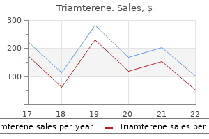
Order triamterene 75mg free shipping
Although the various diseases can be subclassified on the basis of marrow and blood blast cell counts heart attack playing with fire buy on line triamterene, they are considered a single disease entity owing to their similar behavior, which appears to be somewhat independent of blast cell count. The recognition of therapy-related myeloid neoplasms in the late 1970s coincided with the first time patients treated with chemotherapy for cancer actually survived long enough to develop the disease. Alkylating agents were the first to be implicated, with latency periods of 5 to 7 years and a dosedependent effect related to the number of cycles of chemotherapy received. Radiotherapy, alone or in combination with chemotherapy, also increases the risk of therapy-related myeloid neoplasms, although the role of limited field radiation in this disease has been questioned. Other treatments now recognized to be associated with therapy-related disease include autologous hematopoietic cell transplantation and fludarabine chemotherapy. Up to 70% of patients with therapy-related disease had a primary solid tumor, with breast cancer most common, and about 30% had a hematologic malignant neoplasm. The bone marrow may be hypercellular, normocellular, or hypocellular and may have associated fibrosis. Erythropoiesis may exhibit megaloblastic maturation with abnormal nuclear contours or multinucleation; ring sideroblasts are common. Megakaryocytes often display small, abnormal forms with hypolobation or widely separated nuclear lobes. Therapeutic options for patients with therapy-related myeloid neoplasms are limited owing to the high rate of treatment-related mortality, high rate of treatment failure, and early disease recurrence for those patients who respond to therapy. A history of cytotoxic or radiation therapy for a prior neoplastic or non-neoplastic disorder takes precedence over these other categories, and all such cases should be considered therapy-related neoplasms, despite morphologic or cytogenetic similarities to these other disease categories. The exceptions are erythroid leukemia and acute panmyelosis with myelofibrosis, which are defined by different criteria. Twenty percent to 79% of the bone marrow cells are of monocyte lineage, often demonstrated by reactivity with the non-specific esterase stain; however, cytochemical studies are not necessary for diagnosis when the morphologic identity of the monocyte lineage is obvious. Both granulocytic and monocytic differentiation are observed in varying proportions in the bone marrow. In contrast, abnormal immatureappearing monocytes of chronic myelomonocytic leukemia have more condensed chromatin and generally more folded or convoluted nuclear contours. The blasts are cytochemically negative for myeloperoxidase or Sudan black B (<3% positive) and are non-specific esterase negative (<20%), but they may show immunophenotypic evidence of myeloperoxidase expression. In addition, blasts must constitute 90% or more of the non-erythroid marrow cells. Blasts usually have sparse granules and infrequent Auer rods, although the identification of these features does not preclude this diagnosis. Cases may be mistaken for lymphoblastic proliferations without immunophenotyping or cytochemical studies. Blasts express myeloid-associated antigens, but there is no specific immunophenotypic profile. If 80% or more of the monocytoid cells are immature (monoblasts), the case is considered acute monoblastic leukemia; if the cells show evidence of monocytic maturation and less than 80% are monoblasts, it is considered acute monocytic leukemia. Monoblasts are large and have moderately abundant, variably basophilic cytoplasm, which frequently contains delicate peroxidase-negative azurophilic granules or vacuoles. The nucleus is round, with reticular chromatin and one or more prominent nucleoli. The leukemic cells in acute monocytic leukemia manifest more obvious cytologic evidence of monocytic differentiation and maturation. The nuclei have delicate chromatin and a characteristic folded or cerebriform appearance. The promonocyte cytoplasm is less basophilic than that of monoblasts and contains a variable number of azurophilic granules. Blasts more frequently contain cytoplasmic granules or Auer rods but exhibit no specific cytogenetic abnormalities or immunophenotypic profile. The immunophenotype of acute monoblastic and monocytic leukemia is characterized by the expression of monocytic differentiation antigens, but the patterns of expression vary. Monoblastic and monocytic leukemias are associated with a high incidence of organomegaly, lymphadenopathy, and other tissue infiltration. In a significant number of cases, the first clinical manifestations of leukemia result from extramedullary tissue infiltrates. Despite these seemingly unique clinical features, a diagnosis of acute monoblastic or acute monocytic leukemia does not confer prognostic significance. Erythroid-predominant myelodysplastic syndrome previously diagnosed as acute erythroid leukemia. Most cells are dysplastic erythroid precursors, with scattered myeloblasts (arrows) present. Myeloblasts represent less than 20% of all marrow cells but more than 20% of non-erythroid cells. Such features would now result in a diagnosis of myelodysplastic syndrome rather than acute myeloid leukemia. Most patients meeting these criteria present with pancytopenia and nucleated red cells in the blood. There is striking erythroid hyperplasia and dyserythropoiesis characterized by abnormalities of nuclear development, including megaloblastoid changes and karyorrhexis. There is often evidence of a panmyelosis with striking megakaryocytic and platelet abnormalities. In these cases, the erythroid lineage is the only obvious component of acute leukemia; no myeloblast component is apparent. These cells must constitute over 80% of the marrow elements, with proerythroblasts constituting at least 30% of marrow cells. This rare leukemia exhibits a pure population of immature erythroid cells with cytoplasmic vacuoles and no myeloblast proliferation. These cells represent more than 80% of peripheral blood and marrow cells and express erythroid-associated markers of hemoglobin and glycophorin. This patient had no history of chronic myeloid leukemia and did not meet the criteria for another type of acute myeloid leukemia and was therefore diagnosed with acute basophilic leukemia. Myelodysplasia-related cytogenetic abnormalities and multilineage dysplasia are common in these disorders. Pure erythroid leukemias must also be distinguished from several non-neoplastic disorders that manifest marked dyserythropoiesis. These include megaloblastic anemia due to vitamin B12 or folate deficiency, heavy metal intoxication from arsenic, drug effects, congenital dyserythropoiesis, and exogenous erythropoietin administration. In blood and bone marrow smears, megakaryoblasts are usually medium to large cells with a high nuclearto-cytoplasmic ratio. An irregular cytoplasmic border is often noted, and projections resembling budding platelets are occasionally present. Transitional forms between poorly differentiated blasts and recognizable micromegakaryocytes may be observed. In some cases, the majority of the leukemic cells consist of small lymphoidlike blasts. Trephine biopsy sections may reveal morphologic evidence of megakaryocytic differentiation that is not appreciated in the marrow aspirate smears. Identification of a megakaryocyte lineage cannot be made by morphologic features alone and requires immunophenotyping or electron microscopy and ultracytochemistry. Ultrastructural peroxidase activity is found in the nuclear envelope and endoplasmic reticulum and is absent from the granules and Golgi complexes of leukemic megakaryoblasts. This pattern of localization of the ultrastructural peroxidase reaction distinguishes megakaryoblasts from myeloblasts and is the earliest distinctive, recognizable characteristic of megakaryoblasts. Bone marrow cytogenetics may be difficult to obtain owing to the presence of marrow fibrosis. It occurs most commonly in adults with pancytopenia and no splenomegaly (Box 46-15). Marrow myeloblast counts are usually difficult to perform owing to the inability to aspirate marrow as well as the panmyelosis, but most cases have 20% or more marrow blasts. Clonal cytogenetic abnormalities are typically limited to trisomy 21, although non-clonal abnormalities are frequently observed.
Buy triamterene 75mg low cost
Most (95%) of these rearrangements were mutated; however blood pressure instruments discount 75 mg triamterene fast delivery, they mostly carried a low mutational load (97% to 99. The absence of t(11;14) and t(14;18) is also helpful in ruling out these entities. The few studies performed on relatively large series show that adverse clinical prognostic factors are related to high tumor burden and poor performance status. These include the lack of efficacy of 2-chlorodeoxyadenoside,46 the relatively favorable course for patients treated with splenectomy,7,47 and the potential efficacy of fludarabine for patients who relapse after splenectomy or are resistant to chlorambucil. C, On higher magnification, the cells in the follicles are a mixture of centrocytes and centroblasts, typical of germinal-center cells (Giemsa stain). F, Characteristic immunoglobulin D staining of residual mantle cells; the neoplastic cells are negative. Within the spleen and bone marrow, these cases have a characteristic intrasinusoidal pattern of involvement. Most splenic red pulp small B-cell lymphomas (79%) were IgH mutated, with an overrepresentation of V(H)3 and V(H)4 gene families. Bone marrow biopsy shows intertrabecular nodules of small lymphocytes and scattered blasts overrunning residual germinal centers. A marginal-zone pattern can be observed in other small B-cell lymphomas involving the spleen. Splenic marginal zone lymphoma proposals for a revision of diagnostic, staging and therapeutic criteria. Cytogenetic aberrations and their prognostic value in a series of 330 splenic marginal zone B-cell lymphomas: a multicenter study of the splenic B-Cell Lymphoma Group. Over 30% of patients with splenic marginal zone lymphoma express the same immunoglobulin heavy variable gene: ontogenetic implications. Simplification of risk stratification for splenic marginal zone lymphoma: a point-based score for practical use. Splenic lymphoma with circulating villous lymphocytes/splenic marginal-zone lymphoma. The immunophenotype of splenic lymphoma with villous lymphocytes and its relevance to the differential diagnosis with other B-cell disorders. Splenic lym, phoma with villous lymphocytes: clinical presentation, biology and prognostic factors in a series of 100 patients. Regression of, splenic lymphoma with villous lymphocytes after treatment of hepatitis C virus infection. Circulating villous lymphocytes-a link between hyperreactive malarial splenomegaly and splenic lymphoma. Splenic marginal zone lymphoma: clinical characteristics and prognostic factors in a series of 60 patients. Non-malt mar, ginal zone B-cell lymphomas: a description of clinical presentation and outcome in 124 patients. Clonal B-cell lymphocytosis exhibiting immunophenotypic features consistent with a marginal-zone origin: is this a distinct entity Splenic marginal zone lymphoma with increased number of blasts: an aggressive variant Lymph node involvement by splenic marginal zone lymphoma: morphological and immunohistochemical features. Blastic transformation after splenectomy in a patient with nonvillous splenic marginal zone lymphoma with p53 overexpression: a case report. Uncommon cytogenetic findings in a case of splenic marginal zone lymphoma with aggressive clinical course. Splenic marginal zone lymphoma: characterization of 7q deletion and its value in diagnosis. An integrated genomic and expression analysis of 7q deletion in splenic marginal zone lymphoma. Splenic marginal, zone lymphomas are characterized by loss of interstitial regions of chromosome 7q, 7q31. Splenic marginal zone, b-cell lymphomas: two cytogenetic subtypes, one with gain of 3q and the other with loss of 7q. Cytogenetic aberrations and their prognostic value in a series of 330 splenic marginal zone B-cell lymphomas: a multicenter study of the Splenic B-Cell Lymphoma Group. Progression to large B-cell lymphoma in splenic marginal zone lymphoma: a description of a series of 12 cases. Small lymphocytic lymphoma, marginal zone B-cell lymphoma, and mantle cell lymphoma exhibit distinct gene-expression profiles allowing molecular diagnosis. Marginal-zone B cells in the human lymph node and spleen show somatic hypermutations and display clonal expansion. Lack of efficacy, of 2-chlorodeoxyadenoside in the treatment of splenic lymphoma with villous lymphocytes. Long-term follow-up analysis of 100 patients with splenic marginal zone lymphoma treated with splenectomy as first-line treatment. Fludarabine:, an effective treatment in patients with splenic lymphoma with villous lymphocytes. Rituximab, used alone or in combination, is superior to other treatment modalities in splenic marginal zone lymphoma. Risk stratification for splenic marginal zone lymphoma based on haemoglobin concentration, platelet count, high lactate dehydrogenase level and extrahilar lymphadenopathy: development and validation on 593 cases. Splenic involvement by blastic mantle cell lymphoma (large cell/anaplastic variant) mimicking splenic marginal zone lymphoma. Monocytoid/ marginal zone B-cell differentiation in follicle centre cell lymphoma. Splenic small B-cell lymphoma with predominant red pulp involvement: a diffuse variant of splenic marginal zone lymphoma Splenic red pulp lymphoma with numerous basophilic villous lymphocytes: a distinct clinicopathologic and molecular entity Splenic diffuse red pulp small-B cell lymphoma: toward the emergence of a new lymphoma entity. Primary cutaneous lymphomas of germinal center cells are considered a distinct category: primary cutaneous follicle-center lymphoma. In the United States, it is two to three times more common in whites than in blacks. Large mediastinal masses are rare, but large retroperitoneal and mesenteric masses often occur and may cause ureteral obstruction. Pure extranodal presentations are uncommon-9% in one survey3-and extranodal involvement (other than bone marrow) was seen in 20% of cases in another study. Within the gastrointestinal tract, the small bowel and particularly the duodenum are most often involved (see the section on variants of follicular lymphoma later in the chapter). Patients with peripheral lymph nodes that are accessible for open biopsy should have this procedure done. The nuclei appear irregular or angulated in tissue sections; although the term cleaved is used, a distinct nuclear cleft is seldom seen in tissue sections. The chromatin is paler than that of small lymphocytes and is evenly dispersed, giving the nucleus a gray-blue appearance. In most cases, the centrocytes appear more monomorphous than those of normal follicles, with the majority being approximately the same size. The nucleus is vesicular, with a clear center and some peripheral condensation of chromatin; there are one to three basophilic nucleoli, often apposed to the nuclear membrane.
Discount triamterene 75 mg free shipping
Less often there is direct extension into nerves from lymphoma in adjacent tissues blood pressure yeast infection purchase generic triamterene pills. Involvement of multiple nerves with or without involvement of spinal nerve roots, dorsal root ganglia, and meninges (neurolymphomatosis) is more common than involvement of a single nerve. They typically have a subacute onset of neuritic pain, often accompanied by sensory and motor deficits. Physical examination or magnetic resonance imaging reveals a tumor expanding the nerves, sometimes imparting a fusiform contour. Dura Mater Clinical Features Lymphoma arising in the dura mater is uncommon, but welldocumented cases have been described. They present with seizures, headaches, cranial nerve abnormalities, radicular pain, syncope, or a combination of these findings. D, Staining for cytoplasmic lambda light chain is negative (C and D, immunoperoxidase technique on paraffin sections). Therapy varies from case to case, but almost all recently reported patients who have undergone thorough staging and received optimal therapy have done well. The orbital soft tissue is the most common site, followed by the conjunctiva (bulbar or palpebral), lacrimal gland, and then the lacrimal sac. Lymphoid tumors constitute 10% of orbital mass lesions, and lymphoma is the most common orbital malignancy. Conjunctival lymphoma usually produces a salmon-colored plaque that is mobile over the surface of the eye. The orbital soft tissue is involved in the majority of cases, sometimes accompanied by lacrimal gland involvement; the conjunctiva is involved in up to approximately one third of cases. Overall survival at 5 years is approximately 90%, and the 5-year disease-free survival rate is approximately 70%. Inflammatory pseudotumor is a lesion with a variably cellular, polymorphic infiltrate of small lymphocytes, plasma cells, immunoblasts, histiocytes, and sometimes eosinophils or neutrophils, in a stroma with areas that are hyalinized or edematous, or both. Immunohistochemical studies in such cases show a mixture of T cells, B cells, and polytypic plasma cells. In some cases, the plasma cells are predominantly IgG4 positive, suggesting that some inflammatory pseudotumors may be part of the spectrum of IgG4-related disease. A dense, diffuse infiltrate composed predominantly of B cells favors a diagnosis of lymphoma. Such lesions usually express monotypic immunoglobulin and contain clonal B cells on molecular genetic analysis. It consists of the faucial or palatine tonsils, the nasopharynx, and the base of the tongue. Patients have dysphagia, dyspnea, snoring, or a neck mass due to cervical lymphadenopathy. C, High power shows uniform medium-sized round cells with finely stippled chromatin and small nucleoli, numerous mitotic figures, and many admixed tingible body macrophages. However, there is a high rate of distant relapse, particularly in those treated with radiation alone. Although relapses may occur in any lymph node and in a variety of extranodal sites, there is a predilection for spread to the gastrointestinal tract. Infiltration of crypt epithelium by lymphoid cells is normal and does not suggest marginal-zone lymphoma. Clinical Features Paranasal sinus lymphomas affect men more often than women (male-to-female ratio of 1. They affect predominantly middle-aged to older adults81,84 and occasionally children. Also present are scattered reactive follicles and plasma cells, sometimes in large aggregates. In salivary glands other than the parotid, lymphoepithelial lesions may be less conspicuous, but the histologic features are otherwise similar. Follicular lymphoma may also arise in the salivary gland region but usually involves lymph nodes in the vicinity rather than salivary gland parenchyma. The pathologic features are similar to those of other nodal follicular lymphomas (see Chapter 18). Monocytoid B cells confined to lymphoepithelial lesions and even discrete halos around such lesions can be seen in lymphoepithelial sialadenitis, but broad, intersecting bands of monocytoid B cells support a diagnosis of lymphoma. Demonstration of monotypic immunoglobulin in lymphoid cells or plasma cells supports lymphoma. Molecular genetic studies are usually not helpful because B-cell clones are found in more than 50% of cases of lymphoepithelial sialadenitis. Lymphoepithelial lesions are not conspicuous, although large numbers of lymphoid cells may be found within the epithelium of dilated ducts. Chronic sclerosing sialadenitis may have prominent follicular hyperplasia and a dense lymphoid infiltrate with numerous plasma cells and scattered eosinophils, but lymphoepithelial lesions are inconspicuous. There is typically sclerosis, beginning as bandlike, but increasing over time to obliterate parenchyma. IgG4-positive plasma cells are typically numerous; chronic sclerosing sialadenitis is considered a manifestation of IgG4-related disease. Patients are seen with an enlarging mass that is usually painless but is occasionally accompanied by facial nerve paralysis or cervical lymphadenopathy. In a minority of cases, the lymphoma is an infiltrative, ulcerated lesion with raised margins. Staging, Treatment, and Outcome Staging reveals localized disease in approximately 70% of cases. Patients with localized, histologically low-grade lymphomas have an excellent outcome, whereas patients with high-grade lymphoma or disseminated disease have significantly lower survival rates. Oral lymphomas can mimic dental conditions such as periodontal disease, acute necrotizing gingivitis, and dental infections. A, Low power shows crowded follicles within soft tissue beneath squamous epithelium. B, Poorly circumscribed neoplastic follicles contain predominantly centrocytes and are seen adjacent to small acini in the minor salivary gland. Extranodal sites that may be involved include the bone marrow, gastrointestinal tract, lung, liver, and bladder. Reactive follicles; an extrafollicular component of B cells, particularly if they have the morphology of marginal-zone cells; and lymphoepithelial lesions make plasmacytoma unlikely. Blast transformation of neoplastic cells within colonized follicles is more common in the thyroid gland than elsewhere. Thyroid gland with marginal-zone lymphoma (A to E) with large-cell transformation (F). C, In some areas, there is vague nodularity, consistent with colonization of reactive follicles by neoplastic marginal-zone cells. D, the marginal-zone cells are small, with oval to slightly irregular nuclei and a moderate amount of pale cytoplasm. Monoclonal paraproteins are relatively common,139,141 being found in up to 43% of cases. It is very uncommon before age 30 years,139,141-146 although rare cases in younger patients have been reported. The remainder have pulmonary (cough, dyspnea, hemoptysis, chest pain) or constitutional symptoms. A, A dense, diffuse lymphoid infiltrate extends from the bronchial lumen (upper left), past the bronchial cartilage, into the surrounding lung. A, the normal thymic tissue is obliterated by a mottled pale and dark lymphoid infiltrate. Overall, however, patients do well, and survival is good regardless of the therapy given. The more aggressive therapy usually given to patients with these lymphomas could account for the lack of difference in outcome that some have observed. The left ventricle is occasionally involved, but involvement of the left atrium, the most common site of myxoma, is quite uncommon. Two distinctive types have been described: primary effusion lymphoma (see Chapter 29) and pyothorax-associated lymphoma (see Chapter 29).
Generic triamterene 75mg online
A highly curable lymphoma occurs preferentially in the proximal tibia of young patients blood pressure in children purchase triamterene discount. This article reviews the most commonly encountered tumors and non-neoplastic lesions, especially those that can mimic lymphoma, and provides an update on studies useful in distinguishing them. The chapter begins with lymph node metastases because they can present the most diagnostic difficulty and also describes the range of non-neoplastic inclusions included in the differential diagnosis of metastatic tumors. Then mesenchymal and vascular proliferations are discussed, including those that are intrinsic to the lymph node. Histologic Features of Metastatic Tumors Most solid tumors metastasize to regional lymph nodes following invasion of peritumoral lymphatics, with sequential progression down the lymphatic chain. As a result, metastatic deposits in lymph nodes are initially located preferentially in the extranodal vessels and subcapsular sinuses. This localization pattern is diagnostically useful because it is uncommon in lymphoma, with the exception of anaplastic large-cell lymphoma. More extensive metastatic involvement is usually multifocal or geographic, but there is often a discrete boundary separating the tumor from uninvolved areas of lymph node. Often tumor nests or extranodal large vessel invasion in fat may be associated with a lymphoid response and mimic a lymph node, so attention to the presence of a capsule or subcapsular sinus, and a circumscribed versus stellate appearance can be useful. Metastatic solid tumors usually have a cohesive appearance, forming sheets, nests, or islands; undifferentiated carcinoma and melanoma may have a discohesive appearance, mimicking lymphoma. Up to 5% of cancer patients present with lymph node metastasis from an occult primary tumor. Most of these neoplasms are carcinomas; however, 2% of patients with melanoma and a smaller percentage of patients with germ cell tumors and sarcomas may initially present with lymph node metastasis. In this section, we review the histologic features and ancillary tests that can be performed on a metastatic tumor to identify its site of origin. Metastatic papillary tumors of the thyroid gland, kidney, ovary, or lung can show nuclear pseudoinclusions and psammoma bodies; carcinomas of lung and prostate origin often show evidence of partial neuroendocrine differentiation; and foci of necrosis (often with admixed neutrophils and debris) are common in colon adenocarcinoma. Table 59-1 outlines the differential diagnosis of metastatic tumors in each of these morphologic categories. Because of their relatively small cell size and discohesive growth, small-cell tumors are among the most difficult to detect and distinguish from lymphoma; in some instances, immunohistochemistry is required for diagnosis. In the mediastinum, occult lung metastasis of small-cell carcinoma can mimic lymphoblastic lymphoma but typically shows more prominent nuclear molding. For the differential diagnosis to be simplified, metastatic tumors can be divided into those that have an epithelioid (A), anaplastic (B), or spindled (C) appearance. Among epithelioid tumors, metastatic carcinoma and melanoma are the most common non-hematopoietic tumors encountered. Anaplastic tumors presenting in a lymph node have a broad differential diagnosis and can show abnormal antigenexpression patterns. In addition, antigen shedding from infiltrating lymphoid cells or histiocytes can mistakenly make the undifferentiated tumor appear to be positive for leukocyte markers. For example, focal mucin droplets can be present in poorly differentiated adenocarcinoma, intracellular lumens can be seen in vascular tumors, and so-called hallmark cells may suggest anaplastic large-cell lymphoma, but similar cells can be seen in other anaplastic tumors, such as anaplastic thyroid carcinoma. Characteristic Biologic Patterns of Metastasis In addition to histologic features and patient demographic data, the location of an involved lymph node can narrow the possible sources of a metastatic tumor. In cervical lymph nodes, the most commonly encountered occult tumor is squamous cell carcinoma or undifferentiated carcinoma from a head or neck primary tumor. Occult carcinomas originating in the lung and esophagus are the next most commonly encountered metastatic tumors in cervical lymph nodes. Subtle infiltration of the lymph node subcapsular sinus and paracortex by tumor cells in small nests is often observed. The fine nuclear chromatin (described as "smoky" or "dusty") of these small-cell tumors may mimic blastic hematopoietic malignancies, but they usually have more abundant cytoplasm with indistinct borders, as well as large areas of necrosis (not shown). Diffuse replacement of the lymph node by this small-cell neoplasm may be difficult to distinguish histologically from lymphoblastic lymphoma, because diagnostic rhabdomyoblasts may be rare. The lymph node is infiltrated by large germ cells with abundant clear cytoplasm and distinct cell borders. A helpful feature in distinguishing metastatic seminoma from large-cell lymphoma is the admixed granulomatous reaction and small lymphocytes. In inguinal lymph nodes, the most common metastatic tumors are melanoma and prostate carcinoma in men and gynecologic malignancies in women. A review of the prognostic markers is beyond the scope of this chapter, and they are constantly evolving. Suggested diagnostic immunohistochemistry panels for different tumor categories are shown in Table 59-2. In general, the commonly used first-tier diagnostic antibodies are highly specific but variably sensitive for the detection of particular tumor types. Finally, plasmacytomas are notorious for aberrant and false-positive immunoreactivity and can stain for cytokeratin, myeloperoxidase, and T-cell markers, among others. Other markers can be helpful in identifying metastasis from less common primary sites. A punctate or dotlike cytoplasmic staining pattern observed in Merkel cell carcinoma and small-cell carcinoma is characteristic but not completely specific for these tumor types. It should be noted that some lymphomas (approximately 2%) of both mature and lymphoblastic types can show some keratin positivity, most commonly cytokeratin 8. Molecular profiling with a limited array of transcripts of lineage-associated genes has recently shown great promise in accurate classification24-26 and the selection of appropriate therapies. A, Columnar tumor cells that replace the nodal parenchyma exhibit gland formation with central necrosis, typical of colon adenocarcinoma. B, Tumor cells are positive for cytokeratin 20 but negative for cytokeratin 7 (not shown). Electron microscopy has a limited role in the differential diagnosis but can be helpful in the definitive diagnosis of poorly differentiated tumors, for example, by detecting melanosomes in poorly differentiated melanoma or cell junctions that would suggest a carcinoma or dendritic cell neoplasm. In small-cell tumors, electron microscopy is especially useful in detecting muscle filaments in rhabdomyosarcoma. This is particularly common in seminoma, melanoma, and medullary carcinoma of the breast. In mediastinal biopsy specimens, thymoma should always be a diagnostic consideration when numerous small lymphocytes are associated with a spindle cell or epithelioid cell proliferation. The diagnosis of thymoma can be further complicated by the immature thymic immunophenotype of the reactive T-cell component, which can be indistinguishable from lymphoblastic lymphoma by flow-cytometric analysis. In such cases, immunostains can easily detect the extensive cytokeratin-positive tumor meshwork. Undifferentiated nasopharyngeal carcinoma (or undifferentiated carcinoma arising at other sites, such as urothelial tumors) is probably the solid tumor most frequently misdiagnosed as lymphoma. Large nests of cohesive tumor cells are outlined by collagen bands and show multifocal nodal infiltration. Inclusions have been reported in distant nodes such as axillary lymph nodes and can mimic breast cancer. Benign epithelial inclusions resembling glands from nearly all solid organs have been reported in adjacent lymph nodes. Metastatic undifferentiated nasopharyngeal carcinoma-Schmincke or lymphoepithelioma type. Keratin immunostain and Epstein-Barr virus in situ hybridization (inset) were positive in tumor cells. Tumor cell cohesiveness and central necrosis within tumor cell aggregates are helpful clues. A simple cyst in a subcapsular location, lined by cytologically bland cuboid and ciliated columnar epithelium (inset). Cytologically benign nodal reticular cells with fine cytoplasmic cell processes are interspersed between lymphocytes (pankeratin immunostain). Apparent neoplastic transformation of such benign inclusions has also been rarely reported. Similar collections of benign ducts and glands can be observed in perithyroidal, axillary, and perirenal lymph nodes. Bland but occasionally enlarged mesothelial cells can occur as detached groups within the lymph node sinuses, usually in the mediastinum and rarely at other sites.
Purchase genuine triamterene on line
B arteria radicularis magna cheap triamterene 75 mg without a prescription, Re-staining of the smear with Wright-Giemsa confirms the presence of abnormal plasma cells. Subsequent immunohistochemical studies of the core biopsy showed kappa-restricted plasma cell myeloma. If it is not possible for the same pathologist to examine both the marrow aspirate and the core biopsy, each report must note the existence of the other. Although I have not specifically discussed the role of the blood smear in the evaluation of marrow specimens, it is clear that the blood is an integral component in any evaluation of a hematologic abnormality: Not infrequently, abnormalities in the blood are what trigger a bone marrow examination. Occasionally, the blood may show a greater proportion of blasts, more readily apparent dysgranulopoietic features, or a greater degree of differentiation of leukemic cells than the marrow. It is most efficient to obtain blood smears at the time the bone marrow aspiration and biopsy procedure is performed. If a blood smear is not available to the pathologist, at a minimum, hemogram data should be reviewed. When multiple laboratories are involved in the analysis and interpretation of a specimen, a concise summary of the salient results from these contributing laboratories should be provided and integrated into the final diagnosis. Giemsa, which contains azures, is added to the stain to intensify nuclear features and azurophilic and toxic granulation. Move to working Wright-Giemsa buffer solution for 20 to 25 minutes; agitate a few times. Rinse stain off in staining dish of approximately 5 mL methanol/200 mL deionized water. Results Erythrocytes: salmon pink Leukocytes and megakaryocyte nuclei: purple-blue Platelets: purple-blue to lilac cytoplasm containing red-purple granules Notes Because of the difficulty in controlling staining quality, my laboratory no longer uses a flat-rack staining procedure. Be sure the slide holder is dry before placing slides in it; the slide holder can be rinsed in a separate boat of methyl alcohol to clean it and remove water. Change Wright-Giemsa buffer solution after 2 batches of slides or after 1 hour, whichever comes first. If stain is too light, reprocess stained slides in stock WrightGiemsa stain and Wright-Giemsa buffer solution. A, Prussian blue reaction of a crush preparation of the fat-perivascular layer showing increased storage iron within macrophages. B, Dacie stain of a bone marrow aspirate smear showing increased iron within a macrophage. The reaction must take place in an acidic environment to free iron from binding proteins. Specimens Air-dried films of peripheral blood, marrow aspirate (including buffy coat smears), cellular fluids, or cytospins of urine sediment. Crush preparations of the fat-perivascular layer are not suitable since the fat is dissolved by the methanol. Do not dry, as drying at this point before counterstaining in the next step can cause artifact. The nucleated cell count should not include megakaryocytes, macrophages, osteoblasts, osteoclasts, stromal cells, smudged cells, or non-hematopoietic cells such as tumor cells. Lymphoid aggregates, if present, should not be included in the count, but their presence should be commented on. A meaningful interpretation then integrates findings in the marrow aspirate and core biopsy with those from the blood. Rigorous monitoring of the collection, processing, and staining processes ensures specimen adequacy and accurate morphologic observations, which in turn can direct the pathologist to the appropriate ancillary studies to reach an accurate diagnosis. Jatoi, Department of Oncology, Mayo Clinic, Rochester, Minnesota, for her many helpful suggestions. For example, when the bone marrow examination is performed to look for metastatic disease but the hemogram and marrow are otherwise normal, or when there is severe pancytopenia and the marrow is markedly hypocellular, differential counts are not required. Adult patients presenting with pancytopenia: a reappraisal of underlying pathology and diagnostic procedures in 213 cases. World Health Organization Classification of Tumours of Haematopoietic and Lymphoid Tissues. False-negative rates of tumor metastases in the histologic examination of bone marrow. Protocol for the examination of specimens from patients with hematopoietic neoplasms of the bone marrow. Before the development of this technology, the diagnosis of lymphoproliferative diseases depended on classification systems based solely on morphologic differences. The subjective use of morphologically based classification schemes led to difficulty in defining biologically different entities, and the morphologic categories were often difficult to reproduce, even among expert hematopathologists. Such phenotypic markers provide information about the cell lineage and origin of the hematopoietic neoplasm, the production of characteristic oncogenic proteins, and the proliferative characteristics of the tumor. Immunohistochemistry is increasingly being used to identify underlying molecular alterations to aid in diagnosis and guide therapy decisions. In actual practice, the production of an optimally immunostained slide is much more problematic and depends on the condition of the tissue antigen; the type, specificity, and affinity of the primary antibody; and the detection system used. The specific antigenic epitopes present on any given protein or carbohydrate moiety are subject to enzymatic degradation that begins immediately after biopsy or resection and to further conformational changes resulting from fixation. To ensure preservation of the antigen of interest, rapid tissue fixation is important. Some antigenic epitopes, such as those on keratin proteins and other structural proteins of the cell, are relatively resistant to degradation; other antigens, such as phosphoepitopes on signaling proteins, undergo rapid degradation within minutes to hours. This mode of action is potentially deleterious to antigenic structure, and although some antigenic epitopes may not be affected significantly by formaldehyde cross-linking, these chemical modifications clearly have an adverse effect on many antigens. Because formalin penetrates tissues slowly and the chemical reactions are complex, the number of modifications that take place is time dependent. In practice, this means that antigens fall into three basic categories: formalin-resistant epitopes, highly formalinsensitive epitopes, and epitopes with a time-dependent sensitivity to formalin fixation. Although there have been attempts to generate antibodies specifically to formalin-resistant epitopes,4 most of the antibodies found to react with formalinresistant epitopes have been identified through large-scale screenings of available antibody preparations. Over the years, there has been great interest in identifying methods to overcome or reverse the deleterious effects of formalin fixation. The earliest attempts to retrieve antigenicity used proteolytic enzymes,5 which presumably act by breaking formaldehyde-induced methylene cross-links in the antigenic molecules, thereby relaxing some of the conformational constraints on the protein epitopes. Nonetheless, proteolytic methods are difficult to control, and careful attention is needed to optimize their retrieval effect and avoid tissue destruction. However, hydrolytic cleavage of formaldehyderelated chemical groups and cross-links, the unfolding of inner epitopes, and the extraction of calcium ions from coordination complexes with proteins are among the hypothesized mechanisms. Tissue damage can be minimized by ensuring that tissues are optimally fixed, reducing the antigen-retrieval time, or changing the retrieval buffer. Despite this potential problem, the ability to detect otherwise non-detectable antigens far outweighs the potential for tissue damage on occasional tissue sections. Primary Antibodies There are two major categories of primary antibodies used in diagnostic pathology: monoclonal antibodies and polyclonal antibodies. The serum is subjected to antibody purification and sometimes to differential adsorptions to eliminate unwanted reactivity, but it always comprises a spectrum of antibody molecules originating from multiple unrelated antibody-producing cells (hence the term polyclonal). The specificity of a polyclonal antibody preparation is highly dependent on the purity of the initial antigenic preparation and how extensively adsorbed it is. Further, because the antibody response is variable over time and from one individual animal to another, complete standardization of antibody composition is not possible. Monoclonal antibodies, in contrast, are the product of a single immortalized antibody-producing cell, thus avoiding most problems related to antibody heterogeneity and specificity inherent in polyclonal antibody preparations. The hybridoma technology pioneered by Kohler and Milstein14 in the 1970s allows the immortalization of a single antibodyproducing mouse plasma cell by fusing it with a mouse plasmacytoma cell line. Individual hybrid mouse cells can be clonally expanded in tissue culture or in mice as tumors, providing a continuous source of antibody of known composition and reactivity. Because of their high quality and specificity, monoclonal antibodies were rapidly developed as diagnostic reagents in hematopathology, as well as for other clinical applications that require standardized reagents. The specificity advantage of the monoclonal antibody, however, can also be a disadvantage when applied to denatured proteins in tissue sections.
Generic triamterene 75 mg otc
C heart attack zippy demi buy cheap triamterene on line, Lymphoma cells in the peripheral blood vary in size but are larger than normal lymphocytes and have convoluted, "cerebriform" nuclei, less condensed chromatin, and indistinct nucleoli. However, even with immunostains, there is a poor agreement between histologic-immunophenotypic detection of disease and molecular studies for T-cell clonality. The neoplastic lymphocytes may be small or large with variable amounts of cytoplasm. Neoplastic cells with clear cytoplasm typically seen in the extranodal sites are usually not common in the bone marrow biopsy. In some cases, the prominent plasmacytosis may obscure the neoplastic infiltrates. In addition, the heterogeneous infiltrates may also resemble reactive polymorphous lymphohistiocytic proliferation (discussed in Unusual Reactive Lymphoid Infiltrates). Peripheral T-Cell Lymphoma, Not Otherwise Specified Peripheral T-cell lymphoma, not otherwise specified involves the bone marrow in about 20% to 40% of cases at diagnosis. The bone marrow infiltrates are often polymorphous, containing abnormal lymphocytes admixed with reactive polymorphous infiltrates, including plasma cells, histiocytes, and eosinophils; prominent vascularity and reticulin fibrosis may also be present in the lesions. They range from small to large in size, often with irregular and hyperchromatic nuclei. Occasional cases consisting of a monotonous population of medium to large cells or containing large, pleomorphic cells can also be seen. The neoplastic cells with clear cytoplasm typically seen in the lymph node biopsy are uncommon in the bone marrow but are present in this case. B, Rare large lymphoma cells with cytoplasmic vacuoles are present at the feathered edge of the peripheral blood smear. In a study, lymphoma cells were found by immunostaining in 23% of patients with negative bone marrow by routine histology. The lymphoma cells are large but variably sized with irregular nuclei, dispersed chromatin, multiple prominent nucleoli, and abundant basophilic cytoplasm. Focal involvement is present in about 30% of cases; the infiltrates may be single or multiple, random or paratrabecular. Fibrosis is almost always present in the infiltrate and may be prominent, especially in diffuse lesions. The infiltrates are polymorphous and contain Reed-Sternberg cells in a background of small lymphocytes, histiocytes, plasma cells, neutrophils, and occasional eosinophils. B, Discohesive, small carcinoma cells in the bone marrow aspirate smear that resemble lymphoma cells; small cohesive cluster of tumor cells, which is more typical of carcinoma, is also present. A, Focal clusters of carcinoma cells with dispersed chromatin are nestled among the normal hematopoietic elements. In other cases, the metastatic carcinoma may be extensive and diffusely replace the normal bone marrow. Systemic Mastocytosis Systemic mastocytosis involves the bone marrow in at least 90% of cases and shows similar patterns of infiltration as lymphoma, including paratrabecular, perivascular, random, and, rarely, diffuse infiltrates. Note the spindle-shaped mast cells with clear cytoplasm at the periphery of the lesion. B, Immunostaining for tryptase highlights the mast cells surrounding the lymphoid aggregate. Recognition of mast cells in the bone marrow sections is critical to arrive at a diagnosis of systemic mastocytosis. The mast cells show variable morphology and may have round, oval, spindle-shaped, or monocytoid nuclei with abundant, slightly eosinophilic cytoplasm. Exclusively paratrabecular lymphoid infiltrates can occasionally be present in mantle cell lymphoma. Diffuse large B-cell lymphoma may show a discordant lymphoma subtype, often characterized by a low-grade lymphoma in the bone marrow. Low-level monoclonal B-cell populations may be identified in the peripheral blood or bone marrow in "healthy" individuals without evidence of lymphoma. Clonal T-cell populations can be detected in various benign conditions, particularly in autoimmune diseases. Focal lymphoid aggregates (nodules) in bone marrow biopsies: differentiation between benign hyperplasia and malignant lymphoma-a practical guideline. Lymphoproliferative disorders in the bone marrow: histologic criteria for classification and staging. Prevalence of intravascular large B-cell lymphoma with bone marrow involvement at initial presentation. Discordant morphologic features in bone marrow involvement by malignant lymphomas: use of gene rearrangement patterns for diagnosis. Bilateral trephine bone marrow biopsies in lymphoma and other neoplastic diseases. Frequencies and patterns of bone marrow involvement in non-Hodgkin lymphomas: observations on the value of bilateral biopsies. Diagnostic utility of bilateral bone marrow examination: significance of morphologic and ancillary technique study in malignancy. Bone marrow lymphoid aggregates in myelodysplastic syndromes: incidence, immunomorphological characteristics and correlation with clinical features and survival. Bone marrow trephine biopsy involvement by lymphoma: review of histopathological features in 511 specimens and correlation with diagnostic biopsy, aspirate and peripheral blood findings. Clinicopathological characteristics of follicular lymphoma with peripheral blood involvement. Lymphoid aggregates in bone marrow mimic residual lymphoma after rituximab therapy for nonHodgkin lymphoma. A comparison of flow cytometry, bone marrow biopsy, and bone marrow aspirates in the detection of lymphoid infiltration in B cell disorders. Predictive value of blood and bone marrow flow cytometry in B-cell lymphoma classification: comparative analysis of flow cytometry and tissue biopsy in 252 patients. Monoclonal B lymphocytes with the characteristics of "indolent" chronic lymphocytic leukemia are present in 3. Bone marrow histology in monoclonal B-cell lymphocytosis shows various B-cell infiltration patterns. Multiparameter flow cytometry in the differential diagnosis of aberrant T-cell clones of unclear significance. Correlation between molecular and histopathological diagnoses of B cell lymphomas in bone marrow biopsy and aspirates. Gebhard S, Benhattar J, Bricod C, Meuge-Moraw C, Delacretaz F Polymerase chain reaction in the diagnosis. Evaluation of B cell lymphoid infiltrates in bone marrow biopsies by morphology, immunohistochemistry, and molecular analysis. Bone marrow trephines containing lymphoid aggregates from patients with rheumatoid and other autoimmune disorders frequently show clonal B-cell infiltrates. Is clonality equivalent to malignancy: specifically, is immunoglobulin gene rearrangement diagnostic of malignant lymphoma Systemic polyclonal B-immunoblastic proliferation with marked peripheral blood and bone marrow plasmacytosis. Polyclonal systemic immunoblast proliferation: an unusual hematologic entity presenting as a medical examiner case. Atypical angioimmunoblastic T-cell lymphomas masquerading as systemic polyclonal B-immunoblastic proliferation.
Diseases
- Chickenpox
- Psittacosis
- Asphyxia neonatorum
- Microcephaly cleft palate autosomal dominant
- Culler Jones syndrome
- Beriberi
- Skeleto cardiac syndrome with thrombocytopenia
Order triamterene without a prescription
In some of these disorders blood pressure chart for tracking order triamterene 75 mg with visa, there are morphologic and phenotypic features that are either unique or are intrinsically related to the underlying genetic defect that lead us to a diagnosis; in other instances, the histologic changes may be non-specific, but they can help in ruling out any of these syndromes. On physical examination, no lymph nodes are palpable, and imaging studies reveal a lack of thymus shadow. Depending on the genetic defect, when known, or predominant symptom, it is divided into eight broad categories: combined immunodeficiencies (disorders of B cells and T cells), combined syndromic immunodeficiencies, antibody deficiencies, diseases of immune dysregulation, phagocytic disorders, defects of innate immunity, autoinflammatory syndromes, and complement deficiencies. Depending on the prevalent defect and symptoms, some of the inherited disorders are listed under multiple headings. Morphologically, lymph nodes show complete effacement of the architecture with a depleted look and increased number of dendritic cells and eosinophils; they usually lack primary and secondary B follicles. The underlying genetic defect is unknown in the majority of cases, and it remains a diagnosis of exclusion. Incidence ranges from 1: 10,000 to 1: 50,000 (Europe/North America); males and females are equally affected. Clinically the symptoms are very heterogeneous, but two major groups can be broadly recognized based on predominant recurrent infections of the respiratory tract versus inflammatory complications with a variety of autoimmune disorders (22% to 48%) including cytopenias, granulomatous disease, and increased development of malignancy, mainly lymphomas. From the pathology standpoint, the lungs are the major target organ with acute bacterial infections, with possible subsequent development of bronchiectasis and non-infectious immune-mediated changes with lymphocytic interstitial infiltrate, follicular bronchiolitis, and follicular hyperplasia with often "naked" germinal centers and paucity of plasma cells. It presents with recurrent upper and lower respiratory tract involvement due to opportunistic infections (P. Subsequent complications involving the biliary tree and liver are often related to Cryptosporidium- and Giardia-persistent infections of the biliary system leading to sclerosing cholangitis, hepatitis, cirrhosis, and increased gastrointestinal malignancies including cholangiocarcinoma. Morphologically the lymph nodes are characterized by florid follicular hyperplasia with large, expanded germinal centers. A, Small lymph node with prominent paracortical hyperplasia lacking secondary B follicles in the cortex. In addition, prominent lymphoid aggregates consistent with nodular lymphoid hyperplasia are noted. The presence of ill-defined follicular structures may have led in the past to overdiagnosed B-cell lymphomas due to the impression of a distorted architecture. Immunophenotypic studies as well as clonality studies are helpful in clarifying the diagnosis in such cases. It is noteworthy that these patients more frequently also had splenomegaly, granulomatous disease, and enteropathy, which are all associated with decreased overall survival. Immunoglobulin levels were variable (IgG), but usually high for IgM with low IgA; falling levels of IgG were observed over time in some patients. These observations were confirmed by in vitro studies where the B-cell proliferation was within normal limits, but the cells have impaired immunoglobulin production (defects in class-switch recombination). Often these patients had prominent lymphadenopathy involving central and peripheral lymph nodes with compression of the airways in some patients. A, Low-power view showing typical features with "naked" reactive germinal centers and prominent monocytoid B-cell reaction. B, Numerous IgM-positive cells are in the parafollicular areas, and IgG-positive cells are also present within the germinal center (C). Subsequent biopsy (6 years interval) shows similar morphologic features, with large expanded naked germinal centers (D) surrounded by numerous IgM-positive cells (E), and extremely few IgGpositive cells (F), consistent with the serum immunoglobulin levels, simulating a hyper IgM syndrome. In some cases, the monocytoid B-cell reaction was more extensive, raising the possibility of involvement by nodal marginal-zone lymphoma. These patients, in addition, show prominent nodular lymphoid hyperplasia involving the respiratory and gastrointestinal tracts with prominent proliferation of B cells and T cells, often with germinal-center formation without significant colitis. David Purtilo and colleagues described it in 1975 based on a study from a single generation of the Duncan family. It is a rare immunodeficiency with an incidence of approximately 1 to 2 per million males. In rare instances, aplastic anemia, vasculitis, and lymphomatoid granulomatosis have been described in these patients. It is of interest that the diagnosis was frequently delayed even in patients with a positive family history; regardless, nearly all patients became symptomatic before 5 years of age. Autosomal Dominant Immune Dysregulation Syndrome with Heterozygous Germline Mutations of Cytotoxic T-Cell Antigen-4 this disease has been recently characterized. Histologically, the infiltrate was either lymphohistiocytic with scattered plasma cells or mostly lymphoplasmacytic. These disorders have offered the opportunity to better understand the regulatory T-cell function both centrally and peripherally, their development and their maintenance of homeostasis. It has been shown that several patients have diminished and defective Tregs65; however, it remains unclear whether these changes take place in the thymus or in peripheral organs. A and B, Lymphohistiocytic infiltrates involving the white matter with isolated plasma cells are shown. C, A dense plasmacytic perivascular infiltrate extending into the surrounding brain parenchyma; the plasma cells are polyclonal. A progressive B-cell lymphopenia was also noted with an increase of autoreactive B cells. The presence of typical morphologic and immunophenotypic findings represents one of the secondary accessory criteria. Female carriers are asymptomatic, with extreme rare symptomatic cases due to deleterious mutations on the paternally derived X and nonrandom inactivation of the maternally derived X. Spontaneous chimerism due to genetic reversion, which may confer a selective advantage, has been observed in about 11% of patients, but the clinical significance of this finding is unclear. Clinically, patients present with skin rash (eczema) and bleeding (80%) such as petechiae and ecchymoses. In addition, only a 60% penetrance was found among family members carrying the same heterozygous gene mutation, indicating that other genetic differences or modifiers may exist. As expected, nearly all symptomatic patients had chronic adenopathy and splenomegaly. High risk for sepsis was more pronounced in patients that underwent splenectomy at a younger age. Sequencing also has been used in the context of prenatal diagnosis in at-risk couples. Laboratory findings confirm that all arms of the immune system are affected: adaptive, humoral, and innate. T-cell defects also lead to impaired antibody production by B cells, but intrinsic defects in B cells have also been described with hyperresponsiveness and autoantibody production. This deficit in cytotoxic activity, in conjunction with the lymphopenia and intrinsic T-cell and B-cell defects, may be ultimately responsible for the inability to clear infectious agents and may contribute to the development of B-cell lymphomas. Primary immunodeficiency diseases: an update on the classification from the international union of immunological societies expert committee for primary immunodeficiency. Phosphoinositide, 3-kinase delta gene mutation predisposes to respiratory infection and airway damage. A recent review from the French national registry of primary immune deficiencies reported that there is high incidence of cancer (24. Also, other malignancies were very uncommon (rare) like myeloid neoplasms, mature T cell malignancies, and carcinomas involving breast, gastric, liver, and thyroid. The pathology should be interpreted in the context of the clinical presentation, family history, and appropriate immunologic workup. Immunological lossof-function due to genetic gain-of-function in humans: autosomal dominance of the third kind. Guidelines for genetic studies in single patients: lessons from primary immunodeficiencies. Whole-genome sequencing is more powerful than whole-exome sequencing for detecting exome variants. Newborn screening for severe combined immunodeficiency in 11 screening programs in the United States. The expanding clinical and immunological spectrum of severe combined immunodeficiency. History and current status of newborn screening for severe combined immunodeficiency. Analysis of class switch recombination and somatic hypermutation in patients affected with autosomal dominant hyper-IgM syndrome type 2. Epigenetic function of activation-induced cytidine deaminase and its link to lymphomagenesis.

Discount triamterene online
Within the nodules blood pressure instruments buy triamterene 75mg with visa, numerous slitlike round vascular spaces are seen that are lined by plump endothelial cells and pericytes. The fibrosclerotic internodular spaces are composed of myofibroblasts, and a mixed inflammatory infiltrate is commonly seen including lymphocytes, plasma cells, and macrophages. These staining patterns are reminiscent of normal vasculature of red pulp vessels. A background of scattered IgG4-positive plasma cells in the fibrosclerotic stroma is also seen. The nodular pattern with three different vessel types differentiates this proliferation from the others in the differential diagnosis. These proliferations are considered indolent, with no tendency for recurrence after splenectomy. Peliosis Peliosis of the spleen is a rare proliferation of dilated bloodfilled cavities. Secondary conditions such as infections, particularly tuberculosis; malignant conditions such as lymphomas and leukemias; and drug use as in chemotherapy can be associated with peliosis. Sections of the spleen demonstrate multiple round to oval blood-filled cysts with or without sinusoidal endothelial lining cells. Peliosis differs from hemangiomas or hemangiomatosis by the lack of intervening fibrosis. A, the lesion is multinodular, with slitlike spacing surrounded by dense sclerosis. The gross appearance mimics inflammatory pseudotumor, but histologically, the distinction is usually not difficult. Splenic Hamartoma Hamartomas of the spleen are rare benign nodular lesions with an incidence of 0. Clinically, patients may present with splenomegaly, thrombocytopenia, or with other symptoms of hypersplenism; however, 50% of cases will be asymptomatic. They show numerous slitlike vascular channels lined by pulp to flattened endothelial cells, with an absence of normal red pulp cords, lymphatic elements, or organized white pulp elements. Although most cases are benign in behavior, with large lesions there can be a risk for rupture. A, Gross photograph of a splenic hamartoma in a spleen removed for involvement by a low-grade B-cell lymphoma causing the background gross military pattern of infiltration. Clonality has been observed in some of these cases, which further suggests a true vascular origin for at least a subset of these cases. It is unclear whether the division into these subgroups has any clinical significance. Histologically it appears as a bland spindle cell myofibroblastic proliferation with interspersed variable amounts of col- lagenous stroma, and intervening abundant inflammatory cells including lymphocytes, plasma cells, histiocytes, and eosinophils. Splenectomy is curative for symptomatic cases, but cases that are partially resected may recur or metastasize as similar to other dendritic cell neoplasms. Other Proliferations Non-specific areas of fibrosis may mimic inflammatory pseudotumor in the spleen. Because they are generally reactive proliferations, it is important to distinguish them from inflammatory pseudotumor-like dendritic cell sarcoma, which is a true neoplasm that may metastasize if not entirely resected. Areas of scarring after splenic infarction and hemorrhage (Gamna-Gandy bodies) are common in the spleens of children with sickle cell anemia, and fibrotic areas may occur after resolution of a splenic abscess or trauma. Because of confusion caused by the term inflammatory pseudotumor, it is probably best to clarify that the reactive proliferations with fibrosis and inflammation are more inflammatory pseudotumor-like, with a clear statement that they do not represent a neoplastic proliferation. They are unilocular and have a firm, fibrous, trabecular wall that is lined by mesothelial cells or squamous epithelium. Notably, the epithelial lining of primary cysts may be patchy, with denuded areas present that may simulate a secondary cyst. Parasitic cysts, though uncommon, are typically attributable to Echinococcus and are readily identified by the presence of parasite scolices in the cyst contents. Nonparasitic primary cysts appear to arise from congenital inclusions of capsular mesothelium. Secondary cysts represent approximately 80% of splenic cysts and are often associated with a history of abdominal trauma. These are unilocular and thin walled and differ from primary cysts by the complete absence of an epithelial lining and are thus unlikely to recur even if only partially resected. Reported cases of sarcoma include malignant fibrous histiocytoma, fibrosarcoma, leiomyosarcoma, rhabdosarcoma, histiocytic sarcoma, interdigitating dendritic cell sarcoma, and fibroblastic reticular cell tumor. One third of metastatic tumors are only identified on microscopic examination,182 so the frequency of splenic involvement may be underestimated by imaging studies. Lung, gastric, ovarian, and breast carcinoma primaries are the most common to involve the spleen, and splenectomy may be performed for solitary splenic metastases. These are predominantly autosomal recessive conditions whose diagnosis and classification are based on the enzymatic defect characteristic of each disease, often in combination with specific genetic testing. Although most of these conditions are rare, three of the lipid storage diseases are encountered (uncommonly) in surgical pathology practice. The malignant cells are strongly positive with a pan-cytokeratin immunostain (inset). Not uncommonly in these cases, the spleen is removed to confirm the diagnosis or to ameliorate cytopenias. Accumulation of sea-blue histiocytes may be seen in association with lipid disorders, infectious diseases, red blood cell disorders, and myeloproliferative disorders. However, it is also a prominent feature in spleens removed from patients with Hermansky-Pudlak syndrome, a rare, often fatal autosomal recessive condition that is currently classified among the disorders of lysosome-related organelle biogenesis. Because Gaucher cells are macrophages and ingest red blood cells, they frequently stain positive for iron. Ultrastructural studies reveal numerous lysosomes containing characteristic lipid bilayers. The lipid deposits are birefringent and, under ultraviolet light, display yellow-green fluorescence. Electron microscopy reveals lamellated structures resembling myelin figures within lysosomes. In cases of ceroid histiocytosis, smaller histiocytes with more basophilic cytoplasm and vacuoles are characteristically seen. Ceroid is composed of phospholipids and glycosphingolipids and is similar to lipofuscin in its physical and chemical properties. Ultrastructural studies reveal inclusions of lamellated membranous material with 4. None of the cell types identified in storage disorders is specific for a given disease, and their actual diagnosis should be based on biochemical or molecular genetic testing specific for these diseases. Immunodeficiency-associated lymphoproliferative disorders can be confused with malignant lymphoma in the spleen. In adults, common variable immunodeficiency and related disorders can be particularly difficult to distinguish from lymphomas. Myeloid hyperplasia in the red pulp, which can mimic acute myeloid leukemia, may be caused by cytokines, particularly granulocyte colony-stimulating factor. Morphologic and immunohistochemical evaluation of splenic hematopoietic proliferations in neoplastic and benign disorders.

Purchase triamterene 75mg visa
The incidence of drug-induced neutropenia increases with age blood pressure pregnancy range buy generic triamterene 75 mg on line, likely reflecting the higher use of multiple medications in elders. Greater susceptibility of some individuals to idiosyncratic reactions is hypothesized to relate to increased myeloid precursor sensitivity to a normal drug concentration or to gene polymorphisms that alter drug metabolism or drug pharmacokinetics. If a severe, drug-related neutropenia is suspected, all non-essential drugs and overthe-counter medications must be discontinued, with substitution of essential medications. A bone marrow evaluation may be required to exclude a neoplastic process or aplasia. Physical findings and specific immunological tests supporting an autoimmune disorder usually suffice to make the diagnosis. The bone marrow is typically normocellular with a slightly decreased myeloid-toerythroid ratio, due to mild T-cell and cytokine-mediated suppression of granulopoiesis. Infection-Related Neutropenia Infection-related neutropenia may be due to a large number of infectious agents including almost any type of viral infec- tion. A number of different mechanisms are involved in the pathogenesis including infection of progenitor cells or endothelial cells, immunologically mediated bone marrow suppression (particularly viral infections), excessive cellular destruction (especially bacteremia with endotoxemia), increased neutrophil adherence to endothelium, development of antineutrophil antibodies, and enhanced neutrophil utilization at the site of infection. Notable exceptions include megaloblastic anemia (see Table 11-7) and copper deficiency. Neutropenia associated with vitamin B12 or folate deficiency rarely occurs without anemia and macrocytosis. Copper deficiency should be considered when cytoplasmic vacuoles are present in myeloid (particularly promyelocytes and myelocytes) and erythroid precursors. Patients have concurrent normocytic, macrocytic, or microcytic anemia, neurologic disorders, and rarely thrombocytopenia. Copper deficiency occurs secondary to excess zinc intake (through supplements, medications, or denture fixatives), total parenteral nutrition, and gastrointestinal disorders. Mild neutropenia is also observed with severe caloric deprivation such as in anorexia nervosa. Congenital Neutropenia Congenital neutropenia refers to neutropenias with genetic mutations and not simply those that are present at birth. Many of these disorders have intrinsic defects that cause premature apoptosis of cells, ineffective neutrophil production, and recurrent infections. A, Bone marrow smear of severe congenital neutropenia in a 3-year-old boy illustrates a maturation arrest at the promyelocyte stage of development. The marrow exhibits a myeloid maturation arrest before the period of severe peripheral neutropenia. Accelerated apoptosis of bone marrow progenitor cells is found in all stages of the cycle, with insufficient myeloid output. Increased infections are due to neutropenia with impaired neutrophil chemotaxis and, commonly, T-cell and B-cell defects. The diagnosis is made based on identification of exocrine pancreatic dysfunction and intermittent or persistent neutropenia. Disease phenotype varies with time in individuals and between patients, making the diagnosis challenging in some cases. Development of monosomy 7 is more strongly associated with advanced disease or transformation. Faulty neutrophil chemotaxis and function lead to frequent and severe pyogenic infections that characterize this disorder, whereas a deficiency in platelet dense bodies contributes to the tendency for easy bleeding. Accelerated disease develops in up to 85% of patients and is associated with a multiorgan lymphohistiocytic infiltrate that has features of hemophagocytic lymphohistiocytosis. Myelokathexis Myelokathexis is a histologic pattern of increased myeloid cells with excessive neutrophil apoptosis in the bone marrow. They are particularly susceptible to human papilloma virus infection and require careful surveillance. As 80% to 90% of patients develop bone marrow failure, this disorder is more fully described under the bone marrow failure syndromes. The causes of lymphopenia are extensive and include a variety of infectious, drug-related, autoimmune, and congenital processes (Box 11-4). Lymphopenia is seen in a number of viral, fungal, bacterial, mycobacterial, and parasitic infections. Reactive lymphocyte morphology in these disorders provides a clue to underlying infection. The pathogenesis is unclear but may be due to increased apoptosis via the Fas pathway, decreased T-cell production, or defective tumor necrosis factor- or interferon- production. It is encountered frequently in clinical practice, and when severe, is a common cause of hemorrhage. Thrombocytopenia is the result of decreased production, increased destruction or utilization, or abnormal distribution of platelets. Evaluation of megakaryocytes in patients with unexplained thrombocytopenia is one of the most common indications for a bone marrow examination in clinical practice. An important caveat before bone marrow evaluation is to confirm that the platelet count is not spuriously low as a result of in vitro platelet clumping. Blood collection in sodium citrate or alternative anticoagulant may alleviate the problem. Evaluation of bone marrow megakaryocytes is an important initial step in the workup for unexplained thrombocytopenia. Decreased megakaryocytes is indicative of decreased platelet production, whereas normal or increased megakaryocytes point toward ineffective megakaryopoiesis (intramedullary cell death or excessive apoptosis) or loss of platelets from the circulation. Table 11-9 lists the differential diagnosis for thrombocytopenia when megakaryocytes are absent or decreased. Identifying the cause of an underlying bone marrow failure disorder or other process. Evaluation for subtle dysplasia, viral effects, or abnormal cellular infiltrates is required; cytogenetic evaluation, chromosome breakage studies (Fanconi anemia), and molecular analysis. Chemotherapy, toxin exposure, or prolonged use of certain drugs can selectively affect the megakaryocytes in some patients. Causes of thrombocytopenia when the bone marrow megakaryocytes are normal to increased in number are listed in Tables 11-10 and 11-11. Megakaryocytes are often smaller and exhibit less nuclear lobation (left-shifted). In this setting, platelet accumulation in bone marrow sinuses also suggests increased platelet consumption when bone marrow artifacts or a myeloproliferative neoplasm are excluded. Platelet antibody testing is not recommended due to poor sensitivity and specificity. These children usually present without a viral prodrome, and constitutional platelet disorders need to be excluded. B, the bone marrow trephine biopsy section contains increased numbers of megakaryocytes. The immune mechanisms involved are also varied and drug dependent, but they act primarily to increase peripheral platelet clearance, with some drugs also causing marrow suppression. Infection-Associated Thrombocytopenia Infection, especially viral infection, is a frequent cause of thrombocytopenia through direct infection of megakaryocytes, the toxic effects of organism proteins or cytokines, secondary hemophagocytosis, or immune-mediated destruction from antiplatelet antibodies. The latter is due to platelet activation developing within days of heparin exposure that often resolves during continued therapy. Antibodies against heparin/ platelet factor 4 immune complexes engage with platelet and monocyte Fc receptors causing cellular activation and procoagulant microparticle release and thrombin generation.
Buy triamterene 75 mg online
Deeper insights into the downstream consequences secondary to mutational combinations and their interactions with the bone marrow microenvironment as well as epigenetic mechanisms will be critical to understand the pathogenesis and unique clinical behavior of this group of myeloid neoplasms heart attack on plane purchase triamterene with mastercard. Blasts, including promonocytes (which are considered blast equivalents), account for less than 20% of the cells in the peripheral blood and less than 20% of the nucleated cells in the bone marrow. Although absolute monocytosis in the blood is required for diagnosis, there is remarkable variation in the other hematologic parameters. Therefore caution would have to be used in the interpretation of these genetic results. If any of these abnormalities are found, the case should be reassigned to the subgroup of myeloid neoplasms associated with eosinophilia and rearrangement of one of these specific genes. In some cases, myelodysplasia is minimal; in others, all the myeloid lineages have prominent dysplastic changes. Symptoms most often include fever, infection, or hemorrhagic episodes in up to 30% of patients and thrombotic complications in 10% to 15%; in some patients, early satiety due to splenomegaly may be an initial complaint. Monoblasts form a morphologic continuum with promonocytes, from which it may be difficult to distinguish them, but both cell types are considered together in tallying the number of blasts for classification purposes. Eosinophilia may also be seen, and if the eosinophil count is persistently more than 1. Up to 75% of patients are reported to have micromegakaryocytes or megakaryocytes with abnormal nuclear lobation,95,96 although in some cases, enlarged megakaryocytes can be found as well. Peripheral blood smear shows slightly elevated white blood cell count with absolute monocytosis and minimal dysplasia in the neutrophil lineage. A-D, Monoblasts are large, with round to oval nuclei that may be slightly irregular, lacy chromatin, one or more variably prominent nucleoli, and moderate to abundant cytoplasm that may contain a few vacuoles or fine granules. E-H, Promonocytes have more irregular and slightly folded nuclei with fine chromatin, indistinct nucleoli, and moderate to abundant finely granulated cytoplasm that may contain a few vacuoles. However, they have more condensed chromatin; abnormally shaped, irregular, or folded nuclei; and abundant grayish blue cytoplasm, with more cytoplasmic granules and, often, more cytoplasmic vacuoles. A, the bone marrow is hypercellular, with prominent granulocytic and monocytic components and variably sized megakaryocytes. B, On higher magnification, the folded nuclei of monocytes dispersed among granulocytes and large megakaryocytes can be better appreciated. F, Immunohistochemical stain for myeloperoxidase highlights numerous immature and maturing granulocytes. A and B, Peripheral blood smear demonstrates immature granulocytes, circulating blasts, and monocytes. A, Bone marrow aspirate smear demonstrates monocytic and granulocytic components, scattered blasts, and a dysplastic megakaryocyte. A, Bone marrow biopsy shows hypercellular bone marrow with prominent granulocytic and monocytic components, increased blasts, and numerous morphologically variable megakaryocytes, including dysplastic forms. B, On higher magnification, an increase in immature cells and blasts can be better appreciated, and there is a distinct erythroid component. Dysgranulopoiesis, which is usually present, is more often appreciated in aspirate smears than in the peripheral blood. Dyserythropoiesis, particularly megaloblastoid changes or ring sideroblasts, is reported in about 25% of cases. The abnormal megakaryocyte morphology described in the biopsy can be appreciated in the aspirate as well. A, Leukemic infiltrate in the red pulp of the spleen encroaches on the white pulp. Abnormal karyotypes are reported in only 20% to 40% of cases, and the recurring abnormalities most frequently reported include +8, -7, -5, del(12p), del(20q), and complex karyotypes. Mutations cannot be used as a sole evidence of neoplasia because some of these mutations can occur in healthy patients, with appreciable increase in frequency with age, as so-called clonal hematopoiesis of indeterminate potential. Polyclonal hypergammaglobulinemia has been reported in 50% to 60% of patients, and rarely, monoclonal proteins may be detected. B, Bone marrow biopsy demonstrates granulocytic proliferation with marked dysplasia and dysmegakaryopoiesis. Viral, fungal, protozoal, rickettsial, and mycobacterial infections are commonly accompanied by monocytosis, as are autoimmune diseases and other chronic inflammatory disorders. Flow cytometric studies of the peripheral blood or bone marrow monocytes may provide useful information. If dysplasia is lacking or minimal in the blood and bone marrow and there is no myeloid-related karyotypic abnormality or other genetic abnormality that clearly defines the process, it is best to give a descriptive diagnosis and to defer a definitive diagnosis until after an observation period of 3 to 6 months to ascertain that the monocytosis is persistent and that no underlying cause has been discovered. A bone marrow aspirate and biopsy are crucial in distinguishing between these entities because blasts and promonocytes are usually more prominent in the bone marrow than in the blood. Even in the marrow specimens, the blasts may be irregularly distributed, and inspection of the biopsy and aspirate together yields the most useful information. A, Peripheral blood smear demonstrates leukocytosis with monocytosis, circulating granulocytes at all stages of maturation, and basophilia. B and C, Bone marrow aspirate smear (B) and bone marrow biopsy (C) demonstrate monocytosis, with granulocytic proliferation and small, hypolobated megakaryocytes. Symptoms related to anemia or thrombocytopenia are most frequently mentioned, but the chief complaint may also be related to splenomegaly. Neutrophils, many with hypogranular cytoplasm, abnormally lobated nuclei, or abnormally clumped nuclear chromatin, usually predominate. A, Peripheral blood smear shows elevated white blood cell count, with marked granulocytic dysplasia and immature granulocytes. B, Bone marrow aspirate smear demonstrates granulocytic and megakaryocytic dysplasia. C, Bone marrow biopsy is hypercellular due to granulocytic proliferation with dysplasia and dysmegakaryopoiesis. D, On higher magnification, granulocytic and megakaryocytic dysplasia can be better appreciated. Reticulin fibers are increased in some patients at the time of diagnosis and may increase during the course of the disease. Immunohistochemical staining of the marrow biopsy specimen can also be helpful (see later), but it is often not as sensitive as cytochemical techniques. However, the pattern of involvement would be expected to be similar to that observed in other myeloid diseases, that is, mainly red pulp involvement of the spleen and sinus and periportal infiltrates in the liver. A and B, Peripheral blood smear shows marked granulocytic dysplasia, with abnormally lobated nuclei, clumped chromatin, and immature neutrophils. The most common chromosome abnormalities include trisomy 8 and del(20q), but deletions involving chromosomes 12, 13, 14, and 17 are also seen. Splenomegaly has been reported in about 40% cases, and hepatomegaly may also occur. At least 15% of the erythroid precursors must be ring sideroblasts, demonstrated with an iron stain on the aspirate smear. Most patients have normal cytogenetics, although trisomy 8, del(12p), and del(13q) have been mentioned in some cases. Myelodysplastic/myeloproliferative neoplasm with ring sideroblasts and thrombocytosis. B, Bone marrow biopsy from the same patient demonstrates hypercellular bone marrow, with marked erythroid and megakaryocytic proliferation and dyserythropoiesis. C, Megaloblastoid erythroid precursors and atypical megakaryocytes are evident in the bone marrow aspirate smear from this patient. Thus, the distinction between these disorders is important from a clinical, biologic, and prognostic point of view. Nevertheless, because it originates in a pluripotent stem cell, abnormalities of erythroid and megakaryocytic lineages are often present as well.
