

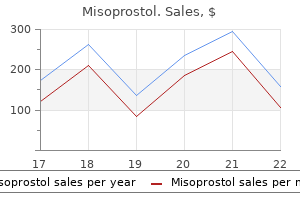
Generic misoprostol 100 mcg on line
Mueller-Hinton Agar With 2% NaCl the addition of 2% NaCl to the basic Mueller-Hinton medium results in a medium that is selective for staphylococci gastritis diet 3-1-2-1 purchase misoprostol 200 mcg. The slower growing resistant cells can be missed in a mixed population with methicillin-sensitive strains. Motility Test Medium the purpose of the motility test medium is to determine whether an organism is motile or nonmotile. This test is particularly useful to identify members of the Enterobacteriaceae, in which two genera, Shigella and Klebsiella, are always nonmotile, and certain Yersinia spp. Nonmotile organisms, which lack flagella, grow only along the stab line, and the surrounding medium remains clear. Motile organisms, which usually possess flagella, move out from the stab line, and the medium appears turbid. A low agar concentration makes the medium semisolid and permits better detection of motility. The medium is inoculated using an inoculating needle to stab the middle of the medium straight up and down once, without going all the way to the bottom of the medium. It is important to be careful and remove the inoculating needle along the initial stab line. For Yersinia, noting the motility reaction at room temperature is particularly useful. This medium will also support the growth of some mycoplasmas as well as Ureaplasma urealyticum. Vancomycin prevents the growth of gram-positive bacteria, colistin inhibits gram-negative rods, and amphotericin B prevents the growth of yeast and molds. Mueller-Hinton Agar Mueller-Hinton agar is a transparent medium useful in testing the susceptibility of organisms to antimicrobial agents. If nitrate has been reduced to nitrite, nitrite will react with these reagents to form a red diazonium dye, p-sulfobenzene-azo-naphthylamine, and the test is positive. If there is no color change, then nitrate was not reduced or the nitrate was reduced to nitrogen gas. However, if nitrate was reduced to nitrogen gas, no color change occurs and the test is interpreted as positive. The presence of gas can be detected by putting a Durham tube into the broth before incubation. Gas produced during nitrate reduction will be captured in the tube and seen as a bubble. The results should be determined immediately after the addition of the reagents because the color fades quickly. Too much zinc can result in the formation of hydrogen gas, which can cause reduction and decrease the color reaction. The medium may need to be supplemented with serum and incubated for up to 5 days in testing for Neisseria organisms. An uninoculated control tube using the reagents should be performed alongside the patient test tube to ensure glassware, reagents, and supplies are free of nitrate. The classic method for using this medium involves the stabbing of two tubes with the organism. The medium in one tube is covered with vaspar (a mixture of petrolatum and paraffin) or melted paraffin. Sterile mineral oil has been used for this purpose but is not recommended because it does not block oxygen as well. Several days of incubation may be required because of the slower growth of some nonfermenting gram-negative rods. A color change to yellow in both tubes means that the organism is fermentative. Color change to yellow in the uncovered tube only means that the organism is oxidative (requires oxygen to use the carbohydrate). If neither tube changes in color or the covered tube shows no change but the uncovered tube turns blue, the organism cannot use the carbohydrate oxidatively or fermentatively and is considered inert. Color change to yellow near the top of the medium only indicates the oxidative use of glucose. Polymyxin B and bacitracin are added to inhibit the growth of most gram-negative and gram-positive bacteria. The medium differentiates between colonies that grow based on the ability or inability of the isolate to ferment lactose. Nutrient Agar Nutrient agar has been used to distinguish between the nonfastidious, less pathogenic Neisseria spp. Nutrient agar contains minimal nutrients and an especially low concentration of protein. Growth of an isolate on this medium means that it is not fastidious and does not require special supplements. The less fastidious neisseriae grow on nutrient agar, whereas the more pathogenic species do not. These enrichments include vitamin K (required for pigment-producing Prevotella and Porphyromonas), yeast extract, hemin, and glucose. Three modifications over traditional media make this medium useful in testing nonfermenting gram-negative rods. A low concentration of peptone prevents the formation of alkaline products that might neutralize the small quantities of acid produced through oxidation. This test also can be used to distinguish phenylpyruvate-positive Moraxella organisms from other Moraxella spp. Protein hydrolysates and meat extracts are not included because these substances contain a variable amount of phenylalanine. At the end of the incubation period, a few drops of 12% FeCl3 are added to the tube so that they run down the slant. If an organism produces phenylalanine deaminase, it converts phenylalanine to the -keto acid, called phenylpyruvic acid. This acid reacts with the added ferric chloride reagent to form a dark green complex. The immediate appearance of a dark green slant on addition of ferric chloride reagent is a positive reaction; no color change on addition of reagent is a negative reaction. This medium can also be used for the differentiation of Enterococcus faecalis, which produces black colonies. Tubes of base medium can be stored and melted to make the complete medium as culture plates are needed. The Staphylococcus colonies are large, glistening, and jet black, whereas those of the gram-negative bacilli and yeasts are dull gray-black but larger than the C. Its name is derived from the original name for Mycoplasma: a pleuropneumonia-like organism. Nutrients in this medium are provided by yeast-enriched peptones, serum, and heart infusion. Agar is added to solidify the medium, but at a lower concentration than in other solid agar plates. With less agar, the medium is softer so that the mycoplasmas can grow into the medium and grow only slightly on top of the plate. Most gram-positive rods will also grow on this medium, except Bacillus anthracis, which is unique among the Bacillus spp. This component inhibits facultative gramnegative rods, especially swarming Proteus spp.
Cheap 200mcg misoprostol
Children are mostly affected by this infection gastritis symptoms baby purchase 200 mcg misoprostol, with mortalityashighas30%;mortalityinadultsismuchlower. The virus is transmitted by Aedes mosquitoes, including Aedes aegypti and Aedes albopictus. Yellow fever, caused by yellow fever virus, is also considered anemerginginfection. However, yellow fever is still epidemic in parts of Africa and South America, where about 80% of the population must be vaccinated to reduce the impact of the disease. Patients bitten by mosquitoes carrying yellow fever virus can develop an asymptomatic or acute infection involving fever, myalgia,backache,headache,anorexia,nausea,andvomiting. In the intermediate cycle,humansandmonkeysarereservoirs,whereasmosquitoes are the reservoirs and vectors in the high-morbidity, low-mortality outbreaks. Atotalof5387casesand 243 deaths were reported; 51% of all cases were described as neuroinvasive. Theremaining20%displaysymptomsofwhatis termed West Nile fever, which includes fever, headache, fatigue, occasionalrashonthetrunk,swollenlymphglands,and/oreye pain. Almost 50,000 of those cases have been laboratory confirmed and 360,000 cases are suspected to be positive. Orthomyxoviridae the influenza viruses are members of the family Orthomyxoviridae. This places the influenza viruses into three genera within the family-Influenzavirus A, Influenzavirus B, and Influenzavirus C. Inthepandemicof1918and1919,influenza killed an estimated 20 million to 50 million people, including morethan500,000intheUnitedStates. However,inthepast90 years, the world has only been able to react to the threat of influenza, rather than vanquish it. In six different years, from 1972 to 1995, influenza deaths in the United States exceeded 40,000. The surface antigens sometimes can change drastically, causing an antigenic shift, resultinginanewHorNantigen. Again,theincidence of human disease was low, but the mortality rate was high, although it is possible that some mild cases went undiagnosed. Byearly 2006,influenzaAvirus(H5N1)hadbeenisolatedfrombirdsin Turkey, Greece, Italy, Germany, Iran, Iraq, Nigeria, and many othercountries. Because many countries stopped counting individual cases, the actual number was much larger. When viruses that normally circulate in animals infect humans, they are termed variant viruses. Thevaccineusuallycontains two different strains of influenzaA virus and a single strain of influenza B virus. The best specimens are nasopharyngeal swabs, washes, or aspirates collected early in the course of the disease. Anumber of rapid kits are commercially available for the diagnosis of influenza in about 30 minutes. These agents, such as zanamivir(Relenza)andoseltamivir(Tamiflu),aremoreexpensive compared with amantadine but provide coverage against infections by both influenzaA virus and influenza B virus. Rhinitis, pharyngitis, laryngotracheitis, tracheobronchitis, bronchiolitis, and pneumonia may result. Serologic assays are more valuable for epidemiologic studies than for diagnostic purposes. Mumps virus, which has a global distribution, is spread through droplets ofinfectedsaliva. The virus infects primarily children and adolescents and usually results in long-lasting immunity. Most of the cases were reported in the Midwestern states among previously vaccinated persons 18 to 25yearsofage. Cross-reactions between soluble and viral antigens can confuse the interpretation of serologic results. Atonetime,measles(rubeola) was the most common viral disease in children in the United States. An average of 500,000 cases of measles were reported annuallyinthe1950s,withanaverageof500deaths. Thedecisiontoadminister a second dose of vaccine to school-age children has drastically reduced the incidence of measles in the United States. Afteranincubationperiodof 7to10days,thereisanabruptonset,withsymptomsofsneezing, runny nose and cough, red eyes, and rapidly rising temperature. Itisthemostcommon cause of severe lower respiratory tract disease among infants and youngchildrenworldwide. Clinical disease ranges from mild upper respiratory tract infection to acute lower respiratory tract infection and includes fever, a nonproductive cough, sore throat, wheezing, congestion,shortnessofbreath,andlethargy. Specimenscanbecollected from the nostrils by using a swab, placed in transport media, and transported on ice to the laboratory for culture or molecular analysis. The respiratory viral panel assay from LuminexMolecularDiagnostics(Toronto,Canada)claims100% sensitivity and 98. Enteroviruseshavealsobeen implicated in early-onset diabetes, cardiomyopathy, and fetal malformations. Viremia can result in the virus spreading from these locationstothespinalcord,heart,andskin. Theclinicaldisease caused by enteroviruses can be neurologic, respiratory, or cardiac, depending on viral spreading and the immune status of the host. Incontrast,neurons are not replaced, which results in neuron death and permanent paralysis. Unfortunately, interruptions in vaccine programs that began in 2003 have resulted in the reemergence of polio, with650casesseenin2011to2012. Enteroviruses have no group antigen, so they must be identified individually by a serum neutralization test. Small painful sores suddenly appear on the tongue, buccal mucosa, and soft palate. More than 150 serotypes of rhinoviruses exist, and they are the major cause of the common cold. Treating symptoms and reducing the spread of the virus in the household isthetypicalresponse. However,prolonged administration has resulted in adverse effects, such as nasal irritation, ulceration, and bleeding. The individual will enter a period of clinical latency, and even though the virus is replicating rapidly in lymphoid tissues, the virus is not detectable in the bloodstream, and the patient remains asymptomatic. Thevirusdestroysthecells(T-helper cells) critical in host immune response to infectious agents. However, in some cases, they may cause painful swelling and be more painful when found in the legs, groin, or skin around the eyes. Theearlydiagnostickits,referred to as first-generation screening tests, used purified viral lysate asantigens. Results are negative, indeterminate, or positive based on the pattern on the strip. Positive corresponds to reactivity to two or more of the following antigens: p24, gp41 or gp120/ gp160. Indeterminate corresponds to the appearance of one or more bands in a pattern that does not satisfy the positive criteria. Inhospitalexposures, a similar strategy is used if a health care worker is exposed to the virus accidentally. Aggressive therapy is initiated after the exposure, and this significantly reduces the risk of contracting theinfection. This titer is considered low for a baby, but because tests so soon after birth were positive, it suggested that the infection occurred in the womb ratherthanduringdelivery. Theinfantreceivedaggressive,threeregimen antiviral therapy, starting about 30 hours after birth, a methodnotcommonlyused. Usually, these assays are performed monthly so that therapy can be adjusted on an individual basis. Rhabdoviridae Rabies is caused by several strains of viruses belonging to the genus Lyssavirus.
Misoprostol 200 mcg
Colonies are gray-white to slightly yellow opaque gastritis not eating misoprostol 100mcg visa, raised, with a irregular "fried egg" morphology; alternatively, colonies may have a "hammered copper" shiny surface. The 9/11 Commission Report referred to it as "a day of unprecedented shock and suffering" noting that "the nation was unprepared. Plans at the national, state, and local levels have been developed to address mass casualties, displaced individuals, long-term health issues, and loss or disruption of government response. Incident command systems have been established, written protocols developed, and interoperability exercises, drills, and cross-training performed to respond more effectively using an "all hazards" preparedness model. The 2009 H1N1 influenza pandemic and the 2015 Texas Ebola case were good tests of U. Importantly, the laboratory community was allowed to fail in relative safety as H1N1, although highly communicable, was not as virulent as it could have been; and in the imported case of Ebola, which caused infection in two health care workers, the virus did not escape containment. Concluding Thoughts Regardless of the type of threat agent, release of a select agent into the general population will certainly challenge the best sentinel laboratory. Biosafety practices aside, the lack of familiarity with rare organisms to have first-hand experience with culturing, biochemical testing, and sensitivity results and techniques could result in delayed recognition of the potential risk. Additionally, a bioterrorism event would suggest large numbers of people seeking care and thus a large influx of patient specimens for analysis. This would certainly require significant resources in terms of media, reagents, and technical and clerical time, as well as security. Patient specimens are evidence in that crime and as such need to be cataloged, processed, and protected in case the evidence is to be used in a court of law. Another discussion point that we should at least mention is that of the psychological stress generated by all of the above. The clinical laboratory scientist is already on the front lines of our battles in health care. Adding the additional stress of working with potential select agents introduces another dimension for which the laboratory scientist should be prepared. As these formations break, cottonlike fragments accumulate at the bottom of the tube (so-called puffballs). Other Biological Agents and Toxins A number of other bacteria, viruses, fungi, and toxins are also select agents (see Table 30. Many of these are highly virulent or cause debilitating disease, even death, or they are less virulent but can be produced in large quantities. Salmonella, Shigella, and Escherichia species are substantial food safety threats. Similarly, Vibrio cholera and Cryptosporidium parvum, as well as various plant and microbial toxins, can be dangerous and costly water safety threats. Although large-scale delivery of many hemorrhagic fever viruses and arboviruses is less likely, their release into the general populations would certainly instill fear and panic. Forensic Microbiology A newer field of study combines forensic science and microbiology. The area of forensic microbiology involves determining the cause of death and identifying those individuals who have committed crimes. This field of study includes the clinical microbiologists who identify infectious agents, as well as public health officials and law enforcement agents. In forensic microbiology, it is not sufficient to identify the genus and species of a bacterial agent, the microbial strain or signature must be determined. The media used to cultivate the bacteria contain isotopes that will be incorporated into the bacteria as they grow. The isotope pattern might be unique to a geographic location, allowing scientists to not only determine if bacteria came from the same source but also identify the location where they were grown. Several infectious agents, including Epstein-Barr virus, cytomegalovirus, Neisseria meningitidis, Haemophilus influenzae, and Streptococcus pneumoniae, have been linked to this syndrome. Merely the detection or isolation of an infectious agent in postmortem tissue does not mean that the agent had a role in the death. It is possible that the bacteria simply colonized the patient before death and spread after death. Forensic microbiology has been investigated as way to help determine drowning as a cause of death. The diagnosis of drowning can be difficult and is often made by the exclusion of other causes. Forensic scientists have started looking for microorganisms, such as diatoms, in the tissue of suspected drowning victims. Diatoms are single-celled organisms found in freshwater, saltwater, soil, air, and food. It is believed that their presence in postmortem tissue, such as the liver, bone marrow, and kidneys, proves the hematogenous spread of the organisms from the lungs from a beating heart before death. However, testing for diatoms is labor-intensive, and diatoms have been notably absent in cases of known drownings. Because of these drawbacks, forensic scientists have recently investigated the detection of fecal bacteria in postmortem tissue. If the same bacteria are found in the tissue as those in the drowning medium, it can be concluded that the likely cause of death was drowning. The American Society for Microbiology provides information on minimal tests for the identification of potential bioterror agents for sentinel laboratories to use. The field of forensic microbiology includes the investigation of bioterror, biocrimes, unexpected deaths, and sometimes drownings. If you wanted to develop a new project to study the pathogenic effects of one of the select agents, what important information would you need to develop a thorough biological risk assessment to protect yourself from exposure What features make a biological agent a potentially ideal weapon as compared with more conventional methods Of the many different possible routes of dissemination, which method is considered the most efficacious, and what specific organisms could be used in that same manner What identifying clinical manifestations of infection with a specific organism will aid the health care provider to determine a useful differential diagnosis so as to order the proper laboratory tests What are the recommended clinical specimens to submit for patients potentially exposed to the following Many major governmental changes occurred as a result of the incidents of 9/11 and anthrax mailings. The Select Agent Program was established to safeguard against the deliberate use of select agents for potential harm to others. What positive or negative effects has this program had on the ability for an individual to perform research studies of select agents Points to Remember Bioterrorism is the use (or threatened use) of biological agents to harm humans, animals, or crops to cause civil unrest. Many of the biological agents (both bacteria and viruses) used in bioterrorarezoonotic. Many biological agents used in bioterror can also be isolated from patients with naturally occurring infections. Many infections caused by the biological terror agents produce similar nonspecific symptoms in patients; therefore, the laboratory scientist plays a key role in the diagnosis. As such, the laboratory scientist must be aware of the select agent rules, proper biosafety containment, and biosecurity and perform a biological risk assessment. Sentinel level clinical microbiology laboratory guidelines for suspected agents of bioterrorism and emerging infectious diseases: Bacillus anthracis. Sentinel level clinical microbiology laboratory guidelines for suspected agents of bioterrorism and emerging infectious diseases: botulinum toxin. Sentinel level clinical microbiology laboratory guidelines for suspected agents of bioterrorism and emerging infectious diseases: clinical laboratory bioterrorism readiness plan. Sentinel level clinical microbiology laboratory guidelines for suspected agents of bioterrorism and emerging infectious diseases: Francisella tularensis. Sentinel level clinical microbiology laboratory guidelines for suspected agents of bioterrorism and emerging infectious diseases: glanders: Burkholderia mallei and Melioidosis: Burkholderia pseudomallei.
Buy genuine misoprostol on line
Nevertheless gastritis diet ������������ buy 100mcg misoprostol, the quality of life for patients and their families is usually much better with drug treatment than without. We sometimes hear laypeople refer to psychiatric drugs with such pejorative terms. In the extreme, it accuses psychiatrists of stripping the creativity and individuality away from the individual with schizophrenia, of making them helpless to control their own destinies. Psychiatric medicine adjusts brain functioning to help patients think clearly and better control their lives. Neuroleptic drugs Neuroleptic or antipsychotic drugs are a group of medicines that have similar chemical properties. Because of their chemical make-up, they can reduce some symptoms of schizophrenia. Doctors usually prescribe them when the affected individual has active, positive symptoms such as delusions or hallucinations. At this stage, the individual is so out of touch with reality that he or she cannot correctly see that efforts are being made to help them. Neuroleptic drugs help break down the emotional and communication barriers that separate patients from their friends, relatives, and therapists. Since the 1950s, when these drugs were first introduced, worldwide studies have shown their effectiveness in treating schizophrenia symptoms. On average, two-thirds of patients show a significant improvement, and about 25% show no or little improvement. Of course, additional medicines may be added to deal with other medical problems or even other psychiatric problems such as mood problems or anxiety. Unfortunately, there is no way for the doctor to be certain that the first choice will be correct. In such cases, we urge the affected individual to not give up on medicine but to try another drug. The workings of the brain are complex, and our knowledge about schizophrenia is incomplete. Although the group of neuroleptic medications is similar to one another, where one fails, another may succeed. Thus, affected individuals and their families should be neither discouraged nor alarmed if their doctor tries a sequence of medications before relief is achieved and side effects are controlled. This is common in the treatment of schizophrenia since we have no way of knowing which specific neuroleptic will be effective for any given patient. Neuroleptic side effects Unfortunately, neuroleptic drugs bring with them the risk of side effects that range in severity from unpleasant to debilitating. Usually, these problems are avoided or controlled if the patient remains in the care of a psychiatrist. Dystonic reactions are involuntary muscle contractions, typically involving muscles of the head and face. Because facial appearance and body posture may be distorted, the patient can be embarrassed in social situations. It may be apparent in pacing, rocking from foot to foot, other motor activity, or insomnia. Ranging from mild to extremely irritating, it, like other side effects, may lead patients to stop taking their medicine. Another possibility is to treat the individual with drugs that are specifically designed to control the side effects. Affected individuals and their families should discuss these symptoms with the psychiatrist. Second, they are usually not a basis for questioning the competence of the psychiatrist. Unfortunately, medical knowledge can predict neither which patients will develop side effects nor how severe these will be. If neuroleptics are used over a long period, a neurological complication, called tardive dyskinesia, may develop. Like extrapyramidal conditions, tardive dyskinesia produces uncontrollable muscle movements, usually of the face. Individuals with tardive dyskinesia repeatedly smack their lips together, stick out their tongue, grimace, and move their chin from side to side. These are not as easily reversed as extrapyramidal symptoms, particularly in older patients. However, in some cases, the syndrome will not stop, even when neuroleptic drugs are taken away. Research suggests that about 20% of neuroleptic-treated patients will develop tardive dyskinesia. However, we have no way of knowing, prior to treatment, which patients will be affected. Thus, neuroleptic treatment must be overseen by a psychiatrist or other physician experienced and skilled in their use. Careful, periodic observations of the patient help the psychiatrist spot tardive dyskinesia in its earliest stages when it is easiest to treat. The clinical signs of the syndrome are fever, a fast heartbeat, muscle stiffness, altered consciousness, abnormal blood pressure, shortness of breath, and sweating. If the psychiatrist suspects neuroleptic malignant syndrome, a blood test will be used to see if it is present. If the affected individual has abnormal levels of specific constituents of blood, then neuroleptic malignant syndrome is likely. Because this syndrome can lead to death, the neuroleptic drug will be taken away from the patient. Low-dose neuroleptic treatment After realizing that neuroleptic side effects were frequent-and that some were severe-clinical scientists sought to develop new dosing strategies. The cornerstone of this new treatment philosophy was that, during their lifetime, patients should take only that amount of medicine that was medically necessary. These scientists quickly learned that excess usage of neuroleptics occurs when the dose needed to help a very ill individual is not reduced after the most severe and distressing symptoms have subsided. When individuals with schizophrenia are very psychotic and agitated, the doctor will usually prescribe a relatively high dose of neuroleptic. However, clinical scientists have shown that these high doses are not always needed after the initial psychosis and agitation goes away. Since these drugs have serious side effects, we cannot justify the extended use of large doses without evidence that lower doses are not effective. The affected individual and family must understand that before treatment, the doctor cannot know what dose is ideal. Owing to differences in physiology, different people may require very different doses to achieve the same clinical effect. Thus, the doctor who changes doses several times is not being erratic, but merely trying to find the optimal dose. Ideally, drug treatment should be reduced soon after the initial symptoms are relieved. We emphasize that, although maintenance treatment is very effective, it cannot guarantee that severe symptoms will not return. After 2 years, about half of individuals with schizophrenia who have been on drug maintenance treatment will relapse. This is a sobering statistic, yet it compares favourably with an 84% relapse rate in patients who have not been treated. During outpatient treatment, two strategies are available: low-dose treatment and intermittent treatment. With a low-dose strategy, patients with schizophrenia are maintained on a dose that is much lower than that initially needed. The maintenance dose of medication that will keep target symptoms at a satisfactory level is highly variable between individuals and, unfortunately, can be found only by trial and error. The intermittent medication strategy withdraws all medication during periods of remission and uses neuroleptics only when the patient appears to be at risk for relapse. This requires frequent observation of the patient by the family and by clinicians so that early signs of a pending relapse will signal a protective increase in medication. Intermittent medication is not common, as it can lead to deterioration of functioning and relapse in more than 50% of individuals with schizophrenia.
Discount misoprostol online visa
With the trichrome stain gastritis symptoms in puppies misoprostol 100mcg cheap, the cytoplasm stains dark green and the central area may stain pale to intensely green, with the nuclei staining dark purple to black. It is resistant to the usual chlorine levels in drinking water, and water has been implicated in transmission of the organism. Tissue Amebae Free-living, thermotolerant amebae can tolerate a wide range of temperatures, pH, and salinity. Although the number of infections caused by these organisms is low compared with those caused by intestinal protozoans, they are very difficult to diagnose and treat and are associated with a high mortality rate. It occurs in healthy, immunocompetent children and young adults with no predisposing condition. A common factor in infection is the report of recent swimming or other water-related activities in warm, artificial lakes or brackish or muddy water or exposure to bottom sediment. Waterskiing, wakeboarding, or other activities that increase the chances of forceful entry of water into the nose may facilitate infection. It is not known why only a few individuals are infected in spite of such common exposure. In recent years, there have been several cases related to sinus irrigation using tap water. The trophozoite enters the nasal cavity through inhalation of contaminated water or soil. The amebic form colonizes the nasal cavity, invades the nasal mucosa, attaches to olfactory nerves, penetrates the cribriform plate, moves along the olfactory nerve to the olfactory bulb, and moves into the arachnoid space. Infections are thought to be related to inhalation of a large number of organisms or forceful entry of water into the nose (microtrauma) and to the virulence of the strain. The organism multiplies in brain tissue, and within 2 to 4 days, the patient can experience drowsiness, confusion, and seizures, and progress to coma. The disease usually is fatal within 1 week of the appearance of clinical symptoms. At autopsy, there will be evidence of trophozoites along with a purulent exudate, edema, and hemorrhagic and necrotic areas of infection in the brain. The invasive properties of the organism are related to its ability to secrete cytotoxic enzymes. Macrophages and neutrophils are the primary host defense against the organism because the trophozoites are relatively resistant to the actions of host cytokines. Because the symptoms resemble those of bacterial meningitis, specific treatment may be delayed, yet the possibility of cure depends on early diagnosis. Aggressive therapy with intravenous and intrathecal administration of amphotericin B has been used. Rifampin, miconazole, or fluconazole and azithromycin have been used in addition to amphotericin B. Survival in several cases has been linked to additional procedures, including use of medically induced hypothermia and the experimental drug miltefosine. Clearing of the agar in thin tracks is evidence of the organism feeding on the bacteria. They have also been identified as the host for several pathogenic bacteria, such as Legionella spp. The condition is characterized by chronic nonhealing lesions that may present as nodules, papules, or ulcerations, especially on the extremities and the face. Lesions may develop at the site of inoculation or may occur as a result of hematogenous dissemination from the lungs. The organism penetrates the blood-brain barrier because of changes in the endothelial cell barrier caused by the interaction of the parasitic enzymes and host cytokines. Symptoms may include drowsiness, seizures, loss of reflex activity, hemiparesis, headache, stiff neck, and personality disorders. Histologic preparations of the brain at autopsy show inflammatory lesions containing many segmented neutrophils, eosinophils, and trophozoites. A specific therapeutic regimen has not been established because most infections have been diagnosed at autopsy. However, disseminated Acanthamoeba infections have been treated with amphotericin B, pentamidine isethionate, fluconazole, ketoconazole, and co-trimoxazole (trimethoprim-sulfamethoxazole). Individuals who wear contact lenses, especially the soft and extended-wear types, are the primary at-risk group, with greater than 80% of the cases identified. Factors in these infections include improper storage and disinfection procedures, a history of corneal trauma, or wearing contact lenses during swimming, all of which may cause corneal trauma and subsequent colonization by the organism. The organism binds directly to corneal epithelium via acanthapodia and produces proteases and other enzymes that cause cell lysis. Patients experience photophobia, blurred vision, inflammation, ring infiltrates, and pain. Because of the similarity in tissue damage, the infection may initially be confused with bacterial or herpes simplex virus infection, delaying treatment. Phase-contrast microscopy of direct wet mount preparations of corneal scrapings can reveal the trophozoite or cyst. Permanent stains, such as trichrome and Giemsa stains, may also demonstrate the trophozoite in clinical specimens. Blunt pseudopods and characteristic spinelike projections of the cytoplasm (acanthopodia) may also be seen on a wet mount. Topical applications of chlorhexidine gluconate and ketoconazole have been used to treat cutaneous infections. Despite treatment, patients with systemic infections have a poor prognosis, and many patients with keratitis lose their sight in the affected eye. Unlike Acanthamoeba, which has a wide distribution, this organism is found primarily in soil. Since 1990, when it was first linked to human illness, this free-living ameba has caused more than 200 cases, with a mortality of more than 95%. It is unknown if there is a genetic predisposition or if the type of work in which the individuals engage increases exposure risk. Humans become infected by inhaling airborne cysts of the organism or by direct inoculation through skin lesions. Once the encephalotropic organism has entered the body through the lungs or skin, it spreads hematogenously. Only rarely will it invade the body nasally and spread along nerve fibers to the olfactory bulb. Once the organism reaches the blood-brain barrier, it is able to bind to microvascular endothelial cells through receptor molecules. The onset is insidious, with fever, headache, stiff neck, vomiting, and photophobia, and progresses to personality changes and seizures. Onset of symptoms can occur weeks to months after infection, but when the brain is affected, the time to death is short. Treatment involves a multiple antimicrobial regimen, including fluconazole, clarithromycin, and sulfadiazine. In most cases, infection is identified at autopsy by finding trophozoites and cysts in the tissue. Serum antibodies can be detected by indirect fluorescent antibody methods using ameba-coated slides. The culture methods using nonnutrient agar seeded with Escherichia coli are nonproductive because this organism will not feed on gram-negative bacteria. Pigs are the natural host for this organism, and humans serve as accidental hosts.
Generic misoprostol 200 mcg otc
Middlebrook 7H10 and 7H11 are similar gastritis and diarrhea trusted 200mcg misoprostol, except 7H11 contains casein hydrolysate, which stimulates the growth of drug-resistant Mycobacterium tuberculosis. Both media contain growth factors, such as amino acids, glycerol, and inorganic salts that encourage recovery of mycobacteria. Albumin is added to inhibit toxic agents that might be present and to provide a source of protein. Thin-pour Middlebrook 7H11 plates are commercially available that have a reduced volume of medium in the plate. These thin-pour plates are used to detect mycobacterial colonies faster than on standard-pour plates. The thin-pour plate is inoculated with a specimen and examined microscopically every 2 days for the appearance of microcolonies. Mitchison 7H11 Selective Agar Mitchison 7H11 selective agar is prepared by adding antimicrobial agents to the Middlebrook 7H11 formulation, thereby making the medium more selective for mycobacteria. Amphotericin B, carbenicillin, polymyxin B, and trimethoprim are typically incorporated to make Mitchison 7H11 selective agar more inhibitory to gramnegative rods in particular, as well as yeast. Members of Enterobacteriaceae can be divided into two groups based on how they metabolize glucose. One group produces large amounts of mixed acids (lactic, formic, succinic, and acetic). When methyl red is added to one of these cultures, a red color is produced because of the acidic pH. The other group produces predominantly neutral end products, acetoin or acetylmethylcarbinol, by the butylene glycol pathway. Cornstarch also is included to absorb inhibitory substances that might be present. Both media contain vancomycin to inhibit the growth of gram-positive bacteria, colistin to inhibit gram-negative rods, and nystatin to prevent the growth of fungi. Because MuellerHinton agar contains animal infusion, casein extract, and starch, it supports the growth of most organisms. In addition, sheep blood may be added to the basic formulation to perform susceptibility testing on streptococci, in particular Streptococcus pneumoniae. The addition of heated or chocolatized sheep red blood cells to Mueller-Hinton agar makes an enriched medium that can be used for susceptibility testing of fastidious organisms, such as Haemophilus and Neisseria. Muller-Hinton agar also can be used for the X and V Factor test for Haemophilus spp. Starch is included in the medium for two reasons-it may protect the organisms against toxic substances and it can also serve as an energy source for some bacteria. Ca2+ and Mg2+ concentrations are critical in the testing of Pseudomonas isolates with aminoglycoside antibiotics. Usually, Mueller-Hinton agar contains sufficient amounts of bivalent cations, but it may be necessary to add these substances to Mueller-Hinton broth. Phenylethyl alcohol is volatile, and plates should be tightly sealed in plastic bags and stored in the refrigerator. Hemolytic reactions are not dependable on this medium because of the action of phenylethyl alcohol on cell membranes. The anaerobic formulation of this medium selects for gram-negative and gram-positive nonsporulating obligate anaerobes while inhibiting the facultatively anaerobic gram-negative rods and other anaerobes. Regan-Lowe Medium Regan-Lowe medium is enriched and selective for the isolation of Bordetella pertussis and Bordetella parapertussis from clinical specimens. The nutritional base is comprised of beef extracts, horse blood, niacin, and pancreatic digests. Charcoal and starch are added to neutralize the inhibitors, especially fatty acids and peroxides that might be present in the medium. The transport medium differs from the isolation medium in that the transport medium uses lysed horse blood, whereas whole horse blood is used in the isolation medium. In addition, the transport medium contains half as much charcoal as the isolation medium. Potassium Tellurite Blood Agar Tellurite blood agar is a selective, differential, enrichment agar useful in isolating Corynebacterium diphtheriae. Some formulations also incorporate cystine to enhance the growth of fastidious organisms further, including C. Potassium tellurite is the selective and differential ingredient responsible for inhibiting the growth of gram-negative organisms, staphylococci, and streptococci while allowing the growth of C. Lactose is the sole carbohydrate source in the medium, and neutral red is the pH indicator. If an organism grows on the medium and ferments lactose, it will produce acid and change the indicator to pink-red. If H2S is produced, it reacts with the ferric ammonium citrate present in the medium, forming a black precipitate in the center of the colony. Salmonella colonies are colorless, with a black center, because these organisms usually make H2S but do not ferment lactose. Pink to red colonies indicate that the organism ferments lactose; if there is a black center, it also produces H2S. In addition, viridans streptococci can be distinguished from Enterococcus (which may sometimes appear -hemolytic) because the viridans streptococci, like group D streptococci, cannot grow in this medium. Sodium chloride broth is prepared from heart infusion broth, a general purpose medium that already contains 0. Sodium chloride broth also contains glucose as a carbohydrate source, and some formulations add bromocresol purple, a pH indicator. If the organism can tolerate this high concentration of salt, it will grow in the medium and produce turbidity. Fermentation of glucose produces acid and can cause the medium to turn from purple to yellow if the pH indicator is present. Any growth in the broth is considered positive, even if the indicator does not change color. To avoid a false-negative result, the broth should be gently mixed before interpretation. Organisms other than enterococci, such as group B streptococci and aerococci, can produce positive results. Schaedler Agar Schaedler agar is an enriched medium used for the isolation of anaerobic bacteria. The growth of more fastidious anaerobes is aided by the addition of vitamin K, sheep blood, and hemin. Facultative anaerobes also will grow on this medium, so aerotolerance testing should be performed on all isolated colonies to determine their oxygen dependency. Fetal bovine serum supplies the cholesterol necessary for the synthesis of sterols for the bacterial membranes, stabilizing these organisms because they lack cell walls. Penicillin is included to prevent the growth of gram-positive bacteria, amphotericin B inhibits fungi, and polymyxin B inhibits gram-negative rods. Biphasic media provide microaerophilic and moist conditions, which some Mycoplasma spp. Selenite Broth Selenite broth is an enrichment broth used for the recovery of low numbers of Salmonella and some strains of Shigella from stool and other specimens containing large amounts of mixed bacteria. The sodium selenite present in this medium inhibits the growth of many gram-negative rods and enterococci but permits the recovery of Salmonella and some Shigella species. Reduction of selenite during bacterial growth produces alkaline products that may inhibit the growth of the salmonellae and also reduce the toxicity of the selenite for other organisms, so lactose and phosphate buffers have been included in this medium to maintain a neutral pH. Lactose fermenters produce acid, which neutralizes these alkaline products and returns the medium to a neutral pH. The broth should be subcultured to enteric media after it has incubated for 12 to 18 hours (some references suggest 6 to 12 hours).
Buy misoprostol 200 mcg low price
The disease in humans is usually mild and selflimiting gastritis diet ��� discount 100 mcg misoprostol fast delivery, lasting anywhere from 6 to 12 weeks in untreated patients. Lymphadenopathy occurs at the lymph nodes involved in the draining of the infected area 1 to 2 weeks after appearance of the lesion. Patients may also experience flulike symptoms, including fever, malaise, and anorexia. One strategy for increasing access is by fostering a relationship with the health care provider, particularly the infectious disease staff. Such efforts are encouraged because they provide communication and increase the likelihood that laboratorians will have access to relevant patient information. Transmission by Direct Contact or Inhalation Anthrax Case Study A 57-year-old male patient, employed as an electrician, had felt ill and feverish before reporting to the hospital and had collapsed at home when he tried to stand. He had been bitten by an insect on the upper left chest the previous day while at work. Some species that infect humans (with vectors and/or animal hosts listed in parentheses) are B. The initial diagnosis was proven wrong when the blood culture samples taken at the time of admission became positive for Bacillus anthracis. Fortunately, the antimicrobials prescribed were appropriate, and the patient made a full recovery. Misdiagnosis occurred in this case largely because of the rarity of this disease and incomplete patient history. However, it was later determined that the patient had recently worked with untreated imported hides, which, coupled with the necrotic ulcer, would have increased the index of suspicion for anthrax. This organism occurs naturally in the soil and is a pathogen of herbivores, such as cattle, sheep, and goats. Human infections occur as the result of direct or indirect contact with animals or animal products. Floods tend to concentrate spores, which remain in the grasses after flood waters drain. Anthrax occurs worldwide, commonly in agricultural regions, such as Central and South America, Africa, and the Middle East. Recurrences of anthrax are prevented by containing the spores and eliminating their spread through the environment. Because the anthrax bacteria in tissues do not sporulate until they are exposed to oxygen, infected animal carcasses are usually incinerated whole or are buried in deep pits. Application of lye or quicklime, a practice that has been used to speed up the decay of carcasses, is no longer recommended because calcium ions are now believed to prolong anthrax spore survival. Clinical Manifestations Human anthrax manifests itself as the cutaneous, intestinal, or pulmonary form. The most common form of anthrax is the cutaneous form (95% of cases), which mimics many other cutaneous infections. After 48 to 72 hours, a papule is formed; it darkens and ruptures, leaving a painless, craterlike ulcer, which progresses to a necrotic eschar. The infection usually remains localized and self-limiting, and the eschar heals without scarring. In about 20% of patients with cutaneous anthrax, the immune system is unable to contain the infection, and B. The gastrointestinal form, the second most common form of anthrax, is also found more frequently in nonindustrialized nations. Once the spores are ingested, they germinate, and the organisms gain entry through preexisting intestinal mucosa lesions. Clinically, the patient will be febrile and have bloody stools and can lose as much as 12 L of fluid per 24-hour period. The pulmonary form is caused by inhaling spores, usually from contaminated animal products. This form of infection usually results in the death of the patient, which, in the absence of treatment, can occur within 24 hours. Administration of antibiotic therapy and supportive care may reduce the fatality rate, but this treatment appears to be effective only when initiated prior to the onset of respiratory symptoms. Necrosis occurs as the result of increased capillary permeability and destruction of the phagocytic cells. Edema can be remarkable and may cause the patient to suffocate by literally swelling the neck shut. Excessive edema of the neck, thorax, and mediastinum signals the beginning of a rapidly fatal course. Treatment options include orally administered doxycycline, quinolones, penicillin, raxibacumab, obiltoxaximab, and human anthrax immune globulin (Anthrasil) (Table 40. Doxycycline and ciprofloxacin may be used in patients who have been exposed to anthrax but have not developed symptoms. He continued to experience pain, general malaise, fever, and vomiting and was admitted to the hospital. Because this was a possible abscess, the physician performed an incision and drainage, but no abscess was found. The patient was treated with intravenously administered penicillin and cloxacillin. Five days after admission, the patient developed shortness of breath and had patchy pneumonic infiltrates of the middle and lower lobes of the right lung. Seven days after admission, lymphangitic streaking on the upper left extremity and tender axillary lymphadenopathy were detected. A swab from the wound was plated onto sheep blood agar and chocolate agar plates and incubated in carbon dioxide. Gram-negative coccobacilli were identified as Francisella tularensis through fatty acid analysis and slide agglutination by using specific antisera. Acute-phase and convalescent-phase serum samples had titers of 1: 800 and 1: 3200, respectively. Types A and B differ from each other biochemically and genetically but not serologically. Clinical Manifestations Tularemia is an acute, febrile, granulomatous disease characterized by rapid onset and flulike symptoms. The most common presentations are ulceroglandular (ulcers and lymphadenopathy), oropharyngeal (pharyngeal ulcer and lymphadenopathy), oculoglandular (conjunctival ulcer and lymphadenopathy), glandular (lymphadenopathy without ulcer), pleuropulmonary (no ulcer, possible lymphadenopathy), and typhoidal (no ulcers or lymphadenopathy). The ulceroglandular, oculoglandular, and glandular types usually occur as the result of direct contact with infected vertebrate or invertebrate reservoirs. Typhoidal and oropharyngeal cases of tularemia usually occur after eating contaminated food. The symptoms associated with tularemia include fever, chills, headache, and myalgia. Typhoidal tularemia, especially when complicated by pneumonia, has a high fatality rate. These infections are usually caused by direct contact with contaminated game or by insect bites. The actual site of the lesion is indicative of the transmission; lesions of the hands or arms often indicate infection by direct contact with infected mammals, whereas lesions on the head or back are suggestive of insect vector transmission. The first isolation of the bacterium was in 1911 during an epizootic outbreak of plaguelike ground squirrel disease. The organism was named Bacterium tularense after Tulare County, California, which was the site of the outbreak. In 1944, researchers defined the role of cottontails and jackrabbits in the transmission of the disease to humans. Since 1945, the proportion of vectorborne (Dermacentor andersoni, Dermacentor variabilis, and Amblyomma americanum) transmission has increased, whereas transmission from vertebrate reservoirs has decreased. In 1959 the genus name of the organism was changed to Francisella in honor of Edward Francis, who first isolated the organism. In North America, the predominant biovar is type A, the strain that is more virulent in humans.
Purchase misoprostol online
Neither should family members stop drug treatment out of fear that their relative will become addicted gastritis test discount misoprostol online visa. Professional help is also needed if ever the individual with schizophrenia talks about committing suicide. The affected individual then needs immediate attention because the risk of suicide among those with schizophrenia is high; any sign of attempted suicide requires emergency care. The close relatives of individuals with schizophrenia may also sometimes feel that life is not worth living. Such pessimistic feelings may affect the capacity to cope with daily routines and work or produce physical symptoms such as loss of weight, sleeplessness, loss of appetite, gastrointestinal upset, and dizziness. Thus, relatives with suicidal thoughts should seek immediate professional attention too. At times, caregivers need their own therapy to help them cope with their affected loved one. The maintenance of a social support network is essential In this article, we have discussed some of the ways in which individuals with schizophrenia and their families can help themselves. We could have mentioned only the positive aspects but felt compelled to discuss difficulties that are likely to arise in order to encourage a realistic approach to schizophrenia and its many challenges. The accumulation of knowledge about schizophrenia is making such an approach increasingly possible. With tolerance and understanding, many individuals with schizophrenia and families successfully cope with living with the disorder. As long as affected individuals, with the support of sympathetic and understanding caregivers, can find the narrow path between over- and understimulation, they can successfully avoid recurrence of positive or negative symptoms. This information should be used in conjunction with advice from health care professionals. Formation of a theca layer and an antrum transition follicles to the antral stage. As puberty approaches, the ovary acquires the full complement of different follicle types at all stages-primordial, growing (preantral and antral), and atretic. The time a follicle takes to develop from the primordial stage to ovulation is up to 12 months in women and approximately 3 and 4 weeks in rodents. The largest portion of that time is taken up by preantral folliculogenesis, whereas the later antral stages that undergo cyclical maturation in waves take only days and are regulated by the hormonal profiles and intrafollicular milieu of growth factors during the follicular phases of the menstrual or estrous cycle. Details of many of the molecular pathways in the oocyte, those responsible for the differentiation and proliferation of the different somatic cell types that make up growing follicles, and how these underpin the interactions between the oocyte and its somatic cell niche, are now understood and are reviewed below. Furthermore, the influence of the oocyte specific factors may underlie the difference between single and multiple ovulations. Selection of Ovarian Follicles the focus of this chapter is the selection of follicles during all stages of folliculogenesis. The primordial follicle pool or ovarian reserve decreases with age for two reasons: the primordial follicles either die mostly by atresia or are selected for growth and enter the pool of growing follicles, throughout which most die, but a select few are ovulated and gain the chance to be fertilized and form a new organism. The oocyte also influences the selection processes by providing checkpoints during folliculogenesis, particularly after antrum formation. In addition to the important intrafollicular role played by the somatic cells of the follicle to nurture and interact with the oocyte, these cells mainly in antral follicles are also essential for production of the sex steroid hormones (estradiol-17, progesterone), which are responsible for the secondary sex characteristics of the female and to prepare the uterus for pregnancy, and provide substrate (androgens) for the production of estradiol. Selection for Death Shortly after the ovarian reserve is established, a large number of primordial follicles are eliminated from the ovary as part of normal, developmentally regulated processes. Given that approximately half this number was assumed to have been activated, it was concluded that death of primordial follicles contributes to greater depletion of the ovarian reserve than activation during the early stages of life [3]. There are a number of possible explanations for the elimination of primordial follicles in the days immediately following their formation. A passive trigger for primordial follicle death may be inadequate growth factor support [5]. Interestingly, the onset of puberty has also recently been identified as a critical developmental window during which primordial follicles are lost in large numbers. Why such a large cohort of primordial follicles are selected for death is not known, but one hypothesis is that a quality control checkpoint exists at puberty to ensure that only those follicles of the highest quality are available for adult reproduction. In this instance, primordial follicles are actively selected for elimination by apoptosis because they have sustained damage that could potentially compromise their ability to give rise to healthy offspring after fertilization [10]. Because of this, the number of primordial follicles initially formed in the ovary, and the rate at which they are activated to begin further development, or triggered to die, greatly influences the female fertile life span. Of note is the observation that in all mammalian species analyzed, there are many more primordial follicles than required for ovulation. For example, in humans 1 million primordial follicles are present at birth and this number is reduced to around 300,000 at puberty, with approximately 1000 remaining when the menopause begins [1,2]. These processes are critical to protect against the introduction of germline mutations. Because the commitment to further development is unidirectional, the rate of primordial follicle activation must be tightly controlled to ensure that sufficient reserves remain in order to support fertility throughout reproductive life [13]. In recent years, significant progress has been made toward characterizing the molecular pathways that govern the transition of follicles from a state of dormancy to growth. Despite an improved understanding of the complex signaling networks responsible for mediating the activation process, the question remains, "What triggers the pathways that result in activation in some follicles but not others In addition to the requirement for activators, primordial follicles are retained in a quiescent state by locally produced inhibitory factors. Primordial follicles were less likely to grow when a neighboring primordial follicle was in close proximity [29], suggesting that, similar to growing follicles, primordial follicles secrete a diffusible factor that prevents nearby primordial follicles from activating. Such an inhibitory molecule would be particularly important for controlling the rate of follicle activation prior to the first wave of folliculogenesis, when growing antral follicles are not yet present. Thus, in addition to the role of specific growth/inhibitory factors, the spatial relationships between follicles, potentially creating morphogen gradients in the ovary, may play an important role in the selection of individual primordial follicles for activation. In 1968, Henderson and Edwards proposed the production-line hypothesis to explain the differential selection of follicles for activation and increased trisomy rate in the offspring of older women [30]. This theory posits that the first oocytes to enter the meiotic prophase during the initial establishment of the ovarian reserve are also the first selected to activate and subsequently ovulate. Secondly, it states that those oocytes that enter meiosis first also have the highest crossover frequency, and thus are less prone to meiotic nondisjunction, thereby implying that "meiotic quality" is a selection factor for activation. This hypothesis was formulated on the basis of studies showing that chiasmata frequency is associated with the timing of meiotic entry in mice. However, a subsequent study in humans could not find any evidence for decreased crossover frequency between oocytes entering meiosis late in fetal development compared to those entering meiosis at earlier time point [31]. Even though the timing of meiotic entry did not correlate with chiasmata number in oocytes, this study does not rule out the possibility that in humans primordial follicles containing oocytes with high crossover numbers activate first, such that in women of advanced maternal age only oocytes with few chiasmata remain. The most simplistic interpretation of these data is that highest quality primordial follicles are selected to activate first and when the supply of healthy primordial follicles becomes reduced, regardless of age, the availability of high-quality oocytes is also reduced. The relationship between the location of follicles in the ovary and the order of activation has also received considerable attention. In separate studies, Hirshfield and Byskov reported that the first follicles to activate after the ovarian reserve is established are those located in the central medulla region of the ovary [33,34]. Recent works extend these earlier observations and propose that there may be two pools of primordial that are distinct in the timing of their formation, their location within the ovary, and their functional roles [35,36]. During endowment of the fetal mouse ovary, the primordial follicles that assemble first are located in the central region of the ovary and these are also the first cohort to activate. Lineage tracing studies in mice showed that most of the primordial follicles making up this first group are used before postnatal day 60 and thus primarily contribute to the establishment of sexual maturity and endocrine cyclicity during early life [35,36]. The second population of primordial follicles is formed in the ovary slightly later, is located in the cortex, and is primarily responsible for supplying follicles to support adult fertility [35,36]. Furthermore, Anderson and colleagues reported the presence of unhealthy primordial follicles, characterized by abnormally large oocytes, in the ovarian cortex of prepubertal girls, which could not be detected in the adult [38].
