

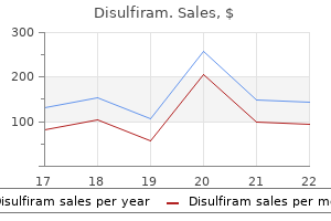
250 mg disulfiram with amex
In cases of methotrexate contraindications (or early intolerance) treatment 24 seven purchase disulfiram 250 mg fast delivery, lelunomide or sulfasalazine may be considered as alternatives to methotrexate as part of the initial treatment strategy. Both sulfasalazine and lelunomide have shown clinical, functional and structural eficacy, and although methotrexate doses in respective comparative trials may not have been optimal, both have shown eficacies similar to methotrexate (Smolen et al. Studies have shown it to be at least as effective as sulfasalazine and methotrexate, and for quality-of-life measures some evidence suggests superiority. However, many centres do not use this loading dose regimen, because patients are unable to tolerate the gastro-intestinal side effects associated with it. The use of lelunomide has been associated with both haematological and hepatotoxic side effects. When lelunomide and methotrexate are used in combination extra caution is advised, and patients should be monitored closely because of the increased risk of hepatotoxicity. Lelunomide can also cause hypertension: blood pressure should be checked before commencing lelunomide and periodically thereafter. Both men and women who are taking lelunomide are required to use adequate contraceptive measures during treatment and for at least 2 years after treatment in women and at least 3 months after treatment in men. If the individual plans to conceive before completion of this period, then a washout protocol must be followed. This involves treatment with cholestyramine or activated charcoal and measuring the concentration of the active metabolite. To reduce the side effect of nausea, the dose is usually titrated upwards from 500 mg daily, increasing at weekly intervals to 1 g twice daily. Haematological abnormalities have occurred rarely with the use of sulfasalazine, and patients should be counselled to report unexplained bleeding, bruising, purpura, sore throat, fever or malaise. Patients should also be warned that sulfasalazine can colour urine red and stain contact lenses. Sulfasalazine is considered safe in pregnancy when given in combination with folic acid. The antimalarial drugs hydroxychloroquine and chloroquine may be used in milder disease. There is a rare risk of retinopathy associated with their use, so patients should have an annual eye check and be informed to report any changes in vision or how they perceive colours. Due to the risk of anaphylaxis, an initial 10 mg test dose should be given in the irst week of treatment followed by a maintenance dose of 50 mg the following week. The maintenance dose of 50 mg every week is given until the irst signs of remission occur. At this point the interval between injections is extended to 2 weeks until full remission occurs. The interval between injections can then be increased progressively to 3 weeks, 4 weeks, and then, after 18 months to 2 years, to 6 weeks. Glucocorticoids Glucocorticoids act by inhibiting cytokine release and give rapid relief of symptoms and decrease inlammation. Depending on the number of affected joints, corticosteroids may be injected directly into the joint (intra-articular), given as an intramuscular depot injection or given as short-term oral therapy. The standard intramuscular dose used is 120 mg methylprednisolone, and oral starting doses can be up to 30 mg once a day of prednisolone. For very severe disease, particularly where there are extra-articular manifestations, pulsed intravenous infusions of methylprednisolone. Treatment with glucocorticoids should ideally be short-term, and they should be gradually withdrawn as soon as it is clinically feasible according to disease activity. Ideally treatment should be gradually reduced and stopped by 3 months (Smolen et al. The long-term side effects of corticosteroids include osteoporosis, peptic ulceration, diabetes mellitus and hypertension. Oral glucocorticoid treatment with greater than 5 mg prednisolone daily can lead to a reduction in bone mineral density and a rapid dose-dependent increase in the risk of fracture (van Staa, 2006). In patients exposed to high-dose and/or long-term corticosteroid, consideration should be given to the need for bone protection with a bisphosphonate and calcium plus vitamin D supplementation. The importance of not stopping glucocorticoid therapy suddenly should be explained to the patient. Agents such as ibuprofen and celecoxib have a lower relative risk of gastro-intestinal complications, while piroxicam and ketoprofen have been shown to have a higher relative risk (Castellsague et al. They are derived from living cells through highly complex manufacturing processes. Monoclonal antibodies may also be fully humanised (fully humanised monoclonal antibody) or partially humanised, where they consist of a combination of both animal and human proteins (chimeric monoclonal antibody). As patents for biologic medicines near their expiry, pharmaceutical manufacturers have taken steps to develop medicines with equivalent therapeutic effect to their branded counterparts, but with a considerably lower acquisition cost. There should be no clinically signiicant difference between the biosimilar and the original reference product. Biosimilar medicines are also in clinical development for other biologics including adalimumab and rituximab. It is important that all prescriptions for a biologic medicine available as both the original reference drug and as a biosimilar speciically state the brand name of the desired product to avoid confusion. Due to the complex nature of these medicines there is a risk that small differences between the products could potentially affect response. At the time of writing this chapter, three biosimilar inliximab products plus two etanercept biosimilar are available (Table 54. Due to it being a fully humanised monoclonal antibody, it is less likely to engender an immune response in the recipient than agents that contain non-human or artiicial sequences. It can be used as monotherapy in patients who are intolerant of methotrexate, or where methotrexate is contraindicated. When given as monotherapy, the dose may be increased to 40 mg once a week where there is a decrease in response. The formulation was developed to address concerns that some of the toxicity associated with inliximab and adalimumab might be caused by the effects of the Fc portion of the antibody such as complement activation and antibody-dependent cell-mediated cytotoxicity. Certolizumab pegol can be given as monotherapy in case of intolerance to methotrexate or when continued treatment with methotrexate is inappropriate. Full clinical response to certolizumab pegol should be achieved within the irst 12 weeks of treatment. If the desired therapeutic response has not been achieved at this point, then treatment with certolizumab pegol should be discontinued. It is given by subcutaneous injection, either 25 mg twice weekly or 50 mg once weekly. The patent for the original reference drug (Enbrel) has now expired, and etanercept is now available as a biosimilar (Benepali and Erelzi). The starting dosage is 50 mg by subcutaneous injection once a month; patients who weigh more than 100 kg should have their dose increased to 100 mg/month if they have not achieved an adequate response after three or four doses. If an adequate response is not achieved following administration of three or four doses of 100 mg, treatment should be discontinued. The initial infusion is given over a 2-hour period; subsequent infusions are given 2 and 6 weeks after the irst. Blood tests are now available for determination of inliximab plasma blood levels; a drop in the plasma level may be indicative of the development of antibodies to inliximab. Due to the availability of biosimilar inliximab, all prescriptions for inliximab should include the brand name of the product to avoid inadvertently switching to an alternative product. Patients should be continued to be monitored for the development of mycobacterial infections during treatment and for 6 months after stopping. Therapy should be stopped if signs or symptoms suggestive of demyelination develop. Use is recommended in combination with methotrexate, but monotherapy can be considered where there is intolerance or a contraindication to methotrexate. It is available as both the original reference drug and as biosimilar formulations. It is available as the original reference drug MabThera and also as the biosimilar brands Rixathon and Truxima.
Diseases
- Pseudo-torch syndrome
- Mucopolysaccharidosis type VII Sly syndrome
- Neurofibromatosis type 3
- Cholestasis, progressive familial intrahepatic 2
- Albinism deafness syndrome
- Asthma
- Dennis Cohen syndrome
- Mycosis fungoides
Buy generic disulfiram line
Biomarkers for the prediction of acute kidney injury: a narrative review on current status and future challenges treatment yersinia pestis order genuine disulfiram line. A new simple and rapid method to monitor the renal function based on pharmacokinetic consideration of endogeneous creatinine=. Evaluation of estimated creatinine clearance before steady state in acute kidney injury by creatinine kinetics. Plasma neutrophil gelatinase-associated lipocalin predicts recovery from acute kidney injury following community-acquired pneumonia. Urinary biomarkers and renal recovery in critically ill patients with renal support. Urinary Neutrophil Gelatinase-associated Lipocalin as a Marker for Identification of Acute Kidney Injury and Recovery in Dogs with Gentamicininduced Nephrotoxicity. Eadon refined further through the concomitant use of either ultrasound or computed tomography guidance as well as the use of a spring-loaded needle. Discuss the indications and contraindications of a renal biopsy in critically ill patients. Understand the difference between performing a renal biopsy as an outpatient as opposed to the critical care setting. Address the risks of a renal biopsy, including their prevalence, practical strategies for prevention, and clinical management of complications. Evaluate the benefits of performing a renal biopsy in the critical care setting and its impact on management strategies. A kidney biopsy should be considered in any patient with reduced kidney function and either unexplained microscopic hematuria or significant proteinuria. Certain common sense prerequisites for a kidney biopsy are applicable to inpatients and outpatients. The differential diagnosis includes diseases associated with significant morbidity, mortality, or a decrement in quality of life. The differential diagnoses have divergent therapeutic strategies that may be entertained only after a tissue diagnosis. This article focuses on the indications, complications, and periprocedural management of nontargeted native kidney biopsies, with a special emphasis on considerations relevant to the critically ill patient. Targeted kidney biopsies for renal masses and protocol kidney transplant biopsies are beyond the scope of this discussion. In the critical care setting, acute kidney injury is a common phenomenon1; however, the cause is often multifactorial or uncertain. The decision to biopsy critically ill patients portends added complexity as compared to commonly performed outpatient renal biopsies. This article outlines valid indications and contraindications, as well as providing practical suggestions to optimize timing, biopsy modality selection, and prevention of complications for patients at higher risk. The presence of proteinuria and hematuria is associated with mortality in critically ill patients independent of other baseline conditions. The evaluation of hematuria frequently is confounded by nonglomerular hematuria, because many patients have Foley catheters, urinary tract infections, or urologic comorbidities. The biopsy provides valuable information about disease activity and chronicity (inflammation, fibrosis) within the kidney or may prove useful in the absence of reduced renal function, because the pathologic information can guide therapy for other involved organ systems. Absolute and relative contraindications to a kidney biopsy are detailed in Table 31. The perception that these patients have increased procedural risk and treatmentassociated adverse events (related to immunosuppression) is thought to outweigh the clinical benefit in this population. Although minimal prospective data are available, one series of renal biopsies in pregnant women revealed that complication rates were low and not significantly different than the general population. The procedure usually is performed by trained nephrologists or interventional radiologists. The technique for native and transplant kidney biopsy is similar except for positioning of the patient. An ultrasound-guided kidney biopsy is performed under local anesthesia with a disposable, automatic, spring-loaded device using 14-, 16-, or 18-gauge needles (outer diameter of 2. The criterion for adequate biopsy tissue typically is considered to be 15 to 20 glomeruli. Adequate tissue is obtained in 95% to 99% of biopsies performed using 14- or 16-gauge needles. Each of these methodologies is associated with specific risks but hold particular merits depending on the clinical scenario (Table 31. In the presence of a solitary kidney, there is concern that a percutaneous biopsy may result in marked bleeding, which in turn may require a nephrectomy and render the patient anephric. Selected case series have revealed this 169 risk may be less than the risk of general anesthesia. Other nonpercutaneous techniques, such as the open or laparoscopic approaches, are preferred. In intensive care patients on mechanical ventilation, the traditional modality of choice for native kidney biopsies is an open surgical approach. It is possible to perform a percutaneous renal biopsy with portable ultrasonographic guidance by placing the patient prone and ventilating manually with a self-inflating (Ambu) bag. The complication rate was similar to the open biopsies performed during the same period. Bleeding Complications in Percutaneous Kidney Biopsies the most common complications of a percutaneous kidney biopsy are bleeding, arteriovenous fistula, and infection. Bleeding is the most common and clinically relevant complication of a kidney biopsy. It is also important to be aware of the effects of postural changes on hemoglobin levels, which are observed commonly after a variety of procedures requiring bed rest. A systematic review and meta-analysis of all adult percutaneous renal biopsy studies from 1980 to 2011 (34 studies with 9474 biopsies meeting inclusion criteria) was done by Corapi et al. Bleeding complications of native kidney biopsy: a systematic review and meta-analysis. The absence of a hematoma was a more useful predictor, because the negative predictive value for developing a complication was 95%. Alternatively, the use of a heparin bridge in the peribiopsy period may be used in patients who cannot safely remain off anticoagulation for multiple days. Platelet transfusions should be given for any patient with a platelet count less than 50,000/mm3. A retrospective single center study of 317 percutaneous kidney biopsies with an immediate postbiopsy ultrasound found that 86% of patients had a detectable hematoma, but only 13% had a hematoma larger than 2 cm. The size of the hematoma did not predict the complication rate, although there was a trend toward association with a hematoma size greater than 3 cm (55% vs. Continuation of antiplatelet agents was associated with a greater absolute decrease in hemoglobin as well as the percentage of patients with a >1 g/dL drop. However, no difference in major complications (requirement for transfusion or radiologic or surgical intervention) was observed between patients undergoing elective (1. Our practice is to hold antiplatelet agents for a minimum of 3 days in the inpatient setting. However, if the procedure cannot be delayed and antiplatelet agents have been administered recently, a transjugular approach should be used. Chapter 31 / Practical Considerations of Renal Biopsies in Critical Care Patients 171 Desmopressin the use of a desmopressin acetate infusion (0. However, desmopressin administration did not result in fewer transfusions or interventions in these patients. Consider a preprocedural platelet function assay or bleeding time if available at your institution. Most anticoagulants or antiplatelet agents are held for 72 hours after a renal biopsy, unless a compelling cardiac or vascular indication requires earlier reinitiation. Arrangements should be made for intravenous heparin infusions, platelet transfusions, fresh frozen plasma transfusions, and desmopressin administration as required.

Buy 250 mg disulfiram with visa
No statistically significant differences were found symptoms exhaustion generic disulfiram 500 mg visa, and no causal relationship with febuxostat was established. Identified risk factors among these patients were a medical history of atherosclerotic disease and/or myocardial infarction, or of congestive heart failure. Treatment with febuxostat in patients with ischaemic heart disease or congestive heart failure is not recommended. Febuxostat treatment should not be started until an acute attack of gout has completely subsided. When initiating treatment with febuxostat, flare prophylaxis should be given for at least 6 months. Colchicine is the agent of choice and should be given at a dose of 500 micrograms once or twice daily for at least 6 months with the febuxostat. The gout flare should be managed concurrently as appropriate for the individual patient. All patients should be advised to remain well hydrated with a water intake of at least 2 L/day; this is especially important for patients who are receiving uricosuric agents because of the risk of uric acid stone formation in the kidneys. It woke him up in the middle of the night, and there is significant pain and swelling. On examination there is palpable tophi on his fingers and his knee is swollen, hot and tender to touch. Oral corticosteroids may have fewer adverse events than other acute treatments when used short-term, particularly in the elderly. In patients with a monoarthritis, an intra-articular corticosteroid injection is highly effective in treating an attack. The use of colchicine is contraindicated in patients with a creatinine clearance less than 10 mL/min. Colchicine should not be co-prescribed with these medicines in the presence of hepatic or renal impairment. The patient is taking atorvastatin, and acute myopathy has been reported in patients given colchicine with statins. These include being male, increasing age, hyperuricaemia, poor renal function, hypertension, type 2 diabetes mellitus and alcohol consumption. The prevalence of gout is much higher in men, and the risk of gout steadily increases in men as they age. Hyperuricaemia is considered the most important risk factor for the development of gout. However, it is important to note that hyperuricaemia alone is not an indicator of gout, and there are a number of contributing factors including genetics and lifestyle which may result in gout developing in an individual. Experts have emphasised the importance of screening for renal disease in individuals with gout and/ or hyperuricaemia due to the strong association between the two conditions (Sivera et al. In addition, men with gout have a twofold higher risk of kidney stones than patients without gout (Jordan et al. Metabolic syndrome is a multiplex risk factor for atherosclerotic cardiovascular disease that consists of atherogenic dyslipidaemia, raised blood pressure, increased blood glucose, and both prothrombotic and pro-inflammatory states. Beer carries the greatest risk, probably because of its high purine content, followed by spirits. However, a moderate consumption of wine is not associated with an increased risk of development of gout (Jordan et al. The mechanism of action involved is thought to be the metabolism of ethanol to acetyl coenzyme A, leading to adenine nucleotide degradation, with resultant increased formation of adenosine monophosphate, a precursor of uric acid. Alcohol also raises lactic acid levels in blood, which inhibits uric acid excretion. It is recommended that males restrict their alcohol consumption to no more than 14 units/week and females to no more than 7 units/ week (Khanna et al. However, for some groups of patients, prophylactic therapy should be instigated after a single attack. Moderate physical exercise can be beneficial; however, intense muscular exercise should be avoided because it can lead to a rise in uric acid levels. Rapid weight loss can precipitate ketosis and a subsequent rise in the urate pool. It is the regular consumption of foods containing purines rather than the absolute purine content of a particular food that is important. Ideally, total daily purine intake should not exceed 200 mg/day, and foods such as shellfish, offal and sardines should be avoided. The consumption of soft drinks sweetened with fructose or sucrose (not diet drinks) should be limited. Acknowledgements the author thanks Professor Philip Conaghan, Leeds Institute of Rheumatic and Musculoskeletal Medicine, University of Leeds, for his invaluable comments regarding the structure and content of this chapter, and Amy Vigar (nee Cunningham) for her valuable contribution in the previous edition. Comparison of triamcinolone acetonide with Indometacin in treatment of acute gouty arthritis. Asymptomatic hyperuricaemia: risks and consequences in the Normative ageing study. Obesity, weight change, hypertension, diuretic use, and risk of gout in men: the health professionals follow-up study. Soft drinks, fructose consumption, and the risk of gout in men: prospective cohort study. Prevalence of metabolic syndrome in patients with gout: the Third National Health and Nutrition Examination Survey. Association of the human urate transporter 1 with reduced renal uric acid excretion in a German Caucasian population. The effects of vitamin C supplementation on serum concentrations of uric acid: results of a randomized controlled trial. Use of oral prednisolone or naproxen for the treatment of gout arthritis: double-blind, randomised equivalence trial. British Society for Rheumatology and British Health Professionals in Rheumatology guideline for the management of gout. Community based epidemiological study on hyperuricaemia and gout in Kin Hu, Kinmen. The interaction between uric acid level and other risk factors on the development of gout among asymptomatic hyperuricaemic men in a prospective study. Comparison of oral prednisolone/paracetamol and oral Indometacin/paracetamol combination therapy in the treatment of acute gout like arthritis: a double blind randomized, controlled trial. Genome-wide association study of clinically deined gout identiies multiple risk loci and its association with clinical subtypes. Management of treatment resistant inlammation of acute or chronic tophaceous gout with anakinra. Canakinumab for treating gouty arthritis attacks and reducing frequency of subsequent attacks (terminated appraisal). Using allopurinol above the dose based on creatinine clearance is effective and safe in chronic gout, including those with renal impairment. Starting dose is a risk factor for allopurinol hypersensitivity syndrome: a proposed safe starting dose of allopurinol. Effects of combination treatment using anti-hyperuriaemic agents with fenoibrate and/or losartan on uric acid metabolism. Multinational evidence based recommendations for the diagnosis and management of gout: integrating systematic literature review and expert opinion of a broad panel of rheumatologists in the 3e initiative. Part 1: systematic non-pharmacological and pharmacological therapeutic approaches to hyperuricaemia. Urate lowering therapy: current options and future prospects for elderly patients with gout. Laser treatment or surgery may be undertaken if the target pressure is not attained with medical therapy.
Order generic disulfiram online
Usefulness of the "Candida score" for discriminating between Candida colonization and invasive candidiasis in non-neutropenic critically ill patients: a prospective multicenter study medicine prescription drugs discount disulfiram online mastercard. In these circumstances, after appropriate life support measures have been instituted, the toxic treatment paradigm is to group signs and symptoms together into a toxidrome. Forty-six percent of poisoning fatalities occurred in persons 20 to 49 years of age. Analgesics were thought to be responsible for most of the fatalities, with the next most common cause attributable to stimulants and "street drugs. Of all of the reported 574 Anticholinergic Toxidrome the most common toxidrome by far is due to anticholinergic toxicity. The young patient with altered state of consciousness may have suffered a primary neurologic event, and a careful neurologic examination looking for focal signs must always be part of the evaluation in such cases. Inonestudy,acetaminophen was the drug most commonly implicated in overdose (54%), but polypharmacy ingestions were the next most frequent (38%). Dermal absorption, as in the case of organophosphate poisoning, poses a particular risk for the rescuer or healthcare worker who attends the patient, without protective clothing, before decontamination. Characteristics of peripheral anticholinergic syndrome, in order of onset, include the loss of ability to salivate, sweat, and lacrimate, followed by blurred vision (caused by decreased ability to accommodate and papillary mydriasis) and then an increase in heart rate and a decrease in bladder motility (leading to urinary retention). An important clinical clue is that tachycardia with dry axillae distinguishes the anticholinergic toxidrome from the adrenergic toxidrome. Antidote Considerations: Physostigmine Little role exists for the routine use of physostigmine in the management of a patient displaying anticholinergic toxicity. Physostigmine is an acetylcholinesterase inhibitor and, unlike neostigmine, crosses the blood-brain barrier. The clinical response to physostigmine can be dramatic, controlling agitation and reversing delirium 96% and 87%, respectively, compared with benzodiazepines, which control agitation in only 24% and have no effect on delirium. Although physostigmine has a short half-life (minutes), the clinical effect is longer. Alternatives such as tacrine, donepezil, rivastigmine, and galantamine do not have sufficient evidence to support use in this setting. Dawson and Buckley4 describe four mechanisms contributing to anticholinergic delirium: 1. Predominant muscarinic antagonists such as atropine, benztropine, and many plants 2. Decreased acetylcholine release after carbamazepine, opiate, and clonidine ingestion 4. Decreased acetylcholine synthesis as a result of thiamine deficiency4 Anticholinergic toxicity can be central, peripheral, or both. Peripheral toxicity may or may not be present before central toxicity develops, and vice versa. Cholinergic Toxidrome the parasympathetic nervous system has acetylcholine as its neurotransmitter at central and peripheral receptors. The sympathetic nervous system uses two neurotransmitters: acetylcholine acts on Nn receptors in the preganglionic central chain, and norepinephrine acts on the peripheral and receptors. The somatic nervous system has acetylcholine as its neurotransmitter, acting on the nicotinic Nm receptor subtype to innervate striped (skeletal) muscle. Thus, in reality, hatters were mad for reasons other than anticholinergic poisoning. Stimulation of central nicotinic receptors affects sympathetic and parasympathetic neurons. An initial excitation phase, manifested by tachycardia and hypertension, may occur as a result of sympathetic nervous system stimulation. After the initial stimulation, however, prolonged ganglionic blockade and adrenal suppression occur (nicotinic receptors also are located in the adrenal medulla). Nicotinic receptor stimulation in the brain leads to altered mental status, with confusion, agitation, restlessness, and vomiting. Acetylcholinergic stimulation of Nm receptors on skeletal muscle causes initial excitation, with fasciculation and tonic clonic jerks, followed by blockade and muscle weakness. These agents inhibit the enzyme acetylcholinesterase, which is responsible for the degradation of acetylcholine. The organophosphate binds to the enzyme, causing it to undergo a conformational change at its binding site to acetylcholine. If the organophosphate does not leave the acetylcholinesterase enzyme within 24 to 48 hours, it is bound irreversibly to the enzyme, which is permanently inactivated; this process is called "aging. Symptomatic treatment with atropine to overcome muscarinic stimulation by acetylcholine. The dose given is that sufficient to "atropinize" the patient-to abolish signs and symptoms (see later). Oximes cleave the organophosphate from acetylcholinesterase and bind circulating free organophosphate. Because of the aging of the organophosphate-acetylcholinesterase complex, the earlier oximes are administered, the earlier acetylcholinesterase can be re-formed. Resolution of symptoms and a rising acetylcholinesterase level indicate response to therapy. In military or disaster scenarios, atropine and an oxime are combined in "autoinjectors. Oxime effectiveness and dosing have been the subject of much discussion because of the lack of randomized trials evaluating organophosphate treatment. A recently the cholinergic toxidrome is manifested by signs of stimulation of the muscarinic and the nicotinic receptors in autonomic and somatic nervous systems. Atropine is a physiologic antidote to the muscarinic features of organophosphate toxicity, acting to competitively inhibit acetylcholine at muscarinic receptors but with no effect at ganglionic or neuromuscular nicotinic receptors. It also may be useful in carbamate toxicity, which may be clinically indistinguishable from organophosphate toxicity. There is no upper limit of dose in the treatment of a severe organophosphate poisoning. Severe toxicity may require extremely large doses to achieve atropinization (up to 1000 mg/24 hours has been used). The dosage is 1 to 2 g (25 to 50 mg/kg in children) given over 30 minutes, followed by an infusion of 200 to 500 mg/hr. Infusions usually must be continued for at least 48 hours in significant exposures. Adverse reactions reported after pralidoxime iodide injection include dizziness, blurred vision, diplopia and impaired accommodation, headache, drowsiness, nausea, tachycardia, hyperventilation, and muscular weakness, but it is very difficult to differentiate the toxic effects produced by the organophosphate compounds from those of the drug. When atropine and pralidoxime iodide injection are used together, the signs of atropinization may occur earlier than may be expected when atropine is used alone, and less atropine may be required. However, similar behavior has been described in cases of organophosphate poisoning that were not treated with pralidoxime iodide injection. A randomized double-blind placebo-controlled trial comparing pralidoxime to saline showed no difference in mortality (28% vs. At the time of publication there has been no change to the World Health Organization recommendations pralidoxime regime. Cardiovascular effects include tachycardia, hypertension, peripheral vasoconstriction, arrhythmias,andmyocardialinfarction. Metabolicdisturbances from increased circulating catecholamines cause elevation of glucose levels and the white blood cell count. Hypokalemia in the absence of vomiting usually does not require correction, because the cause is not a potassium deficit but rather an intracellular shift, which will settle as the toxidrome abates. Other signs and symptoms include bronchodilation, nausea, and vomiting; diaphoresis and rhabdomyolysis also may occur. Managementconsists of lowering body temperature and blood pressure and achieving central sedation, usually with a benzodiazepine and other supportive measures. Hypertension requiring pharmacologic intervention is treated with a specific alpha blocker or smooth muscle antihypertensive. Beta blockers have the potential to precipitate a vasoconstriction crisis by unopposed alpha stimulation. Phenothiazines such as chlorpromazine should be avoided because they lower the seizure threshold and may exacerbate hyperthermia because of anticholinergic activity. Benzodiazepines and barbiturates, anticonvulsants such as valproate, and to some degree, carbamazepine, general anesthetics, and ethanol are some examples. High-dose penicillin, used in animal models to induce seizures, antagonizes the receptor, as does the administration of ciprofloxacin.
Bean Trifoil (Laburnum). Disulfiram.
- How does Laburnum work?
- Are there safety concerns?
- Inducing vomiting.
- Dosing considerations for Laburnum.
- What is Laburnum?
Source: http://www.rxlist.com/script/main/art.asp?articlekey=96346
Purchase disulfiram 250mg without prescription
Streptococcal infections treatment toenail fungus buy disulfiram 250 mg mastercard, particularly pharyngitis, frequently precede the onset of guttate psoriasis. Guttate psoriasis often resolves; however, it may be the irst presentation of psoriasis vulgaris in some individuals. Drugs Certain drugs can exacerbate or induce psoriasis in predisposed individuals. Koebner phenomenon the Koebner phenomenon refers to the development of psoriatic lesions at sites of skin injury, such as surgical cars, burns and trauma. Patients with generalised pustular psoriasis can be systemically unwell with fever and general malaise, and their presentation may be a dermatological emergency. Flares of pustular psoriasis can also be precipitated by the withdrawal of systemic or potent topical corticosteroids in patients with known pustular psoriasis or psoriasis vulgaris. Systemic corticosteroids should therefore be avoided in patients with known psoriasis. This is a dermatological emergency, and acutely unwell patients need urgent hospital admission, supportive medical care and topical treatment. Once a diagnosis is established, often after a skin biopsy, the underlying condition, such as psoriasis, can be treated with appropriate systemic agents. Psoriatic arthropathy Up to 25% of patients with psoriasis will also be affected by an associated psoriatic arthropathy (Zachariae, 2003). There are several distinct patterns of psoriatic arthritis that include asymmetrical mono-/oligo-arthritis, symmetrical polyarthritis, distal small joint arthritis, sacroiliitis and arthritis mutilans. Psoriatic arthritis is also associated with enthesitis and tendonitis, and can present as an acutely swollen inger/toe (dactylitis). These patients are rheumatoid factor negative (seronegative), and the arthritis may precede skin indings. Flexural psoriasis Psoriasis can occur at lexural sites, such as the axillae, submammary areas, groin, perineum and genitalia. Psoriasis at these sites can differ in appearance from classical psoriasis and although well-demarcated, they tend to be red and shiny, rather than scaly. Potent corticosteroids are not advised at these sites because of the risk of skin atrophy. Management is primarily with combined preparations that include mild- to moderate-strength corticosteroids and antifungals, with or without antibiotics. Erythroderma can occur as a consequence of several different skin conditions, which include psoriasis, as well as eczema, drug eruptions and cutaneous T-cell lymphoma. When large areas of skin are inlamed, its function Psoriatic nail disease the nails are frequently affected in psoriasis, and changes seen include nail pitting, nail ridging, onycholysis (separation of the nail from the nail bed), hyperkeratosis under the nail and complete nail destruction. In a small proportion of patients only the nails are affected with no other skin signs. In addition to topical active therapies, patients with psoriasis should use regular emollients to hydrate the skin, remove scale and prevent issuring. When deciding on a topical agent, the prescriber should take into account patient preference, cosmetic acceptability and practical aspects of application such as the extent of psoriasis to be treated. Some topical treatments are used only in a specialist setting, and some patients beneit from periods of intensive therapy administered by trained staff, such as specialist nurses, in a daycare facility. Corticosteroids Topical corticosteroids have an anti-inlammatory effect, and mild-to-moderate strength preparations, when used once or twice daily, are an effective treatment for chronic stable plaque psoriasis. A wide range of corticosteroid preparations are available for psoriasis that are cosmetically acceptable and easy to apply. Reducing acute inlammation may also allow subsequent treatment with other agents that would otherwise irritate acutely inlamed plaques, such as vitamin D analogues or coal tar. Chronic topical corticosteroid use can be limited by irreversible side effects, such as skin atrophy and striae. The use of potent corticosteroids in psoriasis is limited by the risk of systemic absorption and rebound effect (rapid disease relapse) on withdrawal. Potent steroids should only be used on the limbs or trunk under specialist supervision. They are often reserved for localised disease, such as thicker acral sites, and often in combination with salicylic acid if there is hyperkeratosis. Systemic treatments such as methotrexate, when prescribed for generalised psoriasis, may improve nail disease. Vitamin D analogues Vitamin D analogues inhibit keratinocyte differentiation and production, and the most commonly used agent is calcipotriol. Topical calcipotriol applied once or twice daily is an effective treatment for mild localised psoriasis and is commonly used in a primary care setting. Vitamin D analogues can cause some local irritation, and so should not be used, or with caution, on delicate skin sites such as the face and lexures. Hypercalcaemia has been described due to systemic absorption if the maximum weekly dose exceeds 100 g. Eficacy may be enhanced when used in combination with a topical corticosteroid, and some combined corticosteroid and calcipotriol formulations are available. Treatment Many patients with psoriasis have only mild localised disease, for example limited to the elbows. This may only require emollient therapy to prevent drying and issuring of the areas involved. However, a wide range of additional active treatments for psoriasis exists, which range from topical preparations to phototherapy and systemic agents. Other things to consider are patient comorbidities and lifestyle factors, such as their occupation and ability to attend frequent appointments. Coal tar Coal tar is one of the oldest topical treatments for psoriasis and has anti-inlammatory, antibacterial, antipruritic and antimitotic effects. A variety of preparations are available including bath preparations, shampoos, creams and ointment. In addition to being an irritant, coal tar treatment can be limited by its odour and temporary staining of skin and clothing. Although coal tars contain polycyclic aromatic hydrocarbons, epidemiological studies have not demonstrated any association with carcinogenic risk and their topical use. Nails Topical treatments are not effective in treating psoriatic nail disease, which may show some response to systemic therapies. When assessing dystrophic nails in a patient with psoriasis, it is also important to exclude fungal infection. Phototherapy Many patients with psoriasis will report an improvement in their skin after sunny holidays, with only 10% of patients reporting deterioration in symptoms on sun exposure. It is an effective treatment for psoriasis and, with coal tars, is one of the oldest treatments used. Dithranol can irritate normal skin and stain clothing, with treatment starting at lower concentrations that can be titrated up depending on how well it is tolerated. Salicylic acid Salicylic acid is a keratolytic that breaks down the hyperkeratotic component of psoriatic plaques, thereby reducing scale and enabling better penetrance of other topical preparations. Salicylic acid can be used alone or in combination with other topical medications, such as corticosteroids. Higher concentration preparations may be required for particularly hyperkeratotic sites, such as acral sites in palmoplantar psoriasis. The commonest adverse effect is nausea due to systemic psoralens, and the eyes must be photo-protected for at least 12 hours after treatment to reduce the risk of cataracts. Therefore, the total number of treatments and cumulative exposure doses need to be carefully monitored. Topical treatment of psoriasis at special sites Scalp Scalp psoriasis should irst be treated with a potent topical corticosteroid, using an appropriate formulation (shampoo, gel, lotion or foam). Patients should be shown how to effectively use scalp preparations because this can be challenging and lead to poor adherence. If the scalp is scaly, topical corticosteroids should be used in combination with a descaling agent, such as an emollient. Vitamin D analogues, alone or in combination with corticosteroid, are also effective for scalp psoriasis. Coal tar preparations, such as shampoos, are effective in mild disease, but should be used in combination with other treatments for moderate-tosevere scalp psoriasis. Flexures/Genitals Flexural and genital regions are prone to irritation and steroid atrophy.
Purchase genuine disulfiram on-line
Laparoscopy: Examination of the interior of the abdomen by means of a laparoscope counterfeit medications 60 minutes discount 500mg disulfiram with mastercard. Lipohypertrophy: Thickening of subcutaneous tissues at injection sites because of recurrent injections in the same area. Livedo reticularis: A peripheral vascular condition characterised by a reddish blue net-like mottling of the skin and extremities. Lyme disease: A multisystem tick-borne disorder caused by the spirochaete Borrelia burgdorferi. Clinical manifestation includes an erythematous macule followed by systemic disorders, such as arthralgias, myalgias and headache, followed by neurological manifestations, cardiac involvement and a migratory polyarthritis. Maculopapular: An eruption consisting of both macules (areas distinguishable by colour from their surroundings. Malleolus medialis: the rounded protuberance on the medial surface of the ankle joint. Mallory-Weiss tear: Occurs in the mucus membrane at the junction of the lower part of the oesophagus and the upper part of the stomach. Melaena: the passage of dark stools stained with blood pigments or with altered blood. Menorrhagia: Excessive and prolonged uterine bleeding occurring at the regular intervals of menstruation. Miliary tuberculosis is so called because the chest radiograph usually shows miliary speckling. Mycosis fungoides: A rare, chronic, malignant, lymphoreticular neoplasm of the skin and, in the late stages, the lymph nodes and viscera, marked by the development of irm, reddish, painful tumours that ulcerate. Myeloibrosis: Replacement of the bone marrow by ibrous tissue occurring in association with a myeloproliferative disorder or secondary to another disorder. Myometrium: the muscular layers of the uterus that contract spontaneously throughout the menstrual cycle. Necrobiosis lipoidica: A dermatosis usually occurring in individuals with diabetes characterised by necrobiosis (swelling and distortion of collagen bundles in the dermis) of the elastic and connective tissue of the skin, with degenerated collagen occurring in irregular patches, especially in the upper dermis. Nosocomial: Originally taking place in a hospital, or acquired in a hospital, used particularly in reference to an infection. Nystagmus: Involuntary rapid movement of the eyeball, which may be horizontal, vertical, rotatory or mixed. Obligate intracellular pathogen: An organism that cannot be cultured using artiicial media because it requires living cells for growth. Pallidotomy: A stereotaxic surgical technique for producing lesions in the globus pallidus or extirpation of it by other means. Paroxysmal nocturnal dyspnoea: Dificult or laboured breathing at night that recurs in paroxysms. Pericarditis: Inlammation of the ibrous sac (pericardium) that surrounds the heart and the roots of the great vessels. Petechial: Characterised by pinpoint, non-raised, round, purplish red spots caused by intradermal or submucous haemorrhage. The cardinal symptom that represents the increased secretion of adrenaline and noradrenaline is hypertension, which may be persistent or intermittent. Phagocytosis: the enguling of micro-organisms, cells and foreign particles by phagocytes. Polyangiitis: Previously known as Churg-Strauss syndrome is a type of systemic necrotising vasculitis. Polycythaemia rubra vera: A myeloproliferative disorder in which the abnormal bone marrow overproduces red blood cells (white cells and platelets may also be raised). Pompholyx: A skin eruption on the sides of the ingers, toes, palms or soles, consisting of discrete round intraepidermal vesicles 1 or 2 mm in diameter, accompanied by intense itching and occurring in repeated self-limited attacks lasting 1 or 2 weeks. Porphyria: Any of a group of disturbances of porphyrin metabolism, characterised by a marked increase in formation and excretion of porphyrins or their precursors. Pouchitis: Inlammation of the ileal pouch which will have been surgically formed from ileal tissue. Pretibial myxoedema: Localised myxoedema associated with preceding hyperthyroidism and exophthalmus, occurring typically on the anterior (pretibial) surface of the legs where mucin deposits as plaques and papules. Priapism: Persistent, abnormal erection of the penis, usually without sexual desire, and accompanied by pain and discomfort. Pyruvate kinase deiciency: A deiciency in the glycolytic (metabolic) pathway of red blood cells that results in haemolysis. Retroperitoneal ibrosis: Deposition of ibrous tissue in the retroperitoneal space, producing vague abdominal discomfort, and often causing blockage of the ureters with resultant hydronephrosis and impaired renal function. Rhabdomyolysis: Dissolution of muscle associated with excretion of myoglobin in the urine. Rockall score: Scoring system to identify patients at risk of adverse outcome following acute upper gastro-intestinal bleed. Sarcoidosis: A chronic, progressive, generalised granulomatous reticulosis of unknown aetiology, involving almost any organ or tissue. Schoield equation: An equation to predict basal metabolic rate; may be used to estimate the total calorie intake required to maintain current body weight. Sclerotherapy: the injection of sclerosing solutions in the treatment of haemorrhoids or varicose veins. Scotoma: An area of depressed vision within the visual ield, surrounded by an area of less depressed or of normal vision. Sickle cell anaemia: A hereditary haemolytic anaemia occurring almost exclusively in black people, characterised by arthralgia, acute attacks of abdominal pain, ulcerations of the lower extremities and sickle-shaped erythrocytes in the blood. Spherocytosis: the presence of spherocytes (thick, almost spherical red blood cells) characterised by abnormal fragility of erythrocytes, jaundice and splenomegaly. Sympathetic ileus: Failure of gastro-intestinal motility secondary to acute non-gastro-intestinal illness, for example, hyaline membrane disease or septicaemia. Tamponade: Surgical use of the tampon; also pathological compression of a part, as compression of the heart by pericardial luid. Tenesmus: Straining, especially ineffectual and painful straining at stool or in urination. Thalassaemia: A heterogeneous group of hereditary haemolytic anaemias that have in common a decreased rate of synthesis of one or more haemoglobin polypeptide chains, which are classiied according to the chain involved (a, b, g). The heterozygous form (thalassaemia minor) may be asymptomatic or marked by mild anaemia. Tophi: Deposits of monosodium urate crystals, typically in subcutaneous and periarticular areas. Protein malnutrition is usually precipitated by the malabsorption, and anaemia due to folic acid deiciency is particularly common. Tuberoeruptive xanthomas: Groups of lat or yellowish raised nodules on the skin over joints, especially the elbows and knees. Tuberous sclerosis: Congenital familial disease characterised by tumours on the surfaces of the lateral ventricles and sclerotic patches on the surface of the brain and marked clinically by progressive mental deterioration and epileptic convulsions. Tubular cast: A cast formed from gelled protein precipitated in the renal tubules and moulded to the tubular lumen; pieces of these casts break off and are washed out with the urine. Urethral: Pertaining to the urethra, the membranous canal conveying urine from the bladder to the exterior of the body. Indicate conceptual changes that shape current approaches to ventilation assistance. Early ventilation used neuromuscular blocking agents to control respiratory efforts. Today, patient control of ventilation is encouraged after the initial stabilization phase, and awareness of the complications associated with neuromuscular blockade is growing. In patients with severe cardiopulmonary distress and excessive work of breathing, mechanical ventilation effectively offloads the burden otherwise placed on the respiratory muscles.
Buy disulfiram 500 mg on-line
A monoclonal antibody against activated protein C allows rapid detection of activated protein C in plasma and reveals a calcium ion dependent epitope involved in factor Va inactivation symptoms 6dp5dt purchase disulfiram us. Decreased von Willebrand factor protease activity associated with thrombocytopenic disorders. Global tests of haemostasis in critically ill patients with severe sepsis syndrome compared to controls. The scoring system of the Scientific and Standardisation Committee on Disseminated Intravascular Coagulation of the International Society on Thrombosis and Haemostasis: a 5-year overview. Prospective validation of the International Society of Thrombosis and Haemostasis scoring system for disseminated intravascular coagulation. Treatment effects of drotrecogin alfa (activated) in patients with severe sepsis with or without overt disseminated intravascular coagulation. Towards definition, clinical and laboratory criteria, and a scoring system for disseminated intravascular coagulation. Prevalence, management, and outcomes of critically ill patients with prothrombin time prolongation in United Kingdom intensive care units. Anticoagulant factor concentrates in disseminated intravascular coagulation: rationale for use and clinical experience. Safety and dose relationship of recombinant human activated protein C for coagulopathy in severe sepsis. Efficacy and safety of tifacogin (recombinant tissue factor pathway inhibitor) in severe sepsis: a randomized controlled trial. A randomized, controlled, multicenter trial of the effects of antithrombin on disseminated intravascular coagulation in patients with sepsis. Efficacy and safety of lowmolecular-weight heparin in patients with sepsis: a metaanalysis of randomized controlled trials. Chapter 88 / Coagulation Abnormalities in Sepsis Up-Regulation and Thrombomodulin Down-Regulation. Recombinant thrombomodulin protects mice against histone-induced lethal thromboembolism. Omission of early thromboprophylaxis and mortality in critically ill patients: a multicenter registry study. Describe the role of the endothelium dysfunction during sepsis and its interplay with the local environment. Explain how endothelium damage translates into a loss of kidney function: sepsis-induced acute kidney injury. Summarize the therapies targeting renal endothelial cells in sepsis-induced acute kidney injury. Endothelial dysfunction and microcirculation impairment are recognized hallmarks of sepsis-related organ failure. These processes contribute to organ failure with an increased risk of morbidity and mortality for the patient. Moreover, we describe how septic insult to endothelium can lead to a loss of kidney function. Glomerular and Peritubular Endothelium Glomerular endothelial cells are unusually thin; around capillary loops, they have a cell thickness of approximately 50 to 150 nm, whereas in other locations, this thickness is approximately 500 nm. The microcirculation of the kidney presents two specialized capillary beds connected in series: the glomerular capillary bed in the cortex for plasma filtration and the peritubular capillary bed, which forms the vasa recta responsible for electrolyte reabsorption in the outer and inner medulla. Glomerular microcirculation functions via continuous and fenestrated endothelium with no diaphragm, whereas it is more continuous and nonfenestrated in the descending vasa recta in peritubular microcirculation. The vasa recta, connected in series with the juxtaglomerular microvasculature, surround the peritubular cells in the outer and inner medulla and are responsible for solute exchange. This surface is estimated to comprise approximately 1013 cells, covering 4000 to 7000 m2. Many integrins also are involved in the adhesion of polymorphonuclear leukocytes and monocytes in the proximal tubule and serve as transcellular mechanotransducers. The particular composition of the glycocalyx in the fenestrae is likely crucial for its sieving and permeability properties. Importantly, each of these molecules is expressed differentially across the vascular tree, arteries, capillaries, or veins. For every procoagulant response, there is a natural anticoagulant reaction, resulting in the finely tuned balance that is needed to control hemostasis. This complex cascade and interplay among leukocytes, endothelium, and hemostasis is disturbed severely in sepsis and contributes to the pathogenesis of sepsis-induced organ failure. Regulation of Vascular Tone An essential mechanism involved in the vasomotor tone underlying renal autoregulation is endothelium-dependent relaxation. Clinical studies have demonstrated that increased excretion of glycocalyx degradation products in urine was associated with microalbuminuria. At the tissue level, damage to the glycocalyx correlates with increased interstitial fluid and tissue edema. Bleeding is caused by the consumption and subsequent exhaustion of coagulation proteins and platelets because of the ongoing activation of the coagulation system. This sequence of events leads to microcirculation derangement, resulting in plugged microvessels, functional microcirculation shunting contributing to reduced O2 extraction, and renal tissue hypoxemia. This heterogeneous flow generates microareas of ischemia, leading to functional failure of the kidney. The next step is characterized by organ dysfunction, which affects patient outcome. Moreover, many studies have analyzed endothelium dysfunction via perfusion with various fluids and molecules without the addition of leukocytes. Because different microcirculations coexist in the kidney, sepsis-induced disturbances of the microvascular bed may take different forms. Role of Kidney Hypoxia the final common pathway of the pathophysiologic response to sepsis, as discussed above, leads to tissue hypoxia. Lack of oxygen to parenchymal cells directly causes loss of organ function, especially in tubular cells of the kidney. The region of interest is focused on arterioles where vascular smooth muscle cells are present. O2 shunting occurs between the descending and ascending vasa recta and contributes to the high sensitivities of the medulla and corticomedullary junction to decreased O2 supply. Edema and, more generally, fluid overload after fluid resuscitation of septic patients, is a well-recognized contributor to organ dysfunction. The kidney is contained in a noncompliant capsule, increasing the harmfulness of increased interstitial renal pressure secondary to fluid overload. Cellular therapies aim to use cells that are capable of modulating the global inflammatory response by secreting large quantities of proinflammatory and antiinflammatory agents and other types of molecules. The many molecules that can be secreted cells can target a variety of receptors, better preventing/limiting sepsis-induced organ damage, restoring microcirculation, and promoting parenchymal function than a single drug may do. These cell-based therapies also are believed to speed the recovery of renal function and healing. The choice of fluid used during the resuscitation process has been debated extensively. Therapies aimed at restoring endothelial cells function, renal microcirculation, and renal tissue oxygenation are needed to prevent the occurrence or treat sepsis-induced acute kidney injury. Two different types of microcirculatory compartments (tubular and glomerular) exist in the kidney. Therefore the injury to the kidney can occur at different levels, compromising the kidney function at distinct levels. Kidney hypoxia and microcirculatory alterations play a central role in the pathophysiology of sepsisinduced kidney injury. Fluid resuscitation may have deleterious effects on the renal microcirculation irrespective of the type of fluid. Vascular hyporesponsiveness to vasopressors in septic shock: from bench to bedside. The endothelium: physiological functions and role in microcirculatory failure during severe sepsis. Sepsis-induced acute kidney injury revisited: pathophysiology, prevention and future therapies. Endothelial characteristics of glomerular capillaries in normal, mercuric chloride-induced, and gentamicin-induced acute renal failure in the rat. Glomerular endothelial cell fenestrations: an integral component of the glomerular filtration barrier. Glomerular endothelial cells form diaphragms during development and pathologic conditions.
Order disulfiram 250mg without a prescription
The changes in internal structure and function lead to changes in their appearance which are visible under light microscopy medicine lookup buy disulfiram 250mg mastercard, for example, larger and more varied appearance of the cell nuclei and loss of the appearance of specialised cell functions such as gland formation. This allows them to be easily detected, particularly when these lead to changes in the way clusters of cells form structures as a group and relate to normal cells in a tissue or organ. Oncogenes Oncogenes are where the normal gene, called a proto-oncogene, mutates and is then expressed at inappropriately high levels, therefore increasing the function of that gene. Examples of protooncogenes are genes that encode growth factors, signal transducers and transcription factors. Tumour growth A solid tumour represents a population of dividing and non-dividing cells. The time it takes for a tumour mass to double is known, unsurprisingly, as the doubling time. The growth fraction is the percentage of actively dividing cells in the tumour, and this decreases with increasing tumour size. When this function becomes lost due to mutations affecting both copies of the gene in a potentially malignant cell, other genetic mutations have a greater likelihood of progressing to cancer. This pattern of tumour growth kinetics has implications for chemotherapy treatment. Generally, chemotherapy is most successful when the number of tumour cells is low and the growth fraction high, which is the situation in the very early stages of cancer. The patient is usually in the terminal stages of the disease when there are 1012 cells present and a tumour burden of around 1 kg. Less commonly, a tumour may be detected by chance during a routine physical examination or by screening or through identiication of early symptoms. However, if this does not occur or these symptoms are not present or not recognised, then the disease at presentation is commonly at a stage where there is a high risk that metastatic spread has already occurred at a microscopic level even if that is not clinically detectable. Before treatment, each patient must undergo a thorough assessment to establish diagnosis, stage of disease and general itness level. These factors will inluence the choice of treatment and give a guide to prognosis. Diagnosis An accurate diagnosis is usually made from a tissue sample taken from a suspected primary or secondary tumour, according to the initial clinical investigations. This sampling procedure, known as a biopsy, may be obtained by an open operation, or less invasively by endoscopy or image-guided techniques. Such samples may be obtained, for example, during bronchoscopy when lung cancer is suspected or, as in the case of a patient presenting with a breast lump, aspiration through a ine needle under ultrasound guidance. For example, there are two major groups of lung cancer: small-cell and non-small-cell lung cancer. Each of these groups is treated with different combinations of surgery, radiotherapy and chemotherapy drugs. Tumour spread As a primary tumour grows, it both pushes the normal surrounding cells aside and invades between them into normal tissue. It is this property of invasion that characterises a malignant tumour and leads to distant spread of the disease because the abnormal cells often iniltrate through the walls of blood vessels and the lymphatic system. Malignant cells are then released from the iniltrating cluster of tumour cells and are transported by the blood or lymphatic luid to other organs of the body, where they can subsequently form secondary cancers or metastases. The pattern of spread tends to be predictable for different tumour types, inluenced by the anatomy of the primary site and by differences in the local factors affecting the viability of malignant calls when they become deposited elsewhere. For example, breast cancer usually metastasises to the lymph nodes under the arm, lungs and central nervous system, whereas prostate cancer tends to metastasise to bone. Generally, when the primary is irst detected, the larger the tumour mass, the more likely that it has metastasised to other sites. This is a ield of great interest in research for developing methods of early detection of active disease but also indicates that most cells released into the blood by primary tumours do not result in viable secondary tumours. They may be detected in a solid tumour, in circulating tumour cells in peripheral blood, in lymph nodes, in bone marrow or in other body luids. Patient management Clinical assessment Presentation 912 the clinical features of cancer vary with tumour type. Generally, patients most commonly present with either (1) non-speciic Staging investigations Because the cancer is often disseminated at the time of presentation, it is vital that patients undergo thorough staging investigations to establish the extent and nature of disease. Restricted in physically strenuous activity but ambulatory and able to carry out light work 2. Ambulatory and capable of all self-care but unable to carry out any work; up and about more than 50% of waking hours 3. Capable of only limited self-care; confined to bed more than 50% of waking hours 4. Clinical guidelines for staging and management of malignancies of the various body systems have been produced at local and national levels in most developed countries, with relatively minor variations according to local custom and practice. Staging classification Staging is essentially a measure of how far a tumour has progressed in its development at the time of diagnosis, whereas grading is a measure of how aggressive its behaviour is likely to be in future, based on the microscopic appearance of the cells of the tumour. Both measures are relevant to the patient and clinical team in planning the management. Prognostic factors Prognostic factors are those that can predict how the disease is likely to behave and determine an outcome in individual patients. The smaller the tumour bulk when treatment is given, the greater the potential of achieving cure. Childhood malignancies, choriocarcinoma and testicular tumours in adults are most responsive to chemotherapy, and these patients are frequently cured, even with advanced disease. However, for most patients treated with chemotherapy, cure is less likely, and treatment will be given to maximise the probability of cure, prolong survival or be purely palliative. The possibility of medium- to long-term survival justiies aggressive treatment, but with palliative therapy, it is particularly important that the toxicity of treatment is carefully weighed against the potential beneits. Surgery can be curative when solid tumours are conined to one primary anatomical site or region as in localised disease. It can also be used to remove isolated metastatic masses with curative intent in rare circumstances or to deal with anatomical consequences of a tumour as a palliative procedure, such as relief of bowel obstruction by defunctioning colostomy. Surgical techniques may be used to support chemotherapy administration when given by continuous infusion or by the intraperitoneal route. Surgery plays a major role in diagnosis through tissue biopsy or in staging to ascertain the extent of tumour involvement such as in ovarian cancer. In the latter, it may also be used to debulk or reduce the size of the tumour to effect pain relief or to improve the effectiveness of subsequent radiation or chemotherapy. However, with more widespread disease, systemic treatment becomes the mainstay of management, with chemotherapy playing a major role Role of radiotherapy. Radiotherapy can be used to cure cancer, reduce the symptoms of the tumour, reduce the size of the tumour ready for surgery or to prevent re-occurrence of the tumour after surgical removal. A full exposition of the role of radiotherapy is beyond the scope of this chapter, which concentrates on the role of systemic agents. However, the combination of radiotherapy and systemic treatment is becoming increasingly employed as research shows the beneit to disease control and long-term survival, for example, in carcinoma of the uterine cervix (Green et al. Cytotoxic chemotherapy Chemotherapy regimen Although chemotherapy is sometimes administered as a single agent, it is more usual to combine two or more drugs to achieve additive or synergistic effects. Generally, drugs used in combination should have established eficacy as single agents, different mechanisms of action and differing toxicity proiles to allow their use at optimal doses. Treatment guidelines Consensus on the best approach to managing each particular type of cancer is continually evolving based on the evidence from clinical research, most reliably in the form of randomised controlled trials. National guidelines aim to promote equity for patients and ensure a consistent treatment approach that takes account of the boundaries of affordability for the healthcare system. In the absence of clinical consensus, patients should be encouraged to participate in clinical trials.
Purchase 500mg disulfiram mastercard
A mathematical analysis of indirect calorimetry measurements in acutely ill patients medications resembling percocet 512 generic disulfiram 500mg with mastercard. Does indirect calorimetry reflect energy expenditure in the critically ill patient. Indirect calorimetry in critically ill patients: clinical applications and practical advice. Assessing hypermetabolism and hypometabolism in the postoperative critically ill patient. Predicting energy needs in ventilator-dependent critically ill patients: effect of adjusting weight for edema or adiposity. Predictive equation for assessing energy expenditure in mechanically ventilated critically ill patients. Equations for the estimation of energy expenditures in patients with burns with special reference to ventilatory status. Improved equations for predicting energy expenditure in patients: the Ireton-Jones equations. Correlation between measured energy expenditure and clinically obtained variables in trauma and sepsis patients. Validation of 2 approaches to predicting resting metabolic rate in critically ill patients. Comparison of indirect calorimetry, the Fick method, and prediction equations in estimating the energy requirements of critically ill patients. Nutritional requirements of surgical and critically-ill patients: do we really know what they need Accelerated nitrogen loss after traumatic injury is not attenuated by achievement of energy balance. Guidelines for the use of parenteral and enteral nutrition in adult and pediatric patients. Explain the impact of several modalities of renal replacement therapy on metabolism and nutrient balances. Describe the effect of extracorporeal circuits and different types of anticoagulation on the inflammatory state of the patient. Teach how to minimize these side effects and adapt nutrition therapy to compensate therapy-associated changes in nutrient requirements. The loss/increased metabolic use of antioxidants increases nutrition requirements of antioxidative compounds. Last, this increases the risk of developing infections by impairment of immunocompetence. Whether anticoagulation with heparin may contribute to this pattern of side effects and whether this potentially can be mitigated by the use of citrate anticoagulation remains to be shown (below, Metabolic Effects of Continuous Renal Replacement Therapy Modalities). Therapyassociated losses have to be considered when designing a nutrition program and be compensated by an increased intake. In patients not on nutrition support, intradialytic parenteral nutrition may be considered to improve nutrition state. Intradialytic nutrition can reverse the catabolic event hemodialysis into an anabolic situation. Because of the continuous mode of therapy and the currently recommended dose of therapy (dialysate/filtration volumes), these modalities exert a profound effect on metabolism and nutrient balances and are prone to the development of metabolic side effects and serious complications1 (Box 73. However, this hypothermia potentially also can induce untoward effects, such as a disturbance of immunocompetence and increase in infections and impairment of wound healing. Clinicians must be aware of the fact that, depending on the infusion/filtration volume, the use of these solutions is associated with a high infusion of L-lactate and thus increased energy intake. In these conditions the use of lactate-containing solutions can increase plasma lactate concentrations and should be avoided. In many patients additional infusions of phosphate, potassium, and magnesium may become necessary. Among these, prolonged mechanical ventilation, difficulties in the weaning, increase in infections, and an impaired prognosis of critically ill patients have been reported. To prevent the evolution of hypophosphatemia, phosphate has to be substituted in most patients. The glucose eliminated from the body (depending on the filtration volume, up to 40 to 80 g/day) in this situation must be considered when calculating energy requirements and substituted by an increased intake with nutrition. This certainly is an acceptable practice for 432 Section 14 / Metabolism and Nutrition in Critical Illness and Acute Kidney Injury a reasonable approach. Reports on elimination of other trace elements, such as zinc, copper, and chromium, are conflicting. Several available substitution fluids are magnesium free, and thus magnesium has to be supplemented in many patients. The potassium concentration of various substitution fluids is inadequate to maintain stable plasma concentration and potassium has to be supplemented as required. Elimination of Peptides and Short-Chain Proteins ("Mediators") Convective transport during hemofiltration is characterized by an identical clearance of all molecules up to a molecular size, which is determined by the pore size of the filtration membrane. Membranes with a higher cutoff of about 50 kD to eliminate larger molecules have been tested in patients with sepsis. Thus, compared with hemodialysis (= diffusion) during hemofiltration (= convection) also molecules with higher molecular size, "middle molecules," can be extracted from the circulation. Even when a hormone or a "mediator" is eliminated with a sieving coefficient of approximately 1. Examples are the hormones catecholamines and insulin, which have a high elimination in the extracorporeal circuit, but this has no impact on hemodynamic stability or glucose tolerance during treatment. But even if high-volume hemofiltration may have small effects on plasma levels, no relevant impact on the course of disease and prognosis in critically ill patients has been demonstrated. Depending on the mode of therapy, the membrane used, and transmembrane pressure employed, this albumin loss can account for up to 20 g/day. Depending on the mode and duration of therapy, the dose employed and the type of membranes used the losses reported in various studies are highly variable. Because the sieving coefficient for amino acids is approximately 1, losses during postdilution hemofiltration correspond to the mean plasma concentration of amino acids (about 0. Because of electrochemical properties glutamine losses are even higher than expected from the plasma concentration (sieving coefficient > 1) and can account for up to 5 g/day. Endogenous clearance of amino acids is approximately 100 times higher than the exogenous clearance. Specifically, this has been demonstrated for vitamin C, folic acid, and vitamins B1 and B6. Artificial surfaces are rather rapidly saturated, and adsorption decreases within a time frame of 2 to 6 hours. Total energy supply should not exceed the current recommendations for critically ill patients of 20 to 25 (maximum 30) kcal/kg/day. Implications for Nutrition Support For calculating energy intake, peritoneal glucose uptake must be considered. Moreover, these losses can have considerable variations depending on the state of the peritoneum (inflammation), dwell time, and the rate of peritoneal fluid exchanges. This constitutes the loss of many nutrients (such as amino acids, water-soluble vitamins) but also of peptides and proteins. Metabolically most important is the risk of inducing hypophosphatemia because many dialysis/substitution fluids are phosphate free. This is especially true for malnourished patients after initiation of nutrition support (refeeding syndrome). From a metabolic view, even more important, however, is the induction of an inflammatory reaction caused by the extracorporeal circuit and the sustained contact of blood and artificial surfaces. This is associated with multiple consequences and contributes to protein catabolism and the generation of reactive oxygen species and potentially may promote distant organ injury and impair immunocompetence. Because also potentially beneficial molecules are eliminated, the impact on inflammatory reaction and immunocompetence of the organism is unpredictable and ill defined. Clinical implications of this broad pattern of metabolic side effects are twofold. Nutrient balances are affected by losses of various nutrients and peptides/proteins and potentially by an increased uptake of substrates (glucose, lactate, citrate), all of which have to be regarded when designing nutrition therapy.
