

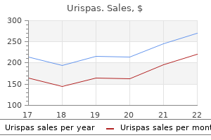
Generic 200mg urispas fast delivery
Intracranial abnormalities detected by three-dimensional magnetic resonance imaging in Prader-Willi syndrome muscle relaxant of choice in renal failure urispas 200 mg fast delivery. Energy expenditure and physical activity in Prader-Willi syndrome: comparison with obese subjects. Laparoscopic sleeve gastrectomy in children and adolescents with Prader-Willi syndrome: a matched-control study. The mini-gastric bypass in the management of [102] [103] [104] [105] [106] [107] [108] [109] [110] [111] [112] [113] [114] [115] [116] [117] [118] morbid obesity in Prader-Willi syndrome: a viable option Management of hypogonadism in adolescent girls and adult women with Prader-Willi syndrome. Multiple forms of hypogonadism of central, peripheral or combined origin in males with Prader-Willi syndrome. Long-term effects of recombinant human growth hormone therapy in children with Prader-Willi syndrome. Growth Hormone Research Society workshop summary: consensus guidelines for recombinant human growth hormone therapy in Prader-Willi syndrome. Beneficial effects of growth hormone treatment on cognition in children with Prader-Willi syndrome: a randomized controlled trial and longitudinal study. Obesity in patients with Bardet-Biedl syndrome: influence of appetite-regulating hormones. Cognitive, sensory, and psychosocial characteristics in patients with Bardet-Biedl syndrome. A new familial syndrome characterized by pigmentary retinopathy, hypogonadism, mental retardation, nerve deafness and glucose intolerance. Incidence and associated endocrine and neurologic abnormalities of optic nerve hypoplasia. Septo-optic dysplasia in childhood: the neurological, cognitive and neuro-ophthalmological perspective. Optic nerve hypoplasia syndrome: a review of the epidemiology and clinical associations. Endocrine and pubertal disturbances in optic nerve hypoplasia, from infancy to adolescence. Neuropathology of "septo-optic dysplasia" (de Morsier syndrome) with immunohistochemical studies of the hypothalamus and pituitary gland. Emotional deprivation and growth retardation simulating idiopathic hypopituitarism. Body mass index and segmental proportion in children with different subtypes of psychosocial short stature. Hamartoma of the hypothalamus and tuber cincereum: a brief review of the literature. Luteinizing hormonereleasing hormone analog treatment of boys with hypothalamic hamartoma and true precocious puberty. Surgical management of primary central nervous system germ cell tumors: proceedings from the Second International Symposium on Central Nervous System Germ Cell Tumors. A comparison of children with suprasellar germ cell tumors and craniopharyngiomas: final height, weight, endocrine, and visual sequelae after treatment. Treatment of chiasmatic/hypothalamic gliomas of childhood and chemotherapy: an update. Prognostic factors for progression of childhood optic pathway glioma: a systematic review. Magnetic resonance imaging as predictor of functional outcome in craniopharyngiomas. Surgical strategies and modern therapeutic options in the treatment of craniopharyngiomas. Neurosurgical treatment of craniopharyngioma in adults and children: early and long-term results in a large case series. Molecular oncogenesis of craniopharyngioma: current and future strategies for the development of targeted therapies. The combined interhemispheric subcommissural translaminaterminalis approach for large craniopharyngiomas. Incidence, treatment and survival of patients with craniopharyngioma in the surveillance, epidemiology and results program. Quality of life and clinical features of long-term survivors surgically treated for pediatric craniopharyngioma. Results after treatment of craniopharyngiomas: further experiences with 73 patients since 1997. Microsurgical resection of extensive craniopharyngiomas using a frontolateral approach: operative technique and outcome. Intraventricular craniopharyngiomas: surgical management and outcome analyses in 24 cases. Comparison of energy expenditure, body composition, metabolic disorders, and energy intake between obese children with a history of craniopharyngioma and children with multifactorial obesity. Prevalence of neurobehavioral, social, and emotional dysfunction in patients [178] [179] [180] [181] [182] [183] [184] [185] [186] [187] [188] [189] [190] [191] [192] [193] [194] [195] treated for childhood craniopharyngioma: a systematic literature review. Elowe-Gruau E, Beltrand J, Brauner R, Pinto G, SamaraBoutsani D, Thalassinos C, et al. Childhood craniopharyngioma: hypothalamus-sparing surgery decreases the risk of obesity. The therapeutic efficacy of fractionated radiotherapy and gammaknife radiosurgery for craniopharyngiomas. Hypothalamic obesity in craniopharyngioma patients: disturbed energy homeostatis related to extent of hypothalamic damage and its implication for obesity intervention. Visual outcome after fronto-temporo-orbito-zygomatic approach combined with early extradural and intradural optic nerve decompression in tuberculum and diaphragma sellae meningiomas. Gamma Knife radiosurgery for sellar and parasellar meningiomas: a multicenter study. Tuberculum sellae meningiomas: functional outcome in a consecutive series treated microsurgically. Suprasellar arachnoid cysts: toward a new simple classification based on prognosis and treatment modality. Endoscopic versus microsurgical resection of colloid cysts: a systematic review and meta-analysis of 1,278 patients. Neuroendoscopic treatment for colloid cysts of the third ventricle: the experience of a decade. Clinical review 136: primary lymphoma of the pituitary: an emerging clinical entity. Pituitary tumours: inflammatory and granulomatous expansive lesions of the pituitary. Hypothalamic-pituitary sarcoidosis with vision loss and hypopituitarism: case series and literature review. Management of adult patients with Langerhans cell histiocytosis: recommendations from an expert panel on behalf of Euro-Histio-Net. The pituitary gland in patients with Langerhans cell histiocytosis: a clinical and radiological evaluation. Incidence of growth hormone deficiency in pediatriconset Langerhans cell histiocytosis: efficacy and safety of growth hormone treatment. Pituitary dysfunction following cranial radiotherapy for adult-onset nonpituitary brain tumours. Effects of low-dose cranial radiation on growth hormone seretory dynamics and hypothalamic-pituitary function.
Discount urispas 200 mg free shipping
Since peripheral tissues have retained their normal sensitivity to the action of cortisol [100 muscle relaxer 75 best urispas 200 mg,101], they appropriately develop features of Cushing disease. No gross abnormality in the nature of the glucocorticoid receptor in the tumoral corticotroph cell has been demonstrated, although loss of heterozygosity at the glucocorticoid receptor gene locus may be frequent [109]. BrgI, a protein important for the glucocorticoid feedback, is misexpressed in many corticotroph cells and this may help explain these observations [110]. Both clinical and experimental observations show that lack of glucocorticoid feedback may induce corticotroph cell mass. A large increase in corticotroph cell area is observed in the anterior pituitary of adrenalectomized rats [124,125]. Glucocorticoids may exert direct inhibitory action on the growth of corticotroph cells [126], their deprivation being a direct stimulus for growth. Quantitatively, responses of the tumor cells have rarely been compared to those of normal human corticotroph cells. This situation is more frequently, but not exclusively, associated with Nelson syndrome, which occurs equally in males and females at relatively early mean age. Growth aggressiveness of the pituitary tumor may be the ultimate prognostic factor [143], and Ki67 labeling index should be assessed on surgical histological specimens to determine a more careful clinical follow-up. This is seldom of clinical significance and most often a chance discovery at routine immunohistochemical staining of the surgically removed tissue. Whether they exert peripheral, autocrine, or paracrine function remains to be demonstrated. Chromogranin A and secretogranin I have potential proteolytic sites that may be natural substrates for maturation enzymes. In several systems [151,152], including AtT-20 cells, pancreastatin [152], a maturation product of chromogranin A, exerts an inhibitory effect on resident hormone release and may be part of an ultrashort autocrine negative feedback. Most often, these tumors present as macroadenomas, revealed clinically because of their space-occupying effect. Thorough electron microscopy may separate various subtypes; some are morphologically indistinguishable from classical basophilic adenoma, others show subtle differences in granule size and loss of some type of microfilaments. Accordingly, an intrinsic traffic or export defect may be responsible, as suggested by reports of increased lysosomal activity [158]. It is likely that variability in the genetic causes of pituitary corticotroph adenomas will be the rule, with a range of consequences. At one end of the spectrum, a given mutation may essentially alter sensitivity to glucocorticoid; at the other end, another mutation may alter growth regulation leading to a silent corticotroph tumor. Further support for this notion is derived from observations that silent corticotroph adenomas frequently behave in a more aggressive fashion than nonfunctioning tumors of similar size [159]. They are likely remnants of the fetal intermediary lobe and may generate cellular cords that penetrate the pituitary posterior lobe [167]. Supporting the hypothesis that some human corticotroph adenomas arise from remnants of the pars intermedia are two animal models of Cushing disease: the dog and the horse both develop the disease spontaneously in association with tumors of the pars intermedia (in at least 30% of the dogs and in all cases of the horses) [58,168,169]. Evidence for an identical subtype of intermediatelike pituitary adenoma in man has been advanced [172]. Based on histologic data showing the close association of argyrophil fibers with nests of tumoral cells, and the hormonal evaluation showing responses more like those expected of an intermediate lobe tumor, it was suggested that these tumors arose from intermediate lobe remnants, were more often associated with hyperplastic lesions, were less amenable to surgical cure, and were driven by a general hypothalamic defect with decreased dopamine turnover. It was hypothesized that highly fluctuating cortisol concentrations were of hypothalamic origin, with low fluctuations of primary pituitary origin [176]. Corticotroph cell hyperplasia has been reported in pituitary glands of patients with Cushing disease [77,78,179,180]. Some adenomas may escape surgical exploration because of an ectopic location, and the difficult histological diagnosis of corticotroph hyperplasia certainly requires that the whole gland be thoroughly examined since corticotroph cells are not scattered randomly in the gland, but rather show clusters of densely aggregated cells [184]. Corticotroph Cell Hyperplasia and Adenoma Formation That hyperplasia often precedes formation of a clonal tumor has long been recognized in animal models and in humans. More interesting are patients whose disease recurs after "successful" removal of a pituitary adenoma [64,194]. Thus, there is no definitive proof that corticotroph cell hyperplasia is a prerequisite condition leading to formation of a pituitary adenoma. The resulting adrenocortical hyperplasia participates in the amplified response of the chronically stimulated gland, and the weight of each gland is greatly increased. Because low amounts of steroid are stored in the gland, increased secretion is a reflection of increased synthesis. Peripheral transformation to testosterone and dihydrotestosterone may lead to a moderate state of androgen excess in females [207]. Dissociation between cortisol and adrenal androgens is observed, however, when patients resume normal corticotroph function after successful pituitary surgery. Yet this action is only transient, since increased concentrations of cortisol in the adrenal cortex inactivate cytochrome P450 11-corticosterone methyl oxidase. Because a small, but definite, amount of -endorphin is formed in the anterior pituitary, the question has arisen of the potential effect of this highly powerful analgesic. Very high levels of authentic nonacetylated, presumably fully bioactive, -endorphin have been measured and chemically identified in the plasma of some patients with Nelson syndrome. These patients showed no evidence of analgesia and had no response after naloxone administration [220]. The two glands are symmetrically (and generally moderately) enlarged, weighing between 5 and 12 g each at operation. The glands are yellow or brown and with expansion of the zonas fasciculata and reticularis, but not the zona glomerulosa. In this form of hyperplasia, small nodular lesions are found, emphasizing the essential continuity of this condition with the nodular hyperplastic form with large, macroscopic nodules [222]. Multinodular Hyperplasia Although this definition is somewhat arbitrary, it is generally used whenever one or several macroscopic (visible to the naked eye) yellow nodules are present [3]. Such glands, in general, have a greater weight than in simple diffuse hyperplasia. The size of the nodules displays an extremely wide range of variation, from a few millimeters to several centimeters. Although as a rule they occur in both glands, marked asymmetry is occasionally seen, which may falsely indicate an autonomous adenoma-like lesion. In contrast to autonomous adenomas, a constant feature is that cortex between the nodules is always hyperplastic. With the light microscope, nodular cells show features not dissimilar to those of hyperplastic regions with alternate collections of compact and clear cells. Adrenal Rests Accessory adrenocortical tissue is often found in ectopic sites, and classically contains only cortical components of the gland. The most usual sites include the celiac plexus, kidney, gonads, broad ligaments, epididymis, and spermatic cord. Capsular extrusions are collections of adrenocortical cells just outside the adrenal capsule. They are thus distinct from adrenal nodules and contain only zona fasciculata cells. Recent data have shown that many are caused by activating somatic mutations in the phophokinase-A catalytic subunit [242]. Cortisol secretion is autonomous and unresponsive to either glucocorticoid deprivation or administration. Whereas benign tumors are usually small and secrete exclusively cortisol, malignant tumors are much larger, and secrete a wide array of steroid precursors and androgens. Removal of a benign adrenal adenoma induces an immediate and definitive cure with often transient, sometimes longlasting, hypocortisolism. Malignant adrenocortical tumors are highly aggressive; in most series the survival rate is only 20% 5 years after diagnosis [243,244]. Cortisol oversecretion is autonomous with hormone dynamics essentially similar to those encountered in cases of adrenocortical adenomas. However, this primary adrenal disorder is driven by bilateral adrenocortical lesions; the two glands harbor numerous nodular lesions that appear typically brown or black.
| Comparative prices of Urispas | ||
| # | Retailer | Average price |
| 1 | Hy-Vee | 480 |
| 2 | IKEA North America | 111 |
| 3 | HSN | 729 |
| 4 | WinCo Foods | 930 |
| 5 | Subway | 492 |
| 6 | Michaels Stores | 583 |
| 7 | Advance Auto Parts | 777 |
| 8 | Rite Aid | 638 |
| 9 | TJX | 943 |
| 10 | Aldi | 357 |
Discount urispas generic
Clinical findings may include cutaneous lesions spasms discount 200 mg urispas free shipping, cardiac disease (usually complete heart block), hepatobiliary disease, or hematologic cytopenias. Many of the lupus-specific skin lesions can occur in patients who have no evidence of extracutaneous disease. The characteristic morphologies of the various lupusspecific skin lesions are largely a function of the depth and intensity of the inflammatory infiltrate, presence or absence of epidermal basal cell damage, involvement of hair follicles, abundance of dermal mucin, and tendency to scar. In practice, there may be some overlap of these features, and there may be more than one type of lesion present in a given patient, which makes classification difficult. Because therapy for most of the lupus-specific lesions is similar, it is not always important to distinguish among the various types of lesions. However, it can be useful to identify conditions that are more likely to scar, to target more aggressive therapy, and to identify conditions that are highly likely or highly unlikely to be associated with systemic disease. Precipitation or exacerbation of lesions by sun exposure is common, and lesions tend to be distributed on the sun-exposed face, neck, extensor arms, and dorsal hands, where the skin over the knuckles is relatively spared. Acute eruptions with considerable focal basal cell damage can result in erythematous papules with dusky centers that clinically mimic erythema multiforme. The phenomenon of lupus skin lesions occurring in the absence of systemic disease has previously been termed discoid lupus by some. However, dermatologists use this term to denote a specific type of skin lesion, regardless of presence or absence of systemic disease. Discoid lupus lesions occasionally appear in a butterfly distribution, where they can result in disfiguring scarring. Lesions may resolve with hypopigmentation or even depigmentation, but they rarely scar. Depending on the morphology of the lesions and the clinical presentation, the differential diagnosis may include psoriasis, tinea, polymorphous light eruption, reactive erythema, and erythema multiforme. It should be noted that many immunofluorescence laboratories do not routinely report particulate epidermal staining. Involvement of hair follicles may be grossly evident as follicular plugs and scarring alopecia. Lesions tend to occur on the scalp, ears, and face but may be widespread and occasionally involve mucosal surfaces. In more difficult cases, biopsy for immunofluorescence may provide additional supporting diagnostic information. Lesions are expected to have granular deposits of Igs at the dermal-epidermal junction. Unless there is concomitant systemic disease, normal skin is expected not to have Ig deposits. Considerable mucin is present in the dermis, which gives the lesions a somewhat boggy look and feel. In some reports, lesions are most common on the face and may be reproduced by phototesting. Usual sites of involvement are the face, scalp, upper trunk, breasts, upper arms, buttocks, and thighs. The correlation of clinical with histologic findings usually serves to establish the diagnosis. Not all bullae related to lupus are due to autoantibodies to basement membrane proteins, however. Treatment of the lupus-specific lesions is relatively similar for most of the subtypes, with some exceptions and modifications. Many or most patients underestimate the amount of sunscreen needed to apply, the potential damage of the seemingly minimal exposure one has in the course of day-to-day activities, and the value of protective clothing. Tobacco use appears to be an exacerbating factor and can prevent response to therapy. Topical or intralesional corticosteroids are the most often used local therapy, but there are some reports of benefit from topical calcineurin inhibitors and topical retinoids. The first-line systemic medication for cutaneous lupus is antimalarial therapy, most often hydroxychloroquine. For skin disease not responsive to hydroxychloroquine, changing to chloroquine or adding quinacrine has been helpful in some patients. For antimalarial-resistant skin disease, a wide variety of medications have been used, but there is no clear second choice when patients have not responded to anti-malarials. When evaluating response to therapy, it is important for the clinician to distinguish active disease, as manifested by erythema or development of new lesions, from "damage," as exemplified by scarring or dyspigmentation. Although dapsone is arguably not helpful in most types of cutaneous lupus, it may be helpful in neutrophil-predominant bullous eruptions. Nonspecific Cutaneous Lesions A wide variety of lupus nonspecific skin lesions has been reported. Many of these, such as vasculitic lesions, are cutaneous clues to the possibility of extracutaneous disease. The netlike erythema of livedo reticularis is a vascular phenomenon caused by lowered oxygenation at the periphery of the area supplied by a particular vessel. This can simply be due to vasoconstriction, such as occurs in a cold environment, and thus can be a benign finding. If livedo is more prominent than usual, not corrected by warming, and persistent, it can indicate lowered flow caused by pathology such as vasculitis, atherosclerotic disease, or sludging. In lupus, livedo reticularis may be a sign of the presence of anti-phospholipid antibodies. The lesions usually appear in infants at a few weeks of age but have been noted at birth in several cases. The natural history of the skin disease is that the lesions last for weeks or months and resolve spontaneously, usually leaving no residuum. Confluent periorbital erythema, which gives the appearance of an erythematous mask, is common and diagnostically helpful. In areas where there is intense destruction of the basal cell layer, lesions may be crusted and look similar to bullous impetigo. Treatment of skin lesions consists largely of sun protection and mild topical steroids. Salivary gland dysfunction causes dry mouth and may result in angular cheilitis and numerous dental caries. Mildly dry mucous membranes and even severely dry skin may be present in a substantial percentage of healthy individuals who live in dry climates, thus the findings should be interpreted in the context of the setting. In most cases, lesions were purpuric, but in some, urticarial vasculitis was the clinical presentation. Patients with cutaneous evidence of vasculitis generally had more severe systemic disease. Patients with involvement of the sides of the fingers are more likely to have interstitial lung disease. The patients with both skin and muscle disease frequently experience resolution of their muscle disease after aggressive treatment with glucocorticoids, with or without immunosuppressives. Patients who do not improve with a single antimalarial can benefit from the addition of quinacrine or a switch from hydroxychloroquine to chloroquine. Patients with active disease can have widespread erythema over the trunk and extremities, with accentuation of the extensor arms and legs, as well as lateral thighs. Damage lesions include postinflammatory hyperpigmentation, poikiloderma, calcinosis, lipoatrophy, and depressed scars. The localized form of the disease occurs as localized or generalized morphea, linear scleroderma, or facial hemiatrophy, otherwise known as Parry-Romberg syndrome. Linear forms of the disease occur more often in children, whereas morphea and scleroderma are more common in adults. Eosinophilic fasciitis involves the fascia below the fat and can be associated with eosinophils in the tissue and is more responsive to glucocorticoids than localized or systemic scleroderma. Nephrogenic systemic fibrosis is related to gadolinium exposure in patients with decreased renal function. Morphea is seen more in adults, with increased incidence with advancing age, whereas linear scleroderma occurs more frequently in children and adolescents. The level of involvement in localized scleroderma can be in the dermis (morphea), fat (subcutaneous morphea), fat and fascia (morphea profundus), and fascia (eosinophilic fasciitis). Morphea typically has round and/or oval, irregular plaques that are initially dull red/violaceous, smooth, and indurated.
Purchase genuine urispas
Attachment to and Interaction with Extra-cellular Matrix Integrins Integrins are key mediators of both cell-to-matrix and cell-to-cell adhesive interactions knee spasms pain purchase 200 mg urispas otc. They are expressed as transmembrane heterodimers containing one - and one -subunit, of which at least 25 combinations are known (Table 14-2). The main adhesion molecules responsible for the attachment of fibroblasts to collagen are 11 and 2 integrins, whereas other 1 integrins such as 41 and 51 integrins mediate attachment of fibroblasts to fibronectin and its spliced variants. Syndecans In addition to conventional integrin-to-ligand binding, additional accessory molecules allow for the integration of adhesive contacts and local growth factor signaling. Cadherins Cadherins mediate homotypic, calcium-dependent adhesive interactions with the same cadherin species expressed by neighboring cells. The cytoplasmic tail interacts with -catenin, which in turn binds -catenin, forming a linkage between the cadherin-catenin complex and the actin cytoskeleton. Tightly regulated expression of cadherins is essential to embryogenesis but is also critical for tissue morphogenesis and tissue-specific cell differentiation. The chemokines specifically recruit the mononuclear cell population, which predominates in persistent inflammatory disease. The exact mechanisms by which different signals cooperate to mediate a specific response of fibroblasts and how this translates into distinct diseases are not yet fully defined. Although these matrix-degrading enzymes are crucial to tissue maintenance and repair, inappropriate overexpression of such enzymes is a key factor in the joint damage, particularly to cartilage, that occurs in inflammatory disease. Three major groups of inducers can be differentiated: pro-inflammatory cytokines, growth factors, and matrix molecules. Role of Specialized Fibroblast Subsets within Tissue Microenvironments Combining surface markers with consistent function has been the key to decades of development in the field of leukocyte biology. By comparison, stromal cell biologists have had remarkably few such stable markers. One example is the murine thymic stroma, in which subsets with both geographic and functional consistency have been identified. Pericytes have been hypothesized to represent the extralymphoid source of mesenchymal progenitor cells and express markers consistent with mesenchymal stem cells. However, when activated by substances released during tissue injury or the products of invading microorganisms, fibroblasts are capable of elaborating a broad repertoire of inflammatory mediators, which fully justifies their classification as immune sentinel cells. The second layer is the sublining layer, which is composed of less densely packed fibroblasts and macrophages in a loose tissue matrix along with blood vessel networks. As mentioned earlier, the unique lining layer barrier function is not supported by a basement membrane and conventional tight junctions but instead by homotypic interactions between cadherin-11 molecules. Cadherin-11 mediates selective association of mesenchymal rather than epithelial tissues, a function that is carried forward after embryogenesis in structures such as the joint, lung, and testis. By virtue of their role in defining the geography of specialized tissues, fibroblasts and other stromal cells exist in living organisms within three-dimensional environments, whereas the vast majority of experiments performed using fibroblasts in the laboratory are still conducted within twodimensional environments. Furthermore, fibroblasts are frequently grown in nonphysiologic stimuli such as serum, to which fibroblasts would not normally be exposed unless tissue damage were to occur. Behavior is significantly different when cells are cultured in artificial three-dimensional environments. This geographic structure is reflected in serial frozen sections of rheumatoid arthritis synovium stained for stromal markers (A-C,E-G). This finding is further evidence of the robustness of epigenetic programming, which determines site and organ specialization. The lining layer undergoes dramatic hyperplasia, sometimes reaching 10 to 20 cells in depth with both type A and type B cell populations expanded and becoming merged with the sublining. T and B lineage cells may remain in diffuse infiltrates or may coalesce into aggregates of cells varying from simple perivascular "cuffs" a few cells in diameter to structures resembling B cell follicles in up to 20% of samples. However, when cadherin-11 knockout mice are evaluated in the K/BxN serum transfer model, invasiveness is reduced with a 50% reduction in inflammation. Similarly, cultured fibroblasts with mutant cadherin-11 constructs also demonstrate impaired invasiveness into cartilage. This unique structural molecule may therefore emerge as a therapeutic target54; because of shared roles in invasive disease, targeting of this molecule in breast cancer is currently in development. The response to tissue damage involves a carefully choreographed series of interactions among diverse cellular, humoral, and connective tissue elements. In order for an inflammatory lesion to resolve, dead or redundant cells that were recruited and expanded during the active phases of the response must be removed. It is becoming increasingly clear that fibroblasts are not only passive players in immune responses but also actively determine the switches that govern progression from acute to chronic inflammation, as well as those governing resolution or the progression to chronic, persistent inflammation. The "switch to resolution" is an important signal that permits tissue repair to take place and enables immune cells to return to draining lymphoid tissues (lymph nodes) in order for immunologic memory to become established. It is known that during wound healing and under profibrotic conditions, some fibroblastlike cells are transformed into myofibroblasts, which are distinct from tissue fibroblasts in terms of both their phenotype and their behavior. This has been shown recently in both human and murine renal fibrotic disease, where hypermethylation of the promoter region of a ras oncogene inhibitor led to gene silencing, ras pathway activation, and hence persistent fibrogenesis. This model has been used to explore the in vivo mechanisms governing invasiveness. This important finding raises the question of which cell populations are grown from the synovium when tissue is digested and adherent cells are cultured in vitro: lining layer cells, sublining cells, or a mixture of both However, we do know from transcriptomic approaches that the phenotype remains more stable in tissue culture than might be expected, with little transcriptional divergence over the first two to four passages and the level of differentially expressed genes between parallel cultures rising to greater than 10% only after passage 7. Interactions of Fibroblasts with Leukocytes Recruitment of Inflammatory Infiltrates into the Joint Stromal elements such as synovial fibroblasts are subject to a pro-inflammatory cytokine network within the inflamed synovium. A number of chemokine receptors have been shown to differ between peripheral blood and synovial leukocytes, suggesting that they are enriched in the synovium either though their selective recruitment by endothelial-expressed chemokines or after upregulation by the microenvironment after their recruitment. Fibroblast Support for Leukocyte Survival Stromal cell support for the survival of leukocyte populations fulfills a physiologic role in certain organs within the body. The selective recruitment and support of hemopoietic subsets is an essential physiologic function of stromal cells in specific microenvironments. The stromal microenvironment plays a crucial role in the maintenance of such survival niches, which are not generic but are highly specific to certain organs and tissues, resulting in site-specific differences in the ability of different stromal cells to support the differential accumulation of leukocyte subsets. In the case of an inflammatory response, successful resolution requires the removal of the vast majority of immune cells that were recruited and expanded during the active phase of the inflammation. A number of studies have shown that during the resolution phase of viral infections, the initial increase in T cell numbers in peripheral blood that is seen within the first few days is followed by a wave of apoptosis occurring in the activated T cells. This situation is mirrored within tissues, where apoptosis induced by the molecule Fas occurs at the peak of the inflammatory response and may be responsible for limiting the extent of the immune response. Recent studies have shown that a failure of synovial T cells to undergo apoptosis contributes to the persistence of the inflammatory infiltrate. Not surprisingly, other leukocyte subpopulations have been shown to derive support from stromal cells. Although fibroblast support for T cell and B cell survival exhibits sitespecific properties, neutrophil survival is dependent on prior cytokine activation of fibroblasts and shows no differences between fibroblasts taken from different anatomic sites. A number of studies have recently reported that the synovial microenvironment contributes directly to the inappropriate retention of T cells within the joint by an active chemokinedependent process. The presence of high levels of inflammatory chemokines, produced by stromal cells, is a characteristic of environments such as the rheumatoid synovium. However, recent data suggest that, paradoxically, constitutive chemokines, which are involved in the recruitment of lymphocytes to secondary lymphoid tissues, are ectopically expressed in immune-mediated inflammatory diseases. This chemokinereceptor pair plays an important role, both in the constitutive traffic of lymphocytes and in the recruitment and retention of hemopoietic cells within the bone marrow. Adhesive interactions consist of integrin-receptor interactions as described in the text and the critical presence of homotypic interaction through cadherin-11. B, Sublining synovial fibroblasts interact with numerous cell types including mast cells and plasma cells (not shown), T cells, B cells, interstitial macrophages, and endothelial cells, leading to their recruitment, retention, activation, and differentiation. Both cell surface receptor interactions and secreted mediators are important in this process. In addition, fibroblasts may activate T cells through antigen presentation, co-stimulatory receptors.
Urispas 200 mg lowest price
Cardiac effects of growth hormone in adults with growth hormone deficiency: a meta-analysis spasms right side abdomen buy cheap urispas. The pituitary could be directly involved by the same processes that affect other organs. Sarcoidosis Sarcoidosis, a chronic multisystemic inflammatory disease of unknown origin, is characterized by noncaseating granulomatous inflammation of the organs involved. Neurosarcoidosis occurs in about 5% of patients; there is evidence of either hypothalamic or pituitary dysfunction in approximately one-third [1]. A diagnosis of neurosarcoidosis is challenging and requires the documentation of characteristic clinical symptoms and radiologic findings after the exclusion of other diseases, including vasculitis, infection, and neoplasm. Noncaseating granulomatous inflammation should be confirmed by histology findings. The value of serum angiotensin-converting enzyme levels in the diagnosis of sarcoidosis remains controversial due to its inadequate sensitivity and specificity. Diffuse extensive leptomeningeal enhancement, with the involvement of the hypothalamus, pituitary stalk, and the optic chiasm has also been reported [4]. Studies using provocative testing have demonstrated a high prevalence of hypothalamic dysfunction, with intact pituitary hormonal responses to releasing factors, but impaired responses to clomiphene, metyrapone, and insulin hypoglycemia [5]. Polyphagia with morbid obesity and temperature dysregulation has also been reported. Transsphenoidal surgery is commonly used to resect advanced pituitary sarcoid lesions refractory to medical management. As histology and imaging are key to diagnosis, a multidisciplinary team approach is valuable for enabling diagnosis and subsequent treatment [6]. Radiologic studies usually show pituitary enlargement (80%), sellar mass with peripheral enhancement, areas of central necrosis, and stalk thickening [11]. Cystic changes in the pituitary, infundibular thickening with contrast enhancement, and the absence of a normal hyperintense signal on T1weighted pituitary images have been described [11]. Spontaneous remission has not been reported in granulomatous or necrotizing hypophysitis. Hyperprolactinemia, observed in some patients, points to a hypothalamic or stalk disturbance, and may contribute to the suppression of gonadotrophin secretion. Since histiocytes may function as antigen-presenting cells, hypothalamic infiltration could lead to immunologically mediated destruction of vasopressin neurons. Granulomatous Hypophysitis Idiopathic granulomatous hypophysitis is rare (,100 cases have been reported) and most often it mimics symptoms associated with a pituitary adenoma at presentation [14]. Clinical presentation often includes headache, with a few patients presenting with isolated hypopituitarism. On imaging, the pituitary is frequently diffusely enlarged and a thickened stalk is infrequently observed. Necrotizing Hypophysitis Necrotizing hypophysitis is extremely rare and may represent a separate disease entity or a variant of other types of hypophysitis. Histologically, diffuse necrosis is surrounded by dense infiltration with lymphocytes, plasma cells, and a few eosinophils with considerable fibrosis. The natural history of any inflammatory hypophysitis remains elusive and treatment is controversial. There has been a dramatic rise in the incidence and now patients are diagnosed much earlier. Most patients are affected in the fifth decade of life, with a slight male preponderance [18]. Clinical manifestations are protean and virtually every organ system can be affected. Other common clinical features include skeletal involvement with typical bilateral osteosclerotic lesions of long bones of the lower limbs, and cardiovascular involvement with circumferential thickening of the aorta and retroperitoneal fibrosis. LyH is associated, in some cases, with other types of endocrine autoimmunity [23]. Antibodies to pituitary tissue have been found in some LyH patients and in a variety of other endocrine autoimmune disorders, as well as in some patients with the primary empty sella syndrome and Sheehan syndrome. However, the functional significance of these antibodies remains unclear [15,23,24]. Immunoglobulin G4-related Hypophysitis Immunoglobulin G4-related hypophysitis is a novel disease, characterized by elevated serum IgG4 concentration and infiltration of IgG4-positive plasma cells in the pituitary. Pathogenesis is poorly understood, with findings consistent with both an autoimmune disorder and an allergic disorder. Although some criteria for diagnosis of IgG4-related hypophysitis have been proposed, they are not fully established; however, on biopsy, the presence of a mononuclear cell infiltrate rich in plasma cells within the pituitary gland appears pathognomonic. The presence of at least 10 IgG4-producing plasma cells has been deemed as required for diagnosis. In other cases, pituitary imaging in addition to the presence of IgG4 lesions in other organs could be viewed as sufficient. Microscopic amyloid deposits are frequently observed in blood vessel walls and interstitial areas in normal human pituitaries derived from elderly subjects. However, amyloid may be deposited in pituitary blood vessels in patients with systemic amyloidosis, usually with intact pituitary function. Use of antiretroviral agents, while lifesaving, has led to the emergence of several comorbidities, including insulin resistance and lipodystrophy. Later in the course of the disease, a pattern of central hypogonadism ensues [35]. Other Infectious Diseases Other infectious diseases affecting the pituitary include tuberculosis, syphilitic infection, Whipple and Chagas disease, and hemorrhagic fever. Tuberculosis has a predilection for the involvement of the basilar meninges and may therefore occasionally involve tissues in the sellar region, sometimes producing anterior or posterior pituitary insufficiency. Radiologically, pituitary tuberculomas may resemble other sellar mass lesions; thickening of the stalk is seen in some cases [37]. Hemorrhagic fever with renal syndrome is a viral illness characterized by fever, hypotension, capillary leak, and acute renal failure; several varieties of Asian and European hantaviruses cause the syndrome. Approximately 20% of infected patients in one series developed pituitary hormone deficiencies, including pituitary atrophy and empty sella. In one series [41], patients presented with complaints and physical findings consistent with a pituitary mass, but rarely with clinical evidence of a serious infection. Headache, endocrine abnormalities, and visual changes were the most common features. Fever, peripheral leukocytosis, and meningismus were also present in one-third of patients. A sellar mass with peripheral enhancement is suggestive of an abscess; however, this finding may be absent in some cases [42]. Interestingly, in most cases, a diagnosis is made when a surgeon discovers a pustular cystic mass during surgery; the rate at which a pituitary abscess is diagnosed before surgery seems however to be rising [43]. Antibiotic therapy is indicated for septic patients, or for patients in whom specific organisms are identified from cultures obtained during surgery. With appropriate treatment, headache, and visual changes may improve; however, endocrine dysfunction likely persists and requires hormonal replacement therapy. Hypopituitarism, particularly corticotrophic insufficiency, seems to be prevalent in a considerable number of middle-aged patients with hereditary hemochromatosis. Female patients seem to be less affected than males, perhaps because of the protective effects of menstrual blood loss. Histopathology studies have revealed that pituitary iron deposits are localized primarily to gonadotrophs and, less frequently, to lactotrophs. Pituitary iron and volume might predict hypogonadism in transfusional iron overload [49]. However, many patients with moderate-to-severe pituitary iron overload retain normal gland volume and function, which represents a potential therapeutic window.
Syndromes
- Removal of burned skin (debridement)
- Coughing
- Hematoma (blood accumulating under the skin)
- Electrolyte changes (abnormal levels of sodium and potassium in the blood)
- The entire penis looks red and swollen
- Congestive heart failure or irregular heartbeat (arrhythmias) - rare
- Numbness or tingling most often on the face or scalp
- Kidney transplant
- CRPS 1 is a chronic nerve disorder that occurs most often in the arms or legs after a minor injury.
- General discomfort, uneasiness, or ill feeling (malaise)
Buy discount urispas 200mg line
The space between this ruffled border and the bone surface is the place where bone resorption occurs muscle relaxant g 2011 cheap 200 mg urispas overnight delivery. It is sealed by a ring of contractible proteins and tight junctions because it represents one of the few regions of the human body where a highly acidic milieu is found. Bone degradation by osteoclasts consists of two major steps: first, demineralization of inorganic bone components, and second, removal of organic bone matrix. To demineralize bone, osteoclasts secrete hydrochloric acid through proton pumps into the resorption lacunae. Consequently, inhibitors of cathepsin K block osteoclast function and slow down bone resorption. Osteoclasts originate from hematopoietic monocytic precursor cells and, upon the influence of specific signals, undergo a series of differentiation steps to become mature osteoclasts. Factors yet unknown, which may likely be of mechanical nature and sensed by the osteocytes, initiate bone remodeling at a specific site. The death of osteocytes and the resulting metabolic changes leading to a lack of silencers for bone turnover, like sclerostin, may govern this activation process. It is followed by a resorptive phase dominated by osteoclast-mediated degradation of the bone matrix, resulting in a resorption lacuna. The naked bone surface inside this lacuna is subsequently populated by mesenchymal cells immigrating from the neighboring bone surface, which start differentiating into osteoblasts and produce the new bone matrix (also termed osteoid). Adults continuously remodel their skeleton, and this process occurs even faster during childhood and adolescence. In adults the entire skeleton is remodeled in 7 to 10 years, which indicates that we fully replace our skeleton several times during our lifetime. Most of the bone remodeling happens in the trabecular bone, which promotes the building of an optimal inner microstructure adapted to the individual mechanical demands. Trabecular bone is the leading structure in the vertebral bodies (constituting up to two-thirds of the bone substance) and in long bones such as the femurs (constituting about 50% of the bone substance). Normal physiologic circumstances ensure a balance between bone formation and bone resorption to maintain skeletal homeostasis. This bone remodeling process requires a tight mutual regulation of bone resorption by osteoclasts and bone formation by osteoblasts, a phenomenon called coupling. The ephrin-A2 system is an autocrine stimulator for osteoclasts and also blocks bone formation. This coupling process involves two main mechanisms; the first is the expression of the essential pro-osteoclastogenic cytokines by the osteoblast lineage, and the second involves the ephrin ligand/ephrin receptor bidirectional signaling. In response to Wnt signaling, osteoblasts slowly lose their supportive activity for osteoclasts when they mature toward more mineralizing cells and then become the boneembedded osteocytes. A second level of coupling involves the expression of ephrin ligands on the surface of osteoclast progenitors that can bind to ephrin receptors and activate their tyrosine kinase activity. The first, ephrin-B2, binds to the receptor EphB4 on osteoblast progenitors, increasing their differentiation and stimulating bone formation. The second, ephrin-A2, acts in an autocrine manner by binding to the EphA2 receptor on the osteoclasts, promoting their differentiation in a Bone Remodeling by the Immune System Aside from the reciprocal regulation of osteoblasts and osteoclasts, bone remodeling is also controlled by the immune system. Insights into the control of bone by the immune system have led to a new research field known as osteoimmunology, which has defined new pathways involved in immune regulation of post-menopausal osteoporosis and bone loss during inflammatory diseases. In addition, two major systemic neuroendocrine regulators of bone homeostasis co-regulate bone, fat, and energy metabolism. Osteocalcin is a hormone produced by mature osteoblasts and acts on the cells of the pancreas to stimulate proliferation and thus insulin production in response to leptin. Although the increased fat mass is certainly linked to the role of leptin in controlling appetite, the effect of leptin on bone and fat can be dissociated. Indeed, bone formation is negatively regulated by leptin through a hypothalamic pathway: the -adrenergic sympathetic nervous system that mediates decreased osteoblast proliferation via the induction of clock genes in osteoblasts. This remodeling process allows the optimal adaptation of the bone architecture to individual demands and tight control of calcium homeostasis. Local factors control the bone remodeling process on the basis of osteoclast-osteoblast interactions, as well as by systemic immune and neuroendocrine factors controlling the bone-resorbing and bone-forming cells. Sato K, Suematsu A, Okamoto K, et al: Th17 functions as an osteoclastogenic helper T cell subset that links T cell activation and bone destruction. Zhao C, Irie N, Takada Y, et al: Bidirectional ephrinB2-EphB4 signaling controls bone homeostasis. Lorenzo J, Horowitz M, Choi Y: Osteoimmunology: interactions of the bone and immune system. Ducy P, Amling M, Takeda S, et al: Leptin inhibits bone formation through a hypothalamic relay: a central control of bone mass. Sato S, Hanada R, Kimura A, et al: Central control of bone remodeling by neuromedin U. The smallest functional unit of muscle, the sarcomere, is composed of an almost crystalline array of filamentous proteins that convert metabolic energy into force and movement. Skeletal muscle contraction is controlled by the central nervous system through depolarization of specific efferent neurons called motor neurons. Motor neurons innervate and depolarize muscle fibers through cholinergic synapses called neuromuscular junctions. Afferent neurons provide the central nervous system with sensory information required for effective control of movement and posture. Force is transmitted to the exterior through two sets of protein cell adhesion complexes: integrins and dystroglycans. Nerves, blood supply, and connective tissue structures that provide support, elasticity, and force transmission to the skeleton (see later discussion) constitute the remaining volume. This elongated shape is determined by the organization of the contractile proteins that occupy most of the sarcoplasm. Each muscle has a limited range of shortening that is amplified into large motions by lever systems of the skeleton, usually operating at a mechanical disadvantage. Variations in geometric arrangements of the fibers-parallel, convergent (fanshaped), pennate (feather-like), sphincter (circular), or fusiform (thick in the middle with tapered ends)-determine some of the mechanical properties. For example, a muscle with fibers aligned parallel to the force-generating axis will have more basic contractile units. Muscles are commonly arranged around joints as antagonistic pairs facilitating bidirectional motion. When one muscle (the agonist) contracts, another (its antagonist) is relaxed and passively extended. Their roles reverse to actively generate the opposite motion, unless it occurs passively by the force of gravity. An extensive network of areolar connective tissue, forming the endomysium, surrounds each muscle fiber. Fine nerve branches and small capillaries, which are necessary for the exchange of nutrients and metabolic waste products, penetrate this layer. The endomysium is continuous with the perimysium, a connective tissue network that ensheathes small parallel bundles of muscle fibers known as fasciculi, intrafusal fibers, larger nerves, and blood vessels. The Approximately 660 skeletal muscles support and move the body under the control of the central nervous system. Most skeletal muscles are fastened by collagenous tendons across joints in the skeleton. The transduction of chemical energy into mechanical work by muscle cells leads to muscle shortening and consequent movement. A high degree of specialization in this tissue is evident from the intricate architecture and kinetics of intra-cellular membrane systems, the contractile proteins, and the molecular components that transmit force extra-cellularly to the basement membrane and tendons. Muscle cells normally exhibit wide variations in activity level and are able to adapt in size, isoenzyme composition, membrane organization, and energetics. This chapter outlines the structure and function of muscle and its relationship to associated connective tissue. It also introduces the basis for the highly adaptive response to altered functional demands and diseases. These layers give the attachment sites great tensile strength and distribute axial force into shear forces over a larger surface area. In any given muscle, part of this adaptation arises from its composition and organization of fiber types. During development, fiber-type specificity may be partially determined before innervation. A, scanning electron micrograph of an motoneuron innervating several muscle fibers in its motor unit.
Generic 200 mg urispas visa
The friction muscle relaxant flexeril 10 mg buy cheap urispas 200mg line, f, of a tendon against the surrounding tissue is in the opposite direction of the tendon motion. The sliding resistance (friction) is the difference between the tension on the distal and proximal ends across the pulley (Fd and Fp respectively). This simple relationship clearly demonstrates the importance of the angle of contact and the friction coefficient. It explains why avoiding awkward joint postures is important ergonomically to reduce repetitive injury of soft tissue. The short head of the biceps brachii tendon is an example of the first scenario, in which the tendon mainly experiences tensile force alone. The second scenario, in which the tendon encounters tensile force and compression together, occurs internally at the bone insertion site of the supraspinatus tendon. The insertional area of the tendon encounters tensile force, as well as compression resulting from the transverse contact of the tendon with the attached bone. The third scenario occurs when the tendon is simultaneously subjected to tension, compression, and gliding. The long head of the biceps brachii tendon, where it passes through the bicipital groove, demonstrates an example of the third scenario. The mode of mechanical tendon loading is potentially correlated to the incidence of tendon degeneration. Tendons commonly affected with tendinopathy are represented in the other two scenarios, in which the affected areas normally sustain compression and shear force with or without gliding in addition to tensile force. In summary, knowledge of biomechanical principles can help explain many commonly encountered situations in the musculoskeletal system that contribute to musculoskeletal disease. Kadaba M, Ramakrishnan H, Wootten M: Measurement of lower extremity kinematics during level walking. Kaufman K, Hughes C, Morrey B, et al: Gait characteristics of patients with knee osteoarthritis. Grood E, Suntay W: A joint coordinate system for the clinical description of three-dimensional motions: applications to the knee. Selvik G: A roentgen-stereophotogrammetric method for the study of the kinematics of the skeletal system, Lund, Sweden, 1974, University of Lund. Spoor C, Veldpaus F: Rigid body motion calculated from spatial coordinates of markers. Myazaki T, Wada M, Kawahara H, et al: Dynamic load at baseline can predict radiographic disease progression in medial compartment knee osteoarthritis. Tissue engineering has adopted the concept of biomimetics of in vivo tissue development. Developmental engineering is the term used to describe novel methodology for the rational and accurate design of robust, well-controlled manufacturing processes of "biological spare parts. Recent advances in regenerative medicine and tissue engineering relevant to rheumatology have entered clinical practice and include the biologic repair of joint surface defects and bone healing. Destruction of joint tissues is ultimately the disabling outcome of most forms of inflammatory or degenerative arthritides. The need for repair and regeneration of joints and joint-associated tissues is becoming more relevant as the dramatic advances of targeted treatments and improved disease management have allowed much more efficient control of inflammation and joint destruction. In view of this situation, other aspects of joint biology deserve more attention-notably, and most importantly, the mechanisms driving tissue response and repair. Introducing regenerative medicine provides a significant opportunity to restore joint homeostasis and thus possibly provide a cure. Targeting repair has entered our discipline, and investigating the potential to activate and enhance joint tissue repair mechanisms has become a prime goal. Nature demonstrates that this goal 90 is achievable, because successful wound healing and fracture repair are processes that happen routinely after birth. We also know, as demonstrated in fetal surgery, that scarless repair is partially dependent on age and context. Thus it is attractive to envision that, with an in-depth understanding of the repair processes at the cellular and molecular level, we may be able to intervene quickly at the time of injury and guide the healing process more appropriately, thereby preventing scar formation. For example, it appears that the process of rebuilding an adult limb, such as seen in the axolotl, but also during fracture healing in higher species, has many similarities with how the limb forms in the first place in the embryo, and particularly requires signaling mechanisms to specify the final pattern. Thus both limb formation and limb regeneration are likely to use the same molecular pathways. These advances not only include our understanding of the mechanisms of body axis formation and organogenesis but also impressive progress in stem cell biology, including the regulation of stemness, stem cell niches, lineage specification and cell differentiation, and the critical molecular pathways involved. The first approach consists of enhancing intrinsic repair mechanisms with stimulation of cell proliferation, differentiation and tissue metabolic activity, and recruitment of endogenous progenitor populations into the damaged tissue. The severity and outcome of disease is determined by the balance between inflammation/destructive processes and anti-inflammatory signals with repair attempts. Although the evidence for the heritability of cartilage repair capacity in humans is still circumstantial, findings of animal studies are more convincing. Comparing the capacity of different inbred mouse strains to heal full-thickness cartilage defects, Eltawil et al. This finding supports the concept that the repair capacity has a genetic component. Thus it was found that both the capacity of healing joint surface defects and the capacity to heal ear wounds were highly heritable and that they correlated with each other. Although Wnt signaling is essential for the development and homeostasis of synovial joints,36-40 genetic studies in humans20 and experimental data22,41,42 demonstrate that excessive/uncontrolled activation of Wnt/ catenin signaling leads to reprogramming of articular chondrocytes toward catabolism or loss of their stable phenotype with subsequent loss of articular cartilage tissue structure and function. In this chapter we select a few examples of how these signaling pathways are involved in joint formation and their role in postnatal joint repair, with a specific focus on some of these pathways that have led to advances in early clinical development in arthritic diseases. It appears that a tight control, both in terms of timing and magnitude, is essential not only to preserve homeostasis but even to induce tissue regeneration. In the clinical context of joint disease and regeneration, the complexity of Wnt signaling and its potential downstream effects may offer the opportunity to target catabolic effects while preserving the homeostatic ones. Better understanding of the downstream signaling mechanism and its regulation will be essential to target only the pathogenic effect of these pleiotropic signaling pathways in cartilage, bone, and the osteochondral junction. Growth Hormone/Insulin-like Growth Factor Axis Other targets besides skeletal developmental pathways are activated by signaling molecules that also play critical roles in postnatal joint tissue homeostasis and turnover. These proteins/pathways can be regarded as potential postnatal "anabolic" agents that contribute to the restoration of joint homeostasis. Stem Cell Niches Stem cells persist in adult life to safeguard tissue maintenance and regeneration. They have the capacity to selfrenew (that is, to produce more stem cells), thereby preserving a constant pool of stem cells, and they have the capacity to differentiate to replace the mature cells that are lost to physiologic turnover, injury, disease, or ageing. Self-renewal and differentiation are regulated by stem cell intrinsic factors and signals from the surrounding microenvironment in which the stem cells reside, called the "stem cell niche. The joint environment is rich in progenitor/stem cells, but our understanding of their anatomic location in apposite niches and the molecular regulation of their functions remains limited. It also must be noted that the term pericyte is an anatomic description of a cell that is abluminal and that its developmental origin is not clear. It is likely that, rather than having a common ancestor, pericytes have distinct ontogeny and biologic properties that are related to their tissues of residence. They could in part derive from the embryonic joint interzone, as lineage tracing studies suggest. The restoration of a functional niche will safeguard durable repair by ensuring lifelong replacement of mature cells. B, details of the dashed box in A, showing cell populations in the synovium of uninjured joints. C, schematic drawing of a synovial joint 12 days after articular cartilage injury in mice (arrowhead). Proliferating cells were detected in both the synovial lining and the subsynovial tissue and were either double positive for idu and chlorodeoxyuridine (Cidu; orange) or single positive for Cidu (red). Targeting Homeostasis of the Joint In the resting condition, articular cartilage has an extremely low rate of turnover. After acute traumatic injury, however, chondrocytes deploy strong adaptive responses that ultimately result in a coordinated sequence of activation and chemotaxis of progenitors within the cartilage itself and in other joint tissues, along with cell proliferation and matrix turnover. In a number of cases, such adaptive responses are sufficient to repair damage and re-establish homeostasis. More accurate molecular analyses have identified several other components that are amenable for targeting. In the entire joint, inflammation is an integral part of a natural response to injury, providing molecular signals and cascades that are likely to have distinct context and tissue-specific beneficial or detrimental effects.
Order urispas with visa
Radiosurgery of growth hormone-producing pituitary adenomas: factors associated with biochemical remission xanax spasms buy 200 mg urispas with visa. The role of stereotactic radiotherapy in patients with growth hormone-secreting pituitary adenoma. Blindness in patients after external beam irradiation for pituitary adenomas: two cases occurring after small daily fractional doses. Multiple radiationinduced intracranial lesions after treatment for pituitary adenoma. An unusual treatment-related complication in a patient with growth hormone-secreting pituitary tumor. Risk of second brain tumour after conservative surgery and radiotherapy for pituitary adenoma. Does pituitary radiotherapy increase the risk of stroke and, if so, what preventative actions should be taken Medical therapy in patients with acromegaly: predictors of response and comparison of efficacy of dopamine agonists and somatostatin analogues. The role of combination medical therapy in acromegaly: hope for the nonresponsive patient. Effective combination treatment with cabergoline and low-dose pegvisomant in active acromegaly: a prospective clinical trial. Opportunities in somatostatin research: biological, chemical and therapeutic aspects. Somatostatin receptor type 5 modulates somatostatin receptor type 2 regulation of adrenocorticotropin secretion. Acromegaly: correlation between expression of somatostatin receptor subtypes and response to octreotide-lar treatment. Comparison between six-year therapy with long-acting somatostatin analogs and successful surgery in acromegaly: effects on cardiovascular risk factors. One-year follow-up of patients with acromegaly treated with fixed or titrated doses of lanreotide Autogel. The clinical response to somatostatin analogues in acromegaly correlates to the somatostatin receptor subtype 2a protein expression of the adenoma. Correlation of in vitro and in vivo somatotropic adenoma responsiveness to somatostatin analogs and dopamine agonists with immunohistochemical evaluation of somatostatin and dopamine receptors and electron microscopy. Clinical review: the antitumoral effects of somatostatin analog therapy in acromegaly. Tumor shrinkage with lanreotide Autogel 120 mg as primary therapy in acromegaly: results of a prospective multicenter clinical trial. Preoperative octreotide treatment in newly diagnosed acromegalic patients with macroadenomas increases cure short-term postoperative rates: a prospective, randomized trial. Perioperative management of patients undergoing transsphenoidal pituitary surgery. Analgesic effect of octreotide in headache associated with acromegaly is not mediated by opioid mechanisms. Significant headache improvement after transsphenoidal surgery in patients with small sellar lesions. Impact of treating acromegaly first with surgery or somatostatin analogs on cardiomyopathy. A comprehensive study of clinical, biochemical, radiological, vascular, cardiac, and sleep parameters in an unselected cohort of patients with acromegaly undergoing presurgical somatostatin receptor ligand therapy. Effects of octreotide on sleep apnoea and tongue volume (magnetic resonance imaging) in patients with acromegaly. Skeletal involvement in female acromegalic subjects: the effects of growth hormone excess in amenorrheal and menstruating patients. Intensity of pituitary adenoma on T2-weighted magnetic resonance imaging predicts the response to octreotide treatment in newly diagnosed acromegaly. Partial surgical removal of growth hormone-secreting pituitary tumors enhances the response to somatostatin analogs in acromegaly. Surgical debulking of pituitary macroadenomas causing acromegaly improves control by lanreotide. Effects of somatostatin analogs on glucose homeostasis: a metaanalysis of acromegaly studies. Hyperglycemia associated with pasireotide: results from a mechanistic study in healthy volunteers. Prospective study of the long-term effects of somatostatin analog (octreotide) on gallbladder function and gallstone formation in Chinese acromegalic patients. Safety and efficacy of long-term octreotide therapy of acromegaly: results of a multicenter trial in 103 patients-a clinical research center study. A novel suspension formulation enhances intestinal absorption of macromolecules via transient and reversible transport mechanisms. Expression of a mutated bovine growth hormone gene suppresses growth of transgenic mice. Long-term effects of pegvisomant on comorbidities in patients with acromegaly: a retrospective single-center study. Long-term treatment with pegvisomant as monotherapy in patients with acromegaly: experience from Acrostudy. Growth hormone receptor antagonist therapy in acromegalic patients resistant to somatostatin analogs. Glucose homeostasis and safety in patients with acromegaly converted from long-acting octreotide to pegvisomant. Pegvisomant improves insulin sensitivity and reduces overnight free fatty acid concentrations in patients with acromegaly. Cotreatment of acromegaly with a somatostatin analog and a growth hormone receptor antagonist. Tumor volume of growth hormone-secreting pituitary adenomas during treatment with pegvisomant: a prospective multicenter study. Combined treatment for acromegaly with long-acting somatostatin analogs and pegvisomant: long-term safety for up to 4. Clomiphene citrate for treatment of acromegaly not controlled by conventional therapies. Comparative effectiveness review of treatment options for pituitary microadenomas in acromegaly. A critical analysis of clinically available somatostatin analog formulations for therapy of acromegaly. Effects of initial therapy for five years with somatostatin analogs for acromegaly on growth hormone and insulin-like growth factor-I levels, tumor shrinkage, and cardiovascular disease: a prospective study. Significant tumour shrinkage after 12 months of lanreotide Autogel-120 mg treatment given first-line in acromegaly. The utility of oral glucose tolerance testing for diagnosis and assessment of treatment outcomes in 166 patients with acromegaly. The ultimate goal of therapy for prolactinomas is restoration or achievement of eugonadism, through the normalization of hyperprolactinemia, and reduction of tumor mass [3]. Successful withdrawal of cabergoline with maintenance of normoprolactinemia may be possible in selected circumstances. Progress in elucidating mechanisms underlying the pathogenesis of prolactinomas may enable future development of novel subcellular therapies for treatment-resistant patients. Giant prolactinomas are defined as greater than 4 cm in diameter and/or those with more than 2 cm suprasellar extension. Supra-, infra-, and parasellar extension occurs when adenomas grow beyond the sellar region. Invasion can be detected radiologically, or microscopically by pathologic examination. The direction and degree of extrasellar extension and invasion are of clinical importance. The larger the tumor and the more invasive it is, the less likely it is that surgery or medical therapy will provide a complete cure [4,5].

Buy generic urispas 200 mg
These may be divided into noninvasive baseline and dynamic tests and invasive dynamic tests muscle relaxant non drowsy urispas 200 mg low cost. In the modern test, serum cortisol is measured at 0 and 148 hours, and a greater than 50% suppression of plasma cortisol from the basal value has been used to define a positive response. The specificity can be improved using a cutoff of cortisol suppression greater than 90%, although a specificity of 100% is never attained [11,359]. In this test, 8 mg of dexamethasone is given orally as a single dose at 23:00 or 24:00 hours and the 08:00 hours or 09:00 hours serum cortisol the next morning is compared with that of the previous (control) day. The proposed cutoff point for a positive response (suppressibility) is a serum cortisol decrease to 50% or less of its control value [360]. This test appeared at least as efficient if not better, with 89% sensitivity and 100% specificity for diagnosis of Cushing disease [361]. That this overnight suppression test reaches higher diagnostic accuracy than the classic test may be explained by the fact that the 8 mg dose is given as a single administration. In the same manner as for the classic test, there is no theoretical reason to fix a rigid cutoff at a 50% decrease. Patients have to restrict fluid intake for the remainder of the day to avoid water overload. Adverse effects are limited to shortlived flushing sensation, transient tachycardia, mild and transient decrease in blood pressure, headache, abdominal pain, or weight increase. It might prove more useful in the postoperative assessment to predict recurrence after pituitary surgery as normal subjects rarely respond to the test [11,367,368]. This discovery opened up new avenues to investigators of corticotroph function in humans [369,370]. A positive response is highly suggestive of Cushing disease, and the stronger the response the higher the probability. This investigation should only be considered and performed in centers where there is a great deal of experience. Unfortunately, the test has a lateralization accuracy of 70% at best, and although successful blind hemihypophysectomies directed by sampling lateralization have been claimed [401,402], this approach is not generally recommended. Although very rare, corticotrophic adenoma should be sought outside the intrasellar region (in the cavernous sinuses, sphenoidal sinuses, nasopharyngeal area) if the pituitary has no focal signal abnormality that is suggestive of adenoma. Falsepositives occur where there is pituitary incidentaloma (10% of the population) and artifact [406]. Skull X-rays Because most pituitary corticotroph adenomas are small, gross deformation of the pituitary sella is rarely encountered in untreated Cushing disease [409]. They may be demonstrated in patients who develop Nelson syndrome, and in the rare patients with an initial macroadenoma. In these circumstances, other tests are advised: salivary cortisol and 24-hour urinary cortisol. This is especially important when using dexamethasone suppression tests as false-positive results may occur as a result of accelerated dexamethasone metabolism [327]. It is essential to undertake adrenal imaging before bilateral adrenalectomy [1,11,328]. Antiglucocorticoids (Mifepristone) Although this drug is primarily an antiprogesterone, it also exerts antiglucocorticoid actions readily observed within a few hours after a single oral administration [416]. As expected, plasma and urinary cortisol are elevated and suppressibility by dexamethasone is altered. Because of the long duration of action of the drug, this state of general glucocorticoid resistance is still noticeable up to 3 days after single-dose administration [417]. The diagnosis may be easily made in the case of an autonomously secreting adrenocortical tumor. Because this enzyme activity is present in the kidney, inhibition induces local cortisol excess, which binds to the kidney mineralocorticoid (or glucocorticoid type I) receptor and exerts a mineralocorticoidlike effect [419]. As a consequence of the blockade of cortisol metabolism in the kidney urinary cortisol is increased; plasma cortisol is unchanged. Thus, urinary cortisol is falsely elevated in subjects taking excess quantities of licorice [420]. This functional hypercortisolemic state (sometimes termed "pseudo-Cushing") is usually mild and transient and regresses with its cause. Plasma cortisol and urinary steroids excretion are increased, and are not suppressed normally on low-dose dexamethasone testing. The hypercortisolemic state that accompanies depression often creates a serious diagnostic challenge. A depressed patient may present with obesity, mild hirsutism, slight hypertension, and moderate glucose intolerance. Classically in depression, the hypercortisolemic state is clinically and biologically mild. Urinary cortisol excretion almost never exceeds three times the upper limit of normal. The circadian pattern of plasma cortisol levels is less disrupted and sometimes a phase-shift phenomenon is merely observed [425]. Anorexia Nervosa Anorexia nervosa is associated with an array of neuroendocrine disorders among which sustained hypercortisolism is frequent. Increased urinary cortisol and lack of normal suppression by the classic low-dose dexamethasone test may be found. An exceptional case has been reported where authentic Cushing disease with a pituitary adenoma found at surgery occurred 2 years after the onset of anorexia nervosa [429]. In contrast with depressed patients, there is generally no clinical confusion for the diagnosis. Abnormal corticotroph dynamics are corrected with calorie replenishment and weight restoration [430]. Alcohol Patients with chronic alcohol dependence may present with clinical and biochemical features of glucocorticoid excess [431,432], with hypothalamicpituitary-adrenal axis activation. Whatever the mechanism involved, alcoholic hypercortisolemia syndrome is a diagnostic challenge. The simplest and most effective way to avoid a false diagnosis is to assess patients with a known history of excess alcohol intake after a period of abstinence. Stress Transient states of glucocorticoid excess without clinical stigmata commonly accompany an array of stressful conditions. Many such situations are encountered, including surgery, test-taking, various acute and chronic illnesses, terminal illnesses, extended burns, and diabetes mellitus [327]. The simple stress of hospitalization has been claimed to increase glucocorticoid secretion. These observations emphasize the absolute need to await the resolution of stressful intercurrent conditions before initiating a proper diagnostic evaluation. Pregnancy Normal pregnancy is associated with significantly altered glucocorticoid homeostasis (see also chapter: the Pituitary Gland in Pregnancy). This generates a parallel rise in plasma cortisol but does not induce a true hypercortisolemic state since plasma free cortisol, and salivary and urinary cortisol remain within the normal range. With time, more significant alterations develop that culminate in the third trimester when unequivocal features of biochemical hypercortisolemia are observed. Mean unbound and salivary cortisol and urinary cortisol excretion show a two- to threefold increase [316,317,436]. Thirty percent of women have 24-hour urinary cortisol excretion above the upper limit of normal, nonpregnant women, and most have an abnormal response to the classic low-dose dexamethasone suppression test [437]. The mechanism and consequences of this mild state of authentic hypercortisolism are not totally understood.
Cheap urispas 200 mg with visa
Bone Regeneration the most promising field to explore the full potential of regenerative medicine approaches in musculoskeletal medicine is bone regeneration muscle relaxant esophageal spasm purchase line urispas. Bone already has a remarkable potential to heal postnatally, which indicates that all the necessary tools are available to obtain full tissue repair, without any tissue scarring, and thus also to obtain tissue integration and remodeling. We should thus understand how nature heals bone fractures postnatally and subsequently mimic these repair processes when bone healing goes wrong, such as in delayed healing and nonunions, or in the repair of large bone defects such as avascular necrosis or after resection of bone tumors. The standard of care in bone repair is the use of autologous bone grafts, which are typically obtained from the iliac crest. However, important developments have moved the boundaries in the field of bone engineering and include the development and improvement of biomaterials and new technologies in the design and production/manufacturing of these new biomaterials. It is not within the scope of this chapter, but suffice it to say that the spectrum of the smart biomaterials for bone engineering is vast, and recent progress has resulted in the production of resorbable and osteoconductive products, with some even displaying osteoinductive properties. In view of this, we and others believe that a combined implant incorporating smart and resorbable biomaterials, stem/progenitor cell populations, and growth factors will be required. Therefore, the generation of tissueengineered viable implants will need to provide tools to enhance vascularization of the implant and promote cell survival to come to full fruition. Thus far, the results of the so-called combination products (scaffold and cells, eventually enriched with growth factors) have been somewhat disappointing. Lack of an underlying scientific basis for the design and manufacturing of these implants is probably responsible for a large part of this disappointment. Therefore, a bio-inspired model called developmental engineering was proposed and extensively described. Menisci the menisci have a poor healing potential largely because their vascularity is restricted to the external third of the tissue. The use of a cell-free biomaterial to replace part of the meniscus relies on repopulation of the scaffold by the host cells from the joint environment. Meniscal transplantation is a valid short-term option in selected patients with symptomatic, totally meniscectomized, aligned knees. Regenerative medicine holds the potential for restoring anatomy and function of meniscal fibrocartilage. They are seeded onto biomaterials, both natural and synthetic, which are anticipated to be biocompatible, biodegradable, bio-instructive (promoting cell differentiation and cell migration if cell free), biomimetic (mimicking architecture and biomechanics of the native meniscus), resistant to mechanical forces, porous (allowing diffusion of nutrients), and user friendly for the surgeon. Natural materials such as small intestine submucosa152 and acellular porcine meniscal tissue153 have high biocompatibility but do not allow varying structure geometry and adequate initial mechanical properties. Isolated tissue components such as collagens and proteoglycans allow generation of custom-made scaffolds with high biocompatibility. Tendons Common tendon injuries are tears of the rotator cuff, Achilles tendon, and flexor tendons of the hand. Unfortunately, current treatment strategies fail to restore the functional, structural, and biochemical properties of the repaired tendons compared with native tendons. Tendon repair has been attempted with platelet-rich plasma, an autologous source of concentrated growth factors relevant to wound repair. With regard to the choice of cell types to use, the harvest of autologous tenocytes may cause secondary tendon defects, and thus dermal fibroblasts have been considered because they are easily accessible and do not cause major donor site morbidity. Cutting-edge processing protocols allow decellularized tendon scaffolds to maintain similar biomechanical properties compared with native tendon tissues. A greater understanding of the molecular mechanisms underpinning tendon development and natural healing coupled with the advances in biomaterials and nanotechnology that allow a closer replication of the native tissues and delivery of growth factors with high spatiotemporal resolution and specificity174 will allow tissue engineers to recapitulate tendon morphogenesis for repair. Researchers are also investigating the possibility of regeneration by triggering/enhancing the natural healing response. This goal could be achieved by using off-the-shelf biomaterials capable of delivering growth factors at specified times to recruit endogenous cells and direct repair to restore native tissue anatomy and function. However, these approaches may still be far from sufficient and "too targeted," and therefore more comprehensive approaches may be needed. Conversely, it becomes clear that the microenvironment will affect the cellular products and influence what they secrete and how they interact with the environment, including their engraftment, proliferation, differentiation, and tissue integration and remodeling. This approach points toward the importance of personalized medicine, and indeed, for regenerative medicine approaches to become cost-effective and successful, identifying the patients at risk and predicting responders to treatment will be of utmost importance. In orthopedics, cell-based tissue repair has entered the routine armamentarium and daily clinical practice. The development of injectable or arthroscopically implantable "regenerative biologics" will soon introduce regenerative medicine to the therapeutic options of rheumatologists. These options will include but are not limited to the repair of damaged joint surfaces, both chondral and osteochondral defects, the regeneration of difficult to heal fractured bones, the repair of damaged ligamentous structures, and the fabrication of a variety of "off the shelf" skeletal tissue structures, such as viable pieces of bones, ligaments, menisci, and other joint tissues. These regenerative treatments will not only be of use for post-traumatic damaged joints and skeletal tissues but also for (post)inflammatory and osteoarthritic joints. Ultimately, the implantation of a biological prosthesis with the potential of full tissue integration and remodeling is a dream target, and proof of principle exists that such a prosthesis may be within reach. It is anticipated that the musculoskeletal field, and thus patients with skeletal disorders and diseases, will benefit from this approach sooner rather than later. In spite of the advances in the field of tissue repair/ regeneration in musculoskeletal conditions, these technologies are not yet in common use, and the National Health Services of several countries do not provide reimbursement. This situation is largely related to the high direct costs associated with such procedures, and cost-effectiveness remains a critical issue. A Swedish study has shown the health economic benefit of regenerative treatments when the reduction of disability, absenteeism, and other indirect costs are taken into account. Some of the emerging approaches include (1) the use of heterologous cell sources such as stem cells; (2) the harnessing of the intrinsic reparative mechanisms and homeostatic pathways through the use of bioactive molecules and/or acellular devices; and (3) the use of physical intervention, such as joint distraction,188 which, by temporarily reducing the mechanical injury, would allow reparative mechanisms to take place. At this stage we have obtained convincing evidence that regenerative musculoskeletal medicine has the potential for long-term, possibly lifelong efficacy in patients. The growing knowledge in the molecular and cellular basis of the processes that drive tissue destruction and repair, disease progression, and homeostasis is paving the path by supplying an increasing number of targets for both pharmacologic interventions, potential biomarkers, and a new generation of advanced therapeutic medicinal products. Ding C, Cicuttini F, Scott F, et al: Natural history of knee cartilage defects and factors affecting change. Zhai G, Ding C, Stankovich J, et al: the genetic contribution to longitudinal changes in knee structure and muscle strength: a sibpair study. Ding C, Cicuttini F, Jones G: Tibial subchondral bone size and knee cartilage defects: relevance to knee osteoarthritis. Yasuhara R, Ohta Y, Yuasa T, et al: Roles of beta-catenin signaling in phenotypic expression and proliferation of articular cartilage superficial zone cells. Tamamura Y, Otani T, Kanatani N, et al: Developmental regulation of Wnt/beta-catenin signals is required for growth plate assembly, cartilage integrity, and endochondral ossification. Enomoto-Iwamoto M, Kitagaki J, Koyama E, et al: the Wnt antagonist Frzb-1 regulates chondrocyte maturation and long bone development during limb skeletogenesis. Zhu M, Chen M, Zuscik M, et al: Inhibition of beta-catenin signaling in articular chondrocytes results in articular cartilage destruction. Yuasa T, Kondo N, Yasuhara R, et al: Transient activation of Wnt/ beta-catenin signaling induces abnormal growth plate closure and articular cartilage thickening in postnatal mice. Brittberg M, Lindahl A, Nilsson A, et al: Treatment of deep cartilage defects in the knee with autologous chondrocyte transplantation. Mithoefer K, Hambly K, Della Villa S, et al: Return to sports participation after articular cartilage repair in the knee: scientific evidence. Fuchs E, Tumbar T, Guasch G: Socializing with the neighbors: stem cells and their niche. Barbero A, Ploegert S, Heberer M, et al: Plasticity of clonal populations of dedifferentiated adult human articular chondrocytes. Aigner T, Zien A, Hanisch D, et al: Gene expression in chondrocytes assessed with use of microarrays. Saris D, Price A, Widuchowski W, et al: Matrix-applied characterized autologous cultured chondrocytes versus microfracture: two-year follow-up of a prospective randomized trial. Ding C, Cicuttini F, Scott F, et al: the genetic contribution and relevance of knee cartilage defects: case-control and sib-pair studies. Loughlin J, Dowling B, Chapman K, et al: Functional variants within the secreted frizzled-related protein 3 gene are associated with hip osteoarthritis in females. Koyama E, Shibukawa Y, Nagayama M, et al: A distinct cohort of progenitor cells participates in synovial joint and articular cartilage formation during mouse limb skeletogenesis.
