

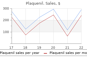
Generic plaquenil 200mg otc
Infant of diabetic mother with amelia of upper limbs rheumatoid arthritis of the eye discount plaquenil 200 mg without a prescription, cleft lip, and caudal dysplasia. Infant of diabetic mother with complete absence of lower half of abdomen and lower extremities. Encephaloclastic Lesions Encephaloclastic lesions can result from insults during the fetal period, at birth, or postnatally. Multicystic encephalomalacia has been reported in twins and is thought to result in multiple emboli occluding cerebral vessels resulting in "Swiss cheese brain. Isolated limb anomalies, especially terminal transverse defects that are sporadic, may be due to vascular disruption. Transverse limb defects, absent limbs, and limb girdles may result from extensive involvement by a hematoma or disruption during early limb bud formation. The limb body wall malformation complex results from a malfunction in the ectodermal placodes. The amnio-ectodermal transition zone plays an important role in the formation of the ventral body wall. A surface ectoderm placode is at the transition zone, depositing mesectodermal cells that will form the mesodermal structures of the body wall. Placodes are specialized parts of the surface ectoderm that add cells to the mesodermal compartment. They are involved in the formation of many organs and structures, including the neural tube, nose, branchial arches, ventral body wall, and limbs. When these ectodermal placodes do not function correctly, the mesoderm remains underdeveloped and severe malformations may be expected. In secondary abdominoschisis the body wall placode is deficient in depositing mesoectodermal cells after the amnioectodermal transition zone has attached to the connecting stalk, and the body cavity has separated from the extraembryonic coelom. The body wall remains very thin and eventually ruptures because of the increase of the abdominal and/or the thoracic organs. The margins of such a body wall defect are smooth and show a transition from skin into the mesothelium of the body cavity. Small developmental defects in the mesodermal compartment can easily result in severe limb malformations due to abnormal function of the limb bud placodes. This is characterized by severe clefts of the abdominal wall with absence of, or very small, umbilical cord, or it is continuous with the placenta. Classically, pleurosomas refers to body wall and upper limb defects, and cyllosomas to body wall deficiency and lower limb defects. In 72% of fetuses, the internal anomalies are recognized to be secondary to vascular disruption. The extrusion of abdominal organs is into the amniotic cavity rather than the extracoelomic space, as occurs in a body wall defect. Gastroschisis appears to result from premature ablation and/or disruption of the embryonic omphalomesenteric artery. The resultant abdominal wall defect leads to extrusion of abdominal contents into the amniotic cavity. Frequent associated anomalies include nonduodenal intestinal atresia or stenosis, atresia of the appendix, atresia of the gallbladder, absence of one kidney, hydronephrosis and hydroureters, and porencephaly. Fetus with thoraco-abdominal wall defect, ectopia cordis with cardiac defects, defect of sternum and diaphragm. Emboli from the placenta to both monogygotis twins, causing death of one twin and structural anomalies from embolic infarction in the surviving twin. Thromboplastin from the demised co-twin causing disseminated intravascular coagulation and structural anomalies in the surviving twin (Swiss cheese brain). Disparate placental blood flow (maternal-placental unit) resulting in altered growth and anomalies from hypo- to hyperperfusion. Adhesive bands are the result of a broad fusion between disrupted fetal parts (mostly cephalic) and an intact amniotic membrane. Most of the craniofacial defects (encephaloceles and/or facial clefts) occurring in these fetuses are not caused by constrictive amniotic bands but are the result of a vascular disruption sequence with or without cephalo-amniotic adhesion. The type of anomalies depends on the stage of embryonic development and the severity of the disruptive event. The amniotic membrane sometimes may become attached to areas of cell death or imperfect histogenesis in the fetus; in this way, amniotic bands can be formed secondary to the malformations. These anomalies most likely result from vascular disruption but amnion rupture may also occur in some cases. The length of the umbilical cord is determined by fetal activity and the tension placed on the cord during growth. A short umbilical cord may be related to omphalocele where traction on a short cord precludes the return of the abdominal contents to the coelomic cavity. Amniotic bands attached to placenta and fetal head resulting in cleft of face and lip. Neonatal gangrene of a limb at the time of birth is rare; most arterial thromboses in infants occur postnatally as a complication of umbilical artery catheters. Amputation of second finger with constriction bands on first, third, and fourth fingers. From the first trimester through 34 weeks, the biparietal diameter is accurate to within 10 days. Other measurements used in the 2nd and 3rd trimesters include fetal abdominal diameter and femur length. It is associated with increased perinatal morbidity and is used as a marker of increased neonatal risk. It is not an ideal marker of fetal growth and development and combines both prematurity and various degrees of growth retardation. In growth-retarded fetuses, the brain development is significantly advanced for the established developmental age. Symmetric growth retardation (A) compared with a normal (B) fetus at 26 weeks gestation. In the symmetric type, the head is reduced in size to the same extent as the body; in the asymmetric type, the head is normal in size and only the body is small. Symmetric growth retardation is seen early in development; asymmetric growth retardation usually is not manifested until after 20 weeks gestation. Timing of the insult to the fetus predisposes to the type of growth retardation: early insults usually result in symmetric growth retardation, probably by restricting fetal cellular hyperplasia. Third trimester insults that restrict cellular hypertrophy usually result in asymmetric growth retardation. Uteroplacental insufficiency and other similar insults result in stresses on the fetus that cause the fetus to redistribute blood flow, maintaining perfusion of the head, heart, and adrenal glands. Particularly severe insults to the fetus may cause asymmetric growth retardation to progress to symmetric growth retardation, as redistribution of blood flow fails to maintain growth of the head. Asymmetric growth retardation in a fetus due to chronic hypoxemia with reduced uteroplacental blood flow. Although dating gestational age by early sonography will tend to miss trisomy 18 fetuses, many such fetuses also will be noted to have increased nuchal "lucency" in the first trimester and subsequently will be found to have other anomalies if evaluated later in pregnancy.
Plaquenil 400mg for sale
Massive malformation of the cerebral hemisphere can constitute the only lesion in congenital varicella syndrome arthritis neck esophagus order plaquenil with a visa. In the acute and generalized infection the skin, liver, and lungs are always involved. Influenza Virus A casual relationship between influenza virus and developmental defects remains unclear. These viral infections have not been associated with an increase in developmental defects. Transplacental passage of Coxsackie viruses is generally detected at term; however, this virus also may be responsible for abortion. Coxsackie virus B infection may cause myocarditis, meningoencephalitis, pneumonitis, and inflammatory foci with necrosis in the liver, pancreas, and adrenals. Pneumonitis and mild myocarditis have been found in A3 and A9 congenital Coxsackie virus infection. Pneumonitis has been described as well as hepatic fibrosis with regenerative nodules, adrenal hemorrhagic necrosis, and foci of myocardial, hepatic, and adrenal calcification. Hepatitis Viruses Hepatitis viruses have not been associated with an increase in fetal loss or in the frequency of developmental defects. Transmission of the virus occurs in one-fourth to one-third of cases and it may occur at any time during gestation. Most of the B-19-associated fetal deaths occur during the second trimester of gestation. Visceral pallor and enlargement of the liver and the spleen are the most frequent internal abnormalities. Adenovirus pneumonia in a newborn with multiple adenoviral inclusions in the alveolar cells. The heart may be normal or symmetrically enlarged and the thymus is abnormally small. It may result in hydrops fetalis due to anemia caused by red blood cell destruction by the virus. Histologic features consist of intranuclear inclusions in erythroid precursors and excessive iron pigment in the liver, hepatitis, a leukoerythroblastic reaction, and eosinophilic nuclear inclusions in hematopoietic cell nuclei. The trypomastigotes are motile, spindle-shaped, and nonmultiplying blood forms of the parasite. Intracellularly they are transformed into amastigotes, which multiply and are later released as a trypomastigotes upon lysis of infected cells. The trypomastigotes enter other cells, initiating a new cycle, or they penetrate the fetal bloodstream, disseminating the infection. The trypomastigotes are present in the blood during the acute phase of the disease but thereafter in the chronic phase of infection, parasitemia is intermittent. Stillborns and newborns with severe disease may present with hydrops and hepatosplenomegaly. Parasites may be detected in most of the organs, but they predominate in the brain, heart, esophagus, intestines, skeletal muscles, and skin. The inflammatory infiltrate consists of mononuclear cells with some neutrophils; less commonly, small granulomas of epithelioid cells are seen. The inflammatory infiltration in the myocardium is generally diffuse and is associated with edema and disruption of the myofibers. The histopathologic diagnosis of congenital Chagas disease is made by finding amastigotes in hematoxylin and eosin-stained preparations. It is believed that fetal hemoglobin protects the fetus against malarial infection, and infection is very low in the first 3 months of postnatal life when the level of fetal hemoglobin is high. Sporozoites introduced into the bloodstream mature in the liver and form the mature schizont, which contains a great number of merozoites. After the release of merozoites the ring stage appears within erythrocytes in the peripheral blood with a subsequent transformation into tachyzoites. Although the parasite is rarely detected in umbilical cord blood, the placenta shows parasitism and sometimes is so affected that it affects the fetus. The reported congenital cases present in the first few months of life with splenomegaly and less frequently with hepatomegaly. Q-Fever Q-fever caused by Coxiella burnetii may cause abortion, prematurity, low birthweight, and fetal death. African Trypanosomiasis Most congenital cases of African trypanosomiasis are not well documented. Considering that the clinical manifestations of this infection generally are of late appearance, it is impossible in endemic areas to consider as congenital late manifestations of the disease. Two cases of congenital disease have been observed in Europe in infants born to infected mothers. Visceral Leishmaniasis (Kala-Azar) Pregnant women with the generalized disease may infect the placenta and possibly the fetus. Borreliosis (Lyme Disease) Fetal infection has been described in Borrelia burgdorferi infection. Spirochetes have been found in brain, spleen, myocardium, and bone marrow, but inflammatory changes are much less than in adult cases and sometimes are absent. The organisms can be demonstrated by silver impregnation by immunofluorescence with monoclonal antibodies. Campylobacter (Vibrio) fetus Infection Campylobacter fetus, a common enteric pathogen in humans, has been described as the cause of prematurity, fetal death, and acute villitis. Fungal Infections Candida results in rounded yellow plaques varying in size from 0. This is usually an incidental finding in an otherwise normal pregnancy that has resulted in a normal newborn. Racult D, Stein A: Q-fever during pregnancy-a risk for women, fetuses and obstetricians. One twin actively perfuses the co-twin (acardiac twin) via large artery to artery and vein to vein anastomosis. The placental parenchyma may be unequally shared by the twins, particularly with respect to venous return. During delivery, the cord of the second twin may prolapse before the birth of the first twin, or it may be around the neck of the first-born twin. Yellow arrows at umbilical cord insertions, black arrows indicate retinal pigment on the left twin and optic vesicle on the right twin. Fetal death of a twin may cause tissue damage to the remaining twin with aplasia cutis and multiple bowel atresias. The pattern of placentation in triplets ranges from one disk with a single inner layer of amnion (monoamniotic) to three separate disks and gestational sacs (trichorionic triamniotic). Eighteen percent of triplets have velamentous insertions, abnormally long cords, and one single-artery cord. Assisted reproduction technology has increased the incidence of quadruplets; fewer than 5% are spontaneous. Tendency of preterm labor occurs earlier and is more resistant to tocolysis with increasing numbers of fetuses. Dicephalus, with two spines and heads, two or more upper limbs, single pelvis, two lower limbs. One axis incompletely expressed (parasite) and attached to complete axis (autosite). The incidence is approximately 1/150,000 births and represents fewer than 1% of monozygotic twins. In these there is sharing of viscera including fusion of multichambered hearts and a common midgut as far as the ileocecal valve. Color doppler studies are useful in viewing arterial circulation particularly in the heart and liver, and fetal echocardiography will determine the cardiac abnormalities. There may be considerable overlap between sacrococcygeal teratoma and fetus in fetu.
Generic plaquenil 400 mg mastercard
Silent myocardial ischemia is commoner than nonsilent ischemia (angina): Beta-blockers are strongly indicated rheumatoid arthritis lupus discount 400 mg plaquenil amex. In the elderly, it is preferable to start with smaller doses, for example, bisoprolol 2. Drug name: Trade namesv Dosage: Carvedilol Coreg, Eucardic Hypertension, initially 12. Drug name: Trade names: Supplied: Dosage: Nebivolol Bystolic; Nebilet, Nebilong, Lobivon 2. Manufacturer advises to avoid if serum creatinine greater than 250 mol/L; see Chap. This highly selective beta1-adrenergic receptor blocker is the only beta-blocker known to induce vascular production of nitric oxide, the main endothelial vasodilator. Nebivolol induces nitric oxide production via stimulates the beta3-adrenergic receptormediated production of nitric oxide in the heart; this stimulation results in a greater protection against heart failure (Maffei and Lembo 2009). The drug stimulates the endothelial L-arginine/nitric oxide pathway and thus causes vasodilation (Cockcroft et al. In contrast with metoprolol, nebivolol improves secondary sexual activity and erectile dysfunction scores (Brixius et al. Although a decade of clinical experience with this drug in Europe provides support to its blood pressure-lowering and antiischemic effects, further clinical trial data are necessary. Maffei and Lembo provided informative information on nitric oxide Chapter 8 / Hypertension 227 mechanisms of nebivolol in an article: Therapeutic Advances in Cardiovascular Disease Maffei and Lembo (2009). But the combination of beta-blocker and valsartan has caused increased cardiac events and is not recommended by the author (see Chaps. Contraindications Hypersensitivity to thiazides or sulfonamides; anuria or severe renal failure; pregnancy and breastfeeding (see Chap. The dose is too large if signs of dehydration or orthostatic hypotension develop, or if the patient has increased urea >10. Hypokalemia occurs in a significant number of patients receiving thiazides and contributes to increased risk of 228 Cardiac Drug Therapy 3. The incidence of hypokalemia can be decreased by the use of low-dose thiazide regimens with K+-sparing diuretics. If mild renal dysfunction exists, hyperkalemia may occur with the use of K+-sparing diuretics; therefore, a plain thiazide is recommended without potassium supplements. Gastrointestinal effects: anorexia, gastric irritation, intrahepatic cholestatic jaundice, and pancreatitis. Central nervous system effects include dizziness, vertigo, paresthesia, and headache. Cardiovascular effects include orthostatic hypotension, low cardiac output, and arrhythmias from hypokalemia. For use in pregnant or nursing mothers, thiazides cross the placental barrier, appear in breast milk, and can cause fetal or neonatal jaundice, thrombocytopenia, decreased vascular volume and placental perfusion, and acute pancreatitis. Precipitation of gout and hyperuricemia is a well-known complication of all diuretics. If gout or hyperuricemia occurs, this is treated in the usual fashion, and the diuretic is discontinued. There is no reason to add allopurinol to the regimen to prevent further episodes of gout. Drug interactions with lithium, steroids, and oral anticoagulants have been reported. Treatment was associated with a 21 % reduction in the relative risk of death from any cause, a 64 % reduction in the relative risk of heart failure, and a 30 % reduction in the relative risk of stroke (Beckett et al. There is no question about the efficacy of diuretics in patients with mild to moderate hypertension, and, when they are combined with other antihypertensive agents, they can be used in all types and degrees of hypertension. Renal failure with increased volume overload is a common cause of resistant hypertension that requires the addition of a diuretic to enhance the efficacy of other agents. Prospective data suggest that in older men and women, the use of thiazide diuretic agents is Chapter 8 / Hypertension 231 associated with a reduction of approximately one-third in the risk of hip fracture. This latter feature creates a hefty depot for chlorthalidone, allowing for a slow streaming effect (red cell plasma) with subsequent gradual elimination from the plasma compartment by tubular secretion (Riess et al. There was good representation for women (47 %) and blacks (35 %); 36 % were diabetics. A study used 24-h ambulatory blood-pressure measurements to study the effects of 25 mg of chlorthalidone daily as compared with 50-mg hydrochlorothiazide daily. Drug name: Trade names: Supplied: Dosage: Hydrochlorothiazide and Amiloride Moduretic, Moduret (C) Tablets containing 50 mg hydrochlorothiazide and 5 mg amiloride Half a tablet daily; see text for further advice the manufacturer indicates up to four tablets daily. A more appropriate combination, co-amilozide, available in the United Kingdom and Europe, contains amiloride 2. This combination-type therapy with Ziac has shown consistently low discontinuation rates because of adverse effects, compared with enalapril. Drug name: Furosemide; [Lasix] Frusemide 234 Cardiac Drug Therapy Furosemide is less effective than thiazides in mild to moderate hypertension, and therefore it is not advisable to use this drug unless the patient has significant renal dysfunction, such as a creatinine level >2. As with thiazides, the combination with a K+-sparing diuretic has proved to be useful in causing further diuresis and in reducing K+ loss. Contraindications: hyperkalemia; concomitant use of potassium-sparing diuretics or potassium supplements. Assess serum potassium before treatment, during initiation, and when dose changed. This action results in: in cardiac work and myocardial oxygen requirement Damage to arteries at branching points: may cause rupture of aneurysms. They are, however, not superior to other agents in diabetic patients, as is often claimed (see Chap. The diminished sympathetic 236 Cardiac Drug Therapy activity causes vasodilation, reduction in afterload, and some decrease in preload. Also, heart rate is not increased by these agents, as opposed to other vasodilators. The accumulation of bradykinin stimulates release of vasodilator prostaglandins that contribute to the decrease in peripheral vascular resistance. Adverse reactions reportedly include severe pruritus and rash in 10 % and loss of taste in 7 % of patients (Jenkins et al. Neutropenia and agranulocytosis are rare and occur mainly in patients with serious intercurrent illness, particularly immunologic disturbances, and in those with an altered immune response, in particular collagen vascular disease. Precipitation of renal failure in patients with tight renal artery stenosis has been reported. Discontinue the following drugs, which alter immune response: steroids, procainamide, hydralazine, probenecid, tocainide, allopurinol, acebutolol, pindolol, and others. Restrictive, obstructive, or hypertrophic cardiomyopathies, constrictive pericarditis, cardiac tamponade, and hypertensive hypertrophic cardiomyopathy of the elderly with impaired ventricular relaxation. Avoid in women aged <45 years who may wish to become pregnant Chlorpromazine therapy because severe hypotension may occur. The usual recommended initial dose in patients not taking a diuretic is 5 mg once daily, except in the elderly or in those with renal impairment, and in suspected high renin states as seen with renal artery stenosis, prior diuretic use, and low-sodium diets. Note that the serum creatinine level may be normal in patients aged >70 years with renal impairment, and caution with dosage is necessary. The maximum doses given in these tables are approximately 20 % lower than those stated by the manufacturer. The combinations are not recommended if there is moderate renal impairment: serum creatinine level >2. A case of an increase from 170/100 to 300/200 mmHg has been reported on sudden cessation of nifedipine therapy (Bursztyn et al. Constipation and bradycardia are limiting considerations, particularly in patients aged >70 years. Drug name: Trade names: Supplied: Dosage: Felodipine Plendil, Renedil Extended-release tablets: 2.
Plaquenil 200 mg discount
Induction of nitrate tolerance in heart failure by continuous infusion of nitroglycerin and reversal of tolerance by N-acetylcysteine rheumatoid arthritis nih purchase discount plaquenil online, a sulfhydryl donor (abstract). Tolerance development during isosorbide dinitrate treatment: can it be circumvented A meta-analysis of 17 randomized trials of a percutaneous coronary intervention-based strategy in patients with stable coronary artery disease. Hemodynamic effects of intermittent transdermal nitroglycerin in chronic congestive heart failure. Of 55 million deaths globally every year, about 30 % are from cardiovascular diseases. There is evidence that a small consumption of alcohol, 1 drink for women and 2 for men, confers some cardiovascular protection. But a Prospective Study and Dose-Response MetaAnalysis indicated that alcohol consumption, even at moderate intakes, is a risk factor for atrial fibrillation. Most city dwellers are exposed to traffic pollution which is now believed to be a major trigger for heart attacks and supersedes other triggers such as physical exertion in relatively sedentary individuals, air pollution, and anger. Importantly, drivers of vehicles in heavy city traffic are exposed to exhaust fumes which circulate within their vehicles. If nitroglycerin is available, one tablet or two puffs sublingual should be used and three chewable aspirins (total 240 mg) are taken while awaiting the ambulance. Most important, women more frequently Chapter 11 / Myocardial Infarction 315 report pain/discomfort in the neck or jaw and back, as well as nausea; they score their pain/discomfort slightly higher than men (Thuresson et al. It is also of no value in areas where only thrombolytic therapy is available and can be given during ambulance transport or within 20 min of arrival at an emergency room. Transient myocardial stunning: stress-related Takotsubo: transient left apical ballooning. Apical four-chamber views in this 62-year-old female who presented with chest pains show (a, b) akinetic apical segments, markedly hypokinetic mid-ventricular segments, with preserved basal segments-a pattern not consistent with coronary artery anatomy. Work-up for acute myocardial infarct- enzymes, electrocardiogram, and cardiac catheterization-was nondiagnostic. In addition, the J-point level almost equals the height of the T-wave in V6 (Reproduced with permission from Khan M Gabriel, Encyclopedia of Heart Diseases, 2nd edition. The contents of a ruptured plaque are highly thrombogenic, and exposed collagen provokes platelet aggregation. Thus, all patients with known coronary heart disease must be strongly advised to chew and swallow this life-saving agent if chest discomfort exceeds 10 min or if chest pain is not relieved by nitroglycerin (glyceryl trinitrate). Aspirin should be taken once the decision has been made to proceed to the nearest emergency room. The combination of aspirin and beta-blockers is life-saving and has proved to be effective. Pain precipitates and aggravates autonomic disturbances, which may cause arrhythmias, hypotension, or hypertension, thus increasing the size of infarction. The dose is reduced or morphine is discontinued if toxicity is observed, that is, depression of respiration, hypotension, or severe vomiting. The drug allays anxiety, relieves pain, causes venodilation, and therefore reduces preload. Caution: Morphine has been shown to decrease clopidogrel absorption, decrease concentrations of clopidogrel active metabolite, and diminish its salutary effects. The drug is avoided or is used under close supervision if severe respiratory insufficiency is present. Respiratory depression can be treated with the narcotic antagonist naloxone (Narcan) at a dose of 0. The drug appears to have a more euphoriant effect than morphine and is preferred physicians in the United Kingdom. Betablockers can be considered important second-line agents for the control of ischemic pain. In some patients, pain has been documented to be relieved by the administration of betablockers without concomitant use of opiates. Unfortunately, despite active public education, triage in the United States, and in many countries, remains difficult, particularly because only approx. Although improvement in survival has been shown in clinical trials to occur with thrombolytic therapy given up to 4 h after the onset of symptoms, the gain is greatest within the first 2 h and then falls off dramatically after the fourth hour. For those who cannot reach a coronary intervention within 2 h or facilities are not available, thrombolytic therapy administered at the earliest moment (<2 h) is of the utmost importance. The delay in the emergency room from the arrival of the patient to the administration of thrombolytic therapy varies from 30 to 90 min. The patient may therefore quickly summon transport by a mobile emergency service that should be equipped with semiautomated defibrillators and provide the use of lifesaving thrombolytic therapy; in the absence of such facilities, the patient should present without delay to the nearest emergency room for early therapy which should be given within the golden 1 h Clopidogrel Antiplatelet therapy should be initiated before diagnostic angiography with clopidogrel (loading dose 600 mg followed by daily maintenance dose 75 mg). Morphine has been shown to decrease clopidogrel absorption, decrease concentrations of clopidogrel active metabolite, and diminish its salutary effects. In particular, omeprazole has been reported to significantly decrease the inhibitory effect of clopidogrel on platelet aggregation. It is advisable to lower the maintenance dose to 5 mg in patients who weigh <60 kg, although the effectiveness and safety of the 5-mg dose have not been studied prospectively (Wright et al. At 1 year, the primary composite end point occurred in fewer patients in the 6,732 ticagrelor-treated group than in the 6,676 clopidogrel group: p = 0. Ticagrelor compared with clopidogrel significantly reduced all-cause mortality at 12 months in all patients (4. Within 30 min, a ticagrelor loading dose of 180 mg results in roughly the same level of inhibition of platelet aggregation as that achieved 8 h after a clopidogrel loading dose of 600 mg (Gurbel et al. There was no significant difference in the rates of fatal bleeding associated with rivaroxaban as compared with placebo (0. Thrombolytic therapy administered at the earliest moment (<2 h) is of the utmost importance. Lives saved / 1000 treated: within 1, 3 and 7 hours respectively: 65 %, 27 %, 8 %. Delays may result from duplication of assessments by different teams of clinicians. The emergency room physician and assistants should have the training and authority necessary to administer a thrombolytic agent. History of cerebrovascular accident with any residual disability, intracranial or intraspinal aneurysm, brain tumor, or arteriovenous malformation. Age over 75 years with suspicion of cerebral arteriosclerotic vascular degeneration, agitation, or confusion. Patients older than age 65 years presenting after 12 h should not be given a thrombolytic drug; there is a high risk of cardiac rupture, particularly in women. A skin test is available and can be done in the emergency room and read in 15 min. Hydrocortisone or methylprednisolone is rarely used, because fatalities have not been reported, and reactions are easy to control with epinephrine and antihistamines. There is an increased risk of hemorrhage in patients who are receiving or who have been recently treated (past 5 days) with anticoagulants, indomethacin, and similar antiinflammatory agents, sulfinpyrazone, allopurinol, and sulfonamides. The risk for major bleeding can be reduced by decreasing the enoxaparin dose to 0. Sreptokinase was the main fibrinolytic used (given to 73 % of those who received lytics). There is no pharmacological reason why the benefits of fondaparinux should differ with different thrombolytic agents. Rivaroxaban can be commenced 12 h after the final dose of fondaparinux (Mega et al. Improvement of coronary diastolic filling is achieved at a lower heart rate (see Chap. Although atenolol has weak cardioprotective properties and is expected to confer less cardioprotection than carvedilol and metoprolol, the drug showed a positive result (see Chap.
Buy cheap plaquenil 400mg on line
The presence of large neck masses may limit mobility of the neck arthritis supplies generic 200mg plaquenil otc, which in turn may contradict breech delivery. Large masses may also necessitate consideration of cesarean delivery for vertexpresenting infants, as has been described for goiter and hemangiomas (Stocks et al. Clefting of the upper lip is relatively easy to assess, while abnormalities of the palate are more difficult to evaluate. If major chromosomal abnormalities are suspected, care should be taken to exclude the possibility of central facial abnormalities. Measurement of inter- and intraorbital diameters and careful evaluation of the nose and mouth are recommended. They occur more commonly in Asians and Native Americans and are uncommon in blacks. The association between facial clefts and aneuploidy varies by the timing of the evaluation. Aneuploidy is found in up to 40% of antepartum evaluations for facial clefting (usually either trisomy 13 or 18) but in only 1% of newborns with facial clefts. These differences occur because of higher pregnancy wastage rates in aneuploid fetuses. The images should be evaluated to identify any bulging, sac-like protrusions, or if abnormal thickening of the skin posterior to the spine is present, which might suggest a neural tube abnormality. If an elevated maternal serum -fetoprotein screen has been identified, or if sonographic markers of neural tube abnormalities such as bitemporal narrowing (the lemon sign), cerebellar fusion (the banana sign), or ventriculomegaly are present, special care should be taken to evaluate for abnormalities of the neural tube. Abdominal Sonography Evaluation of the abdomen should include documentation of the stomach bubble in its proper situs (and concordant with the heart in the thoracic cavity). Inspection of the abdominal contents such as bowel lumen size, ascites, proper appearance of the umbilical cord insertion site, overall contours of the diaphragm and anterior abdominal wall, and appearance of the kidneys is recommended. The primary cross-sectional abdominal image should be obtained in a plane almost perpendicular to the major axis of the spine. The optimal image should include the stomach bubble and hepatic vein in an area close to (but not at) the umbilical cord insertion site and should not include cross sections of the heart, kidneys, bladder, or the actual umbilical cord insertion into the abdomen. This view is best localized by aligning the transducer with the spinal column, rotating the transducer 90 and sliding the transducer cephalad or caudad to obtain a scanning plane inferior to the heart and superior to the renal poles. The stomach bubble is normally found situated on the left of the abdomen, caudad to the heart but with concordant situs. Esophageal atresia and other small bowel atresias are associated with aneuploidy (usually trisomy 21 and 18). Esophageal atresia is also associated with cardiac, gastrointestinal, and genito-urinary abnormalities. A right-sided stomach bubble suggests possible situs inversus or complete transposition of the great vessels (depending on cardiac situs). The size of the stomach increases with fetal swallowing activity and by gestational age. This finding strongly suggests aneuploidy, usually trisomy 21, and necessitates consideration of amniocentesis. Swallowed particulate material within the stomach suggests possible meconium passage in utero. Axial image: "Double-bubble sign" of duodenal atresia (S = stomach, D = duodenum). Esophageal atresia should be suspected if polyhydramnios develops and the stomach bubble cannot be identified. Higher rates would be expected if such infants were assessed earlier in pregnancy. Bowel Echogenicity During abdominal sonography, increased echogenicity of bowel or abdominal structures may be noted. This is a subjective finding that occasionally is associated with karyotypic abnormalities but at times may be overdiagnosed because of technical considerations. The combination of high ultrasound gain and low dynamic range should be avoided when evaluating possibly echogenic bowel to limit falsely positive diagnoses. Potential causes of this sonographic appearance include normal variation, aneuploidy (present in 20% of cases), infections such as toxoplasmosis, meconium ileus due to cystic fibrosis, prior intra-amniotic hemorrhage with ingestion of red blood cells, and uteroplacental insufficiency. Echogenic bowel in association with growth retardation usually is not associated with chromosomal abnormalities. Abdominal calcifications are bright specular intra-abdominal echos with evidence of posterior shadowing. A wide variety of conditions are associated with such calcifications, including infections such as toxoplasmosis and cytomegalovirus, neoplasms (neuroblastoma, teratoma, hemangioma, and hepatoblastoma), fetal cholelithiasis, and complications from meconium peritonitis and meconium plugging. Meconium peritonitis may be focal or diffuse, and meconium pseudo-cysts may develop. Meconium plugs commonly are found with cystic fibrosis and also sometimes occur in association with small bowel atresias and anorectal atresia (very distal lesions may not show dilated loops of bowel). Excessive bowel dilation suggests distal obstruction, and dilated bowel loops are sometimes confused with hydroureters. At 5 weeks of embryonic life, proliferating bowel epithelium obliterates the duodenal lumen, with subsequent restoration of patency within 6 weeks. Failures of vacuolation, vascular accidents, and interruption of the bowel lumen by a diaphragm or membrane may interrupt the recannulation of the duodenum. Atresia is often noted near the ampulla of Vater, and common bile duct obstruction may also be present. Rarely, a central web within the stomach may obstruct flow out of the stomach, leaving a single bubble. Aneuploidy is found in 38% of cases of isolated duodenal atresia, and in 64% of infants with aneuploidy and any other anomalies. Note the cord insertion into the fetal abdomen (arrowhead) lateral to the gastroschisis (L = limb). Mortality from duodenal atresia is approximately 36%, primarily in infants with multiple anomalies. The colon can be visualized by 28 weeks in most fetuses, and it increases in diameter with gestational age, averaging 5 mm at 26 weeks gestation and 17 mm at term gestation (Goldstein et al. This may result from imperforate anus, volvulus, bowel perforation and meconium ileus, and Hirschsprung disease. Dilated bowel does not necessarily constitute an indication for emergency delivery (Sipes et al. Most ventral wall abnormalities are located near the umbilical cord insertion into the abdomen. It occurs in 1/10,000 to 1/15,000 live births and often is found in association with elevation of maternal serum -fetoprotein. Gastroschisis may result from vascular compromise of either the umbilical vein or the omphalomesenteric artery. Ischemic injury to the region of the superior mesenteric artery may explain high rates of jejunal atresia found in association with gastroschisis. Defects in gastroschisis are generally small, <4 cm in diameter, and bowel loops are often covered by an inflammatory exudate. The defects are usually situated in the right paraumbilical area and free-floating bowel often is noted in the peritoneal cavity.
Generic plaquenil 200 mg amex
This obscure trial finding nevertheless led Messerli and colleagues (1998: 13) to state incorrectly that diuretics and not beta-blockers are the treatment of choice in elderly hypertensives arthritis unspecified icd 10 buy plaquenil toronto. A rebuttal with which I concur was made by Kendall and Cohen (1999) regarding the recommendation for treatment with beta-blockers in elderly hypertensives. This risk is higher in patients older than age 60 years than in younger individuals. A trial of the betablocker bucindolol in patients with advanced chronic heart failure. Regional and racial differences in response to antihypertensive medication use in a randomized controlled trial of men with hypertension in the United States. Double-blind placebo-controlled crossover comparison of five classes of antihypertensive drugs. Efficacy and safety of nebivolol and valsartan as fixeddose combination in hypertension: a randomised, multicentre study. A comparison of six antihypertensive agents with placebo: the department of veterans affairs cooperative study group on antihypertensive agents. Medical research council trial of treatment of hypertension in older adults: principal results. Glucose intolerance in hypertensive patients treated with diuretics: a fourteen-year follow-up. Application of ambulatory blood pressure monitoring in differentiating between antihypertensive agents. Randomized trial of a perindopril-based blood pressure lowering regimen among, individuals with previous stroke or transient ischaemic attack. A comparison of the efficacy and safety of a beta-blocker, a calcium channel blocker, and a converting enzyme inhibitor in hypertensive blacks. Trial of secondary prevention with atenolol after transient ischemic attack or nondisabling ischemic stroke. Cardiovascular risk and risk factors in a randomised trial of treatment based on the beta-blocker oxprenolol. Management of essential hypertension in patients with different degrees of left ventricular hypertrophy. Effects of individual risk factors on the incidence of cardiovascular events in the treated hypertensive patients in the hypertension optimal treatment study. For stable angina, the most important feature is the causation of pain by a particular exertional activity and relief within minutes of cessation of the precipitating activity. Catecholamines cause an increase in heart rate, velocity, and force of myocardial contraction that increase oxygen demand and ischemia. Increased heart rate decreases the diastolic interval during which coronary artery perfusion occurs. Ischemia further stimulates catecholamine release, thereby perpetuating the vicious circle. Thus, beta-blocking agents play a key role in the management of patients with myocardial ischemia manifested by anginal pain or silent ischemia. The pathophysiology of unstable angina is more complex and is dealt with later in this chapter. There was rapidity of improvement in health status in both treatment groups; the majority of patients who received optimal medical therapy alone had 286 Cardiac Drug Therapy improved symptoms within 3 months. The improvement was greatest in patients when complete revascularization was achieved (George et al. Beta-Adrenoceptor Blocking Agents All cardiologists now agree that beta-blockers are standard first-line therapy for stable and unstable angina. Calcium antagonists and nitrates cannot reduce mortality or morbidity in these patients and are relegated to second-line therapy. These beta-blockers are chosen because they have been shown to decrease mortality in patients with coronary heart disease (see Chap. Atenolol, a watersoluble drug, is frequently used worldwide for angina and hypertension. The water-soluble, non-lipid-soluble betablockers atenolol and nadolol, also pindolol and acebutolol, are not recommended (see Chaps. Timolol caused an outstanding unmatched 67 % reduction in sudden deaths in post-infarct patients (Norwegian Multicenter Chapter 10 / Angina 289 Study Group 1981), a result that has been lost to the cardiology world. Beta-blockers significantly reduce the number of episodes of angina in more than 75 % of patients. Observations have established that silent ischemia is common and is easily provoked by daily stressful activities (Deanfield et al. Beta-blockers and calcium antagonists have been shown to ameliorate silent ischemic episodes, but beta-blockers are superior. Cardioprotection and Dosage of Beta-Blocker Table 1-4 gives dosages of beta-blockers. The salutary effect of smaller doses is unknown, and larger doses are likely to be nonprotective (see Chap. If side effects occur, the dose is reduced, and a nitrate or calcium antagonist is added. If the maximum cardioprotective dose is used and angina is not controlled, the dose of beta-blocker can be increased, but adverse effects may limit the increase. Some patients do better on an average dose of beta-blocker plus a nitrate or calcium antagonist. Several trials have shown that verapamil is as effective as beta-blockers in the control of angina, but this agent does not prolong life. The hemodynamic, electrophysiologic, and pharmacokinetic effects, adverse effects, and relative effectiveness of calcium antagonists are given in Tables 5-2 and 5-3 and are discussed in Chap. Nitrates Drug name: Supplied: Sublingual glyceryl trinitrate: Spray (Nitrolingual): Nitroglycerin: glyceryl trinitrate Sublingual nitroglycerin: 0. The drug will not be as effective if the patient is lying down; if the patient is standing, dizziness or presyncope may occur. If the systolic blood pressure in routine follow-up is more than 130 mmHg, then it is safe to give 0. The patient must be instructed that nitroglycerin tablets are to be kept in their dark, light-protected bottles; they may be rendered useless after 6 months, or even earlier, if they Chapter 10 / Angina 293 are not protected from light. These two bottles must contain approximately 1-month supply and no cotton wool, to ensure rapid availability in emergencies. At the end of each month, the containers should be emptied and the supply replenished from a third-stock bottle. This will allow a 12-h nitrate-free interval to maintain the efficacy of the drug. Action: Nitrates bind to "nitrate receptors" in the vascular smooth muscle wall that activate guanylate cyclase and thereby stimulate the generation of cyclic guanosine monophosphate, which causes relaxation of the vascular smooth muscle and thus dilation of the veins and, to a lesser extent, arteries. The result is marked dilation of the venous bed and therefore reduction in preload and a minimal decrease in afterload. The large first-pass inactivation of orally administered nitrates causes poor bioavailability to vascular receptors. Cutaneous Nitroglycerins Long-acting or slow-release cutaneous nitroglycerin preparations are available. The advantage of a cutaneous preparation is that the active drug reaches the target organs before it is inactivated by the liver.
Buy plaquenil 200mg low cost
Polytopic Field Defect incorporates more than one developmental field but occurs within the same time during development arthritis in my knee cap buy discount plaquenil 400mg on-line. Three types of structural defects that can result in a chain of defects (sequence) by the time of birth. They are the traits that constitute our morphologic uniqueness, which are also the heritage of ethnic groups and of family inheritance. The morphogenetic lability of the midline increases the probability of multiple midline anomalies in the same individual (as in the trisomy 13 syndromes). Midline morphogenetic events include segmentations (rhombomeres), branchings (lung buds), decussations (corpus callosum), programmed cell death with morphogenetic resorptions (buccopharyngeal and anal membranes, tail), morphogenetic movements (d-looping of cardiac tube), cell migrations (neural crest, primodial cells), etc. Mild Malformations Versus Minor Anomalies Minor anomalies are quantitative changes occurring during phenogenesis. Mild malformations are evident as such per se; apparent minor anomalies must always be evaluated on the basis of the family. Multiple minor anomalies and apparent absence of family resemblance are a highly sensitive indicator of aneuploidy; indeed, many Down syndrome individuals have only growth and minor anomalies. In the third week of development, the mesoderm migrates anterior to the notochord and is responsible for the induction and differentiation of the forebrain and the midline facial structures. Failure of this migration produces a developmental field defect, including arrhinencephaly/holoprosencephaly with midline facial defects. Otocephaly Otocephaly is a first branchial arch developmental field defect that affects structures in the face and upper neck and comprises absence of the mandible and 7. Extreme mandibular hypoplasia, microstomia and contiguous ears (A) beneath the mandible. It has been related to defects of the neural crest cells of cranial origin or to defects in the underlying mesodermal support elements of these cells. Patients with otocephaly may have associated cardiac defects, renal anomalies, bilateral pulmonary agenesis, and esophageal atresia. Most cases have occurred sporadically, although its rare presence in sibs suggests autosomal recessive inheritance in a few cases. The bladder may be massively distended or ruptured, and hydroureters, hydronephrosis, and cystic renal dysplasia may occur. It is a severe developmental field defect of the posterior axis caudal blastema, resulting in apparent fusion of the lower limb buds. It occurs in the primitive streak stage during week 3 of gestation, before development of the allantois, and the allantoic vessels are usually absent. There may be a single umbilical artery that arises directly from the aorta but two symmetrical umbilical arteries have been observed severally. Other defects of the caudal axis include imperforate anus, lower vertebral defects, and genitourinary anomalies. Caudal dysgenesis is a defect that is most commonly seen in infants of diabetic mothers. Renal agenesis or cystic renal dysplasia occurs in virtually every case and is accompanied by pulmonary hypoplasia. The term association should be restricted to the idiopathic occurrence of multiple congenital anomalies apparently not of blastogenetic origin. Absence of the vagina, absence or hypoplasia of the uterus, and renal abnormalities, including agenesis and ectopy, also occur. This newborn infant had colobomas, atresia choanae, retarded growth and ear anomalies. Disruption (See Chapter on Disruptions) A disruption, or secondary malformation, is a morphologic defect of an organ, part of an organ, or larger region of the body resulting from an extrinsic breakdown of, or interference with, an originally normal developmental process (Table 7. Sequence A sequence is a pattern of multiple anomalies derived from a single known or presumed prior anomaly. The lack of amniotic fluid restricts fetal movement and causes fetal compression, producing the typical phenotype of the Potter sequence. Pierre Robin Sequence Robin sequence can be malformational when based on intrinsic mandibular hypoplasia, or deformational when based on constraint. The urinary tract is greatly dilated, usually with urethral or bladder neck obstruction. Megalourethra, megacystis, megaureters, renal hypoplasia, and hydronephrosis may occur. In the Child With Multiple Birth Defects, 2nd ed, Oxford, Oxford University Press, 1997. DiGeorge Sequence the primary defect in DiGeorge sequence involves the fourth branchial arch and derivatives of the third and fourth pharyngeal pouches with defects of the thymus, parathyroids, conotruncal defect of the heart, occasionally the thyroid (Table 7. This complex is associated with congenital absence of the thymus, absence of the parathyroid glands, and immunodeficiency. Because of the role played by the cephalic neural crest in the morphogenesis of the heart, conotruncal heart defects are commonly seen in the DiGeorge sequence. The neural crest has a midline pathogenetic origin and DiGeorge sequence is frequently associated with other midline sequences, schisis anomalies, and arrhinencephaly. Causally, it also may be related to fetal alcohol and fetalAccutane-induced disruptions as well as to the effects of maternal diabetes. A common (1/4,000 liveborn infants) microdeletion of chromosome 22 has been identified as is also seen in the velocardiofacial and conotruncal face syndromes. Dysplasia (See Chapter 9) Dysplasia is the process and the consequences of dyshistogenesis. Blastogenesis refers to all stages of development from the time of karyogamy and the first cell division to the end of gastrulation [(stage 13), day 28]. This is the time of closure of the caudal neuropore and the end of the formation of the intraembryonic mesoderm from the primitive streak. Alcohol teratogenicity Defective migration, proliferation of ectomesenchyme into arches External acoustic meatus Arches Meckel Cartilage. Malleus, incus, ear hillocks Stapes, hyoid (part of), styloid process, stapedial artery Hyoid (majority), proximal third of internal carotid artery Thyroid and laryngeal cartilages. It encompasses the following: Gastrulation, which occurs with the formation of mesoderm and the ap- pearance of the midline, cranial/caudal, right/left, and dorsal/ventral body axes; segmentation; neurulation; and initiation of all developmental processes including neurogenesis, angiogenesis, and (meso)nephrogenesis. Anomalies of blastogenesis tend to be complex, those of organogenesis less complex. Anomalies of blastogenesis tend to be multisystem anomalies or complex polytopic field defects such as the acrorenal field defect; those of organogenesis are more likely to be localized, monotopic field defects. Anomalies of blastogenesis are frequently lethal; anomalies of organogenesis are less commonly lethal. Anomalies of blastogenesis frequently involve defects of placentation or cord formation; except for the presence of a single umbilical artery, the umbilical cord, placenta, and body wall are usually normal in defects of organogenesis. Defects of blastogenesis are frequently associated with monozygotic twinning, which is, by definition, an abnormality of blastogenesis; twinning is less common or not a factor in organogenetic malformations. Sex differences in occurrence appear to be less conspicuous in blastogenetic malformations. In organogenetic malformations there are frequently striking sex differences, an apparent indicator of multifactorial determination. Anomalies of blastogenesis are defects of the embryonic midline; defects of organogenesis are not confined to the midline. Abnormalities of blastogenesis may constitute a cancer risk such as teratomas anywhere along the midline from the skull to the tip of the coccyx; organogenetic malformations are rarely associated with a cancer risk. Multiple congenital anomalies of blastogenesis are usually polygenic field defects or associations; multiple congenital anomalies of organogenesis are more likely to be syndromes representing pleiotropy due to Mendelian mutations and/or chromosome abnormalities.
