

Cheap imuran online
He saw a wall of flame muscle relaxant homeopathic cheap 50 mg imuran with visa, and he felt searing heat and instant pain like nothing he had ever imagined. The skin, or integument, is an important system that we often take for granted until it is compromised. The integumentary system protects the body, helps maintain a constant body temperature, and provides sensory information about the surrounding environment. Because of its visibility, skin reflects our emotions (frowning, blushing) and some aspects of normal physiology (sweating). For example, the bluish skin color associated with hypoxia (oxygen deficiency at the tissue level) is one sign of heart failure as well as other disorders. Abnormal skin eruptions or rashes such as chickenpox, cold sores, or measles may reveal systemic infections or diseases of internal organs, while other conditions, such as warts, age spots, or pimples, may involve the skin alone. So important is the skin to self-image that many people spend a great deal of time and money to restore it to a more normal or youthful appearance. For this reason, if you cut the epidermis there is no bleeding, but if you cut the dermis there is bleeding. Deep to the dermis, but not part of the skin, is the hypodermis (hypo- below), or subcutaneous layer. In adults, the skin covers an area of about 2 square meters (22 square feet) and weighs 4. The hypodermis serves as a storage depot for fat and contains large blood vessels that supply the skin. They are located in the deepest layer of the epidermis, where they contact a tactile (Merkel) disc, a flattened process of a sensory neuron (nerve cell). Tactile cells and their associated tactile discs function together to detect touch sensations. Cells of the Epidermis the epidermis is composed of keratinized stratified squamous epithelium. Keratinocytes also produce lamellar granules, which release a waterproofing sealant that decreases water entry and loss. Once inside keratinocytes, melanin forms a protective veil over the nucleus on the side facing the skin surface. Some cells in this layer are stem cells that undergo cell division to continually produce new keratinocytes. Melanocytes and tactile cells (with their associated tactile discs) are scattered among the keratinocytes of the stratum basale layer. Keratinocytes of the straum spinosum shrink and pull apart when prepared for microscopic examination, so that they appear to be covered with thornlike spines (hence the name "spinosum"). As the keratinocytes of the stratum granulosum move farther from their source of nutrition (the dermal blood vessels), they can no longer carry on vital metabolic reactions, and die. Thus, the stratum granulosum marks the transition between the deeper, metabolically active strata and the dead cells of the more superficial strata. A distinctive feature of this layer is the presence of membrane-enclosed lamellar granules within the keratinocytes. Lamellar granules release a lipid-rich secretion that is deposited in the spaces between cells of the stratum granulosum, stratum lucidum, and stratum corneum. The lipid-rich secretion acts as a water-repellent sealant, retarding loss of water and entry of foreign materials. It consists of four to six layers of flattened clear, dead keratinocytes that contain large amounts of keratin and thickened plasma membranes. The cells are extremely thin, flat, plasmamembrane enclosed packages of keratin that no longer contain a nucleus or any internal organelles. The cells within each layer overlap one another like the scales on the skin of a snake. In this outer stratum of the epidermis, cells are continuously shed and replaced by cells from the deeper strata. Its multiple layers of dead cells help the stratum corneum to protect deeper layers from injury and microbial invasion. Growth of the Epidermis the stratum basale cells are closest to blood vessels in the dermis, and therefore receive the most nutrients and oxygen. These metabolically active cells continuously undergo cell division to produce new keratinocytes. Newly formed cells are slowly pushed from one epidermal strata to the next by the continuing cell division of the stratum basale. As keratinocytes are pushed toward the surface, they receive fewer nutrients from the blood supply and eventually die. Eventually the dead, keratinized cells slough off the surface of the stratum corneum and are replaced by underlying cells. The whole process by which cells form in the stratum basale, rise to the surface, become keratinized, and slough off takes about four weeks. The immature keratinocytes make an abnormal keratin, which forms flaky, silvery scales at the skin surface, most often on the knees, elbows, and scalp (dandruff). Effective treatments- various topical ointments and ultraviolet phototherapy- suppress cell division, decrease the rate of cell growth, or inhibit keratinization. The extensibility of skin can be readily seen around joints and in pregnancy and obesity. The surfaces of the palms, fingers, soles, and toes have a series of ridges and grooves. They appear either as straight lines or as a pattern of loops and whorls, as on the tips of the fingers. Epidermal ridges and dermal papillae fit together like complementary teeth of a zipper forming an extremely strong bond to resist lateral forces that attempt to separate the epidermis from the dermis. The epidermal ridges also increase the surface area of the epidermis and thus increase the grip of the hand or foot by increasing friction. Because the ducts of sweat glands open on the tops of the epidermal ridges as sweat pores, the sweat and ridges form fingerprints (or footprints) upon touching a smooth object. The epidermal ridge pattern is genetically determined and is unique for each individual, and thus can serve as the basis for identification. The second, deeper part of the skin, the dermis, is composed of dense irregular connective tissue containing a woven network of collagen and elastic fibers that provide great tensile strength (resistance to pulling or stretching forces). Because the dermis is typically thinner in women than in men, many women have the appearance of dimples in the skin, referred to as cellulite. Leather, which we use for belts, shoes, and basketballs, is the treated dermis of other animals. The dermis can be divided in to a thin superficial papillary region and a thick deeper reticular region. Dermal papillae contain capillary loops (blood capillaries) and can contain sensory receptors: Corpuscles of touch (Meissner corpuscles) that are sensitive to touch and/or free nerve endings, which initiate signals that produce sensations of warmth, coolness, pain, tickling, and itching. The reticular region (reticul- netlike) is attached to the hypodermis and contains bundles of thick collagen fibers, some elastic fibers, scattered fibroblasts and adipose cells, and various wandering cells (such as macrophages). Blood vessels, nerves, hair follicles, sebaceous (oil) glands, and sudoriferous (sweat) glands occupy the spaces between fibers. The doctor describes Richard as having two types of burns, "partial thickness" and "full thickness. His mother, father, and two sisters came to visit him in the burn unit every day, and his mother stayed overnight. Those burns are third-degree full thickness burns, involving all the layers of the skin down to the underlying subcutaneous layers. Carotene is stored in the stratum corneum and dermis after eating carotenerich foods. Dark-skinned individuals have large amounts of melanin in the epidermis, so their skin color ranges from yellow to reddishbrown to black. Light-skinned individuals have little melanin in the epidermis, making their epidermis appear translucent with a skin color ranging from pink to red depending on the level of oxygen in the blood moving through capillaries in the dermis. The red color is due to hemoglobin, the oxygen-carrying pigment in red blood cells.
Purchase imuran line
The response of a motor unit to a single impulse in its motor neuron is a twitch contraction spasms just before falling asleep 50mg imuran for sale. The three phases of a twitch are the latent period of cell events leading up to contraction; the contraction period of power strokes generating tension; and the relaxation period, during which the muscle is allowed to resume its original length. Multiple stimuli that arrive before the muscle fiber has fully relaxed lead to wave summation. When the frequency of stimulation allows partial relaxation it is called unfused tetanus; rapid frequency of stimulation and sustained contraction is called fused tetanus. Animation-Control of Muscle Tension Exercise-Increase Muscle Tension Clinical Connection-Abnormal Contraction of Skeletal Muscle Clinical Connection-Hypotonia and Hypertonia 2. Motor unit recruitment is the process of increasing the number of contracting motor units. A muscle at rest exhibits muscle tone, a small amount of tension due to involuntary alternating contractions of a small number of its motor units that do not produce movement. Damaged motor neurons that cannot maintain muscle tone cause a muscle to become flaccid. Isotonic contractions involve a change in muscle length without a change in its tension. There are two types: concentric isotonic contractions occur when the muscle shortens; eccentric isotonic contractions occur when the muscle lengthens. An isometric contraction occurs when the load equals or exceeds the muscle tension, and the muscle does not lengthen or shorten. When oxygen is unavailable, anaerobic reactions convert pyruvic acid to lactic acid. Blood removes lactic acid from skeletal muscle and carries much of it to the liver for reconversion to glucose. Muscle fatigue is the inability of muscle to contract forcefully after prolonged activity. Heavy breathing after prolonged muscle activity helps to repay the oxygen debt, more accurately referred to as recovery oxygen uptake. Animation-Muscle Metabolism Exercise-Fueling Contraction and Recovery Concepts and Connections- Muscle Metabolism 2. Skeletal muscle fibers with low myoglobin content appear pale and are called white muscle fibers; skeletal muscle fibers with high myoglobin content have a dark, reddish appearance and are called red muscle fibers. Slow oxidative fibers use aerobic cellular respiration, have a slow speed of contraction, and are fatigue-resistant. Fast glycolytic fibers mainly use glycolysis, contract strongly and rapidly, and are adapted for intense bursts of anaerobic movements, but they fatigue rapidly. Most skeletal muscles in the body are a mixture of all three types of muscle fibers. Training, genetics, and muscle action can slightly alter proportions of the fiber types. Cardiac muscle fibers interconnect by intercalated discs containing desmosomes and gap junctions. Anatomy Overview-Cardiac Muscle Anatomy Overview-Smooth Muscle Tissue 311 Concept 2. There are two types of smooth muscle tissue: (1) visceral (single-unit) smooth muscle tissue is autorhythmic, and the fibers are connected by gap junctions allowing action potentials to spread throughout the network so that cells contract as a single unit; and (2) multiunit smooth muscle tissue acts independently, has few gap junctions, and lacks autorhythmicity. Smooth muscle fibers have tapered ends, one central nucleus, are nonstriated, and lack sarcomeres. Thin and thick filaments of smooth muscle have a sliding mechanism that generates tension, resulting in lengthwise shortening of the fiber. Smooth muscle is involuntary and responds to autonomic nervous system impulses, hormones, and local factors. How is the muscle found in your heart different from the muscle that moves your arms Which connective tissue coat surrounds groups of muscle fibers, separating them in to bundles What prevents an action potential arriving at the synaptic end bulbs of a motor neuron from being transferred directly to a muscle fiber If the lengths of the thick and thin filaments do not change, how does a skeletal muscle fiber shorten during muscle contraction Would muscles of the fingers likely contain many small motor units or a few large ones Which type of contraction can allow you to make a sudden small jerk of your arm when startled In what order are the various types of skeletal muscle fibers recruited when you sprint to make it to the bus stop What are the major functional differences between cardiac and skeletal muscle tissue For a joint to move properly, there needs to be muscles that pull it in different directions. The coaching staff would refer to various muscle groups that the players were using, and Stewart could see how the different muscles worked together to perform some very intricate actions. Now we get to learn about the muscles at the organ level and how they relate to joints. Kendrick continues the lecture on muscle attachments and muscle names, Stewart flips through the pages of the chapter. He works out at the gym regularly and is interested in learning more about the specific muscles that he exercises. He is amazed to realize the logic behind muscle names; he had always used the names but never put much thought in to what they meant. We always talked about working out the biceps, but I never thought about it being a two-bellied muscle. As you study this chapter, pay attention to the action of each muscle and how it interacts with other muscles working on the same joint. Almost 700 individual skeletal muscles, containing both muscle and connective tissues, are found in this important system. A few muscles function mainly to stabilize bones so that other skeletal muscles can execute a movement more effectively. This chapter presents many of the major skeletal muscles in the body, most of which are found on both the right and left sides. While learning the names of skeletal muscles, we will identify their attachment sites and the movements that they produce. Developing a working knowledge of these key aspects of skeletal muscle anatomy will enable you to understand how normal movements occur. This knowledge is especially crucial for professionals, such as those in the allied health and physical rehabilitation fields, who work with patients whose normal patterns of movement and physical mobility have been disrupted by physical trauma, surgery, or muscular paralysis. A useful rule of thumb is that the origin is usually proximal and the insertion distal, especially in the limbs; the insertion is usually pulled toward the origin. The actions of a muscle are the main movements that occur when the muscle contracts. The part of the spring attached to the frame represents the origin, the part attached to the door is the insertion, the coils of the spring are the belly, and closing the door would be the action. Those skeletal muscles that produce movements do so by exerting force on tendons, which in turn pull on bones or other structures (such as skin). When a skeletal muscle contracts, it pulls one of the articulating bones toward the other. The two articulating bones usually do not move equally in response to contraction. You will also see that muscles that cross two joints, such as the rectus femoris and sartorius of the thigh, have more complex actions than muscles that cross only one joint. Fascicular arrangement often represents a compromise between power and range of motion. Pennate muscles, for instance, have a large number of short-fibered fascicles distributed over their tendons, giving them greater power but a smaller range of motion. In contrast, parallel muscles have comparatively fewer fascicles, but they have long fibers that extend the length of the muscle, so they have a greater range of motion but less power. Lever Systems and Leverage A lever is a rigid structure that moves around a fixed point called the fulcrum. In producing movement, bones act as levers, and joints function as the fulcrums of these levers. A lever is acted on by two different forces: the effort, which causes movement, and the load, which resists movement. The effort is the force exerted by muscle contraction; the load is typically the weight of the body part that is moved plus any external object your body is moving. Motion occurs when the effort applied to the bone at the insertion exceeds the load. The force of contraction of the biceps brachii pulling the forearm up is the effort. A lever operates at a mechanical advantage when the load is closer to the fulcrum and the effort is applied farther from the fulcrum; less effort is required to move the load. Conversely, a lever operates at a mechanical disadvantage when the load is farther from the fulcrum and the effort is closer to the fulcrum; more effort is required to move the load.
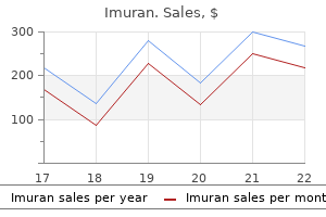
| Comparative prices of Imuran | ||
| # | Retailer | Average price |
| 1 | TJX | 781 |
| 2 | SonyStyle | 992 |
| 3 | True Value | 122 |
| 4 | Gap | 907 |
| 5 | Big Lots | 707 |
| 6 | QVC | 996 |
| 7 | J.C. Penney | 547 |
Discount imuran 50 mg on-line
What is the medication and recommended dilution formula for intracorporeal injection therapy of priapism Typically muscle relaxant recreational use discount imuran, the standard ampule is 10 mg/mL, which is too concentrated to use directly. Vital signs should be checked regularly during this process because of the risk of hypertension when injecting an -agonist. Urological Grab Bag Challenge 613 What is the modality of choice for evaluating the nodal status of penile cancer patients with impalpable nodes True/False: Shockwave therapy has shown slight benefits when used for Peyronie disease. It begins with subacute swelling of the distal extremities and progresses to severe skin induration, which can spread to internal organs. Transmetallation from prolonged exposure to gadoliniumcontrast media is thought to be the underlying cause. Can nephrogenic systemic fibrosis occur in patients without significant renal failure Apparently not, since no confirmed cases have been reported without severe azotemia or end-stage renal disease. True/False: C-reactive protein is an independent prognostic variable for outcomes in renal cell carcinoma and can be used to monitor response to chemotherapy in metastatic disease. True/False: Cryoablation of small renal tumors has similar outcomes to radiofrequency ablation. When performing a partial nephrectomy, what is the maximum recommended warm ischemia time Elastrography is thought to be potentially helpful in identifying prostate cancer. It is performed by compressing prostate tissue, then measuring its tissue strain and degree of elasticity. It is thought that areas of increased stiffness would indicate prostate cancer due to the increased cell density present in cancer tissue. True/False: Penile ultrasonography can be useful in categorizing and following Peyronie disease. Need for nephron-sparing surgery, locally advanced (T4) disease with tumor invasion of surrounding tissues, and significant vena caval tumor thrombus or vena caval wall invasion. What are the characteristics and differential diagnosis of inflammatory myofibroblastic tumors of the bladder The etiology is thought to be inflammatory, but there is evidence of a neoplastic process as well. The main differential diagnosis is sarcoma where the treatment is more surgically aggressive. The correct diagnosis of inflammatory myofibroblastic tumor of the bladder often requires a final histologic confirmation, so this entity must be considered when dealing with bladder masses of undetermined etiology to avoid overtreatment if benign, and potentially dangerous undertreatment if it is ultimately determined to be a malignant sarcoma. This article is intended to provide you with the "secrets" of how to reach the top of Google. When was the last time that you went to the second or third page of a Google search So if you want to get your site and your practice recognized, you have to be on the first page of the Google search and ideally at the very first position. So, how do you get Google to rank your website, in your local area, so it gets in the top 10 which are shown on the first page Consider the fact that Google uses over 200 different algorithms to rank websites, and most of these are not published. To make things harder, many of these algorithms are changed on a regular basis to plug loopholes that programmers and website operators discover to trick or game the system. Only the really important ones are more or less permanent and these are the ones that Google will share and tell us what they want the website owners to do! Whether you want to find a product or service, book a flight, or just research any topic at all, we Google it. Google is still by far the most dominant search engine in the United States followed by Yahoo with 15. The patients are going to be on the Internet and will be mostly looking at the first page of Google, which is why you try to achieve that coveted top spot! Google has the overwhelming ability to send you an endless and enormous supply of patients if you can get on their first page. As we mentioned, there are over 200 different algorithms but here are the top 5 tried and tested steps for search engines to find and rank you: 1. For example, if you are a Urologist and your practice was in Houston the website surfer might type in "Urologist Houston Tx", or simply "Urologist Houston. For "Urologist Houston Tx" Google tells us there are 880 searches per month and for "Urologist Houston" there are 3600 searches per month. Google has an on-line tool that not only tells us how many people are searching a particular keyword every month; they also give you other related keywords that other people are searching for. In that same search above, Google also reports that 480 people are typing in the keyword phrase "Vasectomy Houston. Keywords in Your Website Coding and in the Website Copy Unless you have some experience in this area you might consider hiring a professional for the coding. We recommend that the keyword density should be no more than approximately 3% to 5% of the total copy. This means that if you are writing an article on prostate cancer and the article has 300 words, you should not use the term prostate cancer more than 15 times in the body of article. In-bound Links Google wants users to have a positive experience so that the website surfer will continue to use Google as their primary search engine. Google wants to make sure that the websites they have ranked highly are considered by other websites to be an authority on the subject being searched. The measurement Google uses is the number of websites that have links (your website address on their website) to your website. That means, are the websites that provide a link to your website in a related field. Example: Google would give a high ranking if you had an in-bound link from a medical website such as Mayo Clinic or American Cancer Society. They would penalize you and pull down your rankings if you had an in-bound link from a pest control or a day spay for pets! Getting to the Top of Google in 5 Steps 617 this means you want to submit your website address with a description of your keywords that you want to get ranked for, on a regular monthly basis, to on-line medical directories, to medical publications, and to medical blogs. You should also write a minimum of 250 to 400 word keyword-rich articles with your submissions. New "Keyword Related" Content To position yourself and your website as the knowledgeable expert in your field, add new content that has your keywords in it. Google wants to see an active website, one that has "keyword relevant" updated information. Depending on what your competition is doing your can get an immediate bump in your rankings with your blog. Now this keyword phrase is not as competitive a phrase as "Urologist," but, nonetheless his name and practice was nowhere to be found on the first page. June 2, 2012: He wrote a 300-word blog, "An Ouch In the Pouch-Acute Scrotal Pain and What To Do About It," on his Word Press Blog. The title was "When It Really Hurts Down There- Epididymitis" again with key words "scrotal pain," "testicle," "painful testicle," and "epididymitis" included as tags for the blog. June 10, 2012: A third blog was written, "Pain In the Pouch May Be Coming From Somewhere Else. June 15, 2012: A repeat Google search was conducted on June 15, 2012 which showed, just like magic, that a search for "scrotal pain New Orleans" demonstrated that his practice was at the very top of the Google search page in the first position! Social Media Sites There is a lot of hype about social media sites such as Facebook, Twitter, and YouTube, etc with good reason. First, Google likes them and Google likes them even more when they link back to your website. That means these in-bound links with keyword relevance from Facebook and Twitter are good for your rankings! If you have a social media presence and you regularly add new keyword-related content that is informative, relevant, and fun, over a period of time you will build up followers who will share your information with their friends. These sites give you a forum to position yourself as the expert in your field and will give your rankings a boost! However, if your website does not have any patient conversion processes on it then you will have wasted your time, effort, and money. Success depends on not only getting on the first page of Google so prospective patients will go to your website but also on the conversion quality of your website. Bottom Line All of us have skills and training for diagnosing and treating urologic diseases. However, few of us have the skills necessary to navigate the information superhighway.
Discount imuran 50mg with visa
They are present in several connective tissues spasms mouth order genuine imuran on line, and usually are the most numerous. Fibroblasts migrate through the connective tissue, secreting the fibers and ground substance of the extracellular matrix. In addition, researchers have recently discovered that mast cells can ingest and kill bacteria. However, in response to certain conditions they migrate from blood in to connective tissues to mediate immune system responses. For example, the collagen fibers found in cartilage attract more water molecules than those in bone, which gives cartilage a more cushioning consistency. The bundle arrangement gives the tissue great tensile strength, the ability to resist stretching. Chemically, collagen fibers consist of the protein collagen, which is the most abundant protein in your body, representing about 25 percent of the total protein. Collagen fibers are found in most types of connective tissues, especially bone, cartilage, tendons (which attach muscle to bone), and ligaments (which attach bone to bone). Elastic fibers, which are smaller in diameter than collagen fibers, branch and join together to form a fibrous network within a tissue. Elastic fibers contain the protein elastin, which allows them to stretch up to 150 percent of their relaxed length without breaking. Equally important, elastic fibers have the ability to return to their original shape after being stretched, a property called elasticity. Reticular fibers (reticul- = net) consist of collagen arranged as fine, branching, interwoven fibers that provide support in the walls of blood vessels and form a network around the cells in some tissues, such as areolar connective tissue, adipose tissue, and smooth muscle tissue. Reticular fibers are much thinner than collagen fibers and form branching networks. Reticular fibers are plentiful in reticular connective tissue, which forms the stroma (= bed or covering) or supporting framework of many soft organs, such as the spleen and lymph nodes. The five types of mature connective tissue are loose connective tissue, dense connective tissue, cartilage, osseous tissue, and liquid connective tissue (blood and lymph). Loose Connective Tissue Connective Tissue Cells Each major type of connective tissue contains an immature class of cells with a name ending in blast, which means " to bud or sprout. The types of loose connective tissue include areolar connective tissue, adipose tissue, and reticular connective tissue (Table 4. In and around nearly every body structure (thus, called "packing material" of the body). Hypodermis deep to skin; lamina propria of mucous membranes; around blood vessels, nerves, and body organs. Cell fills up with a single, large triglyceride droplet, and cytoplasm and nucleus are pushed to periphery of cell. Subcutaneous layer deep to skin; around heart and kidneys; yellow bone marrow; padding around joints and behind eyeball in eye socket. Reduces heat loss through skin; serves as an energy reserve; supports and protects organs. Forms stroma of organs; filters and removes worn-out blood cells in spleen and microbes in lymph nodes. There are three types: dense regular connective tissue, dense irregular connective tissue, and elastic connective tissue (Table 4. After an incision is made in the skin, the fat is removed through a stainless steel tube, called a cannula, with the assistance of a powerful vacuum pressure unit that suctions out the fat. The technique can be used as a body-contouring procedure in regions such as the thighs, buttocks, arms, breasts, and abdomen, and to transfer fat to another area of the body. Postsurgical complications that may develop include fat that may enter blood vessels broken during the procedure and obstruct blood flow, infection, loss of feeling in the area, fluid depletion, injury to internal structures, and severe postoperative pain. Forms tendons (attach muscle to bone); most ligaments (attach bone to bone); aponeuroses (sheetlike tendons that attach muscle to muscle or muscle to bone). Walls of elastic arteries; lung tissue; trachea; bronchial tubes; ligaments between vertebrae. Allows stretching of various organs; is strong and can recoil to original shape after being stretched. Elasticity is important to normal functioning of lung tissue (recoils in exhaling) and elastic arteries (recoil between heartbeats to help maintain blood flow). Fine collagen fibers are not visible with ordinary staining techniques; chondrocytes are found in lacunae. Ends of long bones; anterior ends of ribs; nose; trachea; larynx; bronchi; embryonic and fetal skeleton. Reduces friction and absorbs shock at joints, provides flexibility and support; weakest type of cartilage. Pubic symphysis (where hip bones join anteriorly); intervertebral discs, menisci (cartilage pads) of knee; portions of tendons. The extracellular matrix of cartilage forms a strong, firm material that resists tension (stretching), compression (squeezing), and shear (pushing in opposite directions). Because of these properties, cartilage plays an important role as a support tissue in the body. Though bone gradually replaces cartilage during further development, cartilage persists after birth as the growth plates within bone that allow bones to increase in length during the growing years. Cartilage also persists throughout life as the lubricated articular surfaces of most joints. Cartilage in external ear Cartilages in nose Like other connective tissues, cartilage has few cells and large quantities of extracellular matrix. It differs from other connective tissues, however, in not having nerves or blood vessels in its extracellular matrix. There are three types of cartilage: hyaline cartilage, fibrocartilage, and elastic cartilage (Table 4. Key: Bones of skeleton Articular cartilage of a joint Hyaline cartilages Elastic cartilages Fibrocartilages Costal cartilage Cartilage of intervertebral disc Pubic symphysis Epiglottis Larynx Trachea Lung Bronchus Meniscus Respiratory tube cartilages in neck and thorax Cartilage provides flexible, resilient support. The skeletal system supports soft tissues, protects delicate structures, and works with skeletal muscles to generate movement. The extracellular matrix of osseous tissue consists of mineral salts (mostly calcium and phosphates), which give bone its hardness and compressive strength, and collagen fibers, which give bone its tensile strength. The extracellular matrix is arranged in concentric rings or lattice-like thin columns (Table 4. Mature bone cells, called osteocytes, are located within lacunae in the extracellular matrix. Blood is a connective tissue with a liquid extracellular matrix and formed elements. The extracellular matrix is called blood plasma, and consists mostly of water with a wide variety of dissolved substances-nutrients, wastes, enzymes, plasma proteins, hormones, respiratory gases, and ions. Suspended in the blood plasma are formed elements-red blood cells, white blood cells, and platelets (Table 4. Red blood cells transport oxygen to body cells and remove carbon dioxide from them. It consists of several types of cells in a clear liquid extracellular matrix that is similar to blood plasma, but with much less protein. For example, lymph leaving lymph nodes includes many lymphocytes, a type of white blood cell, but lymph from the small intestine has a high content of newly absorbed dietary lipids. Liquid Connective Tissue In a liquid connective tissue, the cells are suspended in a liquid extracellular matrix. Support, protection, storage; houses blood-forming tissue; serves as levers that act with muscle tissue to enable movement. Red blood cells transport oxygen and some carbon dioxide; white blood cells carry on phagocytosis and are involved in allergic reactions and immune system responses; platelets are essential for blood clotting. Tissue engineers have already developed laboratorygrown versions of skin and cartilage using scaffolding beds of biodegradable synthetic materials or collagen as substrates that permit body cells to be cultured. As the cells divide and assemble, the scaffolding degrades; the new, permanent tissue is then implanted in the patient. Other structures currently under development include bones, tendons, heart valves, bone marrow, and intestines. Because epithelial tissues lack blood vessels and form surfaces, they are always found immediately adjacent to blood-vessel-rich connective tissues, which enables epithelial tissues to make exchanges with blood for the delivery of oxygen and nutrients and the removal of wastes that are critical for their survival and function. Major structural differences between epithelial tissue and connective tissue are immediately obvious under a light microscope.
Cheap imuran online visa
In the sympathetic division muscle relaxant tinidazole cheap imuran 50 mg line, cell bodies of preganglionic neurons are in thoracic and lumbar spinal cord segments; in the parasympathetic division, preganglionic neuron cell bodies are in the brain stem and sacral spinal cord segments. Sympathetic trunk ganglia extend on each side of the vertebral column, and their postganglionic axons typically innervate organs above the diaphragm. Prevertebral ganglia lie anterior to the vertebral column, and their postganglionic axons typically innervate organs below the diaphragm. In the thorax, abdomen, and pelvis, sympathetic and parasympathetic neurons form autonomic plexuses adjacent to major arteries. Sympathetic preganglionic axons leave the spinal cord, travel through rami communicantes, enter sympathetic trunk ganglia, and connect to postganglionic neurons; the postganglionic neurons then continue in spinal nerves to effectors. Parasympathetic preganglionic axons leave the brain stem and spinal cord, enter terminal ganglia located near or in visceral organs, and connect to postganglionic neurons supplying local effectors. Sacral parasympathetic preganglionic neurons travel through pelvic splanchnic nerves to reach terminal ganglia. Enteric nerves and ganglia control gastrointestinal tract motility and secretory activities. In an autonomic pathway, neurotransmitters are released at synapses between preganglionic and postganglionic neurons, and between postganglionic neurons and effectors. Cholinergic neurons include all sympathetic and parasympathetic preganglionic neurons, sympathetic postganglionic neurons that innervate sweat glands, and all parasympathetic postganglionic neurons. There are two types of cholinergic receptors: nicotinic receptors on dendrites and cell bodies of sympathetic and parasympathetic postganglionic neurons, and in the motor end plate at the neuromuscular junction; and muscarinic receptors on all effectors innervated by parasympathetic postganglionic axons, and most sweat glands. Adrenergic receptors bind both norepinephrine, released by sympathetic postganglionic neurons, and epinephrine, an adrenal hormone. The two adrenergic receptors are alpha receptors and beta receptors found on visceral effectors. The balance between sympathetic and parasympathetic activity is regulated by the hypothalamus. Activation of the sympathetic division gives rise to a series of physiological responses called the fight-or-flight response. Parasympathetic responses support rest-and-digest activities such as conservation and restoration of body energy during rest and recovery periods, and increased gastrointestinal tract activity. Parasympathetic responses also include decreased heart rate, decreased diameter of airways, and decreased diameter of pupils. Autonomic reflexes regulate controlled body conditions including blood pressure, digestion, defecation, and urination. It is connected to both the sympathetic and the parasympathetic divisions by tracts that relay through the reticular formation of the brain stem. When the meal is finished, the children run and play in the yard, hoping their mothers will linger at the table to talk, delaying their bedtime. He becomes combative, trying to fight off his sons, but he can only use his left arm and leg. His sons pick him up, carry him in to his bedroom, and place him gently on the bed. Read this chapter to see how disruptions in the central nervous system such as the one Mustafa has just experienced can affect the entire body. A group of merchants is discussing the latest political news of the day and wondering about the effects on their businesses. Mustafa returns home to find that dinner has been prepared and is being brought to the patio. The pleasant smell of jasmine wafts over the wall from the public garden next door. Rather, each piece of incoming information is combined with other arriving and previously stored information in a process called integration. Disruption of any of the sensory, motor, or integrative structures or pathways can cause significant disturbances in homeostasis. By learning about the functional role of each of these components, it will be easier to understand the disease processes associated with them. In this chapter we learn more about how information is sensed, the pathways along which information travels, and how sensory information is perceived, modified, and integrated to produce appropriate motor responses. We also introduce two complex integrative functions of the brain: (1) wakefulness and sleep and (2) learning and memory. The Process of Sensation the process of sensation begins in a sensory receptor, which can be either a specialized cell or the dendrites of a sensory neuron. A given sensory receptor responds to only one particular kind of stimulus, a change in the environment that can activate certain sensory receptors. For a sensation to arise, the following four events typically occur: In its broadest definition, sensation is the conscious or subconscious awareness of changes in the external or internal environment. Sensory impulses relayed to the spinal cord may serve as input for spinal reflexes, such as the stretch reflex you learned about in Chapter 14. Sensory impulses that reach the lower brain stem elicit more complex reflexes, such as changes in heart rate or breathing rate. When sensory impulses reach the cerebral cortex, we become consciously aware of the sensory stimuli and can precisely locate and identify specific sensations such as touch, pain, hearing, or taste. Perception, the conscious awareness and interpretation of sensations, is primarily a function of the cerebral cortex. We have no perception of some sensory information because it never reaches the cerebral cortex. For example, certain sensory receptors constantly monitor the pressure of blood in blood vessels. Because the impulses conveying blood pressure information propagate to the cardiovascular center in the medulla oblongata rather than to the cerebral cortex, blood pressure is not consciously perceived. A sensory receptor transduces (converts) energy from a stimulus in to a graded potential. For example, air molecules that we can smell contain chemical energy that is transduced by olfactory (smell) receptors in our nose in to electrical energy in the form of a graded potential. Recall that graded potentials vary in amplitude (size), depending on the strength of the stimulus that causes them, and are not propagated. You seem to see with your eyes, hear with your ears, and feel pain in an injured part of your body because sensory impulses from each part of the body arrive in a specific region of the cerebral cortex, which interprets the sensation as coming from the stimulated sensory receptors. Neurons relaying impulses for touch to the somatosensory area of the cerebral cortex do not transmit impulses for pain. Likewise, impulses from the eyes are perceived as sight, and those from the ears are perceived as sounds. The different sensory modalities can be grouped in to two classes: general senses and special senses. Somatic senses (somat- of the body) include tactile sensations (touch, pressure, vibration, itch, and tickle), thermal sensations (warm and cold), pain sensations, and proprioceptive sensations. Proprioceptive sensations allow perception of both the static (nonmoving) positions and movements of our head and limbs. Visceral senses were introduced in Chapter 14 and will be discussed in association with individual organs in later chapters. Receptors for pain, temperature, tickle, itch, and some touch sensations are free nerve endings. Receptors for pressure, vibration, and some touch sensations are encapsulated nerve endings. These include hair cells for hearing and equilibrium Types of Sensory Receptors Sensory receptors can be grouped in to different classes by structure, location of the receptor or origin of the stimulus, or by the type of stimulus detected. Microscopic Structure Structurally, sensory receptors may be free nerve endings or encapsulated nerve endings of first-order sensory neurons, or separate cells that synapse with first-order sensory neurons. Separate sensory receptors release a neurotransmitter that triggers impulses in a first-order neuron. You will learn more about separate receptor cells for the special senses in Chapter 16. Sensory receptors produce two kinds of graded potentials- generator potentials and receptor potentials-in response to a stimulus. When a generator potential is large enough to reach threshold, it triggers one or more action potentials in the axon of a first-order sensory neuron. By contrast, sensory receptors that are separate cells produce graded potentials termed receptor potentials. The neurotransmitter molecules diffuse across the synaptic cleft and produce a postsynaptic potential in the first-order neuron.
Syndromes
- Nerve damage from diabetes
- Procaine
- Small cell carcinoma (oat cell cancer)
- Infection (a slight risk any time the skin is broken)
- Narrowing of the kidney artery (renal artery stenosis)
- Inhaled medicines to help open the airways
- Preeclampsia -- high blood pressure and protein in the urine that develop after the 20th week of pregnancy
- Toxic shock syndrome
- Return of bleeding after treatment
- Sigmoidoscopy
Generic 50mg imuran otc
Which part of which leg bone is injured when you accidentally bang your shin on a low table The part of which bone forms the bony prominences found on the medial surface of the ankle Virtjean "home" said Virtjean could use the kitchen facility to make special goodies for her great-grandchildren spasms in right side of abdomen order genuine imuran line. Seven children and a lifetime on a farm had made it a requirement as the matriarch of her clan. She had first started noticing intermittent signs of arthritis when she was in her fifties. But she had learned to adapt and she was not going to let anything stop her today. It is an autoimmune disorder in which the immune system attacks the cartilage and linings of freely movable joints (synovial joints). Disorders that affect joints can limit our mobility and reduce our quality of life. As we will see in this chapter, proper joint functioning is important for movement of the body and manipulation of our environment. When we say one bone articulates with another bone, we mean that the bones form a joint. Because most movements of the body occur at joints, you can appreciate their importance if you imagine how a cast over your knee joint makes walking difficult, or how a splint on a finger limits your ability to manipulate small objects. In this chapter we will explore the different types of joints, their structures, and the way in which joints allow the myriad movements that the human body can produce. The following sections present the joints of the body according to their structural classifications. As we examine the structure of each type of joint, we will also explore its functional attributes. Joints are classified structurally based on their anatomical characteristics and functionally based on the type of movement they permit. The structural classification of joints is based on (1) the presence or absence of a space between the articulating bones, called a synovial cavity, and (2) the type of connective tissue that holds the bones together. The functional classification of joints relates to the degree of movement they permit. As previously noted, fibrous joints lack a synovial cavity, and the articulating bones are held together very closely together by dense connective tissue. The three types of fibrous joints are sutures, syndesmoses, and interosseous membranes. The irregular, interlocking edges of sutures give them added strength and decrease their chance of fracturing. Sutures form as the numerous bones of the skull come in contact during development. Some sutures, although present during childhood, are completely replaced in the adult by osseous tissue across the suture line. The dense connective tissue is typically arranged as a bundle (ligament), allowing the joint to permit limited movement. The dense connective tissue between a tooth and its socket is the thin periodontal ligament. Here the articulating bones are tightly connected, either by hyaline cartilage or fibrocartilage. When bone elongation ceases, osseous tissue replaces the hyaline cartilage, and the synchondrosis becomes an immovable synostosis, a bony joint. A symphysis is classified functionally as an amphiarthrosis, a slightly movable joint. Pubic symphysis (b) Symphysis At a cartilaginous joint the bones are held together by cartilage. Because the synovial cavity allows considerable movement at a joint, all synovial joints are classified functionally as freely movable (diarthroses). The bones at a synovial joint are covered by a layer of hyaline cartilage called articular cartilage that provides a smooth, slippery surface that reduces friction Articular Capsule A sleevelike articular (joint) capsule surrounds the synovial joint, encloses the synovial cavity, and unites the articulating bones. Note the two layers of the articular capsule-the fibrous membrane and the synovial membrane. The fibrous membrane usually consists of dense connective tissue (mostly collagen fibers) that attaches to the periosteum of the articulating bones. The flexibility of the fibrous membrane permits considerable movement at a joint while its great tensile strength (resistance to stretching) helps prevent the bones from dislocating. The fibers of some fibrous membranes are arranged in parallel bundles that are highly adapted for resisting strains. Such fiber bundles, called ligaments (ligabound or tied), are one of the principal mechanical factors that hold bones close together in a synovial joint. The inner layer of the articular capsule, the synovial membrane, is composed of areolar connective tissue. Many synovial joints include cushioning accumulations of adipose tissue called articular fat pads. Such damaged cartilage will begin to wear and may cause arthritis to develop unless the damaged cartilage is treated surgically. Currently, surgeons perform a partial meniscectomy, in which only the torn segment of the meniscus is removed. This minimally invasive procedure involves examination of the interior of a joint, usually the knee, with an arthroscope, a lighted, pencil-thin instrument used for visualizing the nature and extent of damage. Arthroscopy is also used to monitor the progression of disease and the effects of therapy. The insertion of surgical instruments through other incisions also enables a physician to remove torn cartilage and repair damaged cruciate ligaments in the knee; obtain tissue samples for analysis; and perform surgery on other joints, such as the shoulder, elbow, ankle, and wrist. Synovial Fluid the synovial membrane secretes synovial fluid (ovegg), which forms a thin film over the surfaces within the articular capsule. This viscous, clear fluid was named for its similarity in appearance and consistency to uncooked egg white (albumin). Synovial fluid consists of hyaluronic acid secreted by fibroblast-like cells in the synovial membrane and interstitial fluid filtered from blood plasma. It forms a thin film over the surfaces within the articular capsule that reduces friction by lubrication of the joint and absorbs shocks; it also supplies oxygen and nutrients to and removes carbon dioxide and metabolic wastes from the articular cartilage. When a synovial joint is immobile for a time, the fluid becomes quite viscous (gel-like), but as joint movement increases, the fluid thins, becoming less viscous. One of the benefits of warming up before exercise is that it stimulates the production and secretion of synovial fluid; more fluid means less stress on the joints during exercise. We are all familiar with the cracking sounds heard as certain joints move, or the popping sounds that arise when a person pulls on the fingers to "crack" the knuckles. According to one theory, when the synovial cavity expands, the pressure inside the synovial cavity decreases, creating a partial vacuum. This suction draws carbon dioxide and oxygen out of blood vessels in the synovial membrane, forming bubbles in the fluid. When the fingers are flexed, the bubbles burst, creating the cracking or popping sound as the gases are driven back in to solution. Accessory Ligaments and Articular Menisci Many synovial joints also contain accessory ligaments that lie outside and inside the articular capsule. Inside some synovial joints, such as the knee, pads of fibrocartilage lie between the articular surfaces of the bones and are attached to the fibrous membrane. By modifying the shape of the joint surfaces of the articulating bones, menisci allow two bones of different shapes to fit more tightly together. Menisci also help to maintain the stability of the joint and direct the flow of synovial fluid across the articular surfaces of the joint. Bursae and Tendon Sheaths the various movements of the body create friction between moving parts. The condition may also be caused by trauma, by an acute or chronic infection (including syphilis and tuberculosis), or by rheumatoid arthritis. Treatment may include oral anti-inflammatory agents and injections of cortisol-like steroids. They are also filled with a small amount of fluid that is similar to synovial fluid. Bursae can be located between the skin and bones, tendons and bones, muscles and bones, or ligaments and bones.
Order generic imuran online
Somatic cell division replaces dead or injured cells and adds new cells during tissue growth spasms catheter 50 mg imuran overnight delivery. Reproductive cell division is the mechanism that produces gametes, the cells needed to form the next generation of sexually reproducing organisms. This process consists of a special Somatic Cell Division Human cells, such as those in the brain, stomach, and kidneys, contain 23 pairs of chromosomes, for a total of 46. When examined under a light microscope, homologous chromosomes generally look very similar. In females the homologous pair of sex chromosomes consists of two large X chromosomes; in males the pair consists of an X chromosome and a smaller Y chromosome. When a cell reproduces, it must duplicate all its chromosomes to pass its genes to the next generation of cells. The cell cycle consists of two major periods: interphase, when a cell is not dividing, and the mitotic (M) phase, when a cell is dividing. Interphase is a state of high metabolic activity; it is during this time that the cell does most of its growing. Virtually all of the cellular activities described in this chapter happen during G1. Once a cell enters the S phase, however, it is committed to go through the rest of the cell cycle. The S phase, the interval between G1 and G2, lasts about 8 hours in a 24-hour cell cycle. As a result, the two identical cells formed during cell division later in the cell cycle will have the same genetic material. The two strands of the double helix separate by breaking the hydrogen bonds (shown as dotted lines) between nucleotides. Not illustrated is cytokinesis, division of the cytoplasm, which usually occurs during late anaphase of the mitotic phase. The G2 phase, the interval between the S phase and the mitotic phase, lasts 4 to 6 hours of a 24-hour cell cycle. During G2, cell growth continues, enzymes and other proteins are synthesized in preparation for cell division, and replication of centrosomes is completed. Once a cell completes its activities during the G1, S, and G2 phases of interphase, the mitotic phase begins. As the chromosomes are pulled by the microtubules of the mitotic spindle during anaphase, they appear V-shaped because the centromeres lead the way, dragging the trailing arms of the chromosomes toward the pole. The identical sets of chromosomes, now at opposite poles of the cell, uncoil and revert to the threadlike chromatin form. A nuclear envelope forms around each chromatin mass, nucleoli reappear in the identical nuclei, and the mitotic spindle breaks up. This process usually begins in late anaphase with the formation of a cleavage furrow, a slight indentation of the plasma membrane, and is completed after telophase. Microfilaments that lie just inside the plasma membrane form a contractile ring that pulls the plasma membrane progressively inward. The ring constricts the center of the cell like a belt around the waist, and ultimately pinches the cell in two. Because the plane of the cleavage furrow is always perpendicular to the mitotic spindle, the two sets of chromosomes end up in separate cells. The sequence of events can be summarized as G1 8n S phase 8n G2 phase 8n mitosis 8n cytokinesis Table 3. The events that occur during mitosis and cytokinesis are plainly visible under a microscope because chromatin condenses in to discrete chromosomes. Mitosis, as noted earlier, is the distribution of two sets of chromosomes in to two separate nuclei. For convenience, biologists divide the process in to four stages: prophase, metaphase, anaphase, and telophase. However, mitosis is a continuous process; one stage merges directly in to the next. As the microtubules lengthen, they push the centrosomes to the poles (ends) of the cell so that the spindle extends from pole to pole. The mitotic spindle is responsible for the separation of chromatids to opposite poles of the cell. Reproductive Cell Division In sexual reproduction, a new organism results from the union of two different gametes (fertilization), one produced by each parent. If gametes had the same number of chromosomes as somatic cells, the number of chromosomes would double at fertilization. Begin the sequence at 1 at the top of the figure and read clockwise to complete the process. As a result of replication, each chromosome consists of two sister (genetically identical) chromatids, which are attached at their centromeres. This replication of chromosomes is similar to the one that precedes mitosis in somatic cell division. Prophase I is an extended phase in which the chromosomes shorten and thicken, the nuclear envelope and nucleoli disappear, and the mitotic spindle forms. Two events that are not seen in mitotic prophase occur during prophase I of meiosis. First, the two sister (genetically identical) chromatids of each pair of homologous chromosomes pair off. Second, parts of the chromatids of two paired homologous chromosomes may be exchanged with one another. Due to crossing-over, the resulting cells are genetically unlike each other and genetically unlike the starting cell that produced them. Crossing-over results in genetic recombination-that is, the formation of new combinations of genes-and accounts for part of the great genetic variation among humans and other organisms that form gametes via meiosis. During anaphase I, the members of each homologous pair of chromosomes separate as they are pulled to opposite poles of the cell by the microtubules attached to the centromeres. The net effect of meiosis I is that each resulting cell contains the haploid number of chromosomes because it contains only one member of each pair of the homologous chromosomes present in the starting cell. These phases are similar to those that occur during mitosis; the centromeres split, the sister chromatids separate and move toward opposite poles of the cell, and a nuclear envelope forms around each chromatin mass. In summary, meiosis I begins with a diploid starting cell and ends with two haploid cells, each with half the number of chromosomes. He has several risk factors associated with cardiovascular disease, including a family history of heart disease, hypertension, and poor diet. As we have learned in this chapter, his heart attack directly impacted cellular processes. The damage to the cellular processes discussed in this story can cause irreversible cell death in the brain within a matter of minutes if oxygen is not available. As you continue your studies of anatomy and physiology, remember that each tissue and organ system of the human body is composed of individual cells, each of which functions to keep you alive. Health and disease are ultimately determined at the cellular and molecular levels. The cell is separated from the external environment by its plasma membrane, a flexible surface that serves as a selective barrier, regulating exchanges of material with the extracellular environment and facilitating communication with other cells. The cytoplasm is the region between the plasma membrane and nucleus; its two components are the cytosol and organelles. The cytosol is the fluid portion containing water, dissolved solutes, and suspended particles. The cytosol surrounds several types of organelles, which have specific shapes and carry out specific cellular functions. It has a fluid mosaic model structure consisting of a sea of fluid lipids containing a mosaic of different proteins. The phospholipids have a hydrophilic, polar phosphate "head," and a hydrophobic, nonpolar part containing two long fatty acid "tails. Integral proteins extend in to the membrane; many of these are transmembrane proteins, extending all the way through the membrane.
Order 50mg imuran fast delivery
Milk is an example of a liquid that is both a colloid and a solution: the large milk proteins make milk a colloid muscle relaxant hiccups discount imuran online visa, and the dissolved calcium salts, milk sugar (lactose), ions, and other small particles make it a solution. The solutes in both solutions and colloids do not settle out and accumulate at the bottom of the container. In contrast, the suspended material in a suspension may mix with the liquid or suspending medium for some time, but will eventually settle out. Water in Chemical Reactions Water serves as the medium for most chemical reactions in the body and participates as a reactant or product in certain reactions. During digestion, for example, decomposition reactions break down large nutrient molecules in to smaller molecules by the addition of water molecules. When freshly drawn from the body, blood has an even, reddish color due to the suspension of red blood cells in the liquid portion of blood. The upper layer, the liquid portion of blood, appears pale yellow and is called blood plasma. Blood plasma is both a solution of small solutes and a colloid due to the presence of larger plasma proteins. In the body, salts such as potassium chloride are electrolytes that are important for carrying electrical currents (ions flowing from one place to another), especially in nerve and muscle tissues. The ions of salts also provide many essential chemical elements in body fluids such as blood, lymph, and the interstitial fluid of tissues. The chemical reactions that take place in the body are very sensitive to even small changes in the acidity or alkalinity of the body fluids in which they occur. The pH scale extends from 0 to 14 and is logarithmic, meaning that a change of one whole number on the pH scale represents a tenfold change in the hydrogen ion concentration. A pH of 6 denotes 10 times more H than a pH of 7, and a pH of 8 indicates 10 times fewer H than a pH of 7 and 100 times fewer H than a pH of 6. A solution with a pH of 7, such as pure water, is neutral-neither acidic nor alkaline. Because the kidneys help remove excess acid from the body, urine can be quite acidic. Even though strong acids and bases are continually taken in to and formed by the body, the pH of fluids inside and outside cells remains almost constant. One important reason is the presence of buffer systems, which function to convert strong acids or bases in to weak acids or bases. Buffers are chemical compounds that convert strong acids or bases in to weak acids or bases by removing or adding protons (H). Important categories of organic compounds include Inorganic compounds are relatively simple. Their molecules have only a few atoms and cannot be used by cells to perform complicated biological functions. Carbon has several properties that make it particularly useful to living organisms. It can form bonds with one to thousands of other carbon atoms to produce large molecules that can have many different shapes. Due to this property of carbon, the body can build many different organic compounds, each of which has a unique structure and function. Moreover, the large size of most carbon-containing molecules and the fact that some do not dissolve easily in water make them useful materials for building body structures. It can bond covalently with a variety of atoms, including other carbon atoms, to form rings and straight or branched chains. Other elements that most often bond with carbon in organic compounds are hydrogen, oxygen, nitrogen, sulfur, and phosphorus. Because organic molecules often are big, there are shorthand methods for representing their structural formulas. Small organic molecules can combine in to very large molecules called macromolecules (macro- large). Macromolecules such as carbohydrates, lipids, proteins, and nucleic acids are assembled in cells via dehydration synthesis reactions. It may be from the acute alcohol intoxication, but t there may be something else going on. Linda, keep an eye on his heart monitor and be alert for any sign of seizure activity. Electrolyte disturbance is common in chronic alcoholics, especially when there is damage to the liver. Although there are exceptions, carbohydrates generally Monosaccharides and Disaccharides: the Simple Sugars Monosaccharides and disaccharides are known as simple sugars. Notice that the formula for sucrose is C12H22O11, not C12H24O12, because a molecule of water is removed as the two monosaccharides are joined. For example, a molecule of sucrose may be hydrolyzed in to its components, glucose and fructose, by the addition of water. Unlike simple sugars, polysaccharides usually are insoluble in water and do not taste sweet. In dehydration synthesis (read from left to right), two smaller molecules, glucose and fructose, are joined to form a larger molecule of sucrose. In hydrolysis (read from right to left), the addition of a water molecule to the larger sucrose molecule breaks the disaccharide in to two smaller molecules, glucose and fructose. Examples of artificial sweeteners include aspartame (Nutrasweet and Equal), saccharin (Sweet `N Low), and sucralose (Splenda). Aspartame is 200 times sweeter than sucrose and it adds essentially no calories to the diet because only small amounts of it are used to produce a sweet taste. Saccharin is about 400 times sweeter than sucrose, and sucralose is 600 times sweeter than sucrose. Both saccharin and sucralose have zero calories because they pass through the body without being metabolized. Artificial sweeteners are also used as sugar substitutes because they do not cause tooth decay. In fact, studies have shown that using artificial sweeteners in the diet helps reduce the incidence of dental cavities. A limited amount of carbohydrates is stored as glycogen in the liver and skeletal muscles. They are found in foods such as pasta and potatoes and are the major carbohydrates in the diet. Like disaccharides, polysaccharides such as glycogen and starches can be broken down in to monosaccharides through hydrolysis reactions. Cellulose is a polysaccharide formed from glucose by plants that cannot be digested by humans, but it does provide bulk to help eliminate feces. Glycogen is made up of glucose monomers and is the stored form of carbohydrate in the human body. The diverse lipid family includes fatty acids, triglycerides (fats and oils), phospholipids (lipids that contain phosphorus), steroids (lipids that contain rings of carbon atoms), fat-soluble vitamins (vitamins A, D, E, and K), and lipoproteins. The proportion of electronegative oxygen atoms in lipids is usually smaller than in carbohydrates, so there are fewer polar covalent bonds. As a result, most lipids are insoluble in polar solvents such as water; they are hydrophobic. Because they are hydrophobic, only the smallest lipids (some fatty acids) can dissolve in watery blood plasma. To become more soluble in blood plasma, larger lipid molecules join with hydrophilic protein molecules. Lipoproteins are soluble because the proteins are on the outside and the lipids are on the inside.
Buy cheap imuran 50mg line
In secondary active transport muscle relaxant not working purchase genuine imuran, a carrier protein simultaneously binds to both Na and another substance and then changes its shape so that both substances cross the plasma membrane at the same time. Plasma membranes contain several antiporters and symporters that are powered by the Na gradient. In each case, sodium ions are moving down their concentration gradient while the other solutes move "uphill," against their concentration gradients. Ca2+ H+ Na+ gradient Extracellular fluid Na+ Na+ Secondary active transport mechanisms use the energy stored in an ionic concentration gradient (here, for Na). A variety of substances are transported in vesicles from one structure to another within cells. Receptor-mediated endocytosis is a highly selective type of endocytosis by which cells take up specific ligands. The receptors are concentrated in recessed regions of the plasma membrane called clathrin-coated pits. The invaginated edges of the plasma membrane around the clathrin-coated pit fuse, pinching off a small piece of the plasma membrane. Almost immediately after the vesicle forms, it loses its clathrin coating as clathrin molecules return to the plasma membrane. These protrusions pinch off, forming transport vesicles that return the receptors to the plasma membrane. These smaller molecules leave the lysosome and become used by the cell to synthesize needed cell components. Transport vesicle 6 Degradation in lysosome Digestive enzymes Lysosome 2 3 Receptor-mediated endocytosis imports materials that are needed by cells. This receptor is present in the plasma membrane of white blood cells called helper T cells. Phagocytosis begins when the particle binds to a receptor on the plasma membrane of the phagocyte. The phagosome fuses with one or more lysosomes, and lysosomal enzymes break down the ingested material. The remaining phagosome and any undigested materials are stored indefinitely as a residual body or secreted via exocytosis. No receptor proteins are involved; all solutes dissolved in the extracellular fluid are brought in to the cell. During bulk-phase endocytosis, the plasma membrane folds inward and forms a vesicle containing a droplet of extracellular fluid. Within the cell, the vesicle fuses with a lysosome, where enzymes degrade the engulfed solutes. The resulting smaller molecules leave the lysosome to be used elsewhere in the cell. In contrast with endocytosis, which brings materials in to a cell, exocytosis releases materials from a cell. Cells carry out exocytosis to release secretions such as digestive enzymes, hormones, mucus, or cellular wastes. During exocytosis, membraneenclosed vesicles called secretory vesicles form inside the cell, fuse with the plasma membrane, and release their contents in to the extracellular fluid. Most cells carry out bulk-phase endocytosis, the nonselective uptake of tiny droplets of extracellular fluid. Transport in vesicles may also be used to successively move a substance in to , across, and out of cell. As vesicles fuse with the plasma membrane during exocytosis, their contents are released in to the extracellular fluid. Transcytosis occurs most often across the endothelial cells that line blood vessels and is a means for materials to move between blood plasma and interstitial fluid. His weight had climbed; he always found an excuse to justify eating the fatty foods he loved. The cells became leaky; sodium slowly began to leak in to the cells, and potassium leaked out. Other types of cytosolic reactions provide the building blocks for maintenance of cell structures and for cell growth. Cytoplasm consists of all cellular contents between the plasma membrane and the nucleus. It has two major components: the cytosol and organelles, tiny structures that perform different functions in the cell. Each type of organelle has its own set of enzymes that carry out specific reactions and serves as a functional compartment for specific biochemical processes. Despite the many chemical reactions going on in a cell at any given time, there is little interference among reactions because they are confined to different organelles. Although they have different functions, organelles often cooperate to maintain homeostasis. Even though the nucleus is a large organelle, it is discussed in a sepa- rate section because of its special importance in directing the life of a cell. In order of their increasing diameter, these structures are microfilaments, intermediate filaments, and microtubules. Aids movement of organelles within the cell, of chromosomes during cell division, and of whole cells such as phagocytes. The cytoskeleton is a network of three types of protein filaments that extend throughout the cytoplasm: microfilaments, intermediate filaments, and microtubules. Microfilaments also provide much of the mechanical support that is responsible for the basic strength and shapes of cells. Because they greatly increase the surface area of the cell, microvilli are abundant on cells involved in absorption, such as the epithelial cells that line the small intestine. Several different proteins can compose intermediate filaments, which are exceptionally strong. They resist mechanical stress, help stabilize the position of organelles such as the nucleus, and help attach cells to one another. The assembly of microtubules begins in an organelle called the centrosome (discussed shortly). They also function in the movement of organelles (such as secretory vesicles), chromosomes (during cell division), and specialized cell projections (such as cilia and flagella). During cell division, centrosomes replicate so that succeeding generations of cells have the capacity to divide. Located near the nucleus, the centrosome consists of a pair of centrioles and pericentriolar material. The microtubules are arranged such that one pair in the center is surrounded by nine clusters of two fused microtubules (doublets). Each cilium is anchored to a basal body just below the surface of the plasma membrane. A basal body is similar in structure to a centriole and functions in initiating the assembly of cilia and flagella. For this reason, smokers cough often to remove foreign particles from their airways. Cells that line the uterine (fallopian) tubes also have cilia that sweep oocytes toward the uterus, and females who smoke have an increased risk of ectopic (outside the uterus) pregnancy. Attached ribosomes synthesize proteins destined for insertion in the plasma membrane or secretion from the cell. The large and small subunits are made separately in the nucleolus, a spherical body inside the nucleus. Once produced, the large and small subunits exit the nucleus separately, then come together in the cytoplasm. Some ribosomes are attached to the outer surface of the nuclear membrane and to an extensively folded membrane called the endoplasmic reticulum. These ribosomes synthesize proteins destined for specific organelles, for insertion in the plasma membrane, or for export from the cell.
Order on line imuran
Simple diffusion through the lipid bilayer is important in the movement of oxygen and carbon dioxide between blood and cells muscle relaxant remedies imuran 50 mg generic, and between blood and air within the lungs during breathing. It is also the route for absorption of some nutrients and excretion of some wastes by cells. Facilitated Diffusion Solutes that are too polar or highly charged to move through the lipid bilayer by simple diffusion can cross the plasma membrane by a passive process called facilitated diffusion. In this process, an integral membrane protein assists a specific substance across the plasma membrane. Most membrane channels are ion channels, integral transmembrane proteins that allow passage of small ions that are too hydrophilic to penetrate the nonpolar interior of the lipid bilayer. In facilitated diffusion, a substance moves across the lipid bilayer aided by a channel protein or a carrier protein. Some gated channels randomly alternate between the open and closed positions; others are regulated by chemical or electrical changes inside or outside the cell. When the gates of a channel are open, ions diffuse in to or out of cells down their electrochemical gradients. The plasma membranes of different types of cells may have different numbers of ion channels and thus display different permeabilities to various ions. The solute binds to a specific carrier on one side of the membrane and is released on the other side after the carrier undergoes a change in shape. The solute binds more often to the carrier on the side of the membrane with a higher concentration of solute. Once the solute reaches equilibrium, arriving at the same concentration on both sides of the membrane, solute molecules bind to the carrier on the cytosol side and move out to the extracellular fluid as rapidly as they bind to the carrier on the extracellular fluid side and move in to the cytosol. The rate of carrier-mediated facilitated diffusion (how quickly it occurs) is determined by the steepness of the concentration gradient across the membrane. Extracellular fluid Channel protein K+ Pore K+ Gate open Cytosol K+ Gate closed Details of K+ channel Channels are integral membrane proteins that allow specific, small ions to pass across the membrane by facilitated diffusion. Water moves by osmosis across plasma membranes from an area of higher water concentration to an area of lower water concentration. Another way to understand this idea is to consider the solute concentration: In osmosis, water moves through a selectively permeable membrane from an area of lower solute concentration to an area of higher solute concentration. During osmosis through a plasma membrane, water molecules can pass directly through the lipid bilayer or move through integral membrane proteins that function as water channels. Osmosis occurs only when a membrane is permeable to water but is not permeable to certain solutes. At the same time, the membrane prevents diffusion of the solute from the right arm in to the left arm. You might think that osmosis would continue until no water remained on the left side, but this is not what happens. In this experiment, the higher the column of solution in the right arm becomes, the more pressure it exerts on its side of the membrane. Pressure exerted in this way by a liquid, known as hydrostatic pressure, forces water molecules to move back in to the left arm. The solution with the impermeable solute also exerts a force, called the osmotic pressure. With enough pressure, the piston could stop the movement of water from the left tube in to the right tube. The amount of pressure needed to maintain the starting condition equals the osmotic pressure. So, in our experiment osmotic pressure is the pressure needed to stop the movement of water from the left tube in to the right tube. Normally, the osmotic pressure of the cytosol is the same as the osmotic pressure of the interstitial fluid outside cells. Because the osmotic pressure on both sides of the plasma membrane is the same, cell volume remains relatively constant. When cells are placed in a solution having a different osmotic pressure than cytosol, however, the shape and volume of the cells change. As water moves by osmosis in to or out of the cells, their volume increases or decreases. Water molecules move through the selectively permeable membrane; the solute molecules in the right arm cannot pass through the membrane. At equilibrium, there is no net osmosis: Hydrostatic pressure forces just as many water molecules to move from right to left as osmosis forces water molecules to move from left to right. In isotonic solutions the concentrations of solutes that cannot cross the plasma membrane are the same on both sides of the membrane. The arrows indicate the direction and degree of water movement in to and out of the cells. Sometimes infusion of a hypertonic solution such as mannitol is useful to treat patients who have cerebral edema, excess interstitial fluid in the brain. Infusion of such a solution relieves fluid overload by causing osmosis of water from interstitial fluid in to the blood. The water in the hypotonic solution moves from the blood in to interstitial fluid and then in to body cells to rehydrate them. Water and most sports drinks that you consume to "rehydrate" after a workout are hypotonic relative to your body cells. Such solutes may be able to cross the membrane by a process called active transport. Active transport is considered an active process because energy is required for carrier proteins to move solutes across the membrane against a concentration gradient. Indeed, carrier proteins that carry out primary active transport are often called pumps. The most prevalent primary active transport mechanism expels sodium ions (Na) from cells and brings potassium ions (K) in. At the same time, the pumps move K in to cells against the K concentration gradient. Because K and Na slowly leak back across the plasma membrane Three Na in the cytosol bind to the pump protein. This chemical reaction changes the shape of the pump protein, expelling the three Na in to the extracellular fluid. Now the shape of the pump protein favors binding of two K in the extracellular fluid. As the pump protein reverts to its original shape, it releases K in to the cytosol. Because sodium ions that diffuse in to a cell or enter through secondary active transport are immediately pumped out, it is as if they never entered. Thus, sodium ions are an important contributor to the tonicity of the extracellular fluid. In secondary active transport, the energy stored in a Na or H concentration gradient is used to drive other substances across the membrane against their own concentration gradients. As a result, the sodium ions have stored or potential energy, just like water behind a dam. Accordingly, if there is a route for Na to leak back in, some of the stored energy can be used to transport other substances against their concentration gradients. In essence, secondary active transport proteins harness the energy in the Na concentration gradient by providing routes for Na to leak in to cells. Ribosomes are also located within mitochondria, where they synthesize mitochondrial proteins. The endoplasmic reticulum is a network of membrane-enclosed sacs or tubules that extend throughout the cytoplasm and connect to the nuclear envelope. In some cases, enzymes attach the proteins to carbohydrates to form glycoproteins.
