

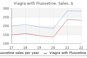
Buy discount viagra with fluoxetine online
However impotence yahoo answers purchase discount viagra with fluoxetine on-line, the main difference between the dysfunction and the failure is the severity and/or acuity of the dysfunction; thus, it is more appropriate that they be grouped into one practice pattern. In this way, particular levels of impairment or function may be used to specifically distinguish ventilatory pump dysfunction from ventilatory pump failure using identifiable characteristics within a continuum of ventilatory pump function. Identifying the primary pathology or impairments is critical to the physical therapist for planning and implementing an effective intervention plan. Practice Pattern 6E would include patients who frequently come from the cardiopulmonary impairment category and may include patients with a number of different primary pulmonary disorders such as asthma, emphysema, chronic bronchitis, and restrictive lung disease, among others. In other words, if the lung disorder causes a patient to work harder than a normal person for their next breath, then the ventilatory pump will be impaired secondary to the lung disorder. Ventilatory pump dysfunction can originate from any of the other pathology and impairment categories (musculoskeletal, neuromuscular, cardiopulmonary, or integumentary). These pathologies and impairments are listed under the "Patient/Clients Diagnostic Classification" section of each practice pattern. Because, pump dysfunction will occur where there is a restriction to the mobility of the chest wall and/or spine (ie, with arthritis, scoliosis, kyphosis, traumas). The resultant restriction will decrease the efficiency and/or the potential for optimal function of the ventilatory muscle pump. Likewise, patients with "neuromuscular disorders" are also listed as patients with potential ventilatory pump dysfunction. Neurologic disease or trauma can affect the strength and motor control of the trunk as well as the respiratory muscles, which may cause ventilatory pump dysfunction (ie, spinal cord injury, brain injuries, cerebral palsy, multiple sclerosis, or Parkinson disease). Last, patients with integumentary disorders, in whom the movement of skin or other connective tissue over the spine, rib cage, and proximal extremity joints (such as the upper extremities, lower extremities, or neck) is limited, may also present with ventilatory pump dysfunction. Patients with burns and connective tissue disorders may have such a ventilatory pump dysfunction. Thus, simply because the practice pattern states that the problem is "ventilatory pump dysfunction," the cause of that dysfunction may not initially stem from the cardiopulmonary system (Table 20-1). Any primary lung dysfunction that results in impaired mechanics of the ventilatory pump. Any musculoskeletal disorder or trauma that results in impaired mechanics of the ventilatory pump. Any neuromuscular disease or trauma that results in impaired mechanics of the ventilatory pump. Any integumentary disease or trauma that results in impaired mechanics of the ventilatory pump. The flattening of the diaphragm is likely to produce two specific changes including (1) a greater use of accessory muscles for inspiration and (2) an inward pull on the lower ribs when the flattened and shortened costal diaphragmatic muscle fibers contract with a different angle of inclination. The greater use of the accessory muscles generates a negative pressure in the upper chest area rather than the normal generation of negative pressure in the abdominal area from the downward descent of the diaphragm. If the diaphragm is unable to generate adequate negative intrathoracic pressure, the abdominal area may actually be sucked in from the negative pressure generated in the upper chest area (producing an abdominal paradox) from the increased activity of the accessory muscles. Under normal conditions, the muscles of the diaphragm contract and pull the central tendon and the dome of the diaphragm caudally. The costal diaphragmatic fibers at the lower rib cage within this zone of apposition produce (1) a cranially oriented force on the lower ribs, (2) outward motion of the lower ribs, and (3) separation of the lower ribs, as the abdominal viscera (with adequate abdominal muscle support) opposes the descent of the diaphragmatic dome. The aforementioned changes may result in the development of inadequate intrathoracic pressure in the abdominal area and inadequate ventilation of the lungs. As a result, the accessory muscles become more active and generate the needed negative pressure to ventilate the lungs. Ventilatory pump failure is commonly diagnosed by an elevation in partial pressure of carbon dioxide (Pco2) and reduction in partial pressure of oxygen, arterial (Pao2). Ventilatory pump failure can result from abnormal lung tissue or poor ventilatory muscle performance. Abnormal lung tissue from a variety of lung disorders may impose a significant workload on the ventilatory muscles that along with poor ventilation and respiration may eventually cause them to fail. It has been suggested that ventilatory pump failure can be recognized by a paradoxical breathing pattern. The patient with neuromuscular pathology will often show a different paradoxical pattern where the belly rises and the chest falls (upper chest paradoxical breathing). The inefficient diaphragmatic descent and subsequent ineffective ventilation of the lungs stimulate the accessory muscles of breathing to play a more active role in breathing. Such a breathing pattern is occasionally seen in patients with spinal cord injury or other neurological disorders. It appears to be less often associated with ventilatory pump failure, but may contribute to ventilatory pump failure if the inward upper chest motion is excessive and contributes to an increased work of breathing and decreased degree of lung ventilation. It is hopefully apparent from these examples that the topic of ventilatory pump dysfunction and failure is so extensive that it would be impossible to discuss every possible patient scenario. Hence, this chapter will focus on one case at length to illustrate cardiopulmonary preferred Practice Pattern 6E. In fact, these impairments can be so significant that respiratory problems have consistently been identified as the major cause of death in America for this population. He will be used to illustrate the cardiopulmonary risks, examinations, and interventions associated with patients who have ventilatory pump dysfunction. Normal Skeletal Support for Breathing the rib cage is an inherently mobile structure designed to move three-dimensionally, freely, and efficiently via skeletal support from the thoracic spine and muscle support from the respiratory and postural muscles. This would occur because the only significant stabilization of the thoracic cage is derived from its posterior attachment to the thoracic spine through the costotransverse ligaments. The costotransverse ligaments, three per spinal segment, are aligned in three different planes of movements and overlap the thoracic segment above and below them (see Chapter 4). This provides a very stable posterior junction, limiting chest wall movements and making dislocations or subluxations of the costotransverse joints a rare event (rib shaft fractures are far more common than rib shaft dislocations). On the other hand, the anterior chest wall is attached only to the sternum, which has no inferior bony attachment and superiorly is only attached to the clavicle. The therapist will find greater anterior chest movement and less posterior chest movement during inhalation. Normal Muscle Support for Breathing: A "Triad" of Support for Inhalation How do the muscles of the trunk allow optimal mechanical alignment of the thoracic cage and thoracic spine in stationary, static postures while still allowing the active movements of breathing and dynamic trunk movements Shape allows for three-dimensional movement that optimizes the potential inspiratory lung volume. Add to that, the importance of two other muscles, the intercostals and the abdominal, which provide the proper pressure support necessary for the diaphragm to move optimally and efficiently, creating a "triad of muscles" for optimal quiet breathing (Table 20-2). Air in the atmosphere rushes into the lungs because of the development of this area of negative pressure during inspiration (movement from a high-pressure to low-pressure area is common in this and many other similar pressure systems). This creates positive pressure within the lungs and chest cavity and therefore forces air out during exhalation. Without the active participation of the intercostal muscles to lift the upper chest upward and outward, the anterior chest wall would collapse inward during inhalation, toward the negative pressure. Clinically, this is seen in patients with spinal cord injury and may result in the development of a pectus excavatum, otherwise known as a concave deformity along the lower sternum. Measurements with a tape measure revealed negative upper chest wall movements of one-fourth in or more of upper chest wall depression during inspiration. The expected positive upper chest wall expansion of resting (or quiet) inspiration did not occur and was replaced with an inward paradoxical motion of the upper chest. The reason that this was observed was likely due to a lack of intercostal muscle contraction that left the upper chest unsupported during inspiration. As the descending diaphragm created greater negative pressure, the upper chest simply fell into the vacuum created by the descending diaphragm. Worse yet, when the patients were asked to take a deep breath (or vital capacity breathing), creating an even greater negative inspiratory pressure, the inward paradoxical movement of the upper chest wall became more severe with as much as two in of depression in many of the patients. Thus, patients must have adequate function of the intercostal muscles to support the negative inspiratory pressures necessary for effective breathing strategies. These muscles provide positivepressure support on the viscera in order to maintain their placement up and under the dome of the diaphragm in upright postures. In addition, without abdominal muscle support (as in spinal cord injury), the central tendon of a descending diaphragm moves excessively in an inferior plane because of the absence of positive abdominal pressure.
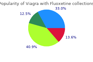
100/60 mg viagra with fluoxetine with amex
Biphasic and poroelasticity constitutive material theories tramadol causes erectile dysfunction purchase genuine viagra with fluoxetine on line, mathematically equivalent under certain assumptions, have been used to analyze the fluid flow paths and the hydrostatic pressure distributions within the disc to draw valuable clinical conclusions (Argoubi and Shirazi-Adl, 1996; Ferguson et al. Finite element analyses have been performed to determine the most sensitive structural parameters (Malandrino et al. Including fluid flow calculations in these studies is relevant for the biomechanics of spinal segments. These models clearly show the link between fluid flow and mechanical response within spinal segments for clinically relevant investigations. The loss of osmotic pressure has clinically relevant consequences, such as a negative effect on the activity of disc cells (Neidlinger-Wilke et al. To incorporate osmotic phenomena, triphasic and quadriphasic mathematical models have been implemented by including the diffusion of ions trough the interstitial fluid and the interaction with the fixed charges. Simpler approaches exist to partially simulate the swelling phenomenon in structural analyses with commercial finite element software, for instance by introducing a fixed pore pressure at the external boundaries of the disc (Argoubi and Shirazi-Adl, 1996; Ferguson et al. They demonstrated that results predicted with the fixed pore pressure boundary and the fixed osmotic pressure in the whole disc models were nearly identical, with the only difference being that the boundary pore pressure model could not simulate differential osmotic pressures in disc regions (Galbusera et al. The swelling model offered the most potential to provide accurate results, provided reliable values for the required coefficients and material properties are available. However, the other approaches were a good compromise between ease of implementation and reliability of results, especially when considering higher loads or when the focus is on global results such as spinal kinematics. At the same time, the annulus presents significant regional differences in mechanical behavior. As a result of several fiber angle distributions in the literature, different fiber patterns could be used as input for models. A circumferential and radial fiber angle variation was introduced in Schmidt et al. The regional variations in fiber angles were studied only circumferentially, and the experimental stress-strain responses were calibrated by varying the fiber stiffness parameters. Tissue in the human body is constantly renewed and maintained by the resident cells, and nutritional aspects that drive structure and functioning of the biological disc machinery were highlighted. One relevant aspect that has emerged is the hydrated nature of the healthy disc material and all intertwined biophysical properties. The importance of hydraulic effects for the local biomechanics as well as for the transport phenomena within the intervertebral disc has become clear. For instance, the local fluid dynamics at the bony part of the endplate was studied via human vertebral bone microtomographic images confirming that the permeability related to the vertebral bony endplate is much higher than the permeability of the cartilaginous endplate, also for very low bone porosities, which again points out the importance of the cartilage at the vertebra-disc interface (Malandrino et al. When analyzing the biomechanics of spinal segment, one fundamental parameter is the effective stiffness of the disc as a whole (Yang et al. Interestingly, these factors, together with the cell density, were also found to be relevant to the diffusion of solutes in a multiscale modeling study that coupled the transport of relevant biomolecule with the local continuum poromechanics (Malandrino et al. Thus a feedback loop of composition, cell nutrition, transport of metabolites, and biochemical properties must be considered (Barthelemy et al. These models ultimately allow relating the local changes in metabolites concentrations with the application of external forces in the spine, for instance relating it to muscle activity and daily tasks (Toumanidou and Noailly, 2015), an ambitious aim in the field of spine biomechanics and mechanobiology. The Effective Stiffness of the Disc Material and Its Maintenance Toward a Multiscale and Multiphysics Comprehension of the Disc Biomechanics In this chapter, we discussed the intricate connections between disc biology, composition, biomechanics, and transport of important solutes related to the disc cell pathways. These studies can finally relate spine mechanics to the stimuli sensed by the avascular disc cells. These mechanochemical models are being combined with medical images and applied to patient-specific geometries and image-derived fluid content and diffusivities to gain a real-life understanding of the mechanical and geometrical effects on metabolic transport and to support clinical decision-making processes related to disc pathologies and regenerative strategies. Direction-dependent resistance to flow in the endplate of the intervertebral disc: an ex vivo study. A computational spinal motion segment model incorporating a matrix composition-based model of the intervertebral disc. Compressive mechanical properties of the human anulus fibrosus and their relationship to biochemical composition. Elastin content correlates with human disc degeneration in the anulus fibrosus and nucleus pulposus. Elastic, permeability and swelling properties of human intervertebral disc tissues: a benchmark for tissue engineering. An anisotropic model for annulus tissue and enhanced finite element analyses of intact lumbar disc bodies. Comparison of four methods to simulate swelling in poroelastic finite element models of intervertebral discs. Bioactive glass serves as a substrate for maintenance of phenotype of nucleus pulposus cells of the intervertebral disc. Role of actin stress fibres in the development of the intervertebral disc: cytoskeletal control of extracellular matrix assembly. Degeneration affects the anisotropic and nonlinear behaviors of human anulus fibrosus in compression. Effects of low oxygen concentrations and metabolic inhibitors on proteoglycan and protein synthesis rates in the intervertebral disc. Effects of degeneration on the biphasic material properties of human nucleus pulposus in confined compression. Statistical factorial analysis on the poroelastic material properties sensitivity of the lumbar intervertebral disc under compression, flexion and axial rotation. Regional annulus fibre orientations used as a tool for the calibration of lumbar intervertebral disc finite element models. The role of endplate poromechanical properties on the nutrient availability in the intervertebral disc. Numerical exploration of the combined effect of nutrient supply, tissue condition and deformation in the intervertebral disc. Poroelastic modeling of the intervertebral disc: a path toward integrated studies of tissue biophysics and organ degeneration. On the relative relevance of subject-specific geometries and degeneration-specific mechanical properties for the study of cell death in human intervertebral disk models. Factors involved in the nutrition of the human lumbar intervertebral disc: cellularity and diffusion of glucose in vitro. Development and validation of a new transducer for intradiscal pressure measurement. Annulus fibrosus functional extrafibrillar and fibrous mechanical behaviour: experimental and computational characterisation. Mechanical stimulation alters pleiotrophin and aggrecan expression by human intervertebral disc cells and influences their capacity to stimulate endothelial cell. Statistical factorial analysis on the material property sensitivity of the mechanical responses of the C4-C6 under compression, anterior and posterior shear. A finite element analysis methodology for representing the articular cartilage functional structure. Confined compression experiments on bovine nucleus pulposus and annulus fibrosus: sensitivity of the experiment in the determination of compressive modulus and hydraulic permeability. Influence of material properties on the mechanical behaviour of the L5-S1 intervertebral disc in compression: a nonlinear finite element study. Tissue engineered intervertebral disc repair in the pig using injectable polymers. Biochemical and structural properties of the cartilage end-plate and its relation to the intervertebral disc. Material property discontinuities in intervertebral disc porohyperelastic finite element models generate numerical instabilities due to volumetric strain variations. Application of a new calibration method for a three-dimensional finite element model of a human lumbar annulus fibrosus. Proteoglycans and collagen in the intervertebral disc of the rhesus monkey (Macaca Mulatta). Musculoskeletal modeling of the lumbar spine to explore functional interactions between back muscle loads and intervertebral disk Multiphysics. Inclusion of regional poroelastic material properties better predicts biomechanical behavior of lumbar discs subjected to dynamic loading. Simulating the sensitivity of cell nutritive environment to composition changes within the intervertebral disc.
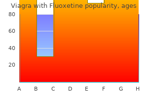
Buy viagra with fluoxetine 100/60mg amex
If tumor thrombus is suspected erectile dysfunction and premature ejaculation underlying causes and available treatments buy viagra with fluoxetine 100/60mg overnight delivery, pre- and postcontrast imaging should be obtained to determine if there is enhancement of the thrombus. Bland thrombus will not enhance, while tumor thrombus will enhance on postcontrast images because of internal vascularity. Conventional splenoportography, mesenteric angiography, or wedged carbon dioxide portography is rarely needed for diagnostic purposes alone. Recanalization of the portal vein can be seen in up to 35% of patients and may take up to 4e6 months after the initiation of therapy. In patients with more severe acute disease, direct catheter-based pharmacologic or pharmacomechanical thrombolysis can be performed via a transhepatic or transjugular approach, with up to 75% of patients showing some degree of lysis. There is an increased risk of significant bleeding if thrombolysis is performed via a transhepatic approach. Indirect thrombolysis is performed by infusing a thrombolytic agent into the superior mesenteric artery. Indirect thrombolysis is more effective than systemic anticoagulation with regard to improving flow through the portal vein [19]. Surgical thrombectomy is not recommended because of the risk of recurrent thrombosis and surgical morbidity and mortality. Occlusion of the hepatic venous outflow results in increased sinusoidal and portal venous pressure. As the portal pressure rises, portal vein flow to the liver decreases, as there is increased hepatic congestion. If the course of the disease is acute, there is inadequate compensation, which results in hypoxic hepatocellular damage and fibrosis. Lab studies will reveal elevated hepatic enzymes, coagulopathy, and hyperbilirubinemia [29]. These patients will typically present with symptoms of portal hypertension, including ascites and splenomegaly. This can be attributed to a decreased obstructive burden relative to more severe disease [23,32,33]. The hepatic veins may have internal echoes or be replaced with a fibrous echogenic cord [23,34e38]. Extrahepatic findings such as ascites and splenomegaly are easily seen with ultrasound. The liver parenchyma can have a normal appearance or show patchy enhancement due to stasis of flow within the sinusoids [37,40,41]. Catheter venography is considered the reference standard for diagnosis [34]; however, it is usually reserved for cases in which a histologic sample is required via transvenous biopsy, or an endovascular therapy is planned. Catheter venography has the advantage of being able to precisely depict the level of obstruction and to determine the hemodynamic significance by measuring intravascular pressures. A combination of intravascular techniques may be needed depending on the level of obstruction to relieve congestion and restore hepatic venous outflow. Technical success rates are reported at 90%, with clinical success in greater than 75% [45,49,52]. Long-term anticoagulation may be required to maintain shunt patency, particularly in those patients with underlying hypercoagulable state [24,25,45]. Clinical manifestations may include abdominal pain, hematuria, nausea, vomiting, and fever. If there is bilateral involvement, patients may present with acute renal failure [59]. The goal of therapy is relief of venous obstruction and preservation of renal function. Several catheter-based techniques have been described, including indirect thrombolysis via a transarterial approach and direct thrombolysis via a transvenous approach. Both infusion-based and pharmacomechanical thrombolysis endovascular therapies have been used for direct thrombolysis. Complications are similar to other thrombolysis techniques used in other parts of the body and include bleeding, thromboembolization, and vascular access complications [59,60]. Presentation of septic thrombophlebitis can range from a benign course affecting localized superficial veins to severe systemic infections resulting in shock and death. Prompt recognition and treatment are necessary to prevent progression and severe complications. Distinct conditions have been described based on the vein and associated organ involved. One such entity is Lemierre syndrome, which is septic thrombophlebitis of the internal jugular veins. Regardless of the location of the involved vessel, the severity of disease is distinguished as either simple or suppurative, with suppurative indicating purulence. Once the infective organism has entered the vein and proliferates, endothelial damage ensues, resulting in inflammation and thrombosis. Patients will present with pain and erythema, which may spread along the course of the infected vessel. Suppurative superficial thrombophlebitis is a rare condition that can be complicated by septic embolization, most commonly to the lungs, which can lead to sepsis, hypoxia, and death. Broad-spectrum antibiotics with coverage of common skin flora should be promptly initiated. Anticoagulation is not routinely used unless there is extension into the deep veins. Burn victims and patients receiving total parenteral nutrition are at increased risk. Patients will present with fever, often without pain or swelling, at the site of catheter insertion. Workup begins with basic lab studies, including complete blood count, chemistry, and coagulation profile. If catheter-related infection is suspected, cultures should be drawn from both a peripheral site and the central catheter for comparison. A catheter tip culture can be helpful; however, caution must be used when removing an infected central catheter as there may be adherent septic thrombus, which could embolize. The natural history of this disease begins with an oropharyngeal infection, which spreads into the lateral pharyngeal space and subsequently the carotid sheath. The organism most commonly implicated is Fusobacterium necrophorum, which is a normal oropharyngeal flora; however, multiple other bacteria have been implicated. Ultrasound is the imaging modality of choice for the extremities as well as the internal jugular veins. This is due to overlying structures, which attenuate the ultrasound beam and limit the acoustic window. Diagnostic venography is not performed in the setting of septic thrombophlebitis unless there is an intention to treat. Antibiotics and anticoagulation are the primary treatment for deep venous septic thrombophlebitis. Data on catheter-directed therapy in the setting of septic thrombophlebitis are limited to case reports. Intravascular manipulation carries a risk of septic embolization and systemic sepsis. Mechanical thrombectomy has been reserved for those patients in whom anticoagulation was contraindicated, or if there was progression of symptoms while adequately anticoagulated. The AngioJet device has been successfully used in cases of deep venous septic thrombophlebitis; however, operators should remain cautious and prepared to manage complications [64]. Recent studies indicate a trend toward higher incidence of this relatively rare condition, probably secondary to increased clinical awareness, improved noninvasive diagnostic imaging techniques, and more inclusive retrospective review strategies [65,67]. Among children, the most commonly affected are neonates who are at increased risk secondary to birth trauma and dehydration [84e86]. Therefore, clinical suspicion should remain high in younger female patients with a combination of the aforementioned risk factors who present with nonspecific clinical symptoms. These symptoms include the following: headache, seizure, papilledema, altered consciousness, and focal neurologic deficits (primarily including paresis, aphasia, diplopia, and visual loss).
Generic viagra with fluoxetine 100/60mg without prescription
However erectile dysfunction latest treatments order 100/60mg viagra with fluoxetine mastercard, stenting can still cause distal embolization, in part due to a "cheese grater" effect of the stent struts. Preventing Embolization Treating coronary lesions with large thrombus may lead to embolization, distally or sometimes even proximally. For example, during attempts to remove a thrombus, it may embolize into a more proximal coronary branch or into the aorta, causing systemic embolism, such as stroke. Thrombus removal before balloon angioplasty and stenting can help decrease the risk for distal embolization. An innovative technique for distal protection (called "forward and backward" aspiration) involves negative aspiration through a deep-seated GuideLiner catheter during balloon angioplasty and stenting. If a large thrombus is located close to an important bifurcation with large branches, protecting both branches with wires may help prevent branch occlusion and facilitate treatment if those branches become obstructed by thrombus. If distal embolization occurs, aspiration thrombectomy may help remove some of the embolized thrombi and restore antegrade flow. Moreover, administration of vasodilators (such as adenosine [20], nicardipine [21], nitroprusside [22], and verapamil [23]) may help improve antegrade flow, which in turn may reduce the risk for additional thrombus formation. Similarly, a retrospective study showed that rheolytic thrombectomy was associated with lower 2-year incidence of major adverse cardiac events in patients in large thrombus burden and acute myocardial infarction complicated by cardiogenic shock [26]. Nevertheless, rheolytic thrombectomy is currently used very infrequently for treating coronary thrombus. The neutral outcome was consistent in all patient subgroups, regardless of baseline clinical or angiographic characteristics. In post hoc analyses of the 180-day follow-up, the composite end point of cardiovascular death, rehospitalization with new myocardial infarction, cardiogenic shock, or new hospitalization for heart failure was similar in the aspiration and no-aspiration groups, as was the incidence of stroke [30]. Moreover, thrombectomy was associated with an increased risk of stroke at 30 days (0. A 2017 patient-level meta-analysis of the above three trials showed that manual thrombectomy was associated with a strong trend for lower incidence of cardiovascular death at 30 days (2. Thrombus Embolization From the Sheath or Guide Catheter Although infrequent, thrombus can form at the tip of arterial sheaths; when a catheter is subsequently advanced through the sheath the thrombus may be carried into the coronary artery causing acute occlusion. Prevention of this complication is key and can be achieved with aspiration of the sheath before insertion of a guide catheter. Suboptimal Antithrombotic Treatment Anticoagulation and antiplatelet therapies are important for preventing coronary thrombus formation. In some patients the venous line may malfunction and the patient may not receive the anticoagulant despite "intravenous administration. This can be facilitated by inserting a small (usually 4 Fr) sheath in a large vein, such as the common femoral vein, or sometimes through the arterial sheath side port. If despite precautions thrombus is formed and causes embolization, subsequent management of the intracoronary thrombus is performed as described under Thrombus as the Cause of Complications. Despite traditional teaching about reversing heparin anticoagulation if a perforation occurs, most operators currently do not administer protamine until after removal of all coronary equipment (guidewires, balloon, etc. Using an 8-Fr GuideLiner, a Graftmaster stent was deployed and (H) the perforation sealed. That is why the use of an embolic protection device in association with an intensive antithrombotic strategy has been advocated for the treatment of lipid-rich lesions. A 64-year-old man presented with stable angina and newly diagnosed decreased left-ventricular systolic function. For example, coronary dissection is treated with stent implantation (ensuring that guidewire position is not lost), stent underexpansion is treated with high-pressure balloon inflations, and equipment loss or entrapment is treated with retrieval of the lost coverage or by covering it with stents. An intensive antithrombotic regimen that combines antithrombin and antiplatelet treatments is critical to preventing and managing thrombus. A stepwise systematic approach to the management of thrombotic lesions could improve the likelihood of restoring antegrade flow, while minimizing the risk of distal embolization or other complications. Jude Medical; research support from Boston Scientific and InfraRedx; spouse is employee of Medtronic. Management of guidewire-induced distal coronary perforation using autologous fat particles versus coil embolization. Successful management of a distal vessel perforation through a single 8-French guide catheter: combining balloon inflation for bleeding control with coil embolization. Covered stent implantation through a single 8-French guide catheter for the management of a distal coronary perforation. Safety and efficacy of intense antithrombotic treatment and percutaneous coronary intervention deferral in patients with large intracoronary thrombus. Safety of lone thrombus aspiration without concomitant coronary stenting in selected patients with acute myocardial infarction. A novel application of GuideLiner catheter for thrombectomy in acute myocardial infarction: a case series. The state of the excimer laser for coronary intervention in the drug-eluting stent era. Randomized evaluation of the TriActiv balloon-protection flush and extraction system for the treatment of saphenous vein graft disease. Intracoronary adenosine administered during percutaneous intervention in acute myocardial infarction and reduction in the incidence of "no reflow" phenomenon. Pretreatment with nitroprusside for microcirculatory protection in saphenous vein graft interventions. Rheolytic thrombectomy for acute myocardial infarction complicated by cardiogenic shock. Coronary stents were then developed to maintain vessel patency postangioplasty, to manage acute occlusion, and to decrease substantially the rate of restenosis. However, stent thrombosis became the most recognized complication of coronary stenting. Probable stent thrombosis includes any unexplained death within 30 days of stent implantation or any myocardial infarction in the territory of the previously implanted stent without angiographic confirmation of stent thrombosis and without any other cause [2]. Possible stent thrombosis is defined as any unexplained death more than 30 days after stent implantation [2]. They were used as a "bailout" technology for management of acute vessel closure during percutaneous coronary angioplasty. In this setting, despite the presence of adequate pharmacotherapeutic systemic anticoagulation, the rate of stent thrombosis remained as high as 20% [3]. The findings indicated that in-hospital stent thrombosis rates were lowered to a rate of 3. Further evaluation regarding the optimal antiplatelet and anticoagulant regimens reported stent thrombosis rates as low as 0. This technological approach also aimed at decreasing the need for coronary artery bypass surgery for lesion recurrence. However, experience with the first generation of these devices raised valid questions concerning the phenomenon of scaffold thrombosis (ScT). Notably, the incidence of ScT remained over time higher than that found with metallic stents, i. Recurrent ScThis another significant concern, with several studies reporting rates of this unwarranted phenomenon in the range of 5. Some patients, in fact, can be asymptomatic despite the presence of stent thrombosis, especially those with collateral vessels supplying the territory at risk [8]. The gold standard diagnosis of stent thrombosis is a filling defect on coronary angiography, indicating the presence of thrombus within the lumen of the stented segment of the vessel [8]. Mortality after stent thrombosis is high, with reported incidence of 11%e42% [15,21e35]. Patient-related factors, pharmacologic factors, lesion- and procedure-related factors, and postprocedural factors can play a role. Overall, the development of thrombus within the lumen of a stent can be the result of one or more of the following: activation of the extrinsic coagulation cascade from exposure to subendothelial tissue, to stent struts, or to the polymer coatings; inadequate inhibition of platelet activation; activation of the intrinsic coagulation cascade from low shear stress due to slow coronary flow; or the presence of a prothrombotic state [8]. Intriguingly, more than one cause accountable for stent thrombosis was identified in 48% of the patients [38]. A majority of these patients had a reduced luminal area (<80% of the reference lumen) due to stent underexpansion and malapposition [38].
Discount viagra with fluoxetine 100/60mg with mastercard
However erectile dysfunction adderall xr purchase viagra with fluoxetine overnight delivery, exhalation can also be active for normal activities, such as in coughing or talking. Active exhalation is achieved by either an eccentric or a concentric contraction of the respiratory muscles. The process is distinctly different and will be discussed in the following section. For normal adults, a typical vocalization of a vowel sound can be sustained for 15 seconds,34 but it is not uncommon for the trained individual (ie, singers, wind-instrument players, long-distance athletes) to sustain a vowel sound for 30 to 60 seconds. There will also be a notable "falling" of the chest wall during exhalation, as the patient cannot slow down the expiratory maneuver. This undesired passive exhalation may be a result of (1) poor motor control, (2) weakness or paralysis of the respiratory muscles, (3) pain, (4) vocal fold dysfunction, or (5) the presence of a tracheostomy tube which prevents the patient from using his or her glottis to aid in the eccentric maneuver. To compensate, they may withdraw socially or respond to questions with short answers. This occurs when the air is forcibly expelled (ie, coughing or yelling), calling into action a concentric contraction of the expiratory muscles. The intercostals and abdominal are the primary muscles recruited for this activity, although the pectoralis and latissimus dorsi muscles can also be recruited. When these muscles contract, they apply significant positive pressure on the thoracic cavity to assist in fast and forceful expirations. Examples of Inspiratory/Expiratory Pump Dysfunction and Failure Stemming From Primary Lung Disease Primary lung diseases can also result in secondary dysfunction of the inspiratory and expiratory muscles. The muscles themselves may be functional and capable of demonstrating passive, concentric, and eccentric patterns, but the disease process demands that the expiratory muscles are activated with every expiratory effort, causing the patient significant fatigue. An extended example of how obstructive and restrictive lung disease can adversely affect the normal biomechanics of breathing is presented in the following paragraphs. Emphysema causes destruction to the alveolar sacs, resulting in the creation of large bulbous distal air sacs which in turn creates (1) excessive compression forces on the conducting airways, especially during exhalation, and (2) abnormal increases in the overall inspiratory lung volumes. The diminished ability to exhale air from the lungs is due to the alveolar damage, which traps air within the lungs. As the pressures continue to increase within the lungs during exhalation, the distal and proximal airways may become compressed and narrowed which will limit the amount of air moving out of them. Thus, even though the muscles and skeletal structures are technically intact, the biomechanics of their breathing is now altered, resulting in an increase in their work of breathing and ventilatory pump impairment. As their disease progresses, the chest wall itself is pushed outward by air trapped in destroyed alveoli and compressed airways, decreasing the efficiency of the diaphragm and accessory muscles of inspiration. At this point, the mechanics of both inspiration and exhalation are significantly impaired. A different presentation is noted in the patient presenting with a restrictive lung disorder such as pulmonary fibrosis or pulmonary hypertension. Thus, even though the muscles are neurologically intact, they must use greater force to generate the same inspiratory lung volumes as patients without restrictive lung disease. However, patients with restrictive lung disease will demonstrate more biomechanical impairments to inspiration, and the patients with obstructive lung disease will demonstrate more impairments to the expiratory mechanics until obstructive lung disease becomes more severe. Applying these concepts extensively to a single case study will allow a more detailed depiction of how ventilatory pump dysfunction and the potential for failure occurs and what clinicians can do to minimize their impact on patient function. The patient tolerated this final step in weaning for the next 48 hours, no longer requiring mechanical ventilation. He was able to assist the nurses and therapists with applying abdominal pressure to cough. Although he remained dependent in bed mobility, he could roll side to side and transfer supine to sitting with moderate assistance from a caregiver. The patient was discontinued from this practice pattern due to successful separation from the mechanical ventilator. Upon review of his admitting paperwork, the doctors determine that he is currently medically stable. For these reasons, as he begins his rehabilitation phase, he clearly falls into Practice Pattern 6E: Impaired Ventilation and Respiration/Gas Exchange Associated With Ventilatory Pump Dysfunction or Failure. What will be the overriding cardiopulmonary concerns and risks for this young man as he begins the rehabilitation phase Impaired Respiratory Mechanics Lee was found to have paralysis of the intercostal and abdominal muscles, which means that, of the core "triad" respiratory muscle groups, only the diaphragm is spared. However, without the muscle support above and below the diaphragm from the intercostals and abdominal muscles, the diaphragm will not be capable of contracting at its highest level of function. In addition to paralysis, his spinal shock is now resolving and spasticity is beginning to develop in his trunk musculature. He says this shortness of breath is particularly evident when he is telling a story, laughing, or any other "breathing activity" that causes him to spontaneously take quicker inspiratory efforts. He also says that his Halo fixation device (cervical fixation device attached to a trunk vest that restricts cervical motion) restricts his chest causing him to feel confined when he tries to inhale. In fact, between his Halo device and the tracheostomy tube, Lee states that he is finding it impossible to bend forward and tuck his chin to cough, making him sense that he "cannot get the secretions out of his lungs. He hates sleeping on his back and has increasingly complained of headaches first thing in the morning. When he recruits his trapezius to take a deep breath, it exacerbates his shoulder pain. However, the most frightening incident that he described was his first bout of autonomic dysreflexia which was short lived, but very disturbing. Autonomic dysreflexia, sometimes called hyperreflexia, does not occur until after spinal shock has resolved; thus, it is more commonly seen in the rehabilitation setting or later. However, lately he has not been drinking adequate fluids due to pain, vocal fold irritation (from being intubated initially after the accident), and depression. It is important to note that respiratory, urinary, and blood infections are some of the most common causes of morbidity in this population. It can improve pulmonary secretion mobility, decrease toxic concentrations in the blood, flush bacteria out of the urinary system, and decrease potential noxious stimuli that may trigger autonomic dysreflexia. Hearing this information, Lee declares that he will definitely drink more water now. If it forms around the spine, shoulder, or pelvis and causes our patient to assume a more kyphotic posture, he will compromise the mechanics of his ventilatory pump, which would cause a decrease in inspiratory lung capacity. This will have to be watched carefully to make sure the connective tissue does not break down completely and cause an open sore. Poor Nutrition/Hydration A secondary complication that has only more recently been recognized is poor nutrition and underhydration. In addition, patients may find swallowing difficult due to cervical bracing, or other traumas that occurred with their injury. He has already pledged to increase his hydration level following our discussion noted previously. Even though he survived the acute phase of his injury and was successfully weaned from the ventilator, he still carries a risk for developing cardiopulmonary problems as long as his paralyzed state remains (Table 20-7). Other Inherent Risks Numerous other cardiopulmonary risks exist, such as advanced age, obesity, other medical problems accrued during the accident, past medical problems, previous lifestyles, etc. In a perfect world, every single possible test would be performed for every single patient. In this section, numerous different assessment tests will be presented with their relative value of information given our particular patient and 6E Practice Pattern. Our patient is currently on antireflux medications, vitamins, a muscle tone relaxant for his spasticity, and a stool softener. Because of its ease of application, low cost, and valuable information, all patients should have ongoing assessments of their vital signs. Medical History A complete medical history is especially important when assessing the complex medical patient. Our patient was healthy prior to his injury; thus, his medical history does not add complicating factors.
Order viagra with fluoxetine master card
Melnyk and colleagues investigated in vitro the effect of shear forces on an unstable model of degenerative spondylolisthesis in a human cadaver spine (Melnyk et al erectile dysfunction other names safe viagra with fluoxetine 100/60 mg. They found that the posterior fixator supported a significantly greater shear force as the specimen was progressively destabilized (facet and disc destabilization) and the implant stiffness increased (Melnyk et al. The internal mechanical loads acting on the implants have been measured in vivo by using instrumented posterior fixators. Several studies investigated the effect of different body positions and tasks on the loading components (Rohlmann et al. Axial forces are mainly compressive and depend on bodyweight, the surgical procedure. Among the common life activities, walking is considered the most critical loading condition for implant fatigue, more because of the high number of repetitions performed every day than for the intensity of the bending moments achieved (Rohlmann et al. Bending moments are also considered the primary cause of fatigue failure due to high repeated stresses on the posterior spinal fixator during clinical use. Based on an estimate of the bending moment on the posterior instrumentation, Cripton warned that, in the case of severe anterior column injuries, the implant may be at risk of fracture, and secondary to reversed bending moments (Cripton et al. Recent numerical studies highlighted the significant role of the anterior support in reducing the internal loads and the maximum stresses on different components of the posterior fixator (La Barbera et al. With an increasing degree of anterior spine instability, the spinal implant could reach much higher loads compared to an intact disc, even during noncritical everyday activities. The predictions of such models agree with the failure modes met during clinical experience (La Barbera et al. Newcomb and colleagues determined with a combined in vitro and in silico approach that pedicle screw trajectory may be optimized to decrease the stress, thus avoiding screw breakage (Newcomb et al. Several numerical models correlated the high internal loads to implant stresses through the elastic modulus of the longitudinal element (Jahng et al. Shih and colleagues compared conical and cylindrical screw designs using bench tests coupled with simple finite element simulations (Shih et al. Amaritsakul performed a similar study showing that a conical design could be further optimized using a dual-core thread shape to increase bending strength (Amaritsakul et al. In this procedure, a posterior access is used with the patient in a prone position. A midline incision is performed and the paravertebral fascia is opened bilaterally to expose the neural arc. Laminectomy, medial facetectomy, and foraminotomy are usually performed to provide canal decompression. Bone chips can be obtained from the same patient (autologous graft); for instance, a significant amount of spongious bone can be harvested from the ilium. The graft material is then placed along the transverse processes and the lateral part of the laminae to stimulate bone growth. Chronic pain at the donor site is a common complication and is often indicative of an unsatisfactory result. Common complications are superficial infection, nerve root injury, and dural tears (Wu et al. Despite lengthening the operation time and the risk of complications, posterior pedicle screw fixation is often used to provide primary stability until solid fusion is achieved (Thomsen et al. A satisfactory outcome is achieved in terms of pain relief and fusion rate, both with and without posterior fixation, but the latter seems to reduce the occurrence of pseudarthrosis (Schwab et al. In fact, because of the loads acting on the anterior spine, the lamina and the facet joints may experience some relatively high motions, while needing a relatively longer time to fuse. Moreover, the use of external orthosis may not be as effective as other internal fixation implants. This second strategy has all the mechanical advantages of posterior fixation alone, providing primary stability in the very short-term after surgery, while shielding the loads on the hardware, particularly after solid fusion is achieved. Another study confirmed the higher complications related to pedicle screw fracture and loosening without adequate anterior support (Wu et al. It involves the use of cages, spacers, or bone grafts that are positioned within the intervertebral space following discectomy and after a careful endplate preparation. In general, surgical options can be classified in two categories taking the transverse process as a reference (Mobbs et al. Clinical evidence did not demonstrate that one specific approach is to be preferred or avoided over the others in terms of fusion rate or clinical outcomes. The presence of nerves, muscles fasciae, and great vessels determines the feasibility of each approach at different spinal levels. However, it is limited for L2/L3 and L3/L4 in cases of extensive peritoneal and kidney retraction and the risk of superior mesenteric artery thrombosis. Contraindications include highgrade degenerative spondylolisthesis without posterior fusion (Malham et al. The anterior access allows maximization of the implant size and surface area, which optimizes the fusion rate (Phan et al. This approach helps to spare spinal muscles and to reduce postoperative pain and disability. Surgery-related complications may include retrograde ejaculation and visceral and vascular injuries (Phan et al. The techniques to access the thoracic disc space include thoracoabdominal, open transthoracic, and thoracoscopic approaches (Patel et al. This approach is well known by the majority of spinal surgeons, who are well trained to perform it. A laminotomy is performed medial to the facets, and the disc space is exposed upon dura retraction. Finally, endplates and disc space are prepared to allow the insertion of an interbody spacer. It can also be used for selected patients with segmental instability, symptomatic stenosis, recurrent disc herniation, and pseudarthrosis (Mobbs et al. The posterior approach is a valuable technique for treatment of the thoracic spine (Smith et al. Posterior exposure ensures excellent visualization of the nerve roots and surrounding structures. The posterior approach facilitates pedicle screw insertion and 360 degree fusion through a single incision. Among the disadvantages of this approach is prolonged paraspinal muscle retraction, which may provoke iatrogenic injury with delayed recovery and mobilization (Fan et al. A midline, bilateral paramedian incision is usually performed with the patient lying in the prone position. A unilateral laminectomy and inferior facetectomy facilitate bone graft positioning. However, it does not allow for coronal imbalance correction and lordosis restoration (McAfee et al. With this technique, the patient is laterally positioned, and a small incision is performed. Neuromonitoring is considered essential to prevent damage to the lumbar plexus and the psoas muscle and bowel injury at lower lumbar levels (Malham et al. The lateral approach guarantees minimal muscle splitting with rapid postoperative mobilization, despite the fact that transient neurological complications have been reported. A high fusion rate can be achieved easily thanks to comprehensive clearance of the disc space (Phan et al. It allows for an effective sagittal and coronal deformity correction, particularly for lumbar degenerative scoliosis with laterolisthesis (Arnold et al. An exhaustive review of the existing literature dealing with fixation and fusion is a challenging task. To the best of our knowledge, very few biomechanical studies have conducted extensive comparisons of the biomechanics of alternative fusion techniques. Moreover, many surgical techniques fall within the same category, even if they exhibit specific peculiarities in terms of implant design and surgical techniques. Therefore it is often very hard to make direct comparisons or to generalize about their biomechanical performances. The posterior fixator is expected to contribute most in the short operative time, whereas the anterior fusion mass.
Viagra with fluoxetine 100/60mg for sale
The prevalence of scoliosis in cerebral palsy patients was reported to range between 15% and 80% (Koop erectile dysfunction at 21 buy cheap viagra with fluoxetine 100/60 mg, 2009), with a large variation due to the differences in the populations being studied. Although these angles may not seem high in absolute terms, the progression of scoliosis in cerebral palsy is still a matter of concern because it does not respond well to conservative treatment and progresses even after skeletal maturity (Saito et al. The coronal deformity often starts at the thoracolumbar junction, subsequently involving the entire thoracolumbar spine and in some cases the pelvis. In contrast with adolescent idiopathic scoliosis, which shows a generally lordotic sagittal alignment, in patients suffering from muscular dystrophy, the tendency to develop a thoracolumbar kyphosis is particularly accentuated (Archer et al. Patients with Becker muscular dystrophy tend to be older and thus show a lower severity and higher stability of the curves in comparison with patients with Duchenne muscular dystrophy (Boos and Aebi, 2008; McDonald et al. Indeed, neuromuscular scoliosis has a greater impact on the functional status of the patient than idiopathic scoliosis does because the former hinders the ability to perform daily activities such as standing, walking, and sitting. In general, the same indications used for idiopathic scoliosis, that is, a curve greater than 50 degrees and the risk of progression, are considered valid for neuromuscular scoliosis (Boos and Aebi, 2008; Cloake and Gardner, 2016; Koop, 2009); however, a comprehensive multidisciplinary preoperative assessment is critical in order to correctly take into account the frequent concurrent pathologies. The aim of the surgery for the treatment of neuromuscular scoliosis is to correct the spinal deformity and restore a correct balance in both the coronal and sagittal planes. Patients with neuromuscular scoliosis have a limited capacity to activate compensatory strategies to achieve a balanced posture, and therefore special care should be taken in order to ensure that physiological alignment of the spine, including the pelvis, is achieved (Shapiro et al. The first instrumentation used to correct neuromuscular deformities was the Harrington rod, which was employed with partial success in patients suffering from poliomyelitis in the 1950s (Harrington, 1988) and opened the way to the development of the modern implantable devices for the treatment of spinal deformities. Until recent years, posterior fixation with sublaminar wires and Luque rods (Luque, 1982) in combination with anterior releases was the most common solution for the correction of neuromuscular scoliosis, because of its capability of achieving a satisfactory but flexible local deformity correction without overloading the anchoring points (Boos and Aebi, 2008). Hooks and pedicle screws have also been used, but showed in the early stages a poorer clinical outcome and higher complication rates in comparison with wiring, mostly due to the complex insertion technique and inadequate purchase in the bone tissue, which is often osteopenic (Archer et al. Hybrid configurations including both wires and posterior instrumentation were then introduced and improved the outcomes (Modi et al. Solutions employing only pedicle screws are now gaining increasing interest because they reduce blood loss and surgical time (Archer et al. The most common proximal fusion level for neuromuscular scoliosis is T2, even if the curve does not extend to such a level in a coronal radiograph (Boos and Aebi, 2008). In fact, the kyphotic deformity is frequently larger than the scoliosis, and a proximal extension ensures a proper correction in the sagittal plane. In order to minimize the risk of curve progression after fixation, fusion at all the involved levels is recommended (Boos and Aebi, 2008). If pelvic obliquity is present, its correction by means of sacral and pelvic fixation is mandatory. The Luque-Galveston technique subsequently evolved into the unit-rod, and provided an improved potential to correct the pelvic obliquity (Bulman et al. In severe cases of rigid kyphosis, the use of technically demanding and risky procedures such as pedicle subtraction osteotomy and apical vertebral resection (Sharrard, 1968) has also been reported. Complications during and after neuromuscular scoliosis corrective surgery are frequent and can be severe. Other reported major complications include respiratory failure, lung collapse, postoperative pneumonia, cardiac arrhythmias, and wound infections (Archer et al. Instrumentation-related complications such as pseudoarthrosis and cutout of the sublaminar wires are not common but have been reported as well (Tsirikos et al. Iliac loosening of the rods after fixation with the Luque-Galveston technique was found in approximately 30% of the patients, but with only a minor clinical impact and no need for re-operation (Nectoux et al. However, despite these complications and the burden for the patient as well as for the caregivers because of the complexity of the surgeries, a high degree of satisfaction has been reported (Jones et al. A key factor in satisfaction is improvement in the shape of the trunk, which facilitates sitting and other daily activities (Koop, 2009). The use of spine surgery aimed at correcting deformities as well as alleviating the symptoms of degenerative disorders such as spinal stenosis has become relatively common in recent years, but high rates of complications and re-operations are still reported in the literature. A widely used scale is the one published by Hoehn and Yahr (1967), which considers five stages from minimal or no functional disability to confinement to bed or wheelchair unless aided. None of these scales includes an evaluation of the posture or of the possible associated spinal deformities, but they take into account their effect on limiting the functional status of the patient. Advanced age and female gender are considered to be risk factors for forward bending (Oh et al. The available literature on this topic is still limited and rather contradictory, but evidence of a higher prevalence of spinopelvic disorders in these patients with respect to age-matched nonpathological subjects seems to have emerged (Watanabe et al. Camptocormia is a large flexion of the trunk observable in the standing posture, which is corrected mostly or completely if the patients lies supine or prone (Djaldetti et al. The standardized diagnostic criterion defines camptocormia as a marked flexion originating in the thoracolumbar spine (minimum 45 degrees) that can be almost completely resolved in the supine position (Doherty et al. In comparison with degenerative sagittal imbalance, camptocormia is therefore substantially more flexible. Its diagnostic definition describes a lateral flexion of at least 10 degrees that can be resolved completely by passive mobilization or in the supine position. Therefore, because there is no structural curve as well as axial rotation of the vertebrae, the Pisa syndrome should not be considered a true spinal deformity. Although many patients have exhibited concomitant sagittal misalignment and scoliosis, an association between the two could not be determined (Choi et al. Early literature based on case reports showed a contralateral scoliosis with respect to the symptoms (Duvoisin and Marsden, 1975; Martin, 1965), whereas subsequent works conducted on human patients showed contrasting findings (Baik et al. Available data about such surgeries, however, are limited, and there is still no strong consensus about the techniques that should be used and the indications for surgery. The techniques used include fixation with pedicle screws and hooks, sacroiliac fixation with screws (Bouyer et al. In all available studies, the rates of complications and re-operations were found to be substantial (Babat et al. Nevertheless, the majority of the patients reported positive satisfaction (Bouyer et al. An aspect that was indicated as critical to achieving a good clinical outcome and limiting the complication rates was the restoration of a correct sagittal balance, including that of the pelvis, with a focus on the lumbar lordosis and the global alignment (Koller et al. In addition to a vitamin D deficiency secondary to hyperparathyroidism and reduced exposure to sunlight (Torsney et al. Other factors that are potentially responsible for complications have been identified. Sarkiss and colleagues found that a poor clinical outcome was associated with being older, having a high level of camptocormia, and having disorders of the hip joint (Sarkiss et al. Association between sagittal balance and scoliosis in patients with Parkinson disease: a cross-sectional study. Posterior spinal fusion for scoliosis in patients with cerebral palsy: a comparison of Luque rod and unit rod instrumentation. Surgical correction of spinal deformity using a unit Rodin children with cerebral palsy. Parkinson disease with old-age onset: a comparative study with subjects with middle-age onset. Luque-Galveston procedure for correction and stabilization of neuromuscular scoliosis and pelvic obliquity: a review of 68 patients. Scoliosis in rats with experimentally-induced hemiparkinsonism: dependence upon striatal dopamine denervation. Longitudinal parental perceptions of spinal fusion for neuromuscular spine deformity in patients with totally involved cerebral palsy. Treatment and complications in flaccid neuromuscular scoliosis (Duchenne muscular dystrophy and spinal muscular atrophy) with posterior-only pedicle screw instrumentation. Scoliosis in patients with severe cerebral palsy: three different courses in adolescents. Results of surgical treatment of adult idiopathic scoliosis with low back pain and spinal stenosis: a study of long-term clinical radiographic outcomes. Progression of scoliosis after skeletal maturity in institutionalized adults who have cerebral palsy. The safety and efficacy of Isola-Galveston instrumentation and arthrodesis in the treatment of neuromuscular spinal deformities.
