

Discount alfuzosin american express
Lymphatic capillaries: Microscopic vessels extending into interstitial spaces in complex networks man health daily shopping category purchase discount alfuzosin on-line. Lymphatic pathways: Tiny tubes formed from lymphatic capillaries that merge to form larger vessels. Lymphatic trunks: Structures that drain lymph from lymphatic vessels and join either the thoracic duct or the right lymphatic duct. Lymphatic vessels: Those that conduct lymph; they are similar to veins, but with thinner walls, and have valves to prevent backflow. Lymphoid tissue: the cells and organs that make up the lymphatic system, including the leukocytes, bone marrow, thymus, spleen, and lymph nodes. Lysosomes: Tiny sac-like organelles that dispose of cell wastes and worn-out cell parts. M M line: A fine dark band in the center of the H zone in the myofibrils of striated muscle fibers. Macromolecules: Large, complex molecules with as few as 100 to over 10,000 amino acids; most proteins are macromolecules. Macrophages: Large, actively phagocytic cells of various types; fixed macrophages or histiocytes are found in certain tissues and organs, such as the microglia of the nervous system and Kupffer cells of the liver sinusoids; free macrophages travel throughout the body, such as alveolar macrophages, which monitor exchange surfaces of the lungs. Macula densa: A group of modified epithelial cells in the distal convoluted tubule that control renin release by relaying information about sodium concentration to the juxtaglomerular cells. Maculae: Equilibrium receptor regions of the saccule and utricle, which are the two membranous sacs suspended in the perilymph of the vestibule of the inner ear; the maculae respond to gravity and transmit impulses concerning changes in head position. Macular degeneration: An eye condition that may cause blurred vision or no vision in the center of the visual field; it is most common in older people and occurs from damage to the macula of the retina. Main pancreatic duct: the primary excretory duct of the pancreas, usually united with the common bile duct, before entering the duodenum at the major duodenal papilla. Major histocompatibility complex: A surface antigen that is important to the recognition of foreign antigens; it plays a role in coordination and activation of the immune response. Male secondary sex characteristics: Increased body hair, decreased hair growth on the scalp (sometimes), enlargement of the larynx, thickening of the vocal folds, thickening of the skin, increased muscular growth, broadening of the shoulders, narrowing of the waist, thickening and strengthening of the bones. Maltose: A disaccharide formed in the hydrolysis of starch, composed of two glucose residues, the fundamental structural unit of glycogen and starch. Mechanically gated channels: They respond to physical distortions of membrane surfaces by opening or closing; important in sensory receptors involving pressure, touch, or vibration. Mechanoreceptors: Receptors that sense mechanical stimulation such as changes in pressure or tension. Medial canthus: the part of the eyelid that is the location of the lacrimal caruncle, which produces rheum or "sleep," the gritty substance often present when awakening. Medial condyles: Processes that, along with the lateral condyles, are flanked superiorly by the lateral and medial epicondyles; the points of articulation between the femur and tibia. Medial epicondyles: the sites of muscle attachment; along with the lateral epicondyles, they superiorly flank the lateral and medial condyles. Medial longitudinal arch: the arch of the foot that curves significantly because of the position of the talus. Median antebrachial vein: A superficial vein of the upper limb, draining the venous plexus on the palmar surface of the hand. Median nerve: the nerve that supplies most muscles in the anterior forearm; commonly injured through compression in carpal tunnel syndrome. Median sacral crest: the structure formed by the fused spinous processes of the sacral vertebrae in the spine. Mediastinum: the medial cavity of the thorax, which contains the heart, its large vessels, the trachea, esophagus, thymus, lymph nodes, and other structures and tissues. Medulla oblongata: the most inferior part of the brain stem; it blends into the spinal cord and contains the cardiovascular center, respiratory centers, and various other brain centers. Medullary cavity: the central cavity in a long bone, surrounded by a thick collar of compact bone; the cavity is called the yellow marrow cavity in adults because it contains fatty yellow marrow. Medullary cords: Structures of lymphatic tissue in the medulla of a lymph node; they include plasma cells, macrophages, and B-cells. Medullary respiratory center: the dorsal and ventral respiratory groups as well as the respiratory group of the pons. Medullary sinuses: Also called sinusoids; vessel-like spaces separating the medullary cords of a lymph node. Mammillary bodies: Two paired, small round masses in the fossa of the midbrain, forming part of the hypothalamus. Mandibular foramen: the opening where the mandibular canal begins, allowing blood vessels and nerves to pass that service the lower teeth. Maple syrup urine disease: An inherited disorder in which certain amino acids cannot be processed; characterized by sweet-smelling urine, poor feeding, vomiting, lethargy, and developmental delays. Mast cells: Cells to which antibodies, formed in response to allergens, attach, bursting the cells and releasing allergy mediators, which cause symptoms. Mastication: Chewing, tearing, or grinding of food with the teeth while it becomes mixed with saliva. Mastoid antrum: A cavity continuous with mastoid cells; it is separated from the middle cranial fossa above by the tegmen tympani. Mastoid process: One of two projections below each external acoustic meatus; it provides a point of attachment, and is attached to certain neck muscles. Matrix: A combination of connective tissue, blood vessels, and minerals that compose bone. Matter: Liquids, gases, and solids both inside and outside of the human body; it takes up space and has weight. Maxillary artery: One of the two larger terminal branches of the external carotid arteries arising from the neck of the mandible, dividing into six branches, and supplying the deep facial structures. Mechanical advantage: A power lever; when the load is closer to the fulcrum (or joint) and effort is applied far away; a small effort exerted over a relatively large distance can move a large load over a small distance. Mechanical barriers: Innate defenses that include the skin, mucous membranes, hair, sweat, and mucus. Glossary Melanocytes: Skin cells that produce melanin; they are found in the stratum germinativum layer of the epidermis, just above the dermis. Melanosomes: Granules within melanocytes that contain tyrosinase and synthesize melanin; they are transferred from the melanocytes to keratinocytes. Melatonin: A catecholamine hormone synthesized and released by the pineal gland; it is involved in regulation of sleep, mood, puberty, and ovarian cycles. Membranous labyrinth: the labyrinth of the ear that is lodged within the bony labyrinth and has the same general form; it is much smaller, however, and separated from the bony walls by the fluid known as the perilymph. Membranous urethra: the part of the male urethra situated between the layers of the urogenital diaphragm; it connects parts of the urethra passing through the prostate gland and penis. Memory cells: Cells that develop from any clone cells that do not become plasma cells; if they encounter the same antigen later, they can cause an almost immediate humoral response. Memory consolidation: A category of processes that stabilize a memory trace after its initial acquisition; it utilizes synaptic and systems consolidation, along with the process of reconsolidation of memories. Memory: the ability to recall information or sensations; it can be divided into short-term and long-term memory. Meningeal branch: A division of each spinal nerve that is very small, reentering the vertebral canal, innervating meninges and blood vessels lying inside. Menstrual cycle: Also called the uterine cycle, it is coordinated with the ovarian cycle, and includes a menstrual phase, proliferative or preovulatory phase, and a secretory, postovulatory phase. Mental foramina: the openings that allow nerves to pass through the mandible, allowing sensory information from the lips and chin to reach the brain. Mesenchymal cells: Stem cells in connective tissues that respond to local infection or injury; they divide, producing daughter cells that differentiate into various connective tissue cells, including fibroblasts and macrophages. Mesentery: A double-layered fold of peritoneal membrane that suspends the jejunum and ileum from the posterior abdominal wall. Mesosalpinx: A short mesentery that supports the peritoneum that externally covers the uterine tubes; it is part of the broad ligament. Mesothelium: the epithelium inside serous membranes, which line the ventral body cavity and cover its organs. Mesovarium: the fold of peritoneum that suspends each ovary in between the uterus and pelvic wall; it is part of the broad ligament. Metabolism: the cellular chemical reactions that break down and build up substances inside living cells. Metarterioles: Small peripheral blood vessels between arterioles and true capillaries that contain scattered groups of smooth muscle fibers in their walls. Micelles: Droplets with hydrophilic portions on the outside; consisting of bile salts, monoglycerides, and fatty acids, in the lumen of the intestinal tract.
Safe alfuzosin 10mg
Haptens are small molecules prostate gland inflammation purchase generic alfuzosin canada, most often metabolites of drugs or environmental xenobiotics, that are incapable of eliciting an immune response unless bound to host carrier proteins. Defective apoptosis might also contribute to the persistence of autoreactive T and B cells in autoimmune diseases. Immune Deviation of Activated T Cells Tcell Receptor Revision in the Periphery Immune deviation refers to the evolution of dominant populations of effector T cells, which alter local immune responses and com promise tolerance [1]. Th1 predominance pro duces greater immunopathology associated with autoimmune diseases that cause tissue damage. The signature cytokines of Th1 and Th2 cells inhibit the proliferation and secre tion of the cytokines of each other, resulting in a dynamic balance. Skewing of this balance contributes to either the maintenance or the loss of tolerance. Thus, polarization of immune responses toward Th1 and Tfh cells greatly increases the consequences of autore active T and B cell activation. Perpetuation of Autoimmune Diseases the primary factor in perpetuation of autoim mune diseases is failure to immunoregulate and terminate the initial activation of B and T cells to autoantigens or their mimics [1]. While the mech anisms already discussed focus on factors leading to autoantigen recognition, it is now clear that epigenetics dictates the subsequent obligatory failure to regulate and terminate 38 Section I Scientific Basis of Clinical Autoimmune Liver Diseases autoimmune reactions. In addition, epigenetics controls suppression of autophagy, which dysregulates immune modulation in effector cells and pro motes persistence of cytokineactivated target cells/tissues in autoimmune diseases. The hypothesis that apoptosis is involved in epitope spreading is attractive because apoptotic blebs do not contain random samples of intracellular constitu ents, but instead contain high concentrations of known autoantigens. By extending the autoreactive Tcell and B cell repertoire mediating autoimmune dis ease, epitope spreading greatly reduces the prospect that host immunoregulatory mech anisms can resume control. Theses cytokines are expressed by both immune and Chapter 2 Concepts of Autoimmunity Relevant to Autoimmune Liver Diseases 39 epithelial cells and result in bilateral stimula tion. These cytokines have a dual potential to immunoregulate innate and adaptive immune responses or to promote immuno pathogenesis. These cytokines are most often pathogenic in autoimmune diseases, perpet uating chronic tissue inflammation. Epithelial Cellinduced Transformation of iTreg to Th17 Cells ation of autoimmunity provide conceptual as well as realistic targets for interventions to prevent and treat autoimmune diseases [1]. However, it may become possible to identify children at risk of autoimmunity and develop strategies to reduce their risk of autoimmune diseases. The development of new immunosuppressive medications, inhibi tion of cytokine production and function, and epigenetic inhibitors increase the probability of controlling a variety of autoimmune dis eases in the near future. Strategies to Prevent Autoimmunity Vitamin D deficiency is epidemiologically associated with risk of autoimmunity [19]. Achieving and maintaining high normal serum levels of vitamin D3 is a realistic and achievable goal, which could reduce the incidence and severity of autoimmune diseases. Prevention of Autoimmunity and Therapeutic Control of Autoimmune Diseases Overview It remains unclear if changes in the fecal microbiota associated with specific autoim mune diseases represent causes or effects [20]. Were causal relationships identified for either initiation or perpetuation of autoim munity, several strategies theoretically could decrease the risk of autoimmunity, espe cially if used during pregnancy and infancy. These include probiotics to shape the evolu tion of the gut microbiota and sustain the mucosal barrier, fecal microbiota transplan tation to create a preventive gut microbi ome, and either deliberate infection with nonpathogenic helminths or ingestion of specific parasite peptides to promote a systemic immunosuppressive Th2 environ ment [28]. Oral Tolerance Our current understanding of the mecha nisms involved in the generation and perpetu Ingestion of antigens leads to absorption and interaction with the reticuloendothelial system of the liver. Repeated ingestion can 40 Section I Scientific Basis of Clinical Autoimmune Liver Diseases result in oral tolerance, defined as the inability to respond to the ingested antigen when given parenterally [29]. The second strategy involves generating antigenspecific iTregs from peripheral blood mononuclear cells ex vivo for infusion. Epigenetic Enhancer Regulation At the time of diagnosis, the initiating events of most autoimmune diseases likely occurred months to years earlier and mechanisms of perpetuation are well established. This poses challenges for clinical management and focuses attention on therapeutic strategies to control inflammation, modify symptoms and signs, and retard progression. Progress in understanding the pathogenesis of autoim mune diseases provides rationales for addi tional conventional and novel therapies (Table 2. Inducible T Regulatory T Cells Studies of two strategies to produce iTregs specific for autoantigens are in progress (Table 2. The theoret ical fear that targeting epigenetic regulatory proteins might cause severe toxicity has not been observed with firstgeneration inhibitors. Intracellular B lymphocyte signalling and the regulation of humoral immunity and autoimmunity. Tolerogenic dendritic cells and Tregulatory cells at the clinical trials crossroad for the treatment of autoimmune disease: emphasis on type 1 diabetes therapy. Approaches and advances in the genetic causes of autoimmune disease and their implications. Chapter 2 Concepts of Autoimmunity Relevant to Autoimmune Liver Diseases 45 19 Dankers, W. Evolving models of the immunopathogenesis of T cellmediated drug allergy: the role of host, pathogens, and drug response. Overlooked mechanisms in type 1 diabetes etiology: 25 26 27 28 29 how unique costimulatory molecules contribute to diabetogenesis. Therapeutic potential of helminths in autoimmune diseases: helminthderived immuneregulators and immune balance. Only a relatively small proportion of overall genetic risk of developing autoimmune liver diseases is explained by the new loci determined so far. In addition, it is generally recommended that genomewide significant associations identified in a "discovery panel" be confirmed by replicating the association in an independent "validation panel" (albeit at a less stringent level of significance). The iChip is an Illumina Infinium array containing 196 524 rare to common variants across 186 known autoimmunity risk loci. Such dense coverage enables fine mapping of risk loci to refine the association signal and pinpoint candidate genes. Highthroughput genetic studies of these conditions undertaken to date are listed in Tables 3. These studies have undoubtedly been successful, identifying numerous risk loci for these disorders that were hitherto unknown, providing fresh insight into the genetic basis of autoimmune liver disease. Discovery panel Study Design Ancestry Cases Controls Validation panel Cases Controls Hirschfield et al. Discovery Study Design Ancestry Cases Controls Validation Cases Controls Karlsen et al. Discovery Study Design Ancestry Cases Controls Cases Validation Controls de Boer et al. Causal variants and genes may be prioritized through functional annotation of risk loci but laboratory experiments linking genotype and molecular phenotype are required to confirm (and explain) the contribution of the variant and gene to disease pathogenesis [50]. The reader is reminded that the potential role of these genes in the pathogenesis of autoimmune liver disease is obvious but also speculative. The mechanism by which causal variants lead to disease is unknown and may be different for each risk locus. It has been observed, however, that many candidate causal variants for complex disorders are regulatory variants, meaning that they overlap regulatory elements. Association is detected with the index variant owing to correlation with the causal variant. The index variant is correlated to numerous other variants, however, making it difficult to know which one is causal. Bioinformatics approaches are helpful for prioritizing candidate genes but functional studies are ultimately required to confirm causality and elucidate mechanism. This suggests that many variants contribute to disease by influencing the regulation of gene expression. They identified 244 independent multidisease signals and showed that comorbidities among the five disorders were best explained by 56 Section I Scientific Basis of Clinical Autoimmune Liver Diseases Box 3. It degrades a broad variety of small peptides, especially those containing neutral bulky aliphatic or aromatic amino acid residues.
Purchase alfuzosin line
The adrenal cortex produces more than 30 steroids as well as hormones such as aldosterone prostate cancer gleason 7 purchase alfuzosin 10mg otc, cortisol, and some sex hormones. Glucagon stimulates the liver to break down glycogen and convert certain noncarbohydrates, including amino acids, into glucose. It decreases blood glucose concentration, promotes amino acid transport into cells, increases protein synthesis, and stimulates adipose cells to make and store fat. The heart secretes atrial natriuretic peptide, which stimulates urinary sodium excretion. The thymus is important in early immunity, secreting hormones called thymosins, affecting production and differentiation of lymphocytes. Thyroid hormones are also required for growth and include thyroxine or T4 and triiodothyronine or T3. Name six hormones from the anterior pituitary gland and five major substances secreted from the adrenal glands. It controls the ovarian cycle Which of the following pituitary hormones controls the release of glucocorticoids from the adrenal cortex The pituitary hormone that triggers the release of thyroid hormone from the thyroid gland is A. Categorize the various white blood cells on the basis of their structures and functions. Overview Blood is pumped by the heart through a closed circuit of blood vessels to the body tissues, returning back to the heart. Blood Components Blood Functions Various components of the blood function are distribution, regulation, and protection. These functions are further described as follows: Distribution: Delivery of oxygen from the lungs, delivery of nutrients from the gastrointestinal tract to all cells in the body, transport of hormones from endocrine organs to target organs, and transport of metabolic waste products from cells to various elimination sites; these include the kidneys, for disposal of nitrogenous wastes in the urine, and the lungs, for the elimination of carbon dioxide. Blood is the only fluid connective tissue, with its cells suspended in a liquid, the extracellular matrix. It is heavier and thicker than water, is the only fluid tissue in the body, and is a homogenous liquid. Most of the formed elements exist in the bloodstream for only a few hours or days before they are replaced by new cells. Also, most of them do not divide and are replaced by stem cells that continuously divide in the red bone marrow. When a blood sample is spun in a centrifuge, the heavier formed elements sink, whereas the plasma rises. It is a clear, straw-colored liquid made up of 90% water, with organic and inorganic biochemicals. It also contains amino acids, carbohydrates, lipids, proteins, hormones, electrolytes, vitamins, and waste materials. Concentrations of major plasma ions are similar to those of the interstitial fluid, differing greatly from the concentrations inside cells. This is because water, ions, and over 100 small solutes are continuously exchanged between plasma and interstitial fluid across the walls of capillaries. Normally, the capillaries deliver more liquid and solutes to tissues than they remove from them. Plasma helps to transport gases, hormones, nutrients, and vitamins and also regulate fluid and electrolyte balance as well as pH levels. Electrolytes such as sodium and chloride are the most prevalent of the solutes in the plasma. Plasma proteins are heavier than electrolytes and are not typically used as energy sources, remaining in the blood and interstitial fluids. By weight alone, the plasma proteins are the most abundant of the plasma solutes, making up approximately 8% of plasma weight. The liver synthesizes and releases more than 90% of the plasma proteins, including all albumins, fibrinogen, and most globulins. The primary difference between plasma and the interstitial fluid involves the concentration of dissolved oxygen and proteins. Albumins are the smallest of the plasma proteins but make up around 60% of these proteins by weight. Plasma proteins are too large to move through capillary walls, so they create an osmotic pressure to hold water in the capillaries, which is known as colloid osmotic pressure. This helps regulate water movement between blood and tissues, to aid in controlling blood volume and blood pressure. Globulins, which include alpha, beta, and gamma globulins, make up around 36% of the plasma proteins. The alpha and beta globulins are produced by the liver, and are mostly transport proteins that bind to fat-soluble vitamins, lipids, and metal ions. The gamma globulins are antibodies released by plasma cells during the immune response. Fibrinogen, which makes up around 4% of the plasma proteins, is important for blood coagulation. Under certain conditions Plasma 425 fibrinogen molecules interact to form large, insoluble strands of fibrin. Fibrinogen is made in the liver and is the largest, in size, of the plasma proteins. Oxygen and carbon dioxide are the most important blood gases, with nitrogen also contained in the plasma. Plasma nutrients include amino acids, nucleotides, lipids, and simple sugars absorbed from the digestive tract. The concentration of glucose in the blood is represented in milligrams per deciliter (mg/dL). When the blood concentration of glucose drops, the potentially dangerous situation called hypoglycemia occurs. Plasma carries amino acids to the liver to manufacture proteins or to be used for energy. Hence, lipids join with proteins to form lipoproteins, which the plasma can carry. All plasma constituents are regulated so their blood concentrations remain mostly stable. Formed Elements the formed elements of blood include erythrocytes, leukocytes, and platelets. Red blood cells make up about 45% of blood volume, which is known as the hematocrit. All formed elements arise from the hematocytoblasts, also called hematopoietic stem cells, which are undifferentiated precursor cells in the red bone marrow. Once a cell becomes committed to a certain blood cell pathway, it is unable to change. The components that make up whole blood can be separated or fractionated to be clinically analyzed. This shape helps them to transport gases by increasing the surface area of the cell, allowing more diffusion. This shape also ensures that the cell membrane is nearer to the hemoglobin, which carries oxygen, inside the cell. Red blood cells are about one-third hemoglobin, a protein that gives them their red color. The formation of erythrocytes, called erythropoiesis, occurs only in the red bone marrow or the myeloid tissue, the tissue that performs hematopoiesis. Erythrocytes have nuclei that are shed as they mature, allowing more room for hemoglobin. They produce adenosine triphosphate through glycolysis because they do not have mitochondria and use none of the oxygen carried in their hemoglobin. Erythrocytes also have nearly no organelles and contain mostly antioxidant enzymes and structural proteins. The structural proteins allow them to change shape and return to their original shape afterward.

Discount 10mg alfuzosin amex
These morpho metric data are consistent with decreased serum levels of osteocalcin prostate meme generic alfuzosin 10mg without prescription, a bone turnover marker of bone formation. Increased production of a fibronectin isoform during liver disease may also participate in the decrease in bone formation. In cholestatic diseases, the con tribution of bilirubin and bile acids may be essential. Both bilirubin and bile acids decrease proliferation, differentiation, and mineralization of osteoblasts. Moreover, sera from jaundiced patients impair osteo blast differentiation, and bilirubin and lith ocholic acid induce apoptosis in osteoblastic cells. Low bone formation is the main mechanism for osteoporosis, related to the detrimental effects of retained cholestatic substances and sclerostin. However, these polymorphisms either do not take part in or have a minor effect on the development of osteoporosis in this cholestatic disease. Elevated bone resorption may also be involved in the pathogenesis of osteoporosis in endstage cholestatic diseases. Accor dingly, reduced trabecular thickness and increased bone turnover proportional to the severity of hepatic dysfunction and chole stasis have been reported. Calcium and vitamin D deficiencies leading to secondary hyperparathyroidism have been proposed as the cause of increased bone turnover in some patients. More over, corticosteroid excess inhibits both oste oblast differentiation and function while at the same time inducing osteoblast apoptosis, resulting in rapid and deep suppression of bone formation. Corticosteroids have early effects on bone resorption as well and there fore they not only compromise the ability of the skeleton to form new bone but also dis turb the regulatory balance between bone formation and resorption, which finally leads to a reduction in bone mass and an increase in fracture risk. In addition to the direct effects of corticosteroids on osteoblasts, osteoclasts and osteocytes, they may indi rectly affect bone metabolism and fracture risk through effects on gonadal hormones and the neuromuscular system (as indicated by an increased propensity for falls in corti costeroidtreated patients). Severe or "established" osteoporosis refers to individuals who meet densitometric criteria and have one or more fragility fractures. Lateral Xrays of the dorsal and lumbar spine should also be car ried out to reveal vertebral fractures. Blood levels of calcium, phosphorus, 25hydroxyvitamin D and parathyroid hor mone should be measured at diagnosis and at 1 or 2year intervals. Consequently, these measurements should be evaluated more frequently in patients with severe cholestasis. The biochemical markers of bone turnover can be determined, but they are mainly helpful for monitoring the response to specific therapy for bone disease. Disturbances in thyroid and gonadal function should be ruled out in particular cases depending on the medical history. This strategy is also recom mended for patients with latestage disease and those eligible for transplantation. Chapter 12 Bone Health in Patients with Autoimmune Liver Diseases 227 Prevention and Treatment of Bone Loss General Measures Factors contributing to bone loss should be reduced to a minimum by stopping alcohol intake and smoking [3]. Physical activity and exercises aimed at improving the mechanics of the spine are recommended. A balanced diet should be prescribed, especially in patients with advanced disease because they may have little appetite and are malnourished. Patients receiving colestyramine should be closely assessed since resins may reduce the absorption of vitamin D. There are no explicit data confirming the efficacy of calcium and vitamin D supple ments for preventing bone loss in patients with liver disease. Thus there have been few clinical trials of the effects of vitamin D sup plementation alone, the optimal dose, and the formulation in patients with cirrhosis. However, other studies did not support the claim that vitamin D supple mentation avoids bone loss. Pharmacologic Agents Bisphosphonates There is no agreement concerning the appro priate time to start treatment of bone disease in patients with autoimmune liver illnesses. At present, patients with established osteo porosis, and therefore with fragility fractures, should be treated to reduce the risk of further fractures. On the other hand, and consid ering that patients with a lumbar spine or proximal femur Tscore lower than -1. Bisphosphonates are antiresorptive drugs that increase bone mass and reduce the incidence of fractures in postmenopausal osteoporosis. A placebocon trolled trial of alendronate (70 mg/week) indicates that this bisphosphonate is able to increase bone mass after one year with no or minor adverse effects. Changes in bone markers were similar in both groups and one patient receiving alendronate devel oped a new vertebral fracture. Serious adverse effects were not observed and potential unfavorable effects of oral bisphophonates, such as esophagitis, were not reported, likely because the patients were carefully instructed to take the medica tion on an empty stomach, swallowing an adequate amount of water and remaining in an upright position for at least 30 minutes. However, the individual data of the patients included in one trial suggest that hormone therapy may worsen cholestasis. Despite these fairly positive results, currently hormone therapy is not considered the most appropriate treatment for osteoporosis as there are other efficacious nonhormonal agents such as bisphosphonates. During the treatment period, humeral fracture at 11 months and latent hypocalcemia was observed in two patients. The pathogenesis of osteoporosis is mainly characterized by low bone formation, although increased bone resorption has also been described in patients with severe cholestasis and advanced liver disease. Osteoporosis is associated with the severity of liver disease, older age and the duration of cholestasis. Lateral Xray of the dorsal and lumbar spine should also be carried out to disclose vertebral fractures, and laboratory measurements of circulating levels of calc ium, phosphorus, 25hydroxyvitamin D and parathyroid hormone should be assessed at diagnosis. For the prevention and treatment of osteo porosis good nutrition is required, as are the suppression of risk factors for osteoporosis and the administration of calcium and vitamin D supplements. The potential of parenteral bisphosphonates such as zole dronic acid or the efficacy of newer agents that are now prescribed in patients with post menopausal osteoporosis and at high risk for developing fractures needs further evaluation in patients with autoimmune liver diseases and low bone mass. Cyclosporin A increases the biochemical markers of bone remodeling in primary biliary cirrhosis. Vitamin Dreceptor genotypes as independent genetic predictors of decreased bone mineral density in primary biliary cirrhosis. Bone disease in primary biliary cirrhosis: independent indicators and rate of progression. Collagen type Ialpha1 and vitamin 11 12 13 14 15 16 17 D receptor gene polymorphisms and bone mass in primary biliary cirrhosis. Severity of cholestasis and advanced histological stage but not menopausal status are the major risk factors for osteoporosis in primary biliary cirrhosis. Low bone mass and severity of cholestasis affect fracture risk in patients with primary biliary cirrhosis. Risk of secondary osteoporosis due to lobular cholestasis in noncirrhotic primary biliary cholangitis. Etidronate versus fluoride for treatment ofosteopenia in primary biliary cirrhosis: preliminary results after 2 years. Alendronate is more effective than etidronate for increasing bone mass in osteopenic patients with primary biliary cirrhosis. Alendronate improves bone mineral density in primary biliary cirrhosis: a randomized placebocontrolled trial. Randomized trial comparing monthly ibandronate and weekly alendronate for osteoporosis in patients with primary biliary cirrhosis. The reported prevalence of recurrence varies between series, in part because of variation in detection and diagnostic criteria. Graft loss is unusual and treatment with ursodeoxycholic acid may slow progression. Recurrence appears more frequent for those on tacrolimus based immunosuppression compared with ciclosporin. Recurrence rates vary across the different studies depending on the length of followup, whether biopsies were done by protocol or per clinical indication, and the criteria used to define recurrence; because of this, factors associated with recurrent disease are inconsistent across the different studies.
Best alfuzosin 10mg
Their upper border is near the 12th thoracic vertebra and their lower border near the 3rd lumbar vertebra; the left kidney is about 1 prostate 3 biopsies cheap alfuzosin line. The kidneys are positioned behind the parietal peritoneum, against the deep muscles of the back, which is described as retroperitoneally. The renal fascia anchors the kidneys Anatomy of the Kidneys 573 Renal column Renal pyramid empty it to the renal pelvis. It moves through the renal pelvis, into the ureter, and then to the bladder for storage. Urine is propelled by peristalsis via the walls of the calyces, pelvis, and uterine smooth muscles. The renal medulla is made of conical tissue called renal pyramids and has striations. Each renal pyramid has a broad base that faces the cortex, whereas each apex or papilla faces internally. The renal cortex encloses the medulla, dipping into it between the renal pyramids to form renal columns. The cortex appears to have granules due to tiny tubules associated with the functional units of the kidneys, the nephrons. Each renal pyramid and the cortical tissue that surrounds it make up a lobe of the kidney. Renal Blood Flow and Nerve Supply the kidneys are continuously supplied with blood from the renal arteries, which arise from the abdominal aorta. While a person rests, the renal arteries carry between 20% and 25% of the total cardiac output, approximately 1,200 mL, into the kidneys every minute. Each kidney has a convex lateral surface and a concave medial side, resulting in a medial depression leading to a hollow renal sinus. Inside the renal sinus, the renal pelvis, a funnel-shaped sac, expands from the superior end of the ureter. Small elevations called renal papillae project into the renal sinus from the renal pelvis walls, and tiny openings leading into the minor calyces pierce each projection. The microscopic blood vessels of the kidney are the main components of how it functions. The right renal artery is the longest, because the aorta lies to the left side of the midline. Corresponding, in general, with the arterial pathways, the venous blood returns through a similar series of vessels. The arcuate artery lies on the boundary between the cortex and medulla of the kidneys. Venous blood returns through a series of vessels that correspond generally to arterial pathways. Blood that leaves each kidney flows from the cortex through the cortical radiate vein, arcuate vein, interlobar vein, and finally, the renal vein. The renal plexus is a varied network of autonomic ganglia and nerve fibers that derives from the celiac plexus. It has many sympathetic fibers as its supply, from the inferior thoracic and first lumbar splanchnic nerves. These nerves trace the renal artery and are sympathetic vasomotor fibers regulating renal blood flow via adjustment of renal arteriole diameter. From where do the renal arteries arise and into what structure does a renal vein drain Structures of the Nephron the functional units of the kidneys are the nephrons, in which 85% are cortical nephrons, located almost totally within the superficial cortex. The renal tubule is a long tube, up to 50 millimeters in length, and begins at the renal corpuscle. The three major sections of each renal tubule are the proximal convoluted tubule, nephron loop, and distal convoluted tubule. It then winds and twists some more to become the distal convoluted tubule, which empties into a collecting duct. The outer wall of the glomerular capsule is lined with a simple squamous capsular epithelium that is continuous with the visceral epithelium covering the glomerular capillaries. The proximal convoluted tubule cells reabsorb water, ions, organic nutrients, and plasma proteins when they are present from the tubular fluid. They are released into the peritubular fluid, which is the fluid that surrounds the renal tubule. Filtrate moves from the renal corpuscle through the proximal convoluted tubule first, then the nephron loop, and finally the distal convoluted tubule. The many twists and turns of the renal tubules means they actually would be much longer if straightened out. Both the renal tubule and collecting duct are made up of just one layer of polar epithelial cells above a basement membrane, but they differ in their histology. The distal convoluted tubule passes between afferent and efferent arterioles and also comes into contact with them. Known as renal ptosis, this can cause kinking of a ureter, resulting in restricting urine, increasing pressure on the renal tissues. Backup of urine is known as hydronephrosis, which can cause severe kidney damage, necrosis, and renal failure. Parts of the Renal Tubule the walls of the proximal convoluted tubule are made up of cuboidal epithelial cells that have large mitochondria. Dense microvilli are found on their luminal apical surfaces, forming a brush border that is similar to the one in the intestines. The brush border increases the surface area and reabsorption capabilities to a large degree. It easily reabsorbs water and solutes from the filtrate while secreting substances into it. The proximal convoluted tubule dips toward the renal pelvis to form the descending limb of the U-shaped nephron loop, formerly called the loop of Henle. It then curves toward the renal corpuscle to form the ascending limb of the nephron loop. The ascending limb has a thick segment in most nephrons, but in others its thin segment extends around a bend to form the ascending thin limb. This returns to the renal corpuscle region, coiling tightly to become the distal convoluted tubule. These tubules, which receive filtrate from several nephrons, merge to form collecting ducts in the renal cortexes. They pass into the renal medulla, resulting in tubes that empty into the minor calyces through openings in the renal papillae. The exchanges between the ascending and descending limbs of the nephron loop are referred to as countercurrent multiplication, because the fluids are moving in opposite directions. The collecting ducts are responsible for maintaining the water and sodium ion balance of the body. Their type A (principal cells) and type B (intercalated cells) play various roles in balancing acids and bases in the blood. Thousands of collecting ducts collect fluid, each one from many nephrons, and move it to the renal pelvis. Nearing the renal pelvis, the collecting ducts become fused, delivering urine to the minor calyces through the papillae of the pyramids. Approximately, 99% of it is reabsorbed by renal tubule cells to be returned to the blood via the beds of the peritubular capillaries. The cortical radiate arteries running through the renal cortex give rise to the afferent arterioles. The efferent arterioles that serve the juxtamedullary nephrons usually do not divide into peritubular capillaries but form bundles of long and straight vasa recta. The vasa recta are vessels extending deeply into the medulla, parallel to nephron loops, which are the longest found there. Modifications of structure exist where the ascending limb and afferent arteriole meet. Each juxtaglomerular apparatus has three types of cells regulating filtrate formation rates and systemic blood pressure: the macula densa, granular cells, and extraglomerular mesangial cells.
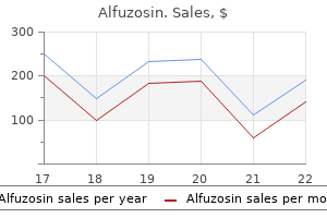
Common Guelder-Rose (Cramp Bark). Alfuzosin.
- Are there safety concerns?
- Cramps, muscle spasms, menstrual cramps, cramps during pregnancy, use as a kidney stimulant in urinary conditions which involve pain or spasms, cancer, hysteria, nervous disorders, and many other conditions.
- What is Cramp Bark?
- How does Cramp Bark work?
- Dosing considerations for Cramp Bark.
Source: http://www.rxlist.com/script/main/art.asp?articlekey=96728
Order alfuzosin online
These cells then mature through several erythroblast stages prostate cancer 6 and 7 purchase alfuzosin us, actively synthesizing hemoglobin. It is released by the kidneys and liver in response to prolonged oxygen deficiency. Breakdown of Erythrocytes Red blood cells bend as they move through blood vessels, but aging causes them to become more fragile. Biliverdin is converted into bilirubin, an orange pigment that is excreted along with biliverdin in the bile. Erythropoietin Regulation Especially during hypoxia, the kidneys produce erythropoietin, which is also called erythropoiesisstimulating hormone. Formed Elements 1 Food nutrients including vitamin B12, folic acid and iron are absorbed from small intestine. If certain kidney cells are deficient in oxygen, enzymes that are oxygen-sensitive cannot function normally. Hypoxia-inducible factor is an intracellular signaling molecule that accumulates as a result, increasing the synthesis and release of erythropoietin. Oppositely, erythropoietin production is depressed by excessive oxygen or excessive erythrocytes in the bloodstream. In the bloodstream, erythropoietin stimulates red bone marrow cells that are already committed to forming erythrocytes, causing them to mature more quickly. Although injection may raise hematocrit from normal levels up to 65%, extreme physical activity can cause blood concentration, because of dehydration. The blood becomes thick and sticky, which can lead to clotting, heart failure, and stroke. Erythrocyte production may also be heightened by many different chemicals released by leukocytes, platelets, and some reticular cells. Red blood cell (erythrocyte) 4 Mature red blood cells are released into the bloodstream and circulate for about 120 days. Liver Hemoglobin Heme Iron Biliverdin Bilirubin 5 Iron from heme is returned to red bone marrow via the bloodstream. Globin Amino acids Iron Amino acids, lipids, and carbohydrates are required for erythropoiesis to occur. The cells of the intestines accurately control iron absorption into the bloodstream as a result of changing amounts of iron that are stored in the body. The liver and spleen store most of the rest of body iron, with small amounts also stored in the bone marrow. They are engulfed and destroyed by macrophages, with the heme portion of their hemoglobin being split from the globin portion. Disorders of Erythrocytes the majority of erythrocyte disorders are classified as either anemia or polycythemia. Anemias the term anemia signifies the blood having an ability to carry oxygen that is too low for normal metabolism. When a person is anemic, he or she is chilled, fatigued, short of breath, and often pale. The three causes of anemia include blood loss, insufficient production of red blood cells, or excessive destruction of red blood cells. When this condition is acute, blood loss is very rapid, and is treated by replacing this depleted blood. Chronic hemorrhagic anemia may be caused by small amounts of blood being lost, but on a persistent basis, such as from a bleeding ulcer or because of hemorrhoids. After the primary condition is resolved, lost blood cells can be replaced by normal erythropoiesis. After losing a fair amount of blood, there would be an increased reticulocyte count. Aplastic anemia-red bone marrow destruction or inhibition because of chemicals, drugs, ionizing radiation, viruses, or most commonly from an unknown cause. Since all formed elements experience altered development, anemia is usually present along with defective blood clotting and immunity. Pernicious anemia-an autoimmune disease that is most common in the elderly, in which the stomach mucosa is destroyed by their own immune systems. In order for intestinal cells to absorb vitamin B12 require intrinsic factor, which is produced by the mucosal cells of the stomach. Treatment of pernicious anemia requires regular intramuscular vitamin B12 injections, or a nasal application of a gel containing this vitamin, once weekly. Also, a lack of dietary vitamin B12 can cause anemia, but this usually only occurs in strict vegetarians, since this vitamin is found in large amounts in fish, poultry, and other meats. Iron-deficiency anemia-this form of anemia usually follows hemorrhagic anemia, but may also occur because of inadequate dietary iron or impaired absorption of iron. Microcytes are produced, which are a type of erythrocytes that are small and pale, due to their inability of synthesizing normal amounts of hemoglobin. Iron-deficiency anemia is treated by increasing dietary iron or by taking iron supplements. Excessive Destruction of Red Blood Cells Hemolytic anemias involve premature rupture or lysis of erythrocytes. Causes include abnormal hemoglobin, certain bacterial or parasitic infections, and transfusions of mismatched blood. When abnormal Formed Elements 433 hemoglobin is produced, there is usually a genetic cause. The two primary examples are thalassemias and sickle-cell anemia, which are serious disorders that may be incurable and fatal. These diseases involve abnormal globin and erythrocytes that are fragile, which rupture prematurely. Thalassemias-most common in people of Mediterranean ancestry, these conditions involve one globin chain that is absent or has abnormal function. A large variety of thalassemias occur, which are classified by their hemoglobin chains that are affected, and where in the chains that this occurs. Some are mild, while others are so severe that they require monthly blood transfusions. Sickle-cell anemia-because of a change in one of the 146 amino acids of a beta chain in the globin molecule, abnormal hemoglobin S (HbS) is formed. When low-oxygen conditions exist, the beta chains link together, forming stiff rods. This interferes with delivery of oxygen, causing severe bone and chest pain, other severe pain, and impaired breathing. A newer treatment involves inhalation of nitric oxide to dilate the blood vessels. Sickle-cell anemia mostly affects Africans living in areas of that country where malaria occurs regularly, and their descendants. Individuals with two copies of the sickle-cell gene have sickle-cell anemia, but those with only one copy of the gene have sickle-cell trait, and are more likely to survive malaria. The cells only sickle when the individual is infected with malaria, or under other abnormal situations. For example, fetal hemoglobin (HbF) does not sickle, even in people likely to develop sickle-cell anemia. Therefore, hydroxyurea can be administered, since it causes the fetal hemoglobin gene to activate. In children with severe disease, bone marrow stem cell transplants are risky but may be totally curative. Newer treatments include orally-administered arginine, which stimulates nitric oxide production and dilates blood vessels, as well as gene therapy. Polycythemias Polycythemia results in the blood to become sluggish, due to excessive amounts of erythrocytes, and elevated hematocrit. Blood volume may be doubled, severely impairing circulation, and the hematocrit may be as high as 80%. Treatment for severe cases involves therapeutic phlebotomy, in which some blood is removed from the body.
Syndromes
- The scrape looks like it may be infected. Signs of infection include warmth or red streaks at the injured site, pus, or a fever.
- Problems swallowing (at all ages)
- Is there anything that relieves the discharge?
- No urine output
- Learn more about eating healthy and eating out.
- Repetitive finger flapping, twisting, or whole body movements
Order 10mg alfuzosin with mastercard
Improvement of autoimmune hepatitis during pregnancy followed by flareup after delivery prostate wiki effective 10mg alfuzosin. Increased risk of preterm birth in women with autoimmune hepatitis: a nationwide cohort study. Pregnancy and birth outcomes in a Danish nationwide cohort of women with autoimmune hepatitis and matched population controls. Chapter 11 Managing Pregnant Women with Autoimmune Liver Disease 215 14 Szczepanska, M. The course and delivery of a pregnancy in a patient with autoimmune hepatitis complicated by cirrhosis of the liver. Autoimmune hepatitis and pregnancy: report of two cases with different maternal outcomes. Pregnant woman with noncomatose autoimmune acute liver failure in the second trimester rescued using medical therapy: a case report. Outcome of pregnancy in patients with autoimmune hepatitis/primary biliary 24 25 26 27 28 29 30 31 32 33 cirrhosis overlap syndrome: a report of two cases. Immunosuppressive effect of pregnancy on autoimmune hepatitis: a case report and review of literature. Outcomes of pregnancies complicated by liver cirrhosis, portal hypertension, or esophageal varices. Impact of cirrhosis and liver transplant on maternal health during labor and delivery. Transjugular intrahepatic portosystemic shunt placement during pregnancy: a case series of five patients. Transjugular intrahepatic portosystemic shunt: a case report of rescue management of unrestrainable variceal bleeding in a pregnant woman. Nonsteroidal anti inflammatory drugs, glucocorticoids and disease modifying antirheumatic drugs for the management of rheumatoid arthritis before and during pregnancy. The safety of 6mercaptopurine for childbearing patients with inflammatory bowel disease: a retrospective cohort study. Pregnancy outcome following in utero exposure to azathioprine: a French comparative observational study. Azathioprine during pregnancy in systemic lupus erythematosus patients is not associated with poor fetal outcome. Azathioprine use during pregnancy: unexpected intrauterine exposure to metabolites. Congenital esophageal atresia and microtia in a newborn secondary to mycophenolate mofetil exposure during pregnancy: a case report and review of the literature. The pathogenesis of osteoporosis is mainly characterized by low bone formation, although increased bone resorption has been described in patients with severe cholestasis and advanced liver disease. For the prevention and treatment of osteoporosis, good nutrition, avoidance of smoking and excess alcohol, weightbearing exercise, treatment of risk factors, and prescription of calcium and vitamin D supplements appear helpful. Osteoporosis is associated with the severity of liver disease, older age, use of corticoste roids, and the duration of cholestasis the pathogenesis of osteoporosis is mainly characterized by low bone formation, although increased bone resorption has also been described in patients with severe cho lestasis and advanced liver disease. Osteomalacia, characterized by poor bone mineralization, is very uncommon and Autoimmune Liver Disease: Management and Clinical Practice, First Edition. The prevalence of bone disease in patients with autoimmune hepatitis is uncertain, although it is assumed that osteoporosis is one of the most frequent complications of corticosteroid treatment. Lateral Xray of the dorsal and lumbar spine should also be carried out to identify vertebral fractures, and laboratory measurements of circulating levels of calcium, phosphorus, 25hydroxyvitamin D, and parathyroid hormone can be assessed at diagnosis. For the prevention and treatment of oste oporosis, good nutrition, avoidance of smoking and excess alcohol, weight bearing exercise, treatment of risk factors, and prescription of calcium and vitamin D supplements appear helpful. Oral bisphosphonates, including weekly alendronate and monthly ibandronate, can be effective for increasing bone min eral density. The efficacy of parenteral bisphospho nates such as zoledronic acid, as well as other newer agents, in patients at high risk for developing osteoporotic frac tures needs ongoing evaluation in patients with autoimmune liver diseases. This is likely due to the lack of bone mineralization as a consequence of the reduced intestinal absorption of vitamin D resulting from the severity and duration of the cholestasis. For many years, up until the 1990s, the diagnosis of bone disease was based on bone Xray and histomorphometry of bone biopsy. However, bone biopsy has only been used in a few cases, as it is an invasive method and also because of the difficulties in obtaining the specimens at the transilial site and performing the analysis of the undecalcified tissue, in addition to the potential complica tions. These problems may explain the reduced information available from this diagnostic tool. However, using bone histo morphometry it was clarified that osteopo rosis but not osteomalacia is the common metabolic bone disease associated with liver (a) diseases. Developments in imaging methods such as bone densitometry have improved the diagnosis of osteoporosis. This article describes the prevalence of metabolic bone disease in these three auto immune liver conditions, and the preva lence and risk for developing fractures. Likewise, the pathogenesis of the associ ated bone disease is reported, as well as an update of the recommendations for diag nosis and treatment. Moreover, in the earlier studies bone disease was assessed by Xray of the spine or by bone biopsy and histomorphometry. Bone mass is now best assessed by bone densitometry of the lumbar spine and proximal femur. A lower prevalence of osteoporosis is observed in patients with less severe disease, while the prevalence is higher in advanced stages and before transplantation [13]. In a series of 185 patients, fractures were detected in 21% of the cases (11% vertebral and 12% peripheral fractures) and associated with osteoporosis and the severity of cholestasis. Thus, one report did not show an increase in the fracture risk, while another populationbased cohort study including Table 12. Severe osteoporosis, defined by the authors as a Zscore less than -2, was found in 33 patients, a prevalence that was 6. Deficiency of vitamin D and calcium was found in 23% and 4% of patients, respectively. Interestingly, osteoporosis was present in 75% of the patients with these three factors, but in only 3% of patients with none of them. This is particularly relevant for patients receiving 30 mg/day, with a cumulative dose higher than 5 g in a year. In an analysis of the fracture risk of oral corticosteroid therapy, it was concluded that intermittent use of highdose oral cortico steroids (daily dose >15 mg, cumulative exposure <1 g) may result in a small increased risk of osteoporotic fracture. Conversely, patients who receive several courses of high dose corticosteroids (daily dose >15 mg, cumulative exposure >1 g) have a consider ably increased risk of fracture. Nevertheless, the pathogenesis is mainly related to osteopo rosis, since osteomalacia is a very infrequent condition that is only present in cases with extremely severe and longlasting cholestasis. Thus, a decreased rate of bone formation and impaired osteoblast function resulting in lower mean wall thick ness and a defect in matrix synthesis has been observed. Similar data are present in patients with advanced cholestatic liver dis ease before transplantation. For example, in a previous study from our group with clinically driven biopsies, we calculated 5 and 10year recurrence rate of 13 and 29% [5], respectively, whereas in a study with protocolguided biopsies, the recurrence rate was higher, as expected, with 5, 10, and 15year rates of 27, 47, and 61%, respectively [6]. Currently, diagnosis is essentially based on histopathologic findings on liver biopsy. Compatible histologic findings include lymphocytic cell infiltrate, lymphoid aggregates, epithelioid granulomas, or florid bile duct lesions. In many of the studies, no multivariable analysis was made to adjust for potential confounders, so the Table 13. Male recipient was significant in one study, but this finding could not be replicated in others studies [5,6]. However, the fact that those observations were inaccurate may be due to short followup and a limited number of patients. Other than that, therapy is focused on addressing biliary complications when present, managing portal hypertensionrelated complications if there is progression to cirrhosis, and considering retransplant in a timely fashion. The most common indication is end stage liver disease with portal hypertension derived complications. Compatible histopathology (lymphoplasmacytic infiltrate with interface hepatitis), with no endotheliitis or ductulitis.
Buy cheap alfuzosin 10 mg on-line
Preganglionic neurons: Visceral motor neurons in the central nervous system whose output controls one or more ganglionic motor neurons in the peripheral nervous system androgen hormone juvenile order alfuzosin with a mastercard. Pregnancy: the condition that begins when the developing offspring implants into the uterine lining; it consists of three trimesters (each about three months in length). Preload: the stretch of ventricular muscle fibers at end diastole; it is reflected by the ventricular pressure and volume in part of the cardiac cycle; cardiac output increases with preload. Premolars: One of eight teeth, four in each dental arch, located lateral and posterior to the canine teeth, in front of the molars. Prepuce: the male foreskin; also, a similar fold of skin around the female clitoris. Presbycusis: Hearing loss that commonly occurs with aging; it is usually linked to heredity, chronic exposure to loud noises, earwax blockage, inner ear damage, infections, and a ruptured tympanic membrane. Primary (olfactory) smell cortex: the main cortex that handles olfactory sensations; located on the medial temporal lobe in the piriform lobe area. Primary auditory cortex: the cortex that receives impulses from the inner ear, interpreting location, loudness, and pitch; it lies in the superior margin of the temporal lobe. Primary bronchi: Branches of the bronchial tree that divide into secondary bronchi, tertiary bronchi, and finer tubes. Primary germ layers: Three cell layers that arise in the embryonic period from which all organs of the body form. Primary immune response: A response constituted by activation of B-cells or T-cells after they first encounter the antigens for which they are specialized to react. Primary oocytes: Derived from an oogonium by differentiation near the time of birth. Primary somatosensory cortex: the cortex within the postcentral gyrus of the parietal lobe, just posterior to the primary motor cortex, that receives input from the somatic sensory receptors of the skin, and input from position sense receptors in the joints, skeletal muscles, and tendons. Primary visual cortex: Also called the striate cortex, it is the largest cortical sensory area, receiving visual information from the retinas of the eyes; it is buried in the calcarine sulcus of the occipital lobe and extends to the extreme posterior occipital tip. Prime mover: An agonist; a muscle that contracts to provide most of a desired movement. Primitive streak: During gastrulation, a groove with raised edges that appears on the dorsal surface of the embryonic disc. Primordial follicles: Structures in developing female fetuses that contain a primary oocyte surrounded by follicular cells. Principal cells: Also called chief cells; they line the gastric glands and secrete pepsinogen and instrinsic factor. Proerythroblasts: the earliest erythrocyte precursors, preceding the basophilic erythroblasts and having a large nucleus with several nuclei, surrounded by a small amount of cytoplasm. Progesterone: A female hormone that promotes changes in the uterus during the reproductive cycle, affects the mammary glands, and helps regulate gonadotropin secretion. The forearm is rotated medially, moving the distal end of the radius across the ulna, forming an X between the two bones. Propagation: the process of spreading to a larger area or greater number; increasing, or dissemination. Proprioceptors: Sensory organs that monitor the position and movement of skeletal muscles and joints. Pubic crest: the raised area of the pubis formed by the thickened anterior border. Pubic tubercle: the lateral end of the pubic crest; it is one of the attachments for the inguinal ligament. Pubis: the part of the hipbone that is superior and partly anterior to the ischium. Pubofemoral ligament: One of the ligaments that reinforces the hip joint capsule; it is a triangular thickening of the inferior area of the capsule. Pulmonary arteries: Those that carry deoxygenated blood from the right side of the heart to the lungs. Pulmonary circuit: the blood flow and blood vessels within the lungs and between the lungs and the heart. Pulmonary plexuses: the combinations of several trunks of the vagus nerve, joined at the root of the lung by branches of the sympathetic trunk and cardiac plexuses. Pulmonary valve: Lying at the base of the pulmonary trunk, this valve has three cusps and allows blood to leave the right ventricle while preventing backflow into the ventricular chamber. Pulmonary veins: Four veins, two on each side, which convey oxygenated blood from the lungs to the left atrium of the heart. Pulmonary ventilation: A measure of the rate of ventilation, referring to the total exchange of air between the lungs and ambient air, usually in liters per minute. Pulp cavity: the space in a tooth bounded by dentin and containing the dental pulp; it is divided into the pulp chamber and pulp canal. Pulp: Also called dental pulp; the richly vascularized and innervated connective tissue inside the pulp cavity of a tooth. Punnett square: A diagram used to determine, for a single trait, the possible gene combinations that would result from the mating of parents of known genotypes. Purines: Organic compounds that are the most widely occurring nitrogen-containing heterocycles; they are water soluble, and found in high concentration in meats and meat products. Purkinje cells: Large, densely branching neurons in the cerebellar cortex of the brain. Purkinje fibers: Consisting of branches of the atrioventricular bundle that spread and enlarge, these fibers are Prostacyclin: A prostaglandin that is a biologically active product of arachidonic acid metabolism in the vascular walls; it is a potent inhibitor of platelet aggregation that inhibits the vasoconstrictive effect of angiotensin and stimulates renin release. Prostaglandins: Lipids made from arachidonic acid that usually act more locally than hormones, are very potent, stimulate hormone secretions, and help to regulate blood pressure. Prostate cancer: A usually slow growing cancer in the prostate gland of the male reproductive system. Prostate gland: A structure that surrounds the proximal urethra; it secretes a milky, alkaline fluid that neutralizes the acidic fluid containing sperm cells. Prostatic urethra: the part of the male urethra from the base of the prostate gland to the point where the urethra emerges from the apex of the prostate gland. Protein synthesis: Assembling of functional polypeptides in the cytoplasm, via the process of translation. Proteins: Substances made up of amino acids that are vital for many body functions, including structures and their functions, energy, and hormonal requirements. Protraction: Moving a part forward, which is a nonangular anterior movement in the transverse plane. Pseudostratified columnar epithelium: Tissue that is layered in appearance and is involved in secretion; lines respiratory system passages. Pseudounipolar neurons: the preferred name for unipolar neurons, since they originate as bipolar neurons. Pterygoid processes: One of three types of processes extending from the sphenoid bone; these are narrow depressions that project inferiorly from where the body and greater wings join, anchoring the chewing-related pterygoid muscles. Pterygopalatine ganglia: the ganglia just posterior to the maxillae, in which preganglionic fibers synapse with postganglionic neurons. Pus: A thick fluid formed from masses of leukocytes, bacterial cells, and damaged tissue. Pyloric antrum: the initial portion of the pylorus, near the bottom of the stomach. Pyloric sphincter: A circular muscle acting as a valve to control gastric emptying. Pyramidal (corticospinal) tracts: Major motor pathways concerned with voluntary movement; they descend from pyramidal cells in the frontal lobes of each cerebral hemisphere. Pyramidal cells: Neurons with pyramid-shaped cell bodies, in the gray matter of the cerebral cortex. Pyrogens: Substances or agents that tend to cause an increase in body temperature. Quadriplegia: Paralysis of all four limbs because of transection of the spinal cord in the cervical region. Radial collateral ligament: One of the two strong capsular ligaments of the elbow joint that restrict horizontal movements; it is triangular and located on the lateral side. Radial nerve: the largest branch of the brachial plexus, which continues from the posterior cord. Radial tuberosity: A process of the radius bone that serves as an attachment for the biceps brachii muscle. Radioisotopes: Also known as radioactive isotopes or radionuclides, they are atoms with unstable nuclei.
Purchase discount alfuzosin on-line
This is the shortest circulation in the entire body and the functional blood supply of the heart mens health gift subscription order alfuzosin cheap. It is important to remember that blood flows through the heart in only one direction, controlled by the four heart valves. The valves open and close as a result of differences in pressure on their two sides. They supply blood to the heart tissues, with openings lying just beyond the aortic valve. Both of these coronary arteries enclose the heart in the coronary sulcus and provide the arterial supply of the coronary circulation. The left coronary artery runs toward the left side of the heart, whereas the right coronary artery runs toward the right side. Right atrium coronary artery is divided into the anterior interventricular artery and the circumflex artery. The anterior interventricular artery is also known as the left anterior descending artery. These arteries have smaller branches with connections called anastomoses between vessels that provide alternate blood pathways. Known as collateral circulation, these pathways may supply oxygen and nutrients to the myocardium when blockage of a coronary artery occurs. The right coronary artery is divided into the right marginal artery and the posterior interventricular artery. The posterior interventricular artery supplies the posterior ventricular walls and runs to the heart apex. It merges or anastomoses near the apex of the heart with the anterior interventricular artery. It empties into the great cardiac vein, middle cardiac vein, and small cardiac vein. The posterior cardiac vein also empties into the great cardiac vein or coronary sinus. Heart Contraction Some cardiac muscle cells are self-excitable, whereas each skeletal muscle fiber requires stimulation by a nerve ending to contract. Certain cardiac muscle cells are interconnected by intercalated discs, at which point interlocking membranes of nearby cells are linked by gap junctions and held together by desmosomes. The intercalated discs transfer the force of contractions from cell to cell and propagate action potentials. There are three major differences between the ways in which cardiac muscle and skeletal muscle contract. First, self-excitable cardiac muscle cells can also initiate the depolarization of the rest of the heart, spontaneously and with rhythm. Second, cardiac muscle has a property in which either all heart fibers contract as a unit or not at all. This is coordinated because all cardiac muscle cells are Coronary Veins the coronary veins are basically located along the same paths as the coronary arteries. On the posterior aspect of the heart, the 458 Chapter 18 the Heart electrically linked together into one contractile unit by gap junctions. All this differs from skeletal muscle, in which impulses do not spread from cell to cell. Third, in cardiac muscle cells, the absolute refractory period lasts over 200 milliseconds (ms), which is almost as long as the contraction. This normally prevents tetanic contractions from occurring, which would stop the heart from pumping. In skeletal muscle cells, contractions last only 15 to 100 ms and have brief refractory periods of 1 to 2 ms. The absolute refractory period is the period of no excitation, in which sodium ion channels remain open or are inactivated. As atrial systole begins, the ventricles are filled to about 70% of their capacity, because of passive blood flow at the end of the previous cardiac cycle. Ventricular Systole Ventricular systole begins after atrial systole, and lasts about 270 ms in an adult who is resting. There are also three steps to ventricular systole: Energy Requirements Cardiac muscle needs much more oxygen for energy metabolism than skeletal muscle and also has more mitochondria. Skeletal muscle differs in that it can contract for a long time via anaerobic respiration and restore oxygen and fuel reserves via excessive oxygen consumption after exercise. Glucose, fatty acids, and other fuel molecules are used by cardiac and skeletal muscle. However, because it is more adaptable, cardiac muscle easily changes metabolic pathways so it can use whatever nutrients are available. Such nutrients include lactic acid, which is generated by skeletal muscle activity. Therefore, a lack of oxygen is much more dangerous to the myocardium than any lack of nutrients. Functions of the Heart Both sides of the heart contract at the same time, and eject equal volumes of blood. However, pressure is lower in the right atrium and ventricle than in the left atrium and ventricle. Early in ventricular systole, the ventricles contract but blood flow has not occurred. The pressures are not yet enough to force the semilunar valves to open, or to push blood into the pulmonary or aortic trunks. At this time, the ventricles contract isometrically, generating tension and increased pressure, yet blood does not flow out. When ventricular pressure exceeds the pressure in the arterial trunks, the semilunar valves open. Blood flows into the pulmonary and aortic trunks, which is the beginning of ventricular ejection. The ventricles then contract isotonically, meaning their muscle cells shorten and there is a near constant production of tension. When a peak is reached, ventricular pressure gradually reduces, near the end of ventricular systole. The right ventricle also experiences isovolumetric contraction and ventricular ejection, yet pressure in the right ventricle and pulmonary trunk are significantly lower. The amount of blood ejected by each ventricle during contraction, 70 to 80 mL of blood, is the stroke volume of the heart. This percentage is called the ejection fraction, and is varied based on the different demands placed on the heart. Blood in the aorta Functions of the Heart 459 and pulmonary trunk begin to flow back to the ventricles, closing the semilunar valves. Once the semilunar valves are closed, pressure increases again, and the elastic walls of the arteries recoil. This short pressure increase causes a "valley" in the pressure tracing, known as a dicrotic notch. Blood flows from the atria into the ventricles, meaning that all these chambers are in diastole. Ventricular Diastole Ventricular diastole lasts for about 430 ms in the current cardiac cycle, yet continues through atrial systole in the next cycle. There are two steps in ventricular diastole: During this period, all heart valves are closed, and the myocardium is relaxing. Ventricular pressures are still higher than that of the atria, and blood cannot flow into the ventricles. However, damage to one or both ventricles, can cause the heart to be unable to pump blood through the peripheral organs and tissues. Conduction System Strands and clumps of specialized cardiac muscle contain only a few myofibrils and are located throughout the heart. This system is also known as the conducting system or nodal system and includes the sinoatrial (S-A) node, atrioventricular (A-V) node, and conducting cells, which are found in intermodal pathways that distribute contractile stimuli to atrial muscle cells.
