

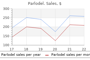
Buy discount parlodel on-line
Convergence treatment hepatitis c order 1.25 mg parlodel visa, divergence and near response is influenced by activity in an area anterior to the frontal eye fields. The superficial layers receive inputs from the optic tract fibers and visual cortex and in turn project to various thalamic nuclei like anterior thalamic nucleus, pulvinar and lateral geniculate body. The neurons of the superficial layer receive inputs from both the eyes and they discharge in response to a stimulus moving quickly in a particular direction. Thus, the superior colliculi coordinate simultaneous bilateral eye movements, like saccades and convergence and keep the eyeball in focus. The deep layers of superior colliculi receive visual inputs from its superficial layers, inputs from the auditory fibers and somatosensory inputs from different body parts. The sensory stimuli are integrated in the deep layers for various reflex activities that involve appro priate head movement and change in eye opposition. Fibers from the optic tract project to the pretectal nucleus and cause activation of the EdingerWestphal nucleus. These fibers mediate pupillary reflexes and are involved in the regulation of visual fixation. It is a small nucleus in the medial hypothalamus, located just above the optic chiasm. It receives fibers from the optic nerve and synchronizes sleepwake cycle, secretion of hormones like cortisol, and other circadian rhythms with lightdark cycle. Information from the visual receptors regarding light intensity during diurnal cycle gets transmitted to the suprachiasmatic nucleus and entrains its biological clock activity. Apart from these areas, some other areas are also activated, such as parts of inferior temporal cortex, posteroinferior parietal cortex, amygdale, caudate nucleus, putamen and claustrum. Effect of lesions at various levels of visual pathway is asked as a Short Question. Name the areas or pathways for processing of visual signal, Macular sparing, What is the on-center and offcenter response of the ganglion cells, What are important features of retinal neurons, Cortical visual areas. Name the visual field defects produced by lesion at different levels of visual pathway. The short est distance, by which two objects can be separated and still be visualized as two different objects, is known as the minimum separable distance. Visual acuity expresses the resolving power of the eye, that is, the extent to which the eye can perceive the details and contours of an object. There are different tests for testing visual acuity for distant vision and near vision. Visual acuity increases as the size of the object increases and distance of the object from eye decreases. But the contrast between the object and the back ground plays a more important role than the colorper se. Factors Affecting Visual Acuity Optical Factors these factors mainly determine the degree of visual acuity. The rows are numbered as 60, 36, 24, 18, 12, 9, 6 and 5 from top to bottom; and the size of the letters in each row gradually decreases. As the cones are concentrated more at the fovea, Chapter 146: Visual Acuity, Visual Field, Light and Dark Adaptations, and Visual Reflexes 1189. The gaps between the lines as well as the width of each line subtend an angle of 1 minute at the nodal angle. The number below each row depicts the distance in meters from which the letters can be read by a normal eye. The subject is asked to read the chart with each eye separately from a distance of 6 meters. Visual acuity is calculated by using the formula d/D, where d is the distance at which letters are read and D is the distance at which letters can be read by a normal eye. For example, if a person can read by his right eye the letters designated by the number 18, the visual acuity for right eye will be 6/18. The visual acuity of 6/5 is called supernormal, where the person can read the last row from a distance of 6 meters. Thus, for any closely placed two points, the minimum separable distance should draw an angle of 1 minute at the nodal angle. Binocular Vision When a person looks at an object with both eyes, the light rays coming from the object stimulate both the retinas. The action potentials transmitted by the neural pathways from both retinas get fused at the visual cortex, so that the brain perceives them as single image. Thus, viewing an object as a single one with both eyes is called binocular vision. The two points on the retinas on which the light rays from an object fall simultaneously to make binocular vision possible, are known as corresponding points. The foveas and all points lying at the same distance and in the same direction from the foveas are corresponding points. For a distant object, the eyeballs move symmetrically and for a near object, the eyeballs converge to allow the light rays to fall on corresponding points. When light rays fall on noncorresponding points, double vision (diplopia) results. For example, while looking at an object if you slightly push one eyeball out of the line, diplopia occurs. When the light rays from any object do not fall on the corresponding points for a prolonged period, especially in children below the age of 6 years, diplopia is not seen as one of the images get suppressed (suppression scotoma). It should be detected and treated early; otherwise there occurs permanent loss of visual acuity in the eye, in which the image is suppressed. Diplopia also occurs when the medial rectus or lateral rectus muscles are damaged or overactivated. The area of the external world that can be seen when the gaze is fixed is known as the visual field of that eye. The visual field is limited on the medial side by the bridge of the nose and superiorly by the roof of the orbit, so that the field is not circular. The visual field of both eyes overlap in the center and is called binocular visual field. An object present in the binocular visual field can be seen by both eyes simultaneously. Each visual field is divided into a nasal (medial) and a temporal (lateral) portion. For each eye, light rays from an object situated in the nasal field stimulate the temporal retina and light rays from an object situated in the temporal field stimulate the nasal retina. Also, light rays from objects located at the upper part of the visual field stimulate the lower part of retina and those from the lower part of the visual field stimu late the upper part of retina. The peripheral visual field is mapped by an instru ment called perimeter and the process is known as perimetry, in which each eye is mapped separately. During the procedure one eye is covered, while the test eye is fixed on a central fixation point. A small contrast target is moved slowly from the periphery to the center in selected meridians along the arc of the instrument, completing the full circle. The scotoma area is mapped by noting the spot where the object disappeared to the point, where again it reappeared. The physiologi cal blind spot due to optic disc is observed on the tem poral visual field. The macular fibers are spared unless the damage is widespread because i) the macular fibers project separately in the visual cortex, and ii) they have a much larger representation compared to rest of the retina. However, when light intensity changes dramatically, the visual system requires some time for adaptation of the eye to respond optimally.
Buy discount parlodel 2.5mg on line
In addition medications by class cheap parlodel 1.25mg free shipping, it has been associated with asthma-like symptoms, though there remains some controversy as to whether these represent true allergic asthma caused by formaldehyde. Aqueous solutions in contact with the skin cause hardening and roughness, due to superficial coagulation of the keratin layer. Ingested formaldehyde attacks the membrane lining of the stomach and intestine, causing necrosis and ulceration. The latter is partly responsible for the metabolic acidosis that is characteristic of formaldehyde poisoning. Circulatory collapse and renal failure may follow the devastating effects of ingested formaldehyde on the gut, leading to death. Toxicity is somewhat less than that of formaldehyde, because of the slow evolution of gas. Hydrogen cyanide gas causes poisoning by inactivating cytochrome oxidase, the final enzyme essential to mammalian cellular respiration. The patient will have signs of severe hypoxia, however, and in some cases may not appear cyanotic. This is due to the failure of hemoglobin reduction in the face of loss of cellular respiration. This will result in a pink or red color to the skin and arteriolization of retinal veins. Unconsciousness and death may occur immediately following inhalation of a high cyanide concentration, respiratory failure being the principal mechanism. Lesser exposures cause a constriction and numbness in the throat, stiffness of the jaw, salivation, nausea, vomiting, lightheadedness, and apprehension. Fixed, dilated pupils, bradycardia, and irregular gasping respiration (or apnea) are typical of profound poisoning. Early symptoms of acute poisoning include headache, dizziness, nausea, vomiting, tremor, slurred speech, and ataxia. The more severe cases of poisoning exhibit myoclonic and generalized tonic clonic seizures, which are sometimes refractory to initial therapy. Residual neurological deficits including myoclonic seizures, ataxia, muscle weakness, tremors, behavioral disturbances, and diminished reflexes may persist in more severely poisoned patients. Exposure to high concentrations may cause central nervous system depression, manifested as fatigue, weakness, and drowsiness. Some absorbed methylene chloride is degraded to carbon monoxide in humans, yielding increased blood concentrations of carboxyhemoglobin. However, concentrations are rarely high enough to cause symptoms of carbon monoxide poisoning. Ingestion has caused death from gastrointestinal hemorrhage, severe liver damage, coma, shock, metabolic acidosis, and renal injury. In laboratory animals, extraordinary dosage has caused irritability, tremor, and narcosis, leading to death. When heated to that point of decomposition, one of the products is the highly toxic phosgene gas that has caused a significant acute pneumonitis. The vapor has a sharp, pungent odor that is irritating to the eyes and upper respiratory tract. Inhalation of high concentrations causes headache, dizziness, nausea, and vomiting. However, homozygous females, who are far less common, will have a similar expression. This illness is most common in non-white African and African-American ethnic groups. It is actually the metabolites of naphthalene that are responsible for the hemoly15 sis. Secondary renal tubular damage may ensue from the naphthol and from the products of hemolysis. In infants, high levels of hemoglobin, methemoglobin, and bilirubin in the plasma may lead to encephalopathy. Kernicterus has been specifically described as a complication of exposure to naphthalene with severe hemolysis and resulting hyperbilirubinemia. Paradichlorobenzene is solid at room temperature, and is now widely used as a moth repellent, air freshener, and deodorizer in homes and in public facilities. Although accidental ingestions, especially by children, have been fairly common, symptomatic human poisonings have been rare. It is used as a fumigant by placing solid aluminum phosphide (phostoxin) near produce or in other storage spaces. Most severe acute exposures have involved ingestion of the solid aluminum phosphide, which is rapidly converted to phosphine by acid hydrolysis in the stomach. Extracellular magnesium levels have been found to be slightly elevated, suggesting a depletion of intracellular magnesium from myocardial damage. In other fatalities, ventricular arrythmias, conduction disturbances, and asystole developed. Sulfur dioxide is a highly irritant gas, so disagreeable that persons inhaling it are usually prompted to seek uncontaminated air as soon as possible. However, laryngospasm and pulmonary edema have occurred, occasionally leading to severe respiratory distress and death. It is sometimes a cause of reactive airways disease in occupationally exposed persons. Although use experience has generally been good, some fatalities have occurred when fumigated buildings have been prematurely reentered by unprotected individuals. Manifestations of poisoning have been nose, eye, and throat irritation, weakness, nausea, vomiting, dyspnea, cough, restlessness, muscle twitching, and seizures. Confirmation of Poisoning There are no practical tests for absorbed alkyl oxides, aldehydes, or phosphine that would be helpful in diagnosis of poisoning. Carbon disulfide can be measured in urine by gas chromatography, but the test is not generally available. Cyanide ion from cyanide itself or acrylonitrile can be measured in whole blood and urine by an ion-specific electrode or by colorimetry. Methyl bromide itself has a short half-life and is usually not detectable after 24 hours. The bromide anion is slowly excreted in the urine (half-life about 10 days), and is the preferred method of serum measurement. The possible contributions of medicinal bromides to elevated blood content and urinary excretion must be considered, but if methyl bromide is the exclusive source, serum bromide exceeding 6 mg per 100 mL probably means some absorption, and 15 mg per 100 mL is consistent with symptoms of acute poisoning. Inorganic bromide is considerably less toxic than methyl bromide; serum concentrations in excess of 150 mg per 100 mL occur commonly in persons taking inorganic bromide medications. In some European countries, blood bromide concentrations are monitored routinely in workers exposed to methyl bromide. Blood levels over 3 mg per 100 mL are considered a warning that personal protective measures must be improved. A bromide concentration over 5 mg per 100 mL requires that the worker be removed from the fumigant-contaminated environment until blood concentrations decline to less than 3 mg per 100 mL. Methylene chloride is converted to carbon monoxide in the body, generating carboxyhemoglobinemia, which can be measured by clinical laboratories. Naphthalene is converted mainly to alpha naphthol in the body and promptly excreted in conjugated form in the urine. Paradichlorobenzene is metabolized mainly to 2,5-dichlorophenol, which is conjugated and excreted in the urine. Large industrial concerns sometimes monitor human absorption of halocarbons by analysis of expired air. These analyses are rarely needed to identify the offending toxicant, because this is known from the exposure history. In managing difficult cases of poisoning, however, it may be helpful to monitor breath concentrations of toxic gas to evaluate disposition of the fumigant. Sperm counts may be appropriate for workers exposed to dibromochloropropane and ethylene dibromide. Some occupational health agencies now urge periodic neurologic and neuropsychologic testing of workers heavily exposed to fumigants and solvents to detect injury to the nervous system as early as possible. This would be particularly desirable in the case of exposures to such agents as methyl bromide and carbon disulfide which have well-documented chronic neurotoxic effects. Flush contaminating fumigants from the skin and eyes with copious amounts of water or saline for at least 15 minutes.

Purchase parlodel online
Isoniazid medications by class generic parlodel 2.5 mg free shipping, an antitubercular antagonizes the action of pyridoxine and therefore, precipitates deficiency of niacin. The major features of pellagra are: dermatitis (erythematous skin rash lead ing to desquamation), diarrhea, dementia (loss of mem ory). Pyridoxine (Vitamin B6) Human beings cannot synthesize vitamin B6; therefore, they need dietary supplementation. It is unique among watersoluble vitamins in that it is stored in large quanti ties in muscles. Yeast, rice polishing, germinal portion of seeds, whole grain cereals, bananas, potatoes, eggyolk, liver, kidney muscle and fish are rich sources. Functions Acetyl CoA is a common substrate for release of energy from carbohydrates, fats and proteins. Along with malonyl CoA, it is utilized for synthesis and elongation of fatty acids. Functions of Vitamin B6 Pyridoxal phosphate is an essential coenzyme for many biological reactions. It acts as a cotransaminase: In protein metabolism, during transamination, deamination and decarboxyla tion reactions, as in the formation of serotonin from tryptophan and noradrenaline from tyrosine it acts as a coenzyme. It serves as a coenzyme for the conversion of muscle and liver glycogen into glycogen phosphate. Therefore, in pyridoxine deficiency niacin synthesis from tryptophan does not occur. Thus, increased level of xanthurenic acid in urine is an index of pyridoxine deficiency. In lipid metabolism, it serves as a coenzyme for the conversion of linoleic acid into arachidonic acid. It acts as coenzyme for the synthesis of various neurotransmitters like norepinephrine, dopamine, hista mine, etc. However, richest sources include kidney, liver, meat, fish, chicken, whole grain cereals and skimmed milk. Daily requirement of pantothenic is; In adults: 5 to 12 mg In children: 4 to 5 mg In infants: 1 to 2 mg Effects of Deficiency As it is present in all food stuffs, its deficiency is usually not reported. Folate is first acted upon by vitamin C to form dihydrofolate which in turn forms tetrahydrofolate. Deficiency of folic acid leads to megaloblastic anemia (for details, refer "Erythropoiesis"). Folate supplement during pregnancy reduces the risk of neural tube defect in newborn. Sources Iron is present in meat, liver, fish, green leafy vegetables, potatoes, legumes and fruits. Vitamin B12 Vitamin B12 is also called cyanocobalamine or extrin sic factor of Castle. Its main function is to bring maturation of red cells (for details, refer "Erythropoiesis"). Dietary deficiency of this vitamin is rare (except in strict vegetarians: vegans) as considerable amount is stored in liver. Therefore, gastric atrophy, chronic gastritis and gastrectomy cause pernicious anemia, which is spe cial type of megaloblastic anemia that occurs due to stom ach diseases. In India, chronic intestinal blood loss due to hookworm infestation demands more iron intake. Deficiency the commonest deficiency among minerals is that of iron which is common even in affluent society, particularly in women in their reproductive years. The minerals that are important in body functions are calcium, phos phorus, iron, iodine, zinc, copper and selenium. Deficiency of minerals occurs due to the same general reasons as vitamin deficiency. Iodine the iodine content of water and food greatly depends on the iodine content of the soil in the region. Iodine deficiency leads to hypothyroidism, which manifests as thyroid swellings, known as goiter (For details of iodine metabolism, refer Chapter 57). Calcium is essential for formation of bones and teeth, blood clotting, neuromuscular excitability and acti vation of several enzymes in the body. Though cereals and legumes contain good amount of zinc, zinc absorption from these sources is limited by presence of phytic acid. Iron An adult has about 4 g irons, two thirds of which exists in the form of hemoglobin and the remaining is present in myoglobin, cytochromes, transferrin, ferritin, and storage iron as hemosiderin. It is also a cofactor 1292 Section 13: Integrative Physiology in the synthesis of collagen. Zinc is essential for normal growth in children, reproductive function, wound healing, and for sense of taste and smell. Balanced Diet A balanced diet is a satisfactory diet that provides ade quate quantities of all essential nutrients. It depends mainly on the age, sex, body weight and level of physical activity of the individual. For example, for a 50 kg moderately active woman, the diet should provide 2225 kcal and at least 50 g protein everyday. As 50g protein provides 200 kcal; the remaining 2025 kcal should be obtained from carbohydrates and fats. For children and pregnant women, some nonvegetar ian food like fish, egg or meat should be included. Deficiency Zinc deficiency is characterized by poor growth and sexual development. Deficiency also causes poor wound healing, loss of appetite, diminished taste and smell sensations and several skin disorders. Zinc deficiency is precipitated by iron supplements as both of them share common intes tinal transport mechanisms. In acrodermatitis enteropath ica there is inherited defect of intestinal zinc absorption. Sources If the soil has adequate selenium, food will have adequate quantity of it. Poor socioeconomic status, infectious diseases, parasitic dis eases, lack of personal hygiene, unsafe drinking water and lack of medical facilities and improper or inadequate nutrition are the etiological factors for malnutrition. Decreased food intake can be due to various reasons such as: decreased food availability, poor dietary habits, food faddism and emotional factors, etc. Malnutrition is common in children, and occurs in two forms: marasmus and kwashiorkor. Sometimes calorie is adequate but food may be grossly deficient in proteins and vitamins; the condition known as kwashiorkor in children. Usually, when edema is pre sent, is called kwashiorkor, and when edema is absent, is called marasmus. Functions Selenium is an important component of the antioxidant system of the body. It is present in glutathione peroxidase, the enzyme that prevents lipid peroxidation. Deficiency Deficiency of selenium is usually seen in low intake and due to long term parenteral nutrition. Selenium defi ciency may also cause Keshan disease, in which cardiac muscle degeneration is an important feature. Kwashior kor occurs in children when the food is devoid of proteins and vitamins, but fills the stomach. So the energy supply is adequate; however, growth and repair of the body are severely restricted.
Purchase on line parlodel
Any slip ups in such circumstances could mean life and death medicine 666 order 1.25 mg parlodel with visa, and hence close attention needs to be paid. Changing strategy: If a business is changing strategy or making big changes within the organization, micromanagement is good. It helps everyone involved to understand the process and how the changes will impact their role and duties. Changing top management: When there is a change in top management, business owners need to micromanage so as to familiarize the new executive to the work environment. When It Is Bad for Business:Excess managing is detrimental to the growth and well-being of an organization. Micromanagement has the potential of too much scrutiny, and this can be counterproductive. It hampers growth and learning: Too much control is not good, for it kills the desire in employees to learn and grow. When an employee knows that he/she is being constantly watched and will be interrupted at every stage, they lose the desire for self-improvement and enhancement. It prevents evaluation of skills: It is difficult to assess the skills of employees who are being micromanaged, as it is unclear what they have done themselves and what they have been directed to do by the micromanager. It affects employees performance: Over scrutinizing is demoralizing and creates self-doubts in the employees, which eventually ends in affecting their performance. The employees know that they will not be allowed to work independently or given credit and hence do not put in any extra efforts or add anything more to the task than is asked. It kills motivation and innovation: Constant criticism and scrutiny also kill initiative. When the micromanager takes over the task, and there is no scope for inputs from anyone else, it kills innovation and creativity and demotivates the team. The micromanager loses control: A micromanager will eventually lose control over the team. Since they use only control to manage their subordinates, soon it becomes ineffective as the employees get used to the bullying, or they leave and seek employment elsewhere. There is a loss of trust and mutual appreciation: Micromanagement breeds distrust and dislike. Such an environment, within an organization, is not conducive to growth and productivity. When the atmosphere at work becomes too oppressive, the productivity drops and good employees leave. It creates dependency: Since a micromanager does not allow initiative and inputs from other people in the team, the employees learn to leave all decision-making to the manager and become totally dependent on him/her. It results in a high attrition rate: One side effect of micromanagement is a high attrition rate, where good employees leave the organization and join rivals or parallel ones. Most creative and hardworking people do not like being under the microscope all the time and prefer to move on. It results in increased workload and burnout: When someone is micromanaging they are essentially taking on work that has been assigned to someone else. So micromanagers end up doing double the work, which they could have easily avoided if they had not micromanaged. This causes the overburdening of one individual, in this case, the micromanager, and can lead to burnout. Managers need to be aware of their employees performance and attitude, but this should be done in a manner that is not hyper critical. They need to be able to deal with people in a respectful and polite manner, and ensure that the inputs that they are giving add to the process and do not unnecessarily bog it down with details. Employees, on the other hand, need to be proactive with their responsibilities, and if they feel they are being micromanaged, do something about it. Whether it is the micromanager or the micromanaged, both need to take stock of the situation. If the micromanagement is becoming restrictive and oppressive, try to remedy it, as sooner or later, it will start to affect the overall productivity of the organization. Florence Stone, "Micromanagement: How to Think More Strategically and Less Operationally," Performance and Profits (American Management Association) 1, no. And What You Can Do to Avoid It," Global Knowledge Training, 2007, Joel Brockner, "Why Its So Hard to Be Fair," Harvard Business Review 84, no. Guillain-Barre syndrome consists of a group of neuropathic conditions characterized by progressive weakness and diminished or absent myotatic reflexes. Guillain-Barre syndrome is believed to result from an aberrant immune response that attacks nerve tissue. The most common form of the disease, acute inflammatory demyelinating polyradiculoneuropathy, presents as progressive motor weakness, usually beginning in the legs and advancing proximally. More than one-half of patients experience severe pain, and about two-thirds have autonomic symptoms, such as cardiac arrhythmias, blood pressure instability, or urinary retention. Diagnosis is based on clinical features, cerebrospinal fluid testing, and nerve conduction studies. Cerebrospinal fluid testing shows increased protein levels but a normal white blood cell count. Patients should be hospitalized for multidisciplinary supportive care and disease-modifying therapy. Supportive therapy includes controlling pain with nonsteroidal anti-inflammatory drugs, carbamazepine, or gabapentin; monitoring for respiratory and autonomic complications; and preventing venous thrombosis, skin breakdown, and deconditioning. Plasma exchange therapy has been shown to improve short-term and long-term outcomes, and intravenous immune globulin has been shown to hasten recovery in adults and children. Neurologic problems persist in up to 20 percent of patients with the disease, and one-half of these patients are severely disabled. For diagnosis, symptoms must reach maximal intensity within four weeks of onset and other possible causes must be excluded. Diagnostic Features to Assist in the Evaluation of Suspected Guillain-Barre Syndrome Feature Required for diagnosis Bilateral symptoms Decreased myotatic reflexes Subacute, weakness and diminished/absent reflexes Supportive of diagnosis Clinical Autonomic involvement Comments Usually begins in the legs Complete areflexia often occurs in affected limbs Peaks within four weeks: peaks by two weeks in 50 percent of cases, and by three weeks in 80 percent of cases2-4 Cranial nerve involvement Relatively symmetrical Sensory involvement Cardiac arrhythmias, orthostasis, blood pressure instability, urinary retention, slowing of gastrointestinal motility; absent in some subtypes Facial weakness occurs in 30 to 50 percent of cases5,6; ophthalmoplegia is common with the Miller Fisher subtype; rarely an initial feature Symptoms may not be absolutely symmetrical in affected limbs or face Usually mild; absent in some subtypes, prominent in others. The location and severity of the inflammation correspond to the clinical manifestations. The extent, progression, and severity of symptoms vary greatly among individual patients. The advancing weakness may compromise respiratory muscles, and about 25 percent of patients who are hospitalized require mechanical ventilation. The weakness typically reaches its peak by the second week, followed by a plateau of variable duration before resolution or stabilization with residual disability. Paresthesia in the feet and hands is common, but sensory symptoms are generally mild, except for in those patients with the acute motorsensory axonal neuropathy subtype. Autonomic symptoms occur in about two-thirds of patients and include cardiac arrhythmias, orthostasis, blood pressure instability, urinary retention, and slowing of gastrointestinal motility. The pain is described as severe, deep, aching, or cramping (similar to sciatica) in Table 2. Because the pain is nociceptive and/or neuropathic, it may be difficult to control. Differential Diagnosis of Guillain-Barre Syndrome tive for human immunodeficiency virus. However, because of the unpredictable course and potential for death or significant disability, all patients with the disease should be hospitalized for multidisciplinary supportive care and disease-modifying therapy. Long-term management with tricyclic antidepressants, tramadol (Ultram), gabapentin, or carbamazepine may be beneficial for chronic pain. Up to 80 percent of patients experience persistent, severe fatigue after resolution of other symptoms. The degree of fatigue does not appear to be related to severity of illness, duration of disability, or patient age. Despite limited evidence, a supervised exercise program is recommended to improve fatigue and functional abilities. Predictive Factors in Guillain-Barre Syndrome Predicts the need for mechanical ventilation24-26 Bulbar symptoms Inability to raise the head or flex the arms Inadequate cough Maximum expiratory pressure: < 40 cm H2O Maximum inspiratory pressure: < 30 cm H2O Time from onset of symptoms to hospital admission is less than seven days Vital capacity: < 60 percent of predicted or < 20 mL per kg Vital capacity, maximum inspiratory pressure, or maximum expiratory pressure reduced by at least 30 percent Predicts long-term disability19,24 Absence of motor response Antecedent diarrheal illness Axonal involvement Campylobacter jejuni or cytomegalovirus infection Inability to walk at 14 days Older age Rapid progression of symptoms Severity of symptoms at their peak Information from references 19, and 24 through 26.
2.5mg parlodel with visa
In vitro studies and animal experimentation have supported the view that the function of the endocrine system may be altered by these interactions symptoms joint pain generic parlodel 2.5mg on line. In addition, some organochlorines may inhibit lactation and may also be developmental toxicants. Headache, dizziness, nausea, vomiting, incoordination, tremor, and mental confusion are also reported. More severe poisoning causes myoclonic jerking movements, then generalized tonic-clonic convulsions. Poisoning by the cyclodienes and toxaphene is more likely to begin with the sudden onset of convulsions, and is often not preceded by the premonitory manifestations mentioned above. Seizures caused by cyclodienes may appear as long as 48 hours after exposure, and then may recur periodically over several days following the initial episode. Because lindane and toxaphene are more rapidly biotransformed in the body and excreted, they are less likely than dieldrin, aldrin, and chlordane to cause delayed or recurrent seizures. Confirmation of Poisoning Organochlorine pesticides and/or their metabolites can sometimes be identified in blood by gas-liquid chromatographic examination of samples taken within a few days of significant pesticide absorption. Such tests are performed by a limited number of government, university, and private laboratories, which can usually be contacted through poison control centers or health departments. Blood levels tend to correlate more with acute toxicity, while levels found in adipose tissue and breast milk usually reflect more long-term and historic exposure. Therefore, a positive finding in a blood sample does not, of itself, justify a diagnosis of acute poisoning. The time of acquisition of the blood level in relation to exposure time must be taken into account when interpreting blood levels. Findings from this study also provide evidence for increased absorption across abraded skin. In the absence of corresponding elevations of blood levels, the amount of stored pesticides is not likely to be of clinical significance. Measurements of urinary metabolites of some organochlorine pesticides can be useful in monitoring occupational exposures; however, the analytical methods are complex, and are not likely to detect amounts of metabolites generated by minimal exposures. Persons exposed to high levels of organochlorine pesticides by any route should be observed for sensory disturbances, incoordination, speech slurring, mental aberrations, and involuntary motor activity that would warn of imminent convulsions. If convulsions occur, place the victim in the left lateral decubitus position with the head down. If jaw movements are violent, place padded tongue blades between the teeth to protect the tongue. Aspirate oral and pharyngeal secretion, and when possible, insert an oropharyngeal airway to maintain an open passage unobstructed by the tongue. Minimize noise and any manipulation of the patient that may trigger seizure activity. Although lorazepam is widely accepted as a treatment of choice for status epilepticus, there are no reports of its use for organochlorine intoxication. Some cases have required aggressive seizure management including the addition of phenobarbital and the induction of pentobarbital coma. For this reason, patients with seizures that do not respond immediately to anticonvulsants should be transferred as soon as possible to a trauma center and will generally require intensive care admission until seizures are controlled and neurologic status is improved. Maintain pulmonary gas exchange by mechanically assisted ventilation whenever respiration is depressed. If organochlorine has been ingested in a quantity sufficient to cause poisoning and the patient presents within an hour, consideration should be given to gastric decontamination procedures, as outlined in Chapter 2. If the patient presents more than an hour after ingestion, activated charcoal may still be beneficial. If the victim is convulsing, it is almost always necessary first to control seizures before attempting gastric decontamination. Activated charcoal administration has been advocated in such poisonings, but there is little human or experimental evidence to support it. Particularly in poisonings by large doses of organochlorine, monitor pulmonary ventilation carefully to forestall respiratory failure. Assist pulmonary ventilation mechanically with oxygen whenever respiration is depressed. Since these compounds are often formulated in a hydrocarbon vehicle, hydrocarbon aspiration may occur with ingestion of these agents. The hydrocarbon aspiration should be managed in accordance with accepted medical practice as a case of acute respiratory distress syndrome which will usually require intensive care management. Do not give epinephrine, other adrenergic amines, or atropine unless absolutely necessary because of the enhanced myocardial irritability induced by chlorinated hydrocarbons, which predisposes to ventricular fibrillation. To control seizures and myoclonic movements that sometimes persist for several days following acute poisoning by the more slowly excreted organochlorines, phenobarbital given orally is likely to be effective. Cholestryamine resin accelerates the biliary-fecal excretion of the more slowly eliminated organochlorine compounds. It should never be given in its dry form and must always be administered with water, other liquids or a pulpy fruit. During convalescence, enhance carbohydrate, protein, and vitamin intake by diet or parenteral therapy. Increased hepatic microsomal enzyme activity from occupational exposure to certain organochlorine pesticides. Interaction of environmental chemicals with the estrogen and progesterone receptors from the oviduct of the American alligator. Organochlorine residues in adipose tissues, blood and milk from Ontario residents, 1976-1985. Of the many living control agents, only the bacterial agent Bacillus thuringiensis will be discussed in detail, since it is one of the most widely used. Many other agents, such as parasitic wasps and insects, are so host-specific that they pose little or no risk to human health. It is an insect growth regulator that interferes with the molting hormone ecdysone. This agent is primarily used and manufactured in India; little use or exposures are expected in the United States. If skin exposure occurs, the skin should be thoroughly washed with soap and water. Due to the severe gastrointestinal irritation, gastric emptying and catharsis are not indicated. Consideration should be given to administration of activated charcoal as outlined in Chapter 2. The bacterial organisms are cultured, then harvested in spore form for use as insecticide. Proteinaceous and nucleotide-like toxins generated by the vegetative forms (which infect insects) are responsible for the insecticidal effect. The spores are formulated as wettable powders, flowable concentrates, and granules for application to field crops and for control of mosquitoes and black flies. Toxicology the varieties of Bacillus thuringiensis used commercially survive when injected into mice, and at least one of the purified insecticidal toxins is toxic to mice. Neither irritative nor sensitizing effects have been reported in workers preparing and applying commercial products. If irritation persists, or if there is any indication of infection, treatment by a physician should be obtained. The patient should be treated symptomatically and fluid support provided as appropriate. Although it works as an anesthetic, in large doses it can cause burns to epithelial surfaces. It is a metabolic product of a cultured fungus, formulated in tablets, granules, and liquid concentrates for application to soil beneath growing plants and trees. Toxicology Experimental animals tolerate large oral doses without apparent adverse effect. If gibberellic acid has been swallowed, there is no reason to expect adverse effects. Very little nicotine insecticide is currently used in the United States, although old preparations of nicotine insecticides may still be found on occasion. Extensive biotransformation occurs in the liver with 70-75% occurring as a first pass effect. Estimates of the half-life of nicotine range from about one hour in smokers to as much as two hours in non-smokers.
Parlodel 1.25 mg on-line
The visual cortex inputs provide necessary feedback regarding perception of orientation and movement of an object medicine technology parlodel 2.5mg with amex. From the cell bodies in layers 1 and 2, the fibers form the magnocellular path way that is concerned with detection of movement, depth and flicker. The fibers form the cell bodies in layers 3-6, carries the impulses regarding color, texture, shape and finer detail. From there, the visual signal is transmitted to a single point in the right visual cortex. It is also known as the visualsensory area as it receives the sensory information regarding vision. The geniculocalcarine tract fibers mainly terminate on the medial part of occi pital cortex, above and below the calcarine fissure. The painted area shows the projection of retinal fibers into the calcarine cortex. The cortical area devoted to receive afferents from macula is much greater than other areas. This layer receives information regarding movement, location, orientation, texture, shape and color of an object. The cells of layers 2,3 are rich in the enzyme cytochrome oxidase and form clusters, known as blobs that receive information regarding color of an object. The neurons that have their cell bodies in layers 2 and 3, project to the other cortical regions and to the neurons in layer 5. The neurons that have their cell bodies in deeper layer 5, project to the superior collic ulus, pulvinar and other brainstem nuclei. But unlike these cells that respond to a point stimulus, the neurons of visual cortex respond to a linear stimulus like lines, edges or bars of light. Thus, the stimulus can be a bar of light against dark background or a dark bar against a light background. The neurons of layer 4 of the visual cortex respond to a stimulus that is positioned at any angle i. But, the cells in other layers are responsive to the orientation of the bar stimulus. Based on this, the cells are classified into simple, com plex and hypercomplex cells. The complex cells show a low resting discharge rate, whereas the simple cells discharge only on stimulation. The complex cells respond best when a bar stim ulus moves laterally without change in its orienta tion. Thus, the complex cells are concerned with movement and velocity of the stimulus and less with its central location. They receive visual information from primary visual cortex and are concerned with interpretation of the visual stimulus. They help in appreciating the finer attributes of the objects like perception of form, texture, shape, depth, location and orientation of the object. Lesions of the association areas produce impairment of these higher visual functions. There are also hypercomplex cells that respond best to a moving bar with a precise orientation but also with a defined length. Some respond better to a stimulus having a specific angle (for example, a Lshaped or tongueshaped stimulus) or having two borders. Thus, the visual cortex receives information regarding orientation, movement, depth, velocity as well as color of an object and help in depth perception and stereopsis (binocular depth perception). Usually, the simple cells project to the complex cells and the hypercomplex cells receive inputs from the complex cells. So, the receptive fields of the simple cells are smaller than those of the complex cells and receptive fields of the hypercomplex cells are larger than those of the complex cells. When a microelectrode is inserted perpendicularly into the visual cortex, the cells that come across have the same orientation (orientation column). All cortical neurons have their receptive fields in the contralateral visual field. Also, the cells receiv ing input from one eye alternate with the cells receiv ing input from the other eye. When radioactive dye is injected into one eye, the dye reaches visual cortex by axoplasmic transport. Many of the simple and complex cells of the visual cortex receive input from both the eyes. The visual system employs several mechanisms that work together in extreme conditions of illumination and help the eye to adapt to achieve optimum efficacy in that situation. The processes involved in visual adaptation can occur in the pupil, in the retina (chemical and neural), or higher up in the nervous system. Hemianopia means half blindness, which can be homonymous (same side of both visual fields) or heteronymous. Lesion of optic nerve cuts off impulse transmission from that eye and produces blindness in that visual field (Refer. Lesion of optic chiasm damages the nasal fibers from both eyes and produces bitemporal (heteronymous) hemianopia (Refer. This usually occurs in tumors of anterior pituitary, especially of the growth hormone secreting somatotrophs, in which the enlarged pituitary presses upon the optic chiasm and produces bitemporal hemianopia, a feature of acromegaly. For example, lesion of right side temporal fibers produces right nasal hemianopia. Lesion of optic tract damages ipsilateral temporal fibers and contralateral nasal fibers resulting in homonymous hemianopia. Lesion of left optic tract produces right homonymous hemianopia; and lesion of right optic tract produces left homonymous hemianopia. Lesion of geniculocalcarine tract damages ipsilateral temporal fibers and contralateral nasal fibers resulting in homonymous hemianopia with macular sparing as macular fibers travel dorsal to the optic radiation and escape the damage. Lesion of occipital cortex damages ipsilateral tempo ral fibers and contralateral nasal fibers with macular sparing. It produces scotoma (blind patches) in the homonymous visual fields, which are usually quadrantic Light Adaptation When a person suddenly moves from a dimly lighted area to broad daylight, initially the light seems very bright and uncomfortable and the image appears blurred. The visual system activates several mechanisms to adjust to the bright light so that vision improves after some time. As an immediate reaction to sudden bright light, the pupil con stricts and the amount of light entering the retina is reduced. Mechanisms of Light Adaptation There are two mechanisms of light adaptation: neural and chemical. Following a light stimulus, the sensitivity of retinal photoreceptors decreases that manifests in the form of decreased burst of activity to a constant stimulus. The horizontal cells may have a feedback inhibitory effect upon the photoreceptors. Besides, with the increase in light intensity, the amplitude of receptor response does not rise proportionately. This mecha nism plays a greater role in quickly bringing down the sensitivity of the photoreceptors, so that light adapta tion is mainly a neural phenomenon. Due to this bleaching, rod responsiveness decreases considerably as rhodopsin becomes less available. Thus, with increased light intensity, rods become deactivated and cones get stimulated. Chapter 146: Visual Acuity, Visual Field, Light and Dark Adaptations, and Visual Reflexes 1191 2.
Cheap parlodel 1.25mg fast delivery
In the past few decades medicine keeper cheap 1.25mg parlodel fast delivery, paraquat has been a popular agent for suicide, but recent experience indicates a decline in such intentional poisonings. Paraquat and diquat are highly toxic compounds and management of poisonings requires a great deal of skill and knowledge of proper management procedures. The primary mechanism is through the generation of free radicals with oxidative damage to lung tissue. Biotransformation of paraquat in these cells results in free-radical production with resulting lipid peroxidation and cell injury. Such a severe impairment of gas exchange causes progressive proliferation of fibrous connective tissue in the alveoli and eventual death from asphyxia and tissue anoxia. Prolonged contact will produce erythema, blistering, abrasion and ulceration, and fingernail changes. This toxicity is manifested by swelling, edema, and painful ulceration of the mouth, pharynx, esophagus, stomach, and intestine. Damage to the proximal renal tubule is often more reversible than the destruction to lung tissue. However, impaired renal function may play a critical role in determining the outcome of paraquat poisoning. Normal tubule cells actively secrete paraquat into the urine, efficiently clearing it from the blood. However, high blood concentrations poison the secretory mechanism and may destroy the cells. Focal necrosis of the myocardium and skeletal muscle are the main features of toxicity to any type of muscle tissue, and typically occur as a second phase. Most paraquat that contaminates marijuana is pyrolyzed during smoking to dipyridyl, which is a product of combustion of the leaf material itself (including marijuana) and presents little toxic hazard. Commercial Products Paraquat Liquid Concentrates: Cekuquat Crisquat Dextrone Esgram Goldquat Gramocil Gramonol Gramoxone In combination with other herbicides: With diquat: Actor Preeglone Preglone Weedol (a 2. Early symptoms and signs of poisoning by ingestion are burning pain in the mouth, throat, chest, and upper abdomen, due to the corrosive effect of paraquat on the mucosal lining. Because the kidney is almost the exclusive route of paraquat elimination from body tissues, renal failure fosters a build-up of tissue concentrations, including those in the lung. Unfortunately, this pathogenic sequence may occur in the first several hours following paraquat ingestion, generating lethal concentrations of paraquat in lung tissue before therapeutic measures to limit absorption and enhance disposition have taken effect. Cough, dyspnea, and tachypnea usually appear 2-4 days following paraquat ingestion, but may be delayed as long as 14 days. Progressive cyanosis and dyspnea reflect deteriorating gas exchange in the damaged lung. In some cases, the coughing up of frothy sputum (pulmonary edema) is the early and principal manifestation of paraquat lung injury. Particularly in concentrated form, paraquat causes localized injury to tissues with which it comes into contact. Fatal poisonings are reported to have occurred as a result of protracted dermal contamination by paraquat, but this is likely to occur only when the skin is abraded, eroded, or diseased, when more efficient systemic absorption can occur. With an intact dermal barrier, paraquat leaves the skin of the hands dry and fissured, can cause horizontal ridging of the fingernails, and may even result in the loss of fingernails. Prolonged contact with skin will create ulceration and abrasion, sufficient to allow systemic absorption. In addition, some agriculture workers can be exposed through prolonged inhalation of spray droplets, and develop nosebleeds due to local damage. However, inhalation has not resulted in systemic toxicity, due to the low vapor pressure and lower concentration of paraquat field formulations. Eye contamination with diquat concentrate or stronger solutions results in severe conjunctivitis and sometimes protracted corneal opacification. The hepatic injury from paraquat may be severe enough to cause jaundice, which signifies severe injury. No other hepatic signs or symptoms are present other than the abnormal laboratory values mentioned in the Toxicology section. Systemically absorbed diquat is not selectively concentrated in lung tissue, as is paraquat, and pulmonary injury by diquat is less prominent. Signs and Symptoms of Poisoning In many human diquat poisoning cases, clinical signs of neurologic toxicity are the most important. These include nervousness, irritability, restlessness, combativeness, disorientation, nonsensical statements, inability to recognize friends or family members, and diminished reflexes. Neurologic effects may progress to coma, accompanied by tonic-clonic seizures, and result in the death of the patient. They include burning pain in the mouth, throat, chest, and abdomen, intense nausea and vomiting, and diarrhea. Intestinal ileus, with pooling of fluid in the gut, has characterized several human poisonings by diquat. Other cardiorespiratory problems may develop, such as toxic cardiomyopathy or a secondary infection such as bronchopneumonia. Diquat is somewhat less damaging to the skin than paraquat, but irritant effects may appear following dermal contamination with the concentrate. There is probably significant absorption of diquat across abraded or ulcerated skin. The great majority of poisonings by paraquat and diquat (discussed below) have been caused by ingestion with suicidal intent in most cases, particularly in Japan11 and many developing countries. Since 1987, there has been a decline in most countries in the total numbers of suicidal deaths attributed to paraquat and diquat. Nearly all of the few poisonings caused by occupational exposure have been survived, but the mortality rate among persons who have swallowed paraquat or diquat remains high. Even though intestinal absorption of dipyridyls is relatively slow, lethal uptake by critical organs and tissues apparently occurs within 18 hours, and possibly within 6 hours, following ingestion of toxic quantities of paraquat or diquat. Once distribution to tissues has occurred, measures to remove bipyridyls from the blood are very inefficient in reducing the total body burden. These include the addition of emetics, stenching agents, gelling substances, and bittering agents such as sodim denatonium. Confirmation of Poisoning: Paraquat and Diquat At some treatment facilities, a simple colorimetric test is used to identify paraquat and diquat in the urine, and to give a rough indication of the magnitude of absorbed dose. Both positive and negative controls should be run to ensure that the dithionite has not undergone oxidation in storage. When urine collected within 24 hours of paraquat ingestion is tested, the dithionite test appears to have some prognostic value: concentrations less than one milligram per liter (no color to light blue) generally predict survival, while concentrations in excess of one milligram per liter (navy blue to dark blue) often foretell a fatal outcome. Although there is less experience with this test in diquat poisonings, the association of bad prognosis with intense color is probably similar. These tests are available in numerous clinical reference laboratories and sometimes by the manufacturing company. Material splashed in the eyes must be removed by prolonged irrigation with clean water. Mild skin reactions usually respond if there is no further contact with the pesticide, but the irritation may take several weeks to resolve. Severe injuries with inflammation, cracking, secondary infection, or nail injury should be treated by a dermatologist. If paraquat or diquat have been ingested, immediate administration of adsorbent is the one therapeutic measure most likely to have a favorable effect. Lavage has not been shown to be effective and should not be performed unless the patient is seen within an hour of ingestion. Later lavage runs the risk of inducing bleeding, perforation, or scarring due to additional trauma to already traumatized tissues. Cathartics and repeat doses of activated charcoal should not be administered if the gut is atonic. Secure a blood sample as soon as possible for paraquat analysis, and urine samples for either paraquat and/or diquat. Serial samples of urine for either agent and plasma for paraquat may be followed for prognostic information.
