

75 mg sinequan otc
Clinical manifestations of iron deficiency stem from anemia and include weakness anxiety symptoms dsm buy discount sinequan on line, lightheadedness, decreased exercise tolerance, and tachycardia. Zinc deficiency results from malabsorption, cirrhosis, alcoholism, nephrotic syndrome, sickle cell anemia, pregnancy, pica, pancreatic insufficiency, use of penicillamine, and chronic diarrhea of any cause. Clinical manifestations of zinc deficiency include growth retardation, scaling skin, alopecia, diarrhea, apathy, night blindness, poor wound healing, and dysgeusia. Copper deficiency in adults is rare and occurs with parenteral nutrition without copper supplements and during penicillamine therapy. Clinical manifestations of copper deficiency the Patient Requiring Nutritional Support 165 include microcytic anemia, leukopenia, neutropenia, and skeletal abnormalities. Selenium deficiency occurs with small bowel causes of malabsorption, fistulae, alcoholism, cirrhosis, acquired immunodeficiency syndrome, and cancer, and with parenteral nutritional formulas without supplemental selenium. Chromium deficiency occurs with short bowel syndrome and in patients who receive poorly supplemented parenteral nutritional formulas. Clinical manifestations include hyperglycemia, insulin insensitivity, encephalopathy, peripheral neuropathy, and weight loss. Iodine deficiency usually is caused by inadequate intake and results in hypothyroidism, thyroid hyperplasia, and hypertrophy. Iodine supplements are rarely needed in parenteral nutritional solutions, presumably because sufficient iodine is present as a contaminant or is absorbed from the skin. Vitamin deficiency states In general, deficiencies of fatsoluble vitamins (A, D, E, and K) take years to develop because large stores are present in adipose tissue. Blood values for fatsoluble vitamins are difficult to interpret because of adipose stores and plasmabinding proteins. Vitamin A deficiency results from decreased intake and fat malabsorption, although impaired carotenoid conversion in mucosal disease, inability to store the vitamin in liver disease, and increased urinary losses. Vitamin A deficiency produces night blindness, xerophthalmia, follicular hyperkeratosis, altered taste and smell, increased cerebrospinal fluid pressure, and increased infections. Vitamin D from fish liver oils, eggs, liver, and dairy products is absorbed by the small intestine. Manifestations of deficiency include hypocalcemia, hypophosphatemia, bone demineralization, osteomalacia in adults, rickets in children, and bony fractures. Vitamin E is a fatsoluble antioxidant and a free radical scavenger found in plants and vegetable oils. Deficiency is rare in humans but may occur with malabsorption in alipoproteinemia, cystic fibrosis, cirrhosis, and malabsorption, and from ingesting excess mineral oil. Vitamin E deficiency elicits hemolysis and a progressive neurological syndrome (areflexia, gait disturbance, decreased vibratory and proprioceptive sensation, and gaze paresis). Deficiency results from malabsorption of fat, diminished liver function or bile secretion, or antibiotic inhibition of bacterial production. Vitamin K deficiency prolongs prothrombin time and increases the risk of hemorrhage. Blood levels of watersoluble vitamins reflect body stores and fall before clinical manifestations of vitamin deficiency develop. Thiamine (vitamin B1) is readily available in the diet, and deficiency presents in alcoholics or in patients with malabsorption, severe malnutrition, or prolonged fever, or on chronic hemodialysis. Thiamine deficiency causes beriberi, which is characterized by easy fatigability, weakness, paresthesias, and highoutput congestive heart failure. Other manifestations include peripheral neuropathy, cerebellar dysfunction, subacute necrotizing encephalomyelopathy, and Wernicke encephalopathy (with mental changes, ataxia, nystagmus, paresis of upward gaze). Deficiency occurs in conjunction with other B vitamin deficiencies in alcoholism and malabsorption. Riboflavin deficiency produces angular stomatitis, cheilosis, glossitis, seborrhealike dermatitis, pruritus, photophobia, and visual impairment. Niacin (vitamin B3) and its precursor, tryptophan, are found in animal proteins, beans, nuts, whole grains, and enriched breads and cereals. Niacin deficiency occurs rarely as a complication of alcoholism, malabsorption, carcinoid syndrome, or Hartnup disease. Niacin deficiency causes pellagra, which presents with a scaly, hyperpigmented dermatitis localized to sunexposed surfaces, diarrhea, and central nervous system dysfunction (irritability and headache progressing to psychosis, hallucinations, and seizures). Pyridoxine deficiency most commonly occurs during treatment with pyridoxine antagonists (isoniazid, hydralazine, and penicillamine) but also occurs in alcoholics and malabsorption. Pyridoxine deficiency produces peripheral neuropathy, seborrheic dermatitis, glossitis, angular stomatitis, cheilosis, seizures, and sideroblastic anemia. Folate deficiency is caused by poor intake or altered small bowel mucosal function in alcoholics, by malabsorption, and during use of sulfasalazine or anticonvulsants. Folate deficiency elicits macrocytic anemia, thrombocytopenia, leukopenia, glossitis, diarrhea, fatigue, and possibly neurological findings. Cobalamin deficiency occurs in some vegetarians and also is caused by pernicious anemia, gastrectomy, ileal disease or resection, or bacterial overgrowth, in which bacteria bind dietary cobalamin so that it cannot be absorbed. Clinical findings of deficiency include macrocytic anemia, anorexia, loss of taste, glossitis, diarrhea, dyspepsia, hair loss, impotence, and neurological disease. Ascorbic acid (vitamin C) is present in fruits (especially citrus) and vegetables. Vitamin C deficiency produces weakness, irritability, aching joints and muscles, and weight loss, which progress to perifollicular hyperkeratotic papules, petechiae, and swollen, hemorrhagic gums. Biotin deficiency occurs in the Patient Requiring Nutritional Support 167 persons whose diet is high in egg whites, which contain a biotinbinding glycoprotein, and in patients who take hyperalimentation solutions without biotin supplements. Biotin deficiency produces anorexia, nausea, dermatitis, alopecia, mental depression, and organic aciduria. Essential fatty acid deficiency Essential fatty acids are longchain compounds that cannot be synthesized by mammals. Because humans synthesize arachidonic acid from exogenous linoleic acid, only linoleic acid (and, to a lesser degree, linolenic) is required in the diet. Parenteral nutritional lipid emulsions consist of soybean or safflower oil, which are predominantly linoleic acid. Essential fatty acid deficiency, caused by fatfree hyperalimentation, appears within three to six weeks as scaly dermatitis, alopecia, coarse hair, hepatomegaly, thrombocytopenia, diarrhea, and growth retardation. Proteincalorie malnutrition From 20 to 60% of inpatients may have proteincalorie malnutrition. Proteincalorie malnutrition produces weakness, impaired immune responses, skin breakdown, infection, apathy, and irritability. If malnutrition is severe, full recovery of cardiac and skeletal muscle function may not occur. Complications Enteral nutrition the major complications of enteral feedings are pulmonary aspiration, nausea and vomiting, abdominal pain, diarrhea, metabolic abnormalities, and infection (pneumonia, gut infections). Diarrhea results from excessive infusion rates, concurrent use of antibiotics and antacids, sorbitolcontaining elixir, inadequate fiber supplementation, too much lipid with fat malabsorption, hypertonic formulations, vitamin or mineral deficiency, or hypoalbuminemia. If no remediable cause of diarrhea is found, loperamide elixir, diphenoxylate with atropine, or tincture of opium may be given. Complications of gastrostomies and jejunostomies include wound infections, leakage, tube migration, ileus, fever, peritonitis, and necrotizing fasciitis. Parenteral nutrition Potential complications of intravenous nutrition include mechanical, infectious, and metabolic problems. Pneumothorax, hemorrhage, brachial plexus injury, air or guidewire embolism, cardiac tamponade, and death may result from 168 Approach to Patients with Gastrointestinal Symptoms or Signs inserting a central venous catheter. Catheters can become occluded by blood, fibrin, intravenous lipid, or precipitated drugs. Vascular catheters are responsible for onethird of nosocomial bacteremias and half of candidemias. Skin flora are the most common pathogens and include Staphylococcus aureus, Staphylococcus epidermidis, Klebsiella pneumoniae, Pseudomonas aeruginosa, Enterobacter species, and Candida albicans. Lipid emulsions can cause pulmonary dysfunction, impaired function of the Table 14. Delayed metabolic consequences include liver dysfunction, bone demineralization, essential fatty acid deficiency, and mineral deficiency or excess. The patient has been experiencing severe diffuse abdominal pain, nausea, and vomiting for the past 48 hours.
Buy sinequan 25 mg fast delivery
Felbamate is not available for administration by intravenous or intramuscular injection anxiety symptoms gad buy sinequan 25 mg. In summary, felbamate is a drug that is effective in treatment of many types of seizures and often exhibits considerable effectiveness for seizures poorly responsive to other drugs, such as atonic seizures characterized by head drops or tonic seizures in infants and children. A Cochrane Review20 failed to find evidence of efficacy as an add-on agent, yet expert opinion and my personal experience indicates felbamate can be dramatically effective in selective patients. With long-term use, felbamate is often well tolerated, and many patients are especially pleased with the alerting, nonsedating properties, decreased appetite, and the associated weight loss. It is absorbed by an active process in the intestine and the brain that to some extent limits how much drug gets in, but once absorbed, it is not changed by the liver. However, due to its relatively short halflife of about 8 hours, it requires multiple daily dosing, which can often be difficult for some. It has also been found to be a very effective medication for the treatment of chronic neuropathic pain. In fact, the majority of the sales of gabapentin are now made for purposes other than the treatment of epilepsy. It rarely produces a rash or other hypersensitivity adverse effects and has almost never been reported to be associated with any toxicity to other organs in the body. A high dose given rapidly, or an excessive dose, may lead to some sleepiness, dizziness, and unsteadiness, but reducing the dose and simply allowing the patient to become accustomed to the gabapentin over time results in clearing of these symptoms in most people. Weight gain is more often reported than patients receiving placebo in clinical trials. The gabapentanoids (including gabapentin and pregabalin) have a very low, but documented, risk of dependence in those with a history of substance abuse. Gabapentin is available in 100, 300, 400, 600, and 800 mg capsules or oral solution. In conclusion, gabapentin is an effective and safe drug to use, particularly for those on multiple medications, or with concomitant neuropathic pain. This may help normalize activation thresholds and decrease pathophysiological neuronal activity. Despite its action, it is also effective as an additive to other sodium channel drugs including carbamazepine, lamotrigine, and phenytoin. Safety appears to be very good, but it is sometimes recommended to obtain a baseline electrocardiogram prior to use if any cardiac conduction defect is a risk. Overall, lacosamide is a safe, fairly well-tolerated medication that is effective as an adjunctive and monotherapy treatment of partial-onset seizures. Alistair Miller and colleagues at GlaxoWellcome (now GlaxoSmithKline), a pharmaceutical company in the United Kingdom. This drug was one of a group of antimicrobials that had properties that counteracted the effects of folic acid. Though folic acid was found to be in low concentrations in epilepsy patients as a result of taking phenobarbital and phenytoin, early studies suggested folate administration seemed to aggravate seizures (not later proven). One of these was lamotrigine, which actually was found to have very weak antifolate properties. However, screening tests revealed it to be a quite potent drug against seizures of multiple types. The pharmacokinetics were generally favorable with a half-life as monotherapy of almost 20 hours. However, it is metabolized by glucuronide conjugation, which is inducible by enzyme-inducing drugs such as the barbiturates, phenytoin, and the dibenzapine carboxamide family. Valproate significantly inhibits the conjugation, causing increased blood levels, and therefore, making it much more likely that a rash would occur. On the other hand, carbamazepine, phenytoin, and phenobarbital all increase the rate at which lamotrigine is changed and eliminated in the body, so that higher doses may need to be given to achieve the desired effect. Finally, lamotrigine is eliminated more rapidly in women taking estrogen-containing oral contraceptives. Similarly, the marked rise of estrogens during pregnancy significantly lowers lamotrigine levels and may increase the risk of seizure breakthrough, requiring more frequent monitoring of blood levels and dose adjustment. This later proved to be incorrect, and the seizure control in comparative monotherapy studies ultimately indicated that by many measures it was similarly effective to the older standard drugs. Lamotrigine has increasingly been used as a substitute for, or in combination with, valproate for these seizure/epilepsy types for safety and tolerability reasons. Although used for absence seizures, it has less efficacy than ethosuximide or valproate. The medication first appeared to have many side effects similar to carbamazepine, but when used alone, it proved to have very few problems in terms of tolerance. It resulted in no cognitive slowing and proved to be generally well liked by patients. In some groups of patients who were also taking valproate (together with rapid dose escalation), the possibility of rash at times rose to 30%. Indeed, it was somewhat less frequent than the occurrence of rash associated with carbamazepine. Although this proved to be very uncommon, it is not an adverse effect that can be ignored. Experience has demonstrated that very slow administration, especially when the patient is also receiving valproate, will result in minimal risk. The immediate discontinuation of lamotrigine with apparent onset of a hypersensitivity response is advised. Specific kits have been developed by the manufacturer (GlaxoSmithKline) to help define the way the drug should be administered. A consequence of the rate of buildup of the drug is that it may take 2 months to achieve an amount in the blood that would be expected to provide seizure control. It is available in chewable 2, 5, and 25 mg tablets, regular tablets of 25, 100, 150, and 200 mg doses, and oral disintegrating tablets. In summary, lamotrigine is a newer drug that has very many positive characteristics. It is a broad-spectrum agent effective for most seizure types, similar to valproate among the older drugs. In addition, it has few side effects when used alone and does not cause sleepiness. Unfortunately, a rash may result, and rarely there may be an allergic reaction of serious consequence in patients who increase the dose too rapidly or are taking valproate at the same time the lamotrigine is started. The serum half-life is rather short, being approximately 8 hours, but clinical trials demonstrated efficacy when administered twice daily. When given for acute seizure control it carries no risk of respiratory or cardiovascular depression, even when given as a bolus in high dose. A large European trial found it to be equally effective as monotherapy compared with carbamazepine, the "gold standard. In animal models, levetiracetam showed promise of being able to prevent the development of epilepsy in addition to controlling seizures; however, this has not been proven in humans. Levetiracetam is unusually free of serious adverse effects, other than some sleepiness or dizziness, which were minimal. Psychiatric and behavioral adverse events (agitation, depression, emotional lability, and irritability) can be a problem and are noted in possibly a quarter of patients treated with levetiracetam in clinical experience. Curiously, such adverse effects were much less frequent in the original controlled trials.

Order 25 mg sinequan visa
NeuN has largely replaced cresyl violet in demonstrating cerebral cortical architecture anxiety 40 year old woman effective 75 mg sinequan. Although formalin-fixed tissues are still the mainstay of diagnosis, banked frozen tissues for adjunct morphological studies or for subsequent molecular or biochemical studies can be useful, particularly in the evaluation of the autoimmune encephalitides or in any unusual case. Frozen tissues are also critical to the researchers seeking a greater understanding of the basic causes of epilepsy and the nature of various epileptogenic pathologies. Preoperative multidisciplinary conferences that include the epileptologist, neuroradiologist, electrophysiologist, and neuropathologist are very helpful in identifying the foci of greatest pathologic interest and the expected pathological findings. Note the hypereosinophilic acutely necrotic pyramidal neurons (long arrows) and reactive microglial cells (short arrows). Postmortem examination of brain has also provided insight into pathology associated with epilepsy. Various methods and terms for grading and classifying the distribution and severity of the findings have been proposed. A typical intact hippocampectomy specimen includes the pes hippocampi and anterior body covered by alveus. Microscopic neuronal loss is characteristically confined to particular sectors, and evaluation by routine hematoxylin and eosin staining can be supplemented by classic Nissl stains such as cresyl violet or by neuronal immunostaining markers such as NeuN. These can cause focal "localization-related" seizures at the time of the injury or later in life, and the seizures can occasionally evolve into intractable epilepsy. In some instances, abnormalities of migration and growth occur in the setting of disruptive ("encephaloclastic") events occurring during early development. Polymicrogyric cortex and nodular heterotopias, for example, may be associated with porencephaly, a circumscribed cavitary defect in the cerebral mantle likely arising following a destructive event during earlier cerebral development. In some cases of intractable and debilitating epileptic activity, even conditions showing extensive or bilateral cerebral involvement such as hemimegalencephaly, polymicrogyria, or tuberous sclerosis may benefit from larger-scale resections, including complete lobectomy or hemispherectomy. Congenital malformations due to abnormalities in neuronal migration, formation of cerebral cortex, and gyration include the agyria/lissencephaly/pachygyria series of malformations, polymicrogyria, and heterotopias. Surgical treatment with resection is seldom employed in individuals with these severe forms of brain malformation. Heterotopias are masses of mature neurons and neuropil in subcortical or periventricular locations. Cerebral heterotopias have been divided into nodular and laminar or band heterotopias. The nodules show a variety of cell patterns, including randomly ordered neurons, discrete clusters, and partly laminated arrays. Although heterotopias may be asymptomatic and discovered as incidental findings, some are associated with epilepsy. More extensive heterotopias can occur in syndromic familial forms and be associated with more widespread neurological dysfunction, as well as seizures. In addition to nodular or laminar heterotopia, the occurrence of increased numbers of subcortical neurons has also been reported as a form of "diffuse" heterotopia in some patients with epilepsy. This finding is difficult to distinguish from the finding of subcortical neurons in substantial numbers in some parts of the brain such as the anterior temporal lobe white matter in nonepileptic patients. Distribution in the middle cerebral artery territory or perisylvian region is common, with bilateral distribution more likely to be syndromic. Both patterns show undulation of cell layers into small convolutions and undulation of the hypocellular molecular layer and small leptomeningeal blood vessels, creating the impression of fusion of adjacent small gyri. Lesions may be difficult to recognize by imaging or gross inspection of the resected tissue. Dysplastic neurons are often abnormally large and vary in size, shape, cytoplasmic appearance and orientation. The malformation occurs in several settings, including in association with systemic conditions such as Proteus syndrome, hypomelanosis of Ito, and tuberous sclerosis, as a rare form referred to as total hemimegalencephaly (also involving the ipsilateral brainstem and cerebellum) and as a sporadic form. The specific pathological findings vary with the type and distribution of the abnormalities in the brain. Microscopically the involved cortex appears dysplastic with variations in cell density and lamination and cytological abnormalities in both neurons and glial cells. The abnormalities may involve the hippocampus and other medial temporal lobe structures as well as neocortex. Both cortical tubers and cortex around tubers may be epileptogenic and result in intractable seizures. Grossly, tubers appear as areas of gyral expansion with poor demarcation from surrounding gray and white matter along with white discoloration and rubbery firmness, due to the dense gliosis ("sclerosis"). In the tubers themselves, the background density is increased, due to dense fibrillary gliosis, which contributes to the discrete appearance and texture of the tuber. Subependymal tubers, sometimes referred to as "candle guttering," consist of similar cellular aggregates, often balloon cells, in a gliotic background along the ependymal surfaces. The presence of the vessels is associated with progressive damage to the underlying cortex and white matter, probably resulting from vascular flow effects that produce neuronal loss and gliosis of the affected areas. Occasional cases are associated with other malformations such as disorders of cortical structure and migrational defects. The progressive cortical damage is associated with the development of focal neurological deficits and seizures progressing to intractable epilepsy. Surgical resection of epileptogenic areas and gliotic calcified cortex can be effective in reducing seizures. Several less common primary neuroectodermal tumors, however, have a close and presumably causal association with epilepsy, frequently early-onset, long-term, and intractable. These tumors are most commonly encountered in children or younger adults and are usually slowly growing tumors that often show neuronal differentiation and directly involve the cerebral cortex, in particular, parts of the temporal lobe. Detailed summaries of the pathological features of these neoplasms are included in the 2016 update of the World Health Organization Classification of Brain Tumors. Gangliocytomas and gangliogliomas are well-differentiated, slowly growing neoplasms composed, respectively, of mature-appearing dysplastic neurons or an intermixture of mature-appearing dysplastic neurons and neoplastic glial cells, the latter usually showing astrocytic features. Both tumor types occur most frequently in children or younger adults with a history of epilepsy and preferentially occur in the temporal lobe. The finding of deformations in the calvarial bone overlying superficially located tumors may reflect the chronic course of the tumor. The tumor exhibits several histological patterns, termed simple and complex, with some cases showing a more diffuse growth pattern. Eosinophilic granular bodies (arrow) and mononuclear inflammation are common (H&E, 200X). Neighboring cortex may contain scattered microscopic clusters of tumor contributing to the multinodular growth pattern. The complex form shows more heterogeneous morphology, often overlapping with patterns seen in astrocytomas. Most tumors contain eosinophilic granular bodies, a variably prominent background of connective tissue fibers, and collections of mononuclear inflammatory cells. Over months, the disease inevitably progresses to an "end" stage with lower seizure activity and fixed deficits. In later stages, with progressive loss of neurons and gliosis, the inflammation subsides, and the cortex is reduced to a thinned spongy layer of residual glial cells and small blood vessels. Pathological examination of brain tissues can also contribute to the diagnosis of the rare autoimmune encephalitides that produce epilepsy. These have included encephalitis associated with systemic neoplasms such as carcinoma of the lung (paraneoplastic encephalitis) and encephalitis occurring in the absence of an underlying neoplasm (nonparaneoplastic encephalitis). Recent awareness and advances in immunological and other diagnostic techniques have identified a growing group of cases of encephalitis associated with a variety of antibodies in both the pediatric and adult populations. The small sample of readily identifiable structural lesions illustrated in this chapter merely scratches the surface and, in many cases probably only identifies one link in the complex chain of events that translates neuronal dysfunction into the chronic clinical condition of intractable epilepsy. The development and introduction of increasingly sophisticated techniques in neuroimaging and molecular genetics will undoubtedly aid both in the identification of more links in the chain and the nature of the connections between them. Towards a clinico-pathological classification of granule cell dispersion in human mesial temporal lobe epilepsies. The epidemiology of clinical neonatal seizures in Newfoundland: a population-based study. A developmental and genetic classification for malformations of cortical development: update 2012.
Order sinequan from india
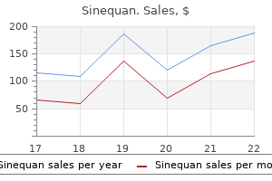
The common use of the term biliary "colic" is a misnomer because the pain is steady and does not fluctuate in intensity anxiety symptoms google buy sinequan 25 mg visa. It is usually epigastric and is often referred to the right shoulder or interscapular region. During attacks, patients are restless and may have associated diaphoresis and vomiting. There is no convincing evidence that ingesting fatty foods precipitates an attack of biliary colic. High fever, hemodynamic instability, and peritoneal signs suggest gallbladder perforation, which is a complication in 10% of patients with acute cholecystitis. Ten to 15% of patients develop jaundice, which is a symptom that may be caused by gallstone obstruction of the common bile duct or by Mirizzi syndrome, which is an obstruction of the common hepatic duct caused by edema and inflammation at the origin of the cystic duct. Chronic cholecystitis Patients with chronic cholecystitis usually have gallstones and have had repeated attacks of biliary pain or acute cholecystitis, which results in a thickened and fibrotic gallbladder. In fact, patients may have few symptoms referable to the gallbladder itself, presenting instead with complications such as cholangitis and gallstone pancreatitis. Acalculous cholecystitis is caused by ischemia and occurs in critically ill patients, often with multiorgan failure, extensive burn injuries, major surgery, and trauma. Diagnostic investigation Laboratory studies Most patients with acute cholecystitis exhibit leukocytosis. Some may have elevations in aminotransferases, alkaline phosphatase, bilirubin, or amylase caused by choledocholithiasis or cystic duct edema with resulting biliary obstruction. Patients with uncomplicated biliary colic usually have normal biochemical profiles. Structural studies Ultrasound is highly sensitive and specific for diagnosing cholelithiasis. Thickening of the gallbladder wall is a nonspecific finding commonly observed in acute and chronic cholecystitis. Pericholecystic fluid and intramural gas are specific ultrasonographic features of acute cholecystitis. Dilation of the intrahepatic or extrahepatic ducts suggests choledocholithiasis; however, ultrasound is insensitive for imaging common bile duct stones. Failure to image the gallbladder within 90 minutes suggests cystic duct obstruction. The gallbladder cannot be visualized in 85% of patients with acalculous cholecystitis. Once symptoms occur, there is a high risk of recurrent attacks of pain and complications such as cholecystitis, pancreatitis, and cholangitis. Although there are many nonsurgical alternatives, cholecystectomy is the definitive treatment for symptomatic cholelithiasis. Laparoscopic cholecystectomy is favored because there are fewer woundrelated complications, shorter hospital stays, and more rapid recoveries. Open cholecystectomy is preferred if acute cholecystitis is evident, if there is extensive scarring from prior abdominal surgery, if exploration of the common bile duct is planned, or if visualization by laparoscopy is inadequate. Given the overall benefits of surgical therapy, dissolution therapy with chenodeoxycholic acid or ursodeoxycholic acid should be reserved for patients who are at high risk of surgery. Candidate patients should demonstrate adequate gallbladder filling and emptying by oral cholecystography. Directcontact dissolution therapy with monooctanoin and methyltertbutyl ether is often successful in days to weeks but it has a high rate of complications and thus remains experimental. Extracorporeal shock wave lithotripsy is 90% successful in achieving stone fragmentation and clearance of solitary, small, radiolucent stones. As with dissolution therapy, it may take months of extracorporeal shock wave lithotripsy to clear the gallbladder of stones. About 20% of patients experience biliary pain for several weeks after fragmentation. Choledocholithiasis In the United States, most bile duct stones are cholesterol stones that have migrated from the gallbladder. The incidence of choledocholithiasis increases with age; onethird of octogenarians who undergo cholecystectomy have coexistent bile duct stones. The prevalence of choledocholithiasis and intrahepatic stones is higher in Asian societies. These populations have higher incidences of pigment stones, which usually are formed de novo in the bile ducts. Clinical presentation Unlike gallbladder stones, most patients with bile duct stones develop symptoms. Some remain asymptomatic for decades while others present suddenly with potentially lifethreatening cholangitis or pancreatitis. Patients with choledocholithiasis often present with biliary pain indistinguishable from the pain of cystic duct obstruction. The pain is steady, lasts for 30 minutes to several hours, and is located in the epigastrium and right upper quadrant. Cholangitis is the result of superimposed infection in the setting of a biliary obstruction. Ten percent of episodes are marked by a fulminant course with hemodynamic instability and encephalopathy. Reynolds pentad refers to the constellation of the Charcot triad plus hypotension and confusion. Diagnostic investigation Laboratory studies Immediately after an attack, levels of serum aminotransferases are often elevated because of hepatocellular injury. Alkaline phosphatase levels are often elevated, mildly in asymptomatic patients, and not more than five times higher than normal in symptomatic patients. Higher elevations of alkaline phosphatase or bilirubin levels suggest malignant obstruction of the biliary tree. Structural studies In contrast to gallbladder stones, bile duct stones are not readily detected by ultrasound due to gasfilled surrounding structures; the sensitivity is less than 20%. Biliary Tract Stones and Cysts 363 There is no consensus on the optimal evaluation of choledocholithiasis in patients undergoing elective cholecystectomy for gallstone disease. If open cholecystectomy is planned, intraoperative cholangiography and common bile duct palpation can be used. If stones are found, the common bile duct should be explored and stones should be extracted. Several alternative strategies are avail able to patients undergoing planned laparoscopic cholecystectomy. Those patients with documented intraductal stones undergo stone extraction laparoscopically or by open cholecystectomy. A second strategy identifies patients preoperatively at high or low risk of coexisting cho ledocholithiasis on the basis of the biochemical profile and the presence or absence of biliary tract dilation on ultrasound. When the stones are cleared from the bile duct, the patient then proceeds to laparoscopic cholecystectomy. Patients with a low risk of choledocholithiasis undergo laparoscopic cholecystectomy with intraoperative cholangiography, as previously described. Benefits and risks are associated with each strategy; the approach is largely determined by the resources available at individual institutions. Management and course Common bile duct stones, even if asymptomatic, generally require therapy because of the high complication rate. Secondary biliary cirrhosis may develop in cases of persistent biliary obstruction.
10 mg sinequan fast delivery
In the B-scan mode symptoms anxiety 4 year old purchase 75 mg sinequan amex, one can investigate the thickness of the dermis, the subcutaneous structure, and possibly musculature as a sign of deeper extension. This means that this investigation must be repeated throughout childhood to detect later formation. Second, shunts from the precapillary part drain directly to the postcapillary veins with a steal effect that reduced the microcirculation followed by a hypotrophy of the affected regions. At the same time, hyperplasia represents the process of enlargement of cells and/or collagen with normal cell numbers in normal mitosis in contrast to neoplasia, where we have a greater number of cells. A Hamburg Organ -intramuscular -subcutaneous/- mucous -parenchymatous -intracavitary -intraosseous/intraaricular -mesenterial Localization -peri-/intraorbital -peri-/intraauricular -peri-/enoral -laryngo/tracheal -remaining face -head/neck -peri-/mammary -anogenital/intraanal/intestinaI -remaining Trunk -acral/Hand/Foot -remaining Extremities -singular -singular I. The Hamburg classification incorporates the embryological origin and provides a tool for indication, in general, and kind of therapy. Therapeutic alogrithm vascular malformation Truncularangiogenetic Capillary arterial venous lymph. The more angiogenetic truncular, the more surgery; the more vasculogenetic extra-truncular, the more interventional procedures. At this time, there are no clinical symptoms, but the thermography is able to detect early hyperperfusion. After three sessions with pulsed alexandrite occlusion of these vessels, reduction of microcirculation and clinical clearance of the Parkes Weber syndrome. A biopsy with histological examination is only necessary in cases of difficult differential diagnosis to exclude malignant tumors. Capillary malformations, hyperceratotic stains, telangiectasias and miscellaneous vascular blots. Peter Berlien 90 To what extent could laser therapy and surgical excision be combined for port-wine stain management Therefore, the destruction of this pathological vasculature network can reduce this hypernutrition and subsequently prevent development of the secondary complications, including the secondary effects on the skin with formation of tuberous transformation as a secondary venous complication and also dermal hypertrophy. It is important to emphasize that due to the fact that this truncular part of this venous malformation remains as the persistent marginal vein on one hand, while hypoplasia or aplasia of the deep and/or saphenous veins coexists on the other hand, this marginal vein fulfills a very important function in venous blood return and cannot simply be removed-the conservation of this marginal vein is essential. This assessment needs to be included in the treatment planning, since venous blood flow from the affected extremity may depend on the marginal vein patency, and obliteration or exclusion of the marginal vein from the circulation carries the risk of impairment of venous return from the affected extremity. However, taking into account that this capillary malformation is a vasculogenic extra-truncular defect, it can never be healed and will recur. In other words, this procedure has to be repeated throughout life as new capillary malformation lesions form. An optimal protocol is to begin the therapy after the first year with sessions under general anesthesia in approximately 3-month intervals until a sufficient symptom resolution is achieved. There are two reasons to support this strategy: one is the reduction of pathological hyperperfusion, and another is the lightening of the "port-wine stain" before the children realize their malformation can have psychological and/or social adverse effects. We strongly advocate against using/considering the term "cosmetic," as these lesions are a part of a much more severe hemodynamic pathology. But when it happens in the vasculogenesis before differentiation to the different vessel types, extra-truncular malformations occur. These children have a disease that causes several impairments that require planning and proper treatment. Therefore, it is important to include psychosocial assessment and adequate management in the treatment algorithm for these patients. Then, if any recurrence should occur, it is recommended to start the next treatment cycle before the children reach school age to reduce the risk of having a difficult situation due to missing school. In adult patients with enlarged fistulas, angiographic embolization is the first procedure. But in contrast to the previous procedure for the other conditions, here instead, a naked bare fiber special applicator. But in contrast to the treatment of conventional varicose veins, here we have the risk of undetected communication to the deep venous system with the risk of uncontrolled washing out of the foam to the circulation. When the marginal vein is massively dilated and/or after thrombophlebitis, only surgical resection can be performed. Sonography shows the exact position of the laser fiber tip in the lumen of the vein; the colorcoded duplex mode signal detects the co2 production during the coagulation as a "color bruit signal" at the end when the vein is occluded. For example, in patients with facial port-wine stain cutis laxa, lipomatous overgrowth and bone extension remain despite laser treatment. For patients where surgical resection is indicated (as described earlier), precise treatment planning is important. Due to the vasculogenic defect in capillary malformations, the formation of pathological vascularization is an inborn error of this region, so that even after the radical resection with free flaps, pathological vessels may occur. Then, in the interval, surgical resection of the cutis laxa and other excessive tissue can be performed. In case of deformity of denture, proper surgery for mandibular bone correction can be added to reconstitute the function. If there is any sign of early recurrence after the healing, laser therapy can be started immediately under thermographic and laser Doppler surveillance. In such cases, an additional pharmacological therapy could be indicated, as described in Chapter 90. In addition to the soft tissue hypertrophy, massive tuberous transformations, not "angiomas," are developed, which have the risk of bleeding from their central artery. After several laser sessions with flashlamp-pumped pulsed-dye laser, and alexandrite for skin clearance, soft tissue hypertrophy on the lip and cheek remains. If it changes rapidly after birth, especially if it is characterized by fast growth, it is never a capillary malformation but a highly aggressive infantile hemangioma that requires immediate therapy specific for this subtype of hemangioma. If the lesion is midfacially located and pale, without changes of dermal structure, and without a thrill, there is a high level of confidence it is a nevus simplex Unna. But, if the lesion is midfacially located and has a pasty structure with bruit and/or thrill, it is suggestive of Wyburn-Mason syndrome. However, clinically it is beneficial, as this provides an opportunity for early intervention to minimize the risks for secondary complications caused by glaucoma and seizure(s). We should not withhold the diagnosis from the patients/guardians (medico-ethico-legally), and all the clinical findings need to be openly and fully discussed prior to any treatment planning. Early elimination of the pathological capillary network can significantly minimize the risk of these complications. Furthermore, the lightening of the vascular birthmark reduces psychosocial impairment. This means that an intensive treatment protocol has to be performed before the patient is 4 years old. In such a case, a thorough inspection of the entire oral cavity is mandatory, and a risk of additional involvement of pharynx and/or larynx should be assessed for the same findings. During this time, neurological and regular ophthalmic examination should be repeated.

Buy sinequan 25 mg without a prescription
Discussion and potential pitfalls Enteral feeding should be initiated as soon as severe acute pancreatitis has been diagnosed because this has demonstrated reduced infectious complications anxiety medication names order 10mg sinequan, decreased hospital days, and a trend toward improving mortality. Each endoscopic procedure has a specific set of indications and contraindications. In general, an endoscopic procedure is indicated only when the results are expected to influence the course of patient management. In some cases, however, the attendant risks of endos copy may outweigh the benefits. Before proceeding with endoscopic interven tion, a patient should give a complete history and have a complete physical examination to establish the indication for the study and exclude the presence of any contraindications. Many procedures require bowel cleansing or prolonged fasting; therefore, the clinician must be aware of comorbid conditions, such as diabetes, heart failure, or renal dysfunction, which may require adjusting the instructions for patient preparation. All patients should be counseled on the risks and benefits of endoscopy; written and verbal informed consent are mandatory. Principles of moderate sedation Most endoscopic procedures require moderate (conscious) sedation to permit a safe and complete examination. The optimal agents and dosages vary, but all carry the risk of cardiopulmonary complications. All patients should be moni tored for changes in blood pressure, heart rate, and respiratory rate throughout the course of sedation. Many centers use pulse oximetry and electrocardio graphic monitoring, but it is uncertain if routine use of these more expensive monitoring procedures improves treatment outcomes. No electronic moni toring can replace clinical judgment; therefore, if significant cardiopulmonary signs or symptoms arise, the procedure should be aborted. Slow titration of the initial dose of the sedative agent is the best way to avoid oversedation. Antibiotic prophylaxis the role of preprocedure antibiotics to prevent endocarditis or bacteremia in patients with vascular or other prostheses is undefined. Based on the docu mented risks of bacteremia with given procedures and the risks of establishing an infection in certain preexisting conditions, the American Society of Gastrointestinal Endoscopy provides guidelines for antibiotic prophylaxis before endoscopic procedures (Table 15. Antibiotics can be costly, and many have a substantial risk of allergic reactions. These issues must be considered when contemplating the use of prophylactic antibiotics. Coagulation disorders Although coagulation abnormalities are not absolute contraindications to endos copy, the use of endoscopic biopsy can be associated with an increased risk of bleeding. Before any therapeutic intervention, including percutaneous gastros tomy tube placement and electrocoagulation for polypectomy or hemostasis, attempts should be made to correct coagulation disorders. Prolongation of prothrombin time unrelated to the administration of warfarin may require parenteral vitamin K therapy. If there is no response to vitamin K or if emergency therapy is necessary, coagulation factors should be supplemented with fresh frozen plasma. Depending on the underlying medical condition, warfarin can often be withheld for five to seven days before the procedure and reinstituted one to two days after therapy. If medical conditions prohibit discontinuation, one of two potential management pathways can be used. In the first, the patient is hospitalized, warfarin is discontinued, and heparin is initiated. When the prothrombin time normalizes, the patient is prepared for the procedure, and heparin is discontin ued four hours before the intervention. Alternatively, warfarin may be stopped five days prior to the procedure and subcutaneous low molecular weight heparin. The last low molecular weight heparin dose is given the night before the procedure and then restarted the evening of the procedure and continued for five days, whereas warfarin is restarted the evening of the procedure and continued as previously taken. This second approach avoids hospitalization because the subcutaneous low molecular weight heparin is selfadministered in an outpatient setting. Therapeutic endoscopy is often indicated for control of variceal and nonvariceal bleeding, dilation of strictures, removal of some foreign bodies, palliation of advanced malignancies with stents or tumor ablation, and placement of a percuta neous gastrostomy tube. The advent of longer endoscopes has expanded the capa bility of upper gastrointestinal endoscopy in diagnosing and potentially treating diseases of the small intestine. Enteroscopy is indicated when investigating chronic bleeding presumed secondary to a source in the small intestine or if visualization or sampling the small intestine is warranted by radiological abnormalities. The major contraindications to upper gastrointestinal endoscopy include per foration, hemodynamic instability, cardiopulmonary distress, and inadequate patient cooperation. Percutaneous gastrostomy tube placement is contrain dicated if the stomach is inaccessible because of a prior gastrectomy or inter posed bowel, liver, or spleen. For complete gastric outlet obstruction, evacuation of the stomach with a nasogastric tube may be necessary. If an emergency endoscopic procedure is required for gastrointestinal bleeding, measures should be taken to avoid aspiration. Moderate (conscious) sedation is typically performed using a combination of a shortacting benzodiazepine. Throughout the procedure, a trained assistant should work together with the endoscopist to monitor the oral secretions as well as the overall clinical condition of the patient. Performance of the procedure the endoscope is introduced under direct visualization by passing the instru ment into the posterior pharynx and instructing the patient to swallow. Direct visualization is preferred because it is less traumatic and provides a view of the larynx. A pediatric colonoscope or push enteroscope can be advanced into the proximal jejunum. Enteroscopy can also be performed with overtubes with inflatable balloons on the tip of the overtube and the tip of the enteroscope to aid advancement of the endoscope into the small bowel. Endoscopic biopsy or brush cytology studies may provide a pathological diag nosis. Upper gastrointestinal endoscopy also pro vides the capability of therapeutic intervention. Dysphagia from esophageal strictures or achalasia can be relieved with endoscopic dilation using pneumatic balloon or sequential bougienage techniques. The safest means of bougienage dilation involves passage of the dilator over a guidewire placed endoscopically into the distal stomach. Although fluoroscopy reduces the complication rate of dilation, radiation exposure and resource limitations have precluded its routine use in many centers. Acute or chronic nonvariceal hemorrhage can be con trolled with electrocoagulation, heater probe application, injection therapy, or laser photocoagulation. Large or bleeding esophageal varices may be treated with injection sclerotherapy or band ligation. Mucosal polyps can be excised 176 Approach to Patients with Gastrointestinal Symptoms or Signs with electrocoagulation using hot biopsy forceps or with snare polypectomy. Large stenosing esophageal or gastric malignancies can be ablated with laser photocoagulation or electrocoagulation. Esophageal malignancies can also be palliated by deploying metallic expandable stents. Complications Diagnostic upper gastrointestinal endoscopy is usually very safe, and rates of serious complications are low. Most complications are related to oversedation, emphasizing the need for preprocedural patient assessment and vigilant patient monitoring throughout the period of sedation. The high rate of wound infec tions associated with gastrostomy tube placement can be substantially reduced by prophylactic antibiotics. The video images are transmitted by radiotelemetry to an array of aerials attached to the body via a recording belt. Relative contraindications include dementia, gastroparesis, and the presence of a pace maker because of potential interference as the capsule traverses the chest.
75mg sinequan
Group I receptors are primarily postsynaptic and their activation is both excitatory and inhibitory anxiety symptoms 50 purchase sinequan without a prescription, modulating the conductance of sodium, potassium, and voltage-gated calcium channels. Within glia, glutamine synthetase converts intracellular glutamate to glutamine, which is then released into the intracellular space. Such seizures are characterized by a "spike and wave" pattern of ictal activity, which is also seen during absence epilepsy. Abnormalities of dopaminergic neural transmission have been implicated in temporal lobe epilepsy, juvenile myoclonic epilepsy, and autosomal dominant nocturnal frontal lobe epilepsy. The nigrostriatal dopamine pathway has been implicated in modulating thalamocortical loops involved in generalized epilepsies. The mesocorticolimbic dopamine projection from the ventral tegmental area to the frontal cortex has been implicated in motor and behavioral symptoms associated with epilepsy. These pathways branch extensively to innervate the entire cerebrum, where they exert an inhibitory influence on seizure activity. Common antiepileptic drugs, including phenobarbital, carbamazepine, valproic acid, and phenytoin, produce an increase in noradrenaline in some brain regions. Similarly, increased noradrenaline has been implicated in the antiepileptic effects of vagal nerve stimulation, perhaps in combination with galanin, neuropeptide Y, and adenosine, which are coreleased with noradrenaline. Kindling results from repetitive seizure activity induced by either chemical or electrical stimulation of a discrete locus within the brain, which spreads with increasing efficiency to associated brain structures via established synaptic pathways. As the propagation of seizure activity via these networks expands, the electrographic activity and accompanying behavioral manifestations become increasingly complex. Once established, the kindled state persists for many months and pathophysiological behavior re-emerges rapidly if additional stimulation is applied to the kindled focus. The progression of seizure propagation associated with the kindling model has given rise to the assertion that seizure activity alone may be sufficient to promote epileptogenesis, that seizures beget seizures. While this is certainly true in the case of status epilepticus resulting in diffuse brain injury, it remains controversial whether brief clinical seizures analogous to those produced during the kindling process are sufficient to promote epileptogenesis. Dysregulation gives rise to several disorders, including tuberous sclerosis, focal cortical dysplasia, and hemimegelencephaly71 and an accompanying intractable epilepsy. These more severe disorders are attributable to highly penetrant single gene or copy number variant mutations. These may reflect a variety of brain pathologies, including cortical tubers, subependymal nodules, and giant cell astrocytomas. This increased excitability within the developing brain often reflects an imbalance between the maturation of excitatory and inhibitory circuits. In general, the risk for developing epilepsy increases proportional to the severity of the developmental pathology. Conversely, in some cases, the epileptic condition precipitates the developmental disorder, particularly those involving impaired cognitive development and/or cognitive decline. Both clinical data and data derived from animal models suggests that these disorders should be treated aggressively to prevent the development of more severe and long-term cognitive impairment. The incidence of epilepsy in children with mental retardation and cerebral palsy is substantially higher than for the general population and the risk for developing epilepsy in this population remains elevated for the first 20 years of life. Epilepsy in this patient population is characterized by early-onset seizures of varying expression and severity, and with a high seizure frequency in the early stages. These seizures also tend to be resistant to drug therapy and have a higher rate of recurrence when therapy is terminated. These disorders are often difficult to diagnose, but should be considered whenever developmental disabilities, cognitive impairment, and epilepsy are present in children. Inborn metabolic defects, in aggregate, amount to a considerable list of conditions promoting epileptogenicity. Small-molecule disorders involve amino, organic, and fatty acids, neurotransmitters, urea cycle constituents, vitamers, and cofactors and are represented by a variety of amino acidopathies, organic acidemias. The large-molecule disorders involve lysosomal storage, peroxisomal and glycosylation disorders, as well as the leukodystrophies. These will involve also a wide variety of metabolopathies, including glycoprotein and membrane transporter deficiencies, ganglioside degradation, and aberrations of lipid metabolism. The effects range from cellular degeneration and dysmyelination to disorders of neuronal migration. Moreover, resection of epileptogenic sites has resulted in reduction of peripheral proinflammatory mediators. Blood levels of most of these proinflammatory mediators had been found to be more detectable in epilepsy patients than in age-matched controls. An understanding of the nature of both the local and wider situations is driving current therapeutic intervention. Electrical properties of neocortical neurons in slices from children with intractable epilepsy. Equal numbers of neuronal and nonneuronal cells make the human brain an isometrically scaled-up primate brain. Selective vulnerability of hippocampal neurons in acceleration-induced experimental head injury. The temporal evolution of electrographic seizure burden in neonatal hypoxic ischemic encephalopathy. Anatomical, physiological, molecular and circuit properties of nest basket cells in the developing somatosensory cortex. Involvement of cortical fast-spiking parvalbumin-positive basket cells in epilepsy. Disynaptic inhibition between neocortical pyramidal cells mediated by Martinotti cells. Chrna2-Martinotti cells synchronize layer 5 type a pyramidal cells via rebound excitation. Impaired excitability of somatostatin- and parvalbumin-expressing cortical interneurons in a mouse model of Dravet syndrome. Epileptiform activity induced by changes in extracellular potassium in hippocampus. Osmolality-induced changes in extracellular volume alter epileptiform bursts independent of chemical synapses in the rat: importance of non-synaptic mechanisms in hippocampal epileptogenesis. Simulation of hippocampal afterdischarges synchronized by electrical interactions. Nonsynaptic receptors and transporters involved in brain functions and targets of drug treatment. Gap junctions in the adult cerebral cortex: regional differences in their distribution and cellular expression of connexins. Inhibition by anandamide of 14 gap junctions and intercellular calcium signalling in striatal astrocytes. Synchronous activity of inhibitory networks in neocortex requires electrical synapses containing connexin36. Focal synchronization of ripples (80-200 Hz) in neocortex and their neuronal correlates. Familial cosegregation of rare genetic variants with disease in complex disorders. Familial mesial temporal lobe epilepsy: a benign epilepsy syndrome showing complex inheritance. Mammalian target of rapamycin pathway mutations cause hemimegalencephaly and focal cortical dysplasia. Mutations in filamin 1 prevent migration of cerebral cortical neurons in human periventricular heterotopia. Mutations in the X-linked filamin 1 gene cause periventricular nodular heterotopia in males as well as in females. Interictal alterations of cytokines and leukocytes in patients with active epilepsy. Accumulation of passively transferred primed T cells independently of their antigen specificity following central nervous system trauma. Markers for cell-mediated immune response are elevated in cerebrospinal fluid and serum after severe traumatic brain injury in humans. Innate and adaptive immunity during epileptogenesis and spontaneous seizures: evidence from experimental models and human temporal lobe epilepsy. Alteration of hippocampal cytokines and astrocyte morphology observed in rats 24 hour after fluid percussion injury.
Discount 75 mg sinequan mastercard
Although present at birth severe anxiety symptoms 247 sinequan 10 mg discount, they are not always clinically evident until later in life and tend to grow in concert with the child and without spontaneous regression. Due to its low incidence and heterogenous presentation of clinical and imaging findings, misdiagnosis is common; therefore, correct classification and terminology are paramount for proper clinical management. To this end, B-mode ultrasonography is particularly useful in being able to discern tumors/hemangiomas from vascular malformations. It is mandatory to also study the arterial vessels, and comparison with the contralateral side should be performed to make sure the vessel is part of the normal anatomy. The B-mode is elicited to discover disorders of the main vessels like hypoplasia, aplasia, and diffuse dilation or aneurysmal dilatation. The B-mode evaluation allows visualization of the dynamic of involved muscles and the presence of major nervous trunks around the malformation. It is also possible to highlight the presence of accessory vessels, as marginal vein, persistent after birth. Thrombosed vessels should be described by the flow pattern, which may be spontaneous, present after augmentation, or absent. The presence of soft tissues calcifications called phlebolith is typical of venous malformations and is the result of multiple episodes of thrombosis. Doppler mode, conversely, is better used to discern hemodynamics, identifying high- or low-flow lesions. Spectral, color, and power Doppler examinations should confirm the flow characteristics (low flow versus high flow); assessment of flow direction under different postural and respiratory conditions should be included in the evaluation. The presence of acoustically shadowing phleboliths postapplication of compression is a strong diagnostic clue for venous lesions. Sonographers should be trained specifically in this field and should appreciate the complexity and the range of conditions they may encounter. This training allows for increased accuracy in the testing and a better-quality laboratory. The limits of the investigation are related to a limited spatial resolution and a limited investigation field. The temporal resolution is very high and allows real-time examinations and precise hemodynamic evaluation. There is some agreement for the necessity to standardize the examination and to perform it according to different depths. The second step is to characterize the malformation according to the hemodynamic parameters (slow flow/high flow) and the vessels involved. It is mandatory to define the malformation according to the embryological development into truncular or extra-truncular malformations. In truncular malformation, hemodynamic impairment is predominant; in extra-truncular malformations, organ involvement is predominant. Hemodynamic evaluation of all the vessels of the anatomical area involved by the malformation is crucial to plan a therapeutic intervention. Low flow vascular malformations of the head and neck: A study on brightness mode, color coded duplex and spectral Doppler sonography. Management of low-flow vascular malformations: clinical presentation, classification, patient selection, imaging and treatment. Venous malformation and haemangioma: Differential diagnosis, diagnosis, natural history and consequences. Venous malformations of the limbs: the Birmingham experience, comparisons and classification in children. Failure to identify the presence of arterial flow can lead to serious adverse events and a catastrophic outcome of the treatment. However, its major limitation is that it is inadequate to determine the true extent of the lesion and to precisely delineate surrounding anatomical structures. Phlebography clearly demonstrates hemodynamic characteristics of a lesion, and it has been used to distinguish low-flow from high-flow lesions with a high degree of accuracy. It is very important to mention that arterial flow characteristically contains flow voids on T1- and T2-weighted sequences. To complete the imaging acquisition, postcontrast T1-weighted spin echo images are acquired after the contrast run. Catheter-based phlebography is limited in its ability to delineate a lesion relative to its adjacent structures. This is a critical limitation, as vascular malformations can infiltrate muscle, circumscribe nerves, or infiltrate neighboring vital tissues. Since not all types of vascular malformation require catheter-based interventions for treatment, the ability to distinguish between high-flow and low-flow lesions noninvasively in order to avoid catheter-related morbidity for low-flow lesions is advantageous. Consensus on controversial issues in contemporary diagnosis and management of congenital vascular malformation-Seoul communication. Use of magnetic resonance imaging for the evaluation of vascular malformations of the lower extremity. Respective rates for specificity and negative predictive value were equal, 90% and 90%. These characteristics allow for planning a future procedure for the untreated segments. This diagnostic algorithm avoids unnecessary imaging and significantly reduces the need for catheterbased venography in most patients. The role of dynamic contrast-enhanced magnetic resonance imaging in the diagnosis and management of patients with vascular malformations. Venous malformations: classification, development, diagnosis, and interventional radiologic management. Soft-tissue venous malformations in adult patients: Imaging and therapeutic issues. The new role of magnetic resonance imaging in the contemporary diagnosis of venous malformation: Can it replace angiography Analysis of the treatment of congenital vascular malformations using a multidisciplinary approach. The technique is commonly used for the functional study of organs-the heart, liver or kidney, and others, by focusing a -camera on the examined organ. A total-body study allows for the visualization of abnormal blood pooling in vascularized areas, a sign of a vascular malformation. Threedimensional images are also possible and can improve the accuracy of the diagnosis. The study can be extended by a new scan on the following day, due to the remaining activity of the radiolabeled erythrocytes, in order to visualize lymphovenous anomalies that are not visible immediately because of the slow recirculation time and mixture among the two pathological districts (venous and lymphatic). Comparison right/left of radiotracer activity in specific areas of the lower limb. Contemporary diagnosis and management of venous and arteriovenous shunting malformation by whole body blood pool scintigraphy. Characterization of congenital vascular malformation in the extremities using whole body blood pool scintigraphy and lymphoscintigraphy. Diagnosis and whole body screening using blood pool scintigraphy for evaluating congenital vascular malformations. Kurosh Parsi and Mina Kang How much is too much for venous malformation management Shortell 149 153 157 163 165 167 32 Do all venous malformations require treatment Examples include the lateral marginal vein, duplicated femoral veins, and venous aneurysms. Typical sites of involvement are skin, subcutaneous layer, muscle, bone, and organs. This should include careful B-mode assessment of the morphology, architecture, and wall thickness. Calcified thrombi, phleboliths, would present as echogenic lesions with a strong shadow artifact. Assessment of deep and superficial vein patency, valvular incompetence and reflux, thrombosis, and/or aneurysmal vein segments.
