

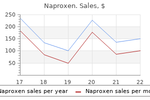
Buy naproxen online
This may be attributed to the narrower acoustic window that exists between spinous processes does arthritis pain get better trusted naproxen 250 mg. The most important thing is to visualize the anterior complex, as this means that the beam has traversed the vertebral canal and that the interlaminar space has been identified. In subjects where image quality is poor, as in the very obese, the anterior and posterior complexes are much less hyperechoic and appear as faint gray rather than bright white lines. The transverse processes and articular processes are additional helpful landmarks in difficult cases, as these lie in approximately the same transverse plane as the interlaminar space. Once an optimal view has been obtained, the depth from skin surface to the posterior complex may be measured. The intersection of these two landmarks indicates a suitable needle insertion point for a midline approach to spinal or epidural anaesthesia. The cephalad angulation required to enter the interlaminar space can also be estimated from the degree of probe tilt required to obtain an optimal transverse interlaminar view. Thoracic Spine the ultrasonographic appearance of the lower thoracic (T9-T12) vertebrae and the lumbar vertebrae are similar, except that the interlaminar spaces tend to be narrower. Imaging in the midthoracic spine, on the other hand, is much more difficult because of the extreme caudad angulation of the spinous processes and the overlapping laminae. This makes it impossible to obtain a transverse interlaminar view and thus the transverse scan provides very little information relevant to central neuraxial blockade. Limitations of Preprocedural Ultrasound Imaging of the Spine Image Quality in the Obese Patient Population In obese patients, anatomical structures are often less distinct due to beam attenuation by the intervening soft tissue. Advances in imaging technology can compensate for this deterioration in image quality, and recent studies support the feasibility of ultrasonography in the obese population. At a minimum the spinous processes (indicating the midline) and interspinous gaps can usually be identified. Successful entry into the interlaminar space is more likely if needle redirections from the initial insertion point are made in very small increments. The use of stiffer, larger-gauge needles, particularly at lengths greater than 90 mm, should be considered, as they are less likely to be deflected from their intended trajectory during insertion. Image Quality in the Elderly Patient Population Poor image quality in elderly patients, on the other hand, is usually due to narrowing of the interspinous spaces and interlaminar spaces due to ossification of the interspinous ligaments and hypertrophy of the facet joints, respectively. Prominent spinous processes in a thin patient can also hinder adequate skin-probe contact and further contribute to poor visualization. Contact may also be improved by generous application of ultrasound gel or by using a probe with a smaller footprint. Inaccuracy of Skin Marking Inaccuracy when marking the needle insertion point on the skin during the preprocedural scan can result from several factors. Many curved-array probes do not have markings that indicate precisely where the ultrasound beam emanates from. Tissue distortion is also common when performing the ultrasound scan, particularly in the elderly, who often have loose and mobile skin. Finally, skin marking does not indicate the caudadto-cephalad angle at which the needle must be advanced in a midline approach. This can only be estimated from the angulation of the probe required to produce an optimal image of the interlaminar space. These factors can, however, be compensated for by meticulous scanning technique and experience. The Learning Curve for Ultrasound Imaging of the Spine Ultrasound imaging of the spine can be a difficult skill to master because of the depth of the structures of interest, and the narrow acoustic windows into the vertebral canal. There is presently only preliminary data on the learning curve associated with ultrasound imaging of the lumbar spine. The criteria for success were also very strict, and the authors noted that most of the errors did not stem from an inability to recognize the relevant anatomy, but rather from imprecision in skin marking and depth measurement. They concluded that these errors could have been avoided by greater meticulousness on the part of the operator. These preliminary studies suggest that having acquired the basic knowledge on ultrasonography of the lumbar spine, experience of 40 or more cases may be required to attain competency in scanning. Further research is also needed to determine the learning curve associated with the actual performance of a successful ultrasound-guided neuraxial block, and the optimal educational strategies. Conclusion There is ample evidence to support the utility of ultrasound imaging of the spine in facilitating central neuraxial blockade. It is particularly useful in patients with challenging surface landmarks, and should be part of the armamentarium of any practitioner of regional anesthesia. Ultrasound imaging improves learning curves in obstetric epidural anesthesia: a preliminary study. Ultrasound imaging facilitates localization of the epidural space during combined spinal and epidural anesthesia. Paramedian access to the epidural space: the optimum window for ultrasound imaging. Use of the ultrasound to determine the level of lumbar puncture in pregnant women. Ultrasonographic control of the puncture level for lumbar neuraxial block in obstetric anaesthesia. Could ultrasonography be used by an anaesthetist to identify a specified lumbar interspace before spinal anaesthesia An evaluation of ultrasound imaging for identification of lumbar intervertebral level. Utility of prepuncture ultrasound for localization of the thoracic epidural space. Identification of cervicothoracic intervertebral spaces by surface landmarks and ultrasound. Ultrasound using the transverse approach to the lumbar spine provides reliable landmarks for labor epidurals. Ultrasound imaging of the lumbar spine in the transverse plane: the correlation between estimated and actual depth to the epidural space in obese parturients. An ultrasound-assisted approach facilitates spinal anesthesia for total joint arthroplasty. Ultrasound imaging of the thoracic spine in paramedian sagittal oblique plane: the correlation between estimated and actual depth to the epidural space. Severe and long-lasting complications of the nerve root and spinal cord after central neuraxial blockade. Ultrasound imaging facilitates spinal anesthesia in adults with difficult surface anatomic landmarks. Combined ultrasound and nerve stimulation-guided thoracic epidural catheter placement for analgesia following anterior spine fusion in scoliosis. Complications of neuraxial anesthesia in an extreme morbidly obese patient for Cesarean section. The level of sedation is titrated to comfort in responsive patients who should be able to acknowledge concordant stimulation, but with minimal procedural pain. After the patient is placed in the prone position, the lower back is prepared and sterile draped. This access needle is somewhat curved at the tip, and the superior articular process of the sacral bone is positioned as much as possible to the middle of spinal column in this oblique fluoroscopic view, negotiating passage of the needle close to the right iliac crest. Change of resistance is felt when the disc annulus is accessed, and noted resistance is then recorded as high, moderate, or low. Injection of water-soluble radiopaque contrast via a manometry measurement device to the targeted disc is initiated without the patient being told that the intradiscal pressure has just increased. During successive disc pressurizations, the volume of the injectate, resistance to injection, pain response, and radiographic appearance are recorded. The presence of control discs is necessary to achieve proper validity of the procedure. It may require an extra craniocaudal angle of the c-arm to provide a better needle access point to the disc nucleus. In addition, an extra bending of the needle tip may further facilitate direct nuclear access of L5-S1 disc. L3-L4 disc retained "cotton ball" shaped contrast spread, mainly visible in normal discs, while inside L4-L5 and L5-S1 discs, contrast spread is wide and irregular, illustrating presence of intradiscal fissures. This was considered a painful, abnormal disc, with obvious presence of posterior intradiscal fissure.
Best order naproxen
Cadaver dissections however have shown significant variability in both the number and location of the ganglia rheumatoid arthritis of spine generic naproxen 250mg mastercard. The vast majority of sympathetic efferent neurons responsible for vascular tone in the lower extremities pass through the paravertebral ganglia at L2 and L3. This separation is consistent, and it is what allows selective sympathetic blockade to the lower extremities without affecting sensorimotor function. The only connection between the sympathetic chain and the somatic nerves is via the gray and white rami communicantes. This must be kept in mind especially when performing neurolytic blocks, as the injectate may track posteriorly along these pathways (or along the path of the needle) and result in somatic nerve injury. The most common indications encountered in interventional pain medicine are sympathetically maintained pain syndromes. Blockade of the lumbar sympathetics is also indicated in conditions associated with limited blood flow within the small vessels of the lower extremities. Other less commonly encountered indications include hyperhidrosis, phlegmasia alba dolens, acrocyanosis, discogenic pain, and vasospasm. Informed consent must be obtained, following detailed discussion about the risks, benefits, and possible alternatives. Patients should be made aware of the rare potential risk of neurological complications, especially with neurolytic blocks. Coagulation parameters (bleeding time, prothrombin time, and platelet count) should be within normal limits. Intravenous fluid pretreatment is necessary to prevent hypotension during the procedure or post procedure secondary to chemical sympathectomy. The patient should remain conversant in order to report unusual pain and for the physician to perform neurological exam for the lower limbs if necessary. It should be advanced until contact is made with the lateral surface of the vertebral body. We prefer to simply use lateral fluoroscopic imaging and advance the needle until the tip is exactly at the anterior border of the vertebral body. Contrast injection with real-time fluoroscopy will ensure adequate contrast spread with the typical honeycomb appearance and allows detection of intramuscular or intravascular spread. Lateral Approach the advantage of this approach is that it allows advancement of the needle with less risk of contacting the transverse process or the exiting segmental nerve. Also, patients with advanced occlusive peripheral vascular disease would not be expected to have as significant a temperature increase compared to patients with normal vasculature. Because of the length and diameter of the needles used for the block, one should not use aspiration of blood or cerebrospinal fluid as a reliable indicator of vascular or subarachnoid needle placement. In one report, the sensitivity of the aspiration test and static radiography were 40. Segmental Nerve Injury As the needle advances laterally to the intervertebral foramen, there is a risk of injury to the exiting segmental nerve. If the patient begins to complain of paresthesia during advancement, the needle should be withdrawn and a new trajectory taken. Infection and Discitis Intervertebral disc puncture may occur with the lateral approaches. Lumbar Plexus Blockade, Psoas Necrosis, and Genitofemoral Neuralgia these complications are more significant when performing neurolytic blockade. When a neurolytic solution is injected into the body of the psoas, muscular necrosis may result. A transdiscal technique was reported in an effort to decrease the risk of genitofemoral neuralgia. A prospective evaluation of psoas muscle and intravascular injection in lumbar sympathetic ganglion block. Lumbar sympathetic block for sympathetically maintained pain: changes in cutaneous temperatures and pain perception. Thermographic correlates of chronic pain: analysis of 125 patients incorporating evaluations by a blind panel. The relative increase in skin temperature after stellate ganglion block is predictive of a complete sympathectomy of the hand. Blood flow, sympathetic activity and pain relief following lumbar sympathetic blockade or surgical sympathectomy. Neurolytic lumbar sympathetic blockade: duration of denervation and relief of rest pain. Post-sympathectomy neuralgia amelioration with diphenylhydantoin and carbamazepine. Transdiscal lumbar sympathetic block: a new technique for a chemical sympathectomy. Published studies have focused on the outcome of surgical sympathectomy (which can be performed either during open surgery or with endoscopic guidance), which provided satisfactory results. In this chapter, first, we review the indications of lumbar sympathetic chain blockade and neurolysis and describe the basic anatomical background. Indications this procedure has been proposed as an alternative to surgical sympathectomy in advanced peripheral arterial disease with excellent results. The relay ganglion located on both sides of the vertebrae in an anterolateral location (much more anterior than the thoracic sympathetic chain), approximately at the junction of the anterior third and the posterior two-thirds of the vertebral body in the angle formed with the psoas muscle. Clinically Relevant Anatomy the lumbar sympathetic chain is an extension of the thoracic sympathetic chain. It is located approximately at the junction of the anterior third and the posterior two-thirds of the vertebral body at the angle formed with the psoas muscle. The psoas muscle separates the sympathetic chain from the lumbar somatic nerves: L1 and L2 (maybe L3) provide white rami communicantes to the lumbar sympathetic chain, and all five lumbar nerves are associated with gray rami communicantes. We therefore start by describing the step-by-step needle positioning common to all three procedures. In younger patients, it is necessary to protect the gonads from radiation by using a lead apron. The targets must be selected so as to maximize the distance from the ureters (may vary from one section to another). The skin entry points (L2 and L4) and safest possible needle trajectory are determined to avoid the kidney and ureters, transverse process, vertebral body, and intervertebral disc. The patient is draped in a sterile fashion, the skin is scrubbed and sterilized, and a local anesthetic is administered at the entry points. A saline injection combined with needle tip lateral displacement and rotation (bevel-edge rotation) is an effective means to avert intervening tissues and enlarge narrow pathways. Note how the ureters gradually get closer to the spine along their route, thus increasing the risk of diffusion to the ureters from L2 to L4. A and B: Right L2 (A) and L4 (B) accurate needle tip placement in a 37-year-old patient. C and D: Left L2 (C) and L4 (D) accurate needle tip placement in a 55-year-old patient. The theoretical needle pathway is possible only on left lumbar (dotted line) target site (arrow). Indeed, at this level, the right urinary tract is too close to the right target site (arrowhead), thus, the risk of urinary tract necrosis is high. In case of significant spread posteriorly, the needle is advanced slightly anterior to avoid contact of anesthetic mixture to the lumbar nerve roots. A series of control slices is performed to verify the appropriate spread in the space located within the vertebra-psoas muscle angle. A series of control slices is performed to verify the spread of the mixture, making sure it does not spread to the ureter or backward to the spinal lumbar nerve root, in which case the needle is advanced anteriorly. A new scan is performed to monitor the diffusion once again with 1 mL of the same mixture. The hypodense alcohol mixes with the hyperdense contrast medium (this mixture should not come into contact with the ureter). If the anesthetic-contrast mixture or the alcohol diffuses to the ureter at the L4 level, the alcohol injection should be immediately stopped. The inguinalfemoral nerves originate at the L1-L2 level, the femoral nerve and obturator nerve at the L2-L4 level, and the obturator nerves and at the sciatic nerve at the L4-L5 level. Stimulation test mode is performed: the patient describes a sensation of posterior lumbar pain.
Syndromes
- Osteogenesis imperfecta
- Abdominal x-rays
- Reactions to medications
- Stomach (gastric juice)
- Corn
- Tube through the nose into the stomach to wash out the stomach (gastric lavage)
- Chest pain or pressure
Order genuine naproxen on line
Implanted systems use up to 10 electrodes arthritis in hands cheap naproxen online american express, with either shoulder or wrist motion control. The brace fixes the wrist in neutral, making it applicable primarily to C5 level tetraplegic individuals who do not have a tenodesis grasp. Functional performance was assessed in at least four tasks, which included pouring water from a can, opening a jar, opening a bottle and inserting and removing a video tape. The results demonstrated that all four of the subjects could perform at least two tasks independently using the Handmaster that they could not perform without assistance using their hand with a splint. Three of the subjects demonstrated improvement in pouring from a can and opening a bottle. A matrix of electrodes was placed over the forearm and stimulation applied to electrically excite the underlying muscles. This enabled the most superficial muscles of that are anatomically located in the forearm to be excited. The use of percutaneous electrodes for motor neuroprostheses was pioneered by Peckham and Mortimer101. The implantation is minimally invasive, requiring needle insertion only, with no surgical exposure. The system used up to 16 electrodes to provide palmar and lateral grasp for both C5 and C6 complete spinal cord injured individuals. However, for chronic clinical applications, percutaneous systems have been abandoned in favor of implanted systems due to the need for regular upkeep of the percutaneous electrodes and percutaneous sites. This system was primarily used in stroke for therapeutic applications, and is not available commercially. Percutaneous electrodes are used experimentally for many research applications to demonstrate clinical feasibility of new concepts, and are a powerful tool to investigate new neural interfaces in human volunteers. Jim Jatich who had sustained a C5 spinal cord injury, was the first pioneer to receive an implanted motor system neuroprosthesis. He used this system for 28 years before his death in 2013 of unrelated medical causes. A radio frequency inductive link provided the communication and power to the implant receiver-stimulator. The results showed that the neuroprosthesis produced increased pinch force in every patient. The direct impact of the neuroprosthesis in the performance of activities of daily living was 520 K. Each participant was tested in 6 to 15 tasks, which included eating with a fork, drinking from a glass, writing with a pen, dialing a phone, using a computer diskette, and brushing teeth. Follow-up surveys indicated that usage patterns are maintained at least four years post implant. This was developed in response to user requests to have the sensor implanted and to provide more channels of stimulation for control of the finger intrinsic muscles (first and second dorsal interossei). The sensor provided the control source of grasp-release, forearm pronation, and elbow extension to persons with cervical level spinal cord injury with wrist extension proportionally generating stimulation to provide grasp, and wrist flexion providing finger and thumb extension. All subjects demonstrated increased grasp strength and range of motion, increased ability to grasp objects, and increased independence in the performance of activities of daily living, and used the systems in daily home use. The importance of this advance was to show the advantage of an implanted control source. More recently, the second generation of the original Freehand neuroprosthesis was further developed to enable implanted myoelectric control (as an alternative to joint angle control) from two voluntarily controlled muscles and to provide stimulation of 12 paralyzed muscles. The use of myoelectric control enabled the selection of more control sites, one for proportional control and one for toggling functions on and off, and stimulation of additional muscles such as triceps for elbow extension and activation of additional muscle motor units other muscles. Epimysial and intramuscular stimulating electrodes were used, and a bipolar epimysial or intramuscular recording electrode was developed based upon the stimulating electrode design. Myoelectric control also enabled bilateral implantation such that users could control both limbs. Three of these individuals have both limbs implanted, with the first bilateral implant in 2004. Skeletal Motor Neuroprostheses 521 Their levels of spinal cord injury were high C5 to low C6. The flexible selection of muscles for use in myoelectric control and the availability of multiple channels of stimulation is an important principle that has been learned from the configuration of this systems in patients with voluntary control of considerably different muscles and muscles available for stimulation and surgical intervention. Restoring functional control in high level (C4 and above) spinal cord injury has proven to be an extremely complex undertaking. A mobile arm support was needed to support the mass of the arm during functional activities. One individual was able to perform several activities of daily living with some limitations due to spasticity, while the second individual was able to partially complete two activities of daily living. While these interventions demonstrate feasibility, they also identify the significant impediments that must be overcome. Multiple degrees of freedom (joints movements) must be controlled with a high degree of accuracy, and the muscles may be weak or denervated due to damage of the anterior horn cells from the spinal cord injury. Additionally, the control signal demands require control of a substantial number of degrees of freedom and the control sites and control information are limited with high level injuries. The neuroprosthesis increased active range of finger extension, increased lateral pinch force, increased the number of objects that the participant could grasp and place in a Grasp-Release Test, and increased scores on the Arm Motor Abilities Test. The upper limb FuglMeyer score increased from 27 at baseline to 36 by the end of the study. It was found that stimulated finger extension was reduced if the participant attempted to assist the stimulation or when stimulation followed voluntary flexion. Recent Advances Advances in function in people with spinal cord injury have been made in many areas. If an effective neuroprosthetic intervention for spinal cord injury is to be made available, it must address these needs and realize that the target regions for intervention are throughout the body (arms, trunk, chest, legs). The current design of all neuroprostheses is essentially characterized as one device for each function. For bilateral hemispheric deep brain stimulation, only two leads in the same cortical region are available from a single device. Extension of the strategy of using a single device for each function is not infinitely expandable, because of the safety and complexity of having a large number of devices in the body. Furthermore, that strategy is highly unlikely to achieve commercial viability of being cost effective. Because the nature of spinal cord injury is a multi-system injury that affects multiple organ systems, this new technology strategy must address many or all of the body organs with a single expandable device. A technology platform that would address multiple applications has been a conceived in two substantially different designs: the Bion and the Networked Neuroprosthesis. The Bion is a pill shaped device that was designed for being implanted in distinctly separated regions of the body. The fundamental concept of this Skeletal Motor Neuroprostheses 523 innovative design was a capsule with electrodes at each end of a hermetic enclosure that contains the electronics. Stimulation flowed from one end of the device and the return pathway was to the electrode at the other end. This design was an exciting contribution, but whose clinical applications have not been successful to date. It has been used in some clinical trials, where it was reported to migrate in the tissue, and even small amount of migration may significantly alter the tissue excitation. This design is a modular, scalable, fully implanted reprogrammable design that allows for both recording and stimulation, where the sites for control signal acquisition and stimulation may be remote from one another, and can be located throughout the body. The basic configuration of this device is a central unit that communicates and powers stimulating and control modules that are remotely placed at the target end position in the body. The current design of the remote modules enables two channels of bipolar recording from each sensing module and four channels of monopolar biphasic stimulation from each stimulation module. The design of the circuitry and packaging allows straightforward implementation of alternative circuit designs or electrode technology without redesign of the entire platform. This design is expandable to allow a large number of remote modules to be controlled by a single central "power module", which prioritizes traffic on the network.
Buy naproxen 250 mg low cost
This is usually done by placing a sterilized coil over the receiver in the operating room arthritis medication for horses buy naproxen with paypal. Threshold currents of each electrode should be determined by gradually increasing stimulus amplitude until a diaphragm twitch is observed. With the Atrotech and MedImplant devices, threshold and suprathreshold current should be assessed for each lead combination. If threshold values are high or the difference between the lowest and highest thresholds among leads exceeds 1 mA, the electrode leads may need to be re-positioned around the phrenic nerve. Both the Avery and Atrotech manufacturers provide onsite technical support during the implantation procedure. Pacing schedules It is important to note that phrenic nerve pacing is a life support system and must be instituted carefully to avoid complications. Consequently, a gradual switch from mechanical ventilation to phrenic nerve pacing should be performed under the supervision of experienced physicians and respiratory therapists to provide optimal results. These include stimulation thresholds (minimum stimulus amplitude which results in visible or palpable diaphragm contraction), maximal stimulus amplitudes (values just sufficient to result in 580 A. Changes in airway pressure development during airway occlusion are also a useful indicator of diaphragm force generation. With the development of airway secretions or atelectasis, inspired volume production may fall whereas airway pressure generation will be maintained. With the Atrotech and MedImplant systems, threshold and maximal amplitudes, inspired volumes and pressure development must be determined for each lead combination. Inspired volume and pressure measurements should be made in both the supine and sitting postures. Diaphragm excursions will be less in the sitting posture due to the higher lung volume and shorter diaphragm length, resulting in smaller inspired volumes. It is important to note that many tetraplegics are maintained in a hyperventilated state with relatively large tidal volumes while on mechanical ventilation. Secondary to the acidosis, patients may experience significant dyspnea suggesting insufficient inspired volume generation during pacing to maintain ventilatory support. Both tidal volume and respiratory rate on the ventilator should be adjusted to values expected during pacing to allow as smooth a transition as possible. Full-time ventilatory support cannot be achieved immediately following implantation of the pacing system since the diaphragm has undergone atrophic changes in ventilator dependent tetraplegics. The initiation of pacing is likely to be associated with the development of muscle weakness and fatigue. There are no definitive guidelines in terms of pacing schedules to achieve fulltime ventilatory support. General recommendations are to provide phrenic nerve pacing for 10-15 min each hour initially and to gradually increase this time, as tolerated. While the conditioning phase may take 8-10 weeks or longer, it is possible to bring some Neuroprosthetic Control Of Respiratory Muscles 581 patients up to full-time support within 3-4 weeks. At the beginning of each week, the tolerable stimulation time is re-determined and the patient is stimulated for this new time period every hour. After full-time pacing is achieved during waking hours, pacing is provided during sleep and gradually increased until full-time pacing is achieved. Objective evidence of fatigue including reductions in inspired volume and increased patient effort are usually present, as well. It is advisable to monitor oxygen saturation with pulse oximetry throughout the reconditioning process. In contrast, low frequency electrical stimulation causes favorable biochemical, structural and physiologic alterations. Consequently, the conditioning phase should be applied with chronic low frequency stimulation. However, as the conditioning process progresses, vibrating contractions are gradually replaced by smooth coordinated contractions due to reductions in the fusion frequency and fiber type transformation. With the application of low frequency stimulation at low respiratory rates, the total number of diaphragm contractions per respiratory cycle is less, resulting in a lower propensity for the development of fatigue. Creasey of stimulus frequency is required due to the initial application of low stimulation frequencies via sequential nerve stimulation. The sequential nerve stimulation methodology is designed to mimic the natural activation of skeletal muscle, allowing individual nerve-muscle compartments to be stimulated at low frequencies and providing time for recovery even during muscle contraction. There have been no controlled trials however to compare these different methods of nerve stimulation. Our own experience, however, suggests that fulltime pacing with the quadripolar stimulation system can be achieved in 6-8 weeks, which compares favorably with the previously reported 12-16 week conditioning phase with the unipolar stimulation systems. With the Synapse system, initial stimulus frequencies are in the range of 20 Hz and gradually reduced, as tolerated. Once full-time pacing has been achieved, current intensity, stimulus frequency and respiratory rate should always be set to the lowest level that provides adequate ventilation. This can be alleviated to a significant degree, however, by the use of a snug fitting abdominal binder, which reduces the change in abdominal girth and secondarily diaphragm length which occurs when patients assume the sitting posture. The Atrotech system has low, normal and high settings, which allow for convenient changes in stimulus parameters, which may also be necessary to accommodate for posture change. With careful patient selection, appropriate use of stimulus parameters, adequate patient monitoring, and involvement of experienced professionals, the incidence of complications should be very low. Nonetheless, complications do arise and appropriate precautions must be taken and remedial action instituted promptly, when necessary. Phrenic nerve pacing systems may fail to provide adequate ventilatory support due to a variety of factors. As with any life support system, therefore, careful monitoring of the phrenic nerve pacing system is also required. Inspired volume generation should be checked on a routine basis to ensure adequate minute Neuroprosthetic Control Of Respiratory Muscles 583 ventilation. Most patients are able to perceive small decrements in inspired volume generation and alert their caregivers to troubleshoot potential problems. Since this sensation may be somewhat less intense during sleep, some manufacturers have recommended continuous pulse oximetry as a monitoring system during the night. Since pacemaker systems do not have internal alarms to monitor inspired volume, all patients should have easy access to a patient triggered alarm system and caregiver attendance. All patients should have a readily accessible alternative method of providing ventilator support. Some systems are equipped with a low battery alarm system to alert patients and caregivers. With the radiofrequency activated systems, another common cause of mechanical failure is breakage of the antenna wires. This usually occurs at stress points, either near the connection to the transmitter or connection to the receivers. Receiver failure was a common occurrence with older systems in which body fluids could seep into the receiver capsule and result in system failure, but this is much less common with current systems due to improvement in housing materials. Iatrogenic injury to the phrenic nerve may occur at the time of electrode implantation. This complication is preventable by meticulous dissection technique, which minimizes manipulation 584 A. Malfunction can also occur at the nerve-electrode interface, due to tissue reaction around the electrode developing either early or late following implantation. The development of scar tissue at the electrode site may also be manifested by changes in stimulus threshold values. With the Synapse system having wires emanating from the skin, superficial wound infections, skin irritation and wire breakage have been noted. Discomfort during stimulation has also been observed but usually relieved by reducing stimulus amplitude. Increases in airway resistance or decrements in respiratory system compliance will also result in reductions in inspired volume generation. Due to their inability to cough, most ventilator dependent tetraplegics have tracheostomies in place and require routine evacuation of their secretions by suctioning or other means.
Generic naproxen 500 mg without prescription
One of the principal functions of somatosensation is to guide our interactions with objects does arthritis in dogs come on suddenly purchase genuine naproxen line. One can think of the tactile or exteroceptive system as providing information about the state of the external environment as it interacts with the body, and of the proprioceptive system as providing information about the orientation of the body in that environment. Anatomically, distinctions between mechanoreceptor types are based on the size and myelination state of the axons that innervate them, the location of the nerve endings in the skin. Physiologically, distinctions are made on the basis of the structure to which the mechanical stimulus needs to be applied. The correspondence between the anatomical and physiological classifications stems from the fact that function follows form: the location of the receptor ending determines what needs to be deformed to activate it, and the structure of the nerve ending determines what types of mechanical stimuli will be effective in activating it. People who classify cutaneous mechanoreceptors fall into two classes: lumpers and splitters. Splitters assign significance to small, but reliable, differences in the way subsets of the members of the sets assigned by the lumpers respond to mechanical stimuli, such as the range of frequencies effective in activating vibration sensitive receptors. A lumper would be inclined to merge, for example, the field receptor subgroups into one, and might use different terminology to identify the groups. Tonic mechanoreceptors respond to both the displacement of the innervated structure from its rest position and the rate of change of displacement (velocity), and are 136 S. Horch better activated by movements away from the rest position than by movements back toward the rest position. Phasic mechanoreceptors are not always directionally sensitive: many respond nearly equally well to movements toward the rest position as to movements away from the rest position. The T1 and T2 receptors are tonic, continuing to respond to maintained skin indentation or stretch, respectively. Most cutaneous receptors have a well developed phasic component with only a few having a pronounced tonic component to their response. In general, phasic receptors respond better to high frequency vibration than do more tonic receptors. The response properties of cutaneous mechanoreceptors are sufficiently rich and complex to provide for the variety of sensations elicited by cutaneous stimulation, which range from faint, evanescent tickles to strong and persistent feelings of skin indentation and pressure. In hairy skin the picture would be Somatic Sensation 137 similar, except that Pacinian corpuscles would be absent, replaced by richly innervated hair follicles. Major cutaneous mechanoreceptor types in human glabrous skin and their properties. Shown are typical receptive field sizes, receptor densities, and locations in the skin. Somatic Sensation 139 Cutaneous temperature sensations arise from two classes of thermoreceptors in the skin: cold receptors and warm receptors. These are innervated by small myelinated or unmyelinated nerve fibers, show a mixed phasic and tonic response profile to changes in skin temperature, and acclimate over time to chronically maintained skin temperatures within the normal physiological range. This latter phenomenon shifts the sensitivity of the receptors to make them more responsive to changes in skin temperature than to absolute temperature. Interestingly, hot is a synthetic sensation caused by the simultaneous activation of warm and cold receptors, a result of the so-called paradoxical response of cold receptors to temperatures above about 45 C. Like the thermoreceptors, these are innervated by small myelinated or unmyelinated nerve fibers. The former give rise to fast, sharp pain that typically has a distinct focus and tends to go away once the painful stimulus is removed. Activation of nociceptors innervated by unmyelinated fibers tends to produce slow, burning pain of ill defined focus that may persist even after the stimulus is removed. That is, the same neural signals can be decoded in different ways to obtain different types of information. As mentioned above, all afferents respond better to dynamic stimuli than to static ones, so a tactile stimulus tends to feel more intense to the extent that it is dynamic (that is, when the degree to which it indents the skin changes over time). Of course, different fiber types are differentially sensitive to mechanical stimulation of the 140 S. Importantly, under normal circumstances, even a basic sensory dimension such as intensity involves the integration of signals from a large number of afferents spanning all classes. Shape: When we interact with an object, different parts of our hand touch different parts of the object. At each contact point, the pattern of skin deformation reflects the contours of that object at that location. The spatial pattern of activation of afferents under the contact area also reflects the spatial configuration of the stimulus over that area. As a result, certain stimulus features are enhanced (edges, corners) and others obscured (small internal stimulus features). In downstream structures, the extraction of information about object shape is remarkably analogous to its visual counterpart. Texture: When we run our fingers across a surface, we acquire information about its surface microgeometry and material properties. Again, a variety of receptors contribute to our remarkable sensitivity to texture. Given the density of innervation of the skin, this spatial mechanism cannot account for our ability to discern fine textural features, which are orders of magnitude more tightly packed than are receptors. Proprioceptive Receptors Like cutaneous mechanoreceptors, proprioceptive mechanoreceptors provide position or displacement signals and movement or transient signals. The former relay information about joint angle or muscle length continuously during a maintained position of a limb or digit, while the latter are available only during movement of a limb or digit. Movement-related signals can include information about the rate (velocity) at which a limb or digit changes its position and about acceleration or higher order derivatives. According to this scheme, static position signals (meaning signals continuously available while a limb or digit is stationary) can come only from slowly adapting receptors and are thought to give rise to static position sense. Dynamic position sense (awareness of limb or joint position during movement) is more perceptually salient than static position sense. Similar to their cutaneous counterparts, kinesthetic receptors probably use both dedicated channels and pattern codes to provide information about the positions and movements of the limbs and digits. The current view is that proprioceptive information can be derived from several sources: neural signals from the skin, joints, and muscle are all potential sources of information about limb posture and movements. Humans can detect the occurrence of a movement without necessarily sensing its direction or speed, and they can independently judge the direction and the speed of a movement and the position of a limb. It seems reasonable, therefore, to expect that these features are either encoded 142 S. This suggests that the role of cutaneous mechanoreceptors in proprioception is supportive but not informative. Put simply, cutaneous input may be needed to support a body image of having, for example, a hand, but is not sufficient to provide reliable information about the configuration of the hand in space. In the ligaments, slowly adapting responses arise mainly from Golgi type endings formed by a profuse branching of the nerve terminals. Thus, ligament receptors seem incapable of encoding position or movement of passively rotated or positioned joints, except perhaps to signal the extremes of flexion and extension. Ruffini endings appear very similar to the Golgi receptors, though a bit smaller, and they respond to stretch of the capsule. Rather, joint receptors likely play a protective role, Somatic Sensation 143 such as providing a fast mechanism to prevent hyperextension of the joint during a vigorous movement. Muscles contain two types of slowly adapting mechanoreceptors: the Golgi tendon organs and the muscle spindles. Golgi tendon organs lie in series with the main muscle fibers, well situated to measure tension in the muscle. Muscle spindles lie in parallel with the main muscle fibers, well situated to measure muscle length and rate of change of length. The Golgi tendon organ consists of a thinly encapsulated bundle of small tendon fascicles with a fusiform (spindle-like) shape, innervated by a single largediameter group Ib nerve fiber. This receptor ending responds to stretch of the tendon fascicles, but due to the very low compliance of tendon, tension seems the more appropriate variable. In addition, the spindle receives 6 to 12 small diameter motor fibers in the gamma range, the gamma efferents. Activity in the gamma efferent fibers can substantially alter the activity in the sensory fibers. There are two main types of muscle spindle sensory endings, the primary found at the middle of the intrafusal fibers and the secondary found just offset from the middle.
Buy naproxen in united states online
The injected current is measured and equals the amount of current passing through the membrane at the command potential arthritis knots best naproxen 500mg. They thus allow users to alter neuron transmembrane potentials and describe how transmembrane currents change in response. In this technique the only limitations are the amount of current the current-passing electrode can pass, and how large the gain of the feedback circuit, which determines how closely the neuron transmembrane potential tracks the user-set potential (Halliwell and Whitaker 1987), can be made without the electronic circuit oscillating. To achieve maximum possible gains, modern equipment has circuits that can be set to compensate for electrode resistance and capacitance. Again, high switching frequencies are important to optimize the clamp, but it is essential to ensure that no current passes through the electrode during transmembrane potential recording. In this case, given the smaller resistances of patch pipettes, 16 Neurobiology of Motor Control: Fundamental Concepts and New Directions the difficulties of achieving adequate clamp are much reduced. Patch Clamp In patch clamp recording the cell membrane under the pipette is kept intact (it is the situation in the Whole Cell Patch Clamp Recordings part of Section 2. As such, absent information from intracellular electrodes, the experimenter has no direct knowledge of cell transmembrane potential. It is thus impossible to clamp cell transmembrane potential to a user-set potential. In the absence of the patch-clamp apparatus, currents flowing through the membrane under the pipette would change the potential of the interior of the pipette. To maintain the potential of the pipette interior at the user-set value, the voltage clamp injects a current equal and opposite to the transmembrane currents. An important technical issue is that in patch clamp how one changes transmembrane potential is reversed. Consider, for example, the case in which one desires to bring the transmembrane potential of the membrane under the pipette to 0 mV. In patch clamp, alternatively, one controls only of the potential of the pipette interior. In the literature this "reversal" is typically taken into account, and the above procedure would be referred to as a transmembrane (or membrane) depolarization, not a pipette hyperpolarization. The fact that in patch clamp the pipette does not have access to the cell interior leads to a limitation of the technique. As such, data from patch clamp experiments taken from intact cells (cell-attached patches) attempting, for instance, to describe the voltage-dependence of channel opening, suffer from one having to calculate the transmembrane potential using an assumed value of cell resting potential. A technique to overcome this difficulty is to perform experiments in which the membrane under the pipette is removed from the cell, and thus the potential on the external side of the membrane is the potential of the external solution, typically zero. Since the potential in the pipette is being clamped, in this case the transmembrane potential of Electrophysiological Recording Techniques 17 the patch is unambiguously known. The portion of membrane under the patch remains attached to the pipette, resulting in an "inside-out" patch, since the side of the membrane that was originally on the interior of the cell now faces the external solution. In the second way the membrane under the pipette is first ruptured and the pipette is then pulled away. The cell membrane that pulls away with the pipette then comes together and forms a new, continuous membrane across the pipette lumen, but in this case the membrane that faced the external solution when in the cell again faces the external solution in the detached patch (hence called an "outside-out" patch). If one instead switches to the patch clamp circuit explained above, the voltage clamp attempts to clamp the interior of the pipette to the user-set command potential. It therefore injects whatever currents are required to charge the electrode capacitance, establish the appropriate voltage across the electrode resistance, charge the cell membrane capacitance, and establish the appropriate voltage across the cell membrane resistance, including the changes in cell membrane resistance that occur as a result of changes in cell transmembrane potential, necessary to keep the pipette interior at the command potential. It should be clear from the discussions above about electrode resistance and capacitance that simultaneously fulfilling all these tasks is difficult (Ogden and Stanfield, 1994). As current flows through the resistance of the extracellular space, a potential difference is produced that can be measured by extracellular electrodes (see Section 2. Depending on placement, 18 Neurobiology of Motor Control: Fundamental Concepts and New Directions the electrodes can record the sum of the action and synaptic potentials of only a few neurons or even one, or of large ensembles of neurons (field potentials). Extracellular electrodes are often made of metal, typically steel or tungsten, insulated but for the tip. Blunt glass pipettes filled with saline as an electrolyte can also be used, either by simple placement near or in the tissue of interest or, often with an attached plastic tube heat-pulled to the appropriate diameter, as suction electrodes. A reference electrode is always placed either close to the first electrode in contact with the same tissue, or somewhere in the body cavity or saline. Extracellular electrodes pick up action potentials from neurons in their vicinity. In early work considerable effort was devoted to understanding the basis of extracellularly recorded action potential shape, and whether these differences could be used to gain insight into neuron and axon properties (Section 2. With special techniques, it is also possible to use extracellular electrodes to measure absolute transmembrane potential (Section 2. However, all extracellular recording techniques are highly susceptible to small changes in recording technique and extracellular amplifier filter settings. With the invention of intracellular recording, in modern work extracellular recording is therefore primarily used to detect action potential discharge patterns and measuring action potential frequencies. One important advantage of extracellular recording is that it is often possible to record the activity of multiple neurons simultaneously from single electrodes. Signal amplitude depends on the distance between the electrode and the active axon and how large the neuron or the axon diameter is. Large neurons or large diameter axons produce large electric fields, and hence large extracellular action potentials, because more current flows along and out of the axon. The use of multiple electrodes, and more advanced spike sorting techniques to identify the activity of individual neurons in more complex situations, are covered in Chapter 3. Extracellular electrodes are also used to stimulate single neurons or neuron ensembles, i. The amplitude of the peaks depends on both the number of axons in each class, and the amplitude of the individual action potentials (with larger diameter axons, as noted above, having larger amplitudes). Neuron Recordings In some preparations the activity of single or a few neurons can be recorded with suction electrodes. These electrodes are saline-filled blunt micropipettes attached to a device that allows applying negative pressure (in which sense they are similar to patch-clamp electrodes). Suction electrodes are used to record activity from tracts in slice preparations or superficial neurons in invertebrate ganglia. Suction electrodes have also been used to record action potentials from single identified leech neurons by placing the electrode on the cell body (Cymbalyuk et al. Hook electrodes are commonly used to record en passant from invertebrate nerves or connectives containing axons of interest. Such recordings can be made from nerves in intact, semi-intact, or isolated in vitro preparations. Often, the nerve is positioned on the hook-like ending of a single electrode while the reference electrode is positioned close by in the body cavity. It is critically important to isolate the electrode and recording site from the surrounding saline or body fluid to prevent a short circuit between the recording and reference electrodes. This is often done by lifting the hook and its overlain nerve into a layer of petroleum jelly (Vaseline) or viscous silicon oil. To achieve better isolation, particularly of fine nerves that are easily damaged, a device in which a plastic tube is situated above the hook and nerve, and from which highly viscous grease is extruded that forces the nerve firmly against the hook, and saline away from nerve and hook, can be used (Schmitz et al. Generally, when stimulating a nerve extracellularly large diameter fibers are activated more readily than small diameter fibers. Large diameter fibers have a lower excitation threshold mainly because of their low longitudinal resistance. In vertebrates, selective activation of small axons can be achieved using a multi-contact electrode array placed along the nerve (Lertmanorat et al. Nerve activity can be recorded en passant or from a stump sucked into the electrode. Alternatively, Vaseline is used to form a well separating a saline pool from the surrounding saline. The activity in a nerve running through this pool can be recorded by pushing a pin into the pool (the pin need not touch the nerve). In particular, we do not cover the specialized and extensive literature on extracellular recording of heart activity (electrocardiography).
Yucca aloifolia (Yucca). Naproxen.
- Arthritis, migraines, digestive disorders, diabetes, high blood pressure, high cholesterol, high triglycerides, poor blood circulation, skin problems, and other conditions.
- What is Yucca?
- How does Yucca work?
- Are there safety concerns?
- Dosing considerations for Yucca.
Source: http://www.rxlist.com/script/main/art.asp?articlekey=96718
Buy discount naproxen on-line
Other neurons rheumatoid arthritis diet reviews discount naproxen on line, known as motor neurons and sensory neurons, with cell bodies located in the brain and in or near the spinal cord (within the spinal column), extend their axons outside the central nervous system and form the efferent and afferent component of peripheral nerves, respectively. The ensuing sections will introduce different kinds of neuronal cells located throughout the central nervous system. Among their tasks is the provision of insulation or myelin for some axons which acts to increase the conduction velocity of electrical impulses, and the recycling of excess neurotransmitters released between cells. The sagittal plane passes lengthwise through the body and divides it into left and right portions. The term lateral refers to locations away from the midline in the sagittal plane while medial refers to locations close to the midline. The term ipsilateral means the same side in the sagittal plane while contralateral means the opposite side. The frontal or coronal plane passes lengthwise through the body and divides it into front and back portions. The terms dorsal and posterior refer to locations in the back part of the body in the frontal plane while ventral and anterior refer to locations in the front part. The horizontal, transverse or cross-sectional plane divides the body into upper (rostral or superior) and lower (caudal or inferior) portions. The roof (tectum) of the midbrain is the site of the superior and inferior colliculi, two important nuclei for vision and audition (see Chapters 1. As one travels "up" the system, progressing from the mesencephalon to the diencephalon (from which various forebrain structures extend), this simple linear progression breaks down, and various lateral branches, such as the cerebellum, make the anatomical system much more interesting and complicated. The hypothalamus plays a critical role in the regulation of many autonomic nervous system functions (see Chap. The basal ganglia or cerebral nuclei consist of a collection of structures, some of which are related to the limbic system, but the majority of which are major players in the control of muscle tone and voluntary movement. The limbic system is a collection of cortical and subcortical structures responsible for functions such as learning and memory, emotional states, and behavior associated with drives. Located most rostrally in the central nervous system is the cerebral cortex which is comprised of four lobes in each hemisphere, frontal, parietal, temporal and occipital. The wrinkled outer surface (composed of fissures or sulci and folds or gyri) that covers the majority of the brain is the cerebral cortex. In panel (A) labels illustrate some of the areas of localized function in the cerebral cortex including primary motor cortex on the precentral gyrus (M1) and primary somatosensory cortex on the postcentral gyrus (S1). The cerebrum is divided in two hemispheres which are connected through the corpus callosum and anterior commissure. Though the chosen structures are segregated into individual sections, the reader is reminded that these structures are extensively interconnected. The reader is encouraged to examine the more specific descriptions on sensory, motor, and other systems that are covered in detail in subsequent chapters. We start with an examination of anatomy from a top-down perspective (cerebral cortex downward), with a focus on different functions that these structures play a role in mediating. The majority of these discussions will describe neural cells and how to interface with those cells, but the reader is reminded that the non-neural cells, glia or neuroglia, are a large majority of the cells in the brain. Regarding specific non-neuronal cells, microglia are the resident "firstresponders" of the brain that respond to inflammation or other "invaders". Astrocytes are key in maintaining the immune privilege of the brain, working with pericytes and epithelial cells to govern the blood brain barrier. In other words, a cell from layer 1 has to progress through layers 6-2 to get to its final location. Another cell type is the oligodendrocyte that provides myelination of axons, much like Schwann cells in the peripheral nervous system. One notable difference between Schwann cells and oligodendrocytes is that Schwann cells interact with only one axon or cell where oligodendrocytes can interact with several axons or cells. The constant generation and maintenance of this fluid helps maintain a stable environment for the brain. It also provides mechanical benefits Central Nervous System 45 by suspending the brain in the skull to minimize mechanical loading while at rest or during movement. While recent research has shown the interplay of neuronal and non-neuronal cells are vital in key brain functions and function of neurons themselves, further research is needed to tease apart the importance of all the relationships. Forebrain anatomy - overview the forebrain, or prosencephalon, contains some of the most evolutionary advanced structures in neuroanatomy. Most notably, the cerebral cortex, often referred to as the cerebrum, neocortex, or just the cortex, is often associated with many of our higher reasoning capabilities. The forebrain can be split into two different groups of structures: the telencephalon that contains the cerebral cortex, hippocampus, basal ganglia, among others and the diencephalon that contains the thalamus (as well as hypothalamus, subthalamus, epithalamus). It is thought to be the foundation of consciousness, central processing of sensory information, integration of that information, memory, decision making, and generation of motor output commands, among other tasks. The undulating surface of the brain is composed of gyri (outfolds) and sulci (infolds). The folding increases the surface area of the cortex while not increasing skull volume, resulting in additional cortical areas not present in smaller brains. This difference may impact which animal model is most relevant for preclinical work. Another difference to bear in mind is the ratio of white matter to gray matter volume. Packed into this large volume are 6 layers of neural cells that predominantly have different functions. Motor areas are located in the frontal lobe, in front of the central sulcus, while the somatosensory cortex lies in the parietal cortex immediately behind the central sulcus. Each of these lobes can be further subdivided into areas that are specific to certain functions. Several different maps of the cortex and its lobes are available, some which will be covered here. In humans, he specified 43 areas, labeled 1-52 with areas 12-16 and 48-51 not present in his original human maps, but present in his maps of Old World monkeys. The work by these researchers and others suggested that different areas of the cerebral cortex were specialized to perform discrete functions. Other techniques can build upon these atlases and segmentation to map out taskspecific activation of brain areas and how different areas of the brain are connected to each other, also known as the connectome. In addition to these anatomical and functional driven maps, there are many other maps associated with the brain. One such map, examines the blood supply (and outflow) to the cortex and the brain. Bottom row, inflated surface projections of those viewed in top row, with shading and text of each region. While the brain can be split up in many ways, distinct regions of the brain have a mapping on their own within their subsection. While cortical maps are the focus of this section, it should be noted that different maps of the body exist in other neural structures outside of the cortex. These include the motor and sensory maps in the cerebellum and thalamus, among others. For vision, there are many translations from the visual field to the visual cortex, but one of the main features is the enlarged projection of images to cortex Central Nervous System 49 for items in the center of the field of view (near the fovea). Process to map brain networks relies on segmentation of the brain into regions or nodes, first. Then, in parallel, structural and functional networks are studied, resulting in matrices mapping connections. The final result is the merging of both networks to develop a structure showing regions of connectivity during certain tasks. Regardless of the map or location, the reader should be warned that while the mapping and assignment of these areas tend to depict clear boundaries, these boundaries are never as sharp as drawn. While certain functions are fairly reliable to delineate (motor and sensory functions), some higher-level functions (like 50 D. Some of the imprecision may be due to lack of true understanding of the function of these higher-level functions. The somatosensory area is primarily input whereas the motor area that is primarily output. In the somatosensory area, there are more layer 4 cells that receive primary input from the thalamus.

Purchase naproxen without a prescription
Neurons and networks are tuned on an individual animal basis and are extremely plastic getting rid of arthritis in fingers purchase generic naproxen pills. This expectation was wrong: virtually identical motor outputs can be generated in multiple ways (Prinz et al. This is an important lesson that needs to reach investigators studying mammalian neural networks. All work to date indicates that mammalian motor networks are every bit as complex as invertebrate ones. As such, it is almost certain that the neuron conductances of mammalian neural networks in general are individually and homeostatically regulated, and that the networks rely on multiple rhythm generating mechanisms, contain large neural ensembles that are sparsely connected and dynamically regulated, and interact not only with a staggering number of neuromodulators but also with glial cells. A connectome is Neural Networks for the Generation of Rhythmic Motor Behaviors 253 only predictive when its multiple membrane and ionotropic and metabotropic synaptic conductances are added, when the strengths of these conductances are homeostatically fine-tuned, and when it is embedded in its network of neuromodulators. The ball and stick diagrams used to explain interactions between identified invertebrate neurons are inadequate to describe these highly complex networks, and new computational approaches will be required to describe the dynamic network interactions that underlie their rhythm generation. Many of these processes have been described in smaller invertebrate networks, and we have numerous examples that show that lessons learned in smaller networks can be applied to more complex ones. Bouhadfane M, Tazerart S, Moqrich A, Vinay L, Brocard F (2013) Sodium-mediated plateau potentials in lumbar motorneurons of neonatal rats. Brocard F, Tazerart S, Vinay L (2010) Do pacemakers drive the central pattern generator for locomotion in mammals Duysens J (2006) How deletions in a model could help explain deletions in the laboratory. Fenelon V, Le Feuvre Y, Bem T, Meyrand P (2003) Maturation of rhythmic neural network: role of central modulatory inputs. Gola M, Selverston A (1981) Ionic requirements for bursting activity in lobster stomatogastric neurons. Gosgnach S (2011) the role of genetically-defined interneurons in generating the mammalian locomotor rhythm. Hutcheon B, Yarom Y (2000) Resonance, oscillation and the intrinsic frequency preferences of neurons. Kjaerulff O, Kiehn O (1996) Distribution of networks generating and coordinating locomotor activity in the neonatal rat spinal cord in vitro: A lesion study. Llinas R, Sugimori M (1980) Electrophysiological properties of in vitro Purkinje cell somata in mammalian cerebellar slices. Neural Networks for the Generation of Rhythmic Motor Behaviors 259 Mulloney B, Smarandache-Wellmann C (2012) Neurobiology of the crustacean swimmeret system. Pearson K, Duysens J (1976) Function of segmental reflexes in the control of stepping in cockroaches and cats. Schwindt P, Crill W (1999) Mechanisms underlying burst and regular spiking evoked by dendritic depolarization in layer 5 cortical pyramidal neurons. Thoby-Brisson M, Ramirez J-M (2001) Identification of two types of inspiratory pacemaker neurons in the isolated respiratory neural network of mice. Turrigiano G (2012) Homeostatic synaptic plasticity: local and global mechanisms for stabilizing neural function. Zhang Y, Khorkova O, Rodriguez R, Golowasch J (2009) Activity and neuromodulatory input contribute to the recovery of rhythmic output after decentralization in a central pattern generator. Living on land adds the additional challenges of gravity and functioning at the interface between a low-drag atmosphere and a textured substrate. Friction must be minimized to move quickly over this substrate, which can be accomplished by having a polarized, axial body structure raised on segmentally specialized, jointed legs. This set of adaptations was achieved by both vertebrates and invertebrate arthropods, including insects, crustaceans, and arachnids. Legged animals face the additional challenges of maintaining body posture against gravity and external perturbations both when stationary and when moving. The common body plan presents common control problems to both sets of animals: how to produce stable postures, how to produce coordinated and stable locomotor gaits, and how to shift efficiently between these states in a noisy, changing environment. In both sets of animals descending motor commands excite similar motor circuits to produce similar leg and body movements. Feedback from similar sensors then provides information on the immediate consequences of the motor commands, which is particularly important in shaping their postural and locomotor behaviors. These common solutions have emerged despite large differences in the sizes, skeletal structures, and habitats of vertebrates and arthropods. In the last 50 years, human engineers have discovered the same strategies in their efforts to build devices that respond adaptively to the environment. Chapter Plan We begin with a brief history of feedback control and a review of classical control theory. We then show how that theory informs our general understanding of the control of limb movement and posture. We then describe feedback control of posture and locomotion in arthropods and vertebrates, and interpret that in light of control theory. As engine speed increased, the plane of the fly-ball rotation rose and reduced the flow of the steam that drove the engine. The valve openings and sensitivity of the system could be adjusted to enable it to rotate at a near constant speed independent of steam pressure. In the mid-19th century, Claude Bernard identified negative feedback as being responsible for maintaining the constancy of the internal body environment despite changes in outside conditions (Gross 1998). Early in the 20th century, Charles Sherrington showed how spinal reflexes used negative feedback to resist perturbations to maintain limb positions and body postures (Sherrington 1910). He showed that the reflex response consisted of several parallel pathways that excited ipsilateral muscles to resist the perturbation and inhibited their antagonists, while also exciting and inhibiting appropriate contralateral muscles to maintain body posture. Sherrington also showed how these reflexes could be chained in sequences to generate rhythmic locomotor movements (Sherrington 1913). An initial leg movement would trigger a reflex response in the opposing direction, and the resulting movement would trigger another reflex response in the original direction. Recent work in many animals has established the importance of feedback-evoked reflex responses in helping centrally generated rhythmic motor patterns adjust to the physical demands of locomotion (Duysens et al. Work on this problem led to the development of what is now called "classical" control theory, which showed how desired patterns of output could be evoked from any system in a noisy environment despite large differences between the power of the control signal and that of the output. In his book "Cybernetics: Command and Control in the Animal and the Machine", Wiener (1961) showed that the same control problems are faced by animals. The power required to change the angle of the ship is much greater than the power needed to turn the steering wheel, and thus the steering wheel signal, I, must be amplified greatly. Arrow leaving a rectangle is its output, the product of the rectangle and its input. Output of a summing node (a circle with a ) is the sum of its inputs, according to the sign adjacent to the input arrow. This means that despite perturbations produced by noise, system output will equal system input. When this is not true, interesting (and occasionally catastrophic) things can happen. Assume, for instance, that it takes some time, for the input to activate the plant and generate an output, but that the feedback from that output is immediately subtracted from the input. If the input varies with a period twice the time needed to produce the output, then an output peak will produce a peak in feedback that is subtracted from the input at a time when the input is in a trough. As a result, the delay has transformed the negative feedback into positive feedback, where the error signal grows with each cycle of the feedback loop. However, sometimes there is a limit to how fast the output can be produced in response to an input. In cats the shortest delay of the ankle muscle monosynaptic stretch reflex is about 12 ms, in humans about 30 ms. Unaided, this system will blow up if the frequencies of the input to the motorneurons are near f, 42 Hz for cats and 17 Hz for humans. However, there is also an instantaneous negative feedback to postural perturbations caused by intrinsic muscle properties-so-called short-range muscle stiffness-in which muscle stretch increases muscle force. This instantaneous negative feedback can be made to work for both flexing and extending perturbations by co-contracting opposing muscles. It may also leave a residual difference between input and output after steady-state is achieved. The residual error may be both large enough to impair system effectiveness and too small for the feedback to reduce further. These oscillations are greater than would be observed in real limbs because the muscle models each have the same single time-constant and are each excited by single motorneurons. Despite these oscillations, the motorneuron activity produced a steady 30 flexion of the limb.
Buy discount naproxen online
What seems to be crucial is for the regenerated axons that grow into the microchannel device to be able to establish contact with appropriate target end organs arthritis back pain remedies purchase naproxen overnight delivery, which must be provided or somehow mimicked. Since the trunk nerves are generally still viable in the above elbow amputee, for example, researchers have sought to overcome this problem by employing direct nerve based interfaces such as sieve or intrafasicular electrodes. It is possible, however, to surgically connect a motor nerve or a mixed trunk nerve (such as the median and ulnar nerve) from the residual limb to a nearby muscle and allow the efferent nerve fascicles to establish contact with that muscle. It should be noted that the muscle segments involved are only being used as a form of "biological signal amplifier", and not mechanical actuators intended to produce joint motions. Special care was taken to clear away as much Peripheral Nerve Recording Electrodes and Techniques 417 subcutaneous fat as possible so that the nerves were as close as possible to the surface. After the surgical procedure the patient was trained using a prosthesis custom built for this purpose. This improvement has been credited, by the patient, to the more natural and intuitive operation of the device as opposed to the standard prostheses with touch pad control that he had previously used. The successful demonstrations of simultaneous control of multiple joints in advanced prosthetic arms depends heavily on being able to derive an independent control signal for each respective joint axis and direction by accessing (interfacing to) the motor nerve which would normally control the muscle that moved that joint in the respective direction. Theoretically, for an upper extremity amputees to control an artificial arm that contained all of the degrees of freedom and matched the dexterity of a human arm and hand, a neural interface would have to be capable of sampling the activity of the motor nerves to every arm and hand muscle independently. To this end, the development of techniques to achieve higher selectivity from neural interface devices is a prominent goal among researchers. In this case designed to record from a single muscle sliver that would be innervated by a motor nerve that subserved a single joint movement. A separate compartment is also illustrated that could be filled with de-epithelized cutaneous tissue to provide a target for any cutaneous fibers that might be present with the motor nerve fibers, preventing them from invading the muscle compartment and "stealing" muscle target sites away from the regenerating motor nerve fibers. Bonding sites visible at the top left and right of the polyimide insert are used to connect fine wire cables leading to a percutaneous connector for chronic in-vivo studies in research animals. For example, a drawback with transferring an amputated nerve stump to a normal muscle is that it sacrifices the original function of the host muscle to create the interface. In addition, depending on the composition of the transferred nerve, the host muscle could become innervated by a mix of nerve fascicles originally targeted at multiple other muscles which may have subserved several different limb or joint motions. The implanted device was formed by gluing two short lengths of silicon tubing side by side and placing wire electrodes within each in a tripolar recording arrangement. The tibial and peroneal components of the sciatic nerve were then separated and each was introduced into the compartment that contained a slice of muscle which they formerly innervated. While the data in the figure show several instances where there was coactivation of the two nerve-muscle systems, there are clear epochs that show independent activity as well. The rational for this is to provide a suitable skin target for regenerating sensory cutaneous afferents that are present in many mixed peripheral nerves. In the absence of finding suitable cutaneous end organs, transected cutaneous nerve fibers would compete with the regenerating motor nerve fibers in an attempt to connect to available muscle tissue. An additional motivation Peripheral Nerve Recording Electrodes and Techniques 421 for the inclusion of a compartment that contains cutaneous tissue, is the possibility to provide cognitive tactile feedback via electrical stimulation of the cutaneous afferents that innervate that compartment. The goal of harnessing the electrical activity that an amputee can generate in the muscles and nerves of the residual limb in order to control advanced prosthetic limbs is being pursued using at least two additional techniques. Instead a whole muscle (extensor digitorum longus) is excised and freely transplanted to the thigh region. The nerve-muscle-electrode system is then secured in place by first wrapping it with decelluarized gut tissue and then suturing that package to the fascia of the surrounding musculature. Deficiencies that were noted included mainly mechanical failures that developed over time such as the epimysial electrodes pulling away from the muscle surface and lead breakage on route to the head mounted connector. The electrode array with its imprisoned cells is then implanted into the neural tissue where the neural communication is ultimately desired. The strategy is to select the appropriate neurons for incorporation into the electrode array and then expect them to grow axon collaterals into the host tissue using their natural mechanisms of axonal guidance to achieve this. Instead, bioactive substances are employed that are known to promote neuronal attachment and axon outgrowth, or to inhibit neuronal attachment. Regions can then be patterned on a pre-fabricated substrate to control where seeded neurons will adhere, and to restrict the directions and paths along which growing neurites will travel. A glass cover slip with a layer of glia cells grown on it is placed over the Peripheral Nerve Recording Electrodes and Techniques 423 neuron probe structure with the glia facing the neurons to provide nutrients and sustain the neurons. The networks of cultured neurons are kept separate from each other by adhesive treatment of the substrate. After implantation, the host tissue is expected to form functional connections with the neuronal colonies. Design Considerations There are many factors which must be taken into account when designing and using nerve recording electrodes. Both the needs of the biology and the needs of the implant must be weighed against one another and balanced. If non-biocompatible materials are implanted, they must be hermetically sealed or passivated with a biocompatible material. Although new biomaterials are constantly being developed, the selection of available biomaterials is restricted to a small subset of the available engineering materials. The iono-electric properties and the ionic byproducts must be further considered for the electrically conductive components of the electrode, as they must remain functional not only under passive conditions, but also under an electrical bias. Similarly, ions and oxidation byproducts of commonly used electrical conductors such as copper and aluminum are toxic and should be avoided. Materials such as gold or stainless steels containing nickel, Peripheral Nerve Recording Electrodes and Techniques 425 previously thought of as inert, have recently been found to cause allergic reactions when implanted in the body. The final barrier limiting the selection of biomaterials is the threat of litigation. Even though a prospective material is electrically and mechanically ideal and all evidence shows it to be biocompatible, the producer of the material may prohibit its use in biomedical applications because of the threat of litigation in case the material proves to be non-biocompatible in the future. Methods to place and secure the active site of the electrode on or in the nervous tissue, while at the same time minimizing the injury to the tissue and surgical time, must be found. As we saw in discussing the theory of recording, the amplitude of the recorded signal is related to the distance of the electrode active site to the active nerve fiber. Generally speaking, electrodes tend to preferentially record from the nerve fibers closest to the electrode active site, but at the same time the nerve fibers most susceptible to injury are those closest to the electrode. For example, micromovements such as pulsatile vasodilation may contribute to continuous injury at the implant site. It is also possible that everyday normal motion may contribute to continuous irritation if the material properties of the implant mismatch the tissue. Depending on the severity of the injury, the injured fibers may either be repaired by the support cells in the peripheral nerve (such as the Schwann cells) or else they undergo Wallerian degeneration. Glial scaring or foreign body reactions that encapsulate implanted materials tend to form around the electrode structure. The general effect is that the overall distance between the electrode active site and the active nerve fibers increases, resulting in a decrease in the recorded signal amplitude. In general, nerve recording techniques are more sensitive to changes in the relationship between active site and nerve fiber than stimulating techniques. Such changes, however, result in an increase in the stimulation threshold and possibly also a loss of stimulation specificity. Such compensatory techniques do not apply for recording situations - signal amplitudes and specificity are simply degraded without any recourse for recovery. The challenge of keeping the electrode active site fixed relative to the active nerve fibers is made more difficult by the peripheral nerve being loosely fixed to the surrounding soft tissue in the body. The electrode site must move in tandem with the nerve as the nerve moves within the body. Non-coupled movements or vibrations risk disrupting the electrode double layer at the chemo-electric interface, and can result in movement artifacts and movement related shot noise that can overwhelm the signal. Glass pipette intracellular electrodes for acute animal work use "the tank" approach. Since the electrode tolerates very little vibration or movement, everything and anything is done to minimize the amount of vibration or mechanical movement of the preparation and electrode. All components are solidly anchored onto a solid structural mass to ensure that there is little to no movement. The anchoring mass or table top is often suspended on an air table to minimize mechanical vibrations coming from the floor. If an animal is used, instead of excised tissue, its body is clamped onto the structural anchor using bone pins and clamps.
Discount 250 mg naproxen free shipping
Also arthritis suitable diet purchase 500 mg naproxen with amex, this method may be less effective in obese subjects due to the high electrical resistance of adipose tissue. The potential benefit of this device has also not been evaluated in clinical trials. In a clinical trial involving 9 patients, 4 mm disc electrodes were positioned in these locations, in addition to a ground electrode, and then connected to a radiofrequency receiver (Finetech Medical) implanted subcutaneously on the anterior chest wall. Stimulation was achieved by activating a small portable external control box connected to a rubberized transmitter placed directly over the implanted receiver. Stimulation at each individual site alone resulted in high peak airflow rates and large airway pressures in the range of 6. Combination of any 2 electrodes resulted in substantially greater values in the range of 7. Stimulation at all 3 sites did not result in any further increases in airflow rate or airway pressure generation. In several patients there was evidence of mild autonomic dysreflexia following the initial application 594 A. These occurrences were asymptomatic but characterized by hypertension and bradycardia. It is important to mention that this method can only be applied in individuals with absent sensation in the region of the lower chest and abdominal wall as the high stimulus currents would activate sensory fibers resulting in significant discomfort. Spinal cord stimulation system with external transmitter and implanted stimulator-receiver attached to epidural electrodes. Neuroprosthetic Control Of Respiratory Muscles 595 Since initial publication, the clinical trial was extended to 17 patients and in each subject the degree of difficulty in raising secretions improved markedly and the need for alternative methods of secretion removal was virtually eliminated. Subjects also reported greater control of breathing issues and enhanced mobility, resulting in improvements in life quality. Importantly, the incidence of acute respiratory tract infections fell significantly and the level of trained caregiver support related to secretion management decreased significantly. The improvements in each of the measures related to airway clearance persisted and alternative methods of secretion removal were unnecessary. The need for trained caregivers to provide other means of secretion management and the incidence of respiratory tract infections remained significantly below pre-implant levels. All subjects reported reduced anxiety and embarrassment related to secretion clearance, greater mobility and consequent improvement in social interaction and improvement in life quality. Future studies are planned to evaluate this technique using wire electrodes, which can be placed employing minimally invasive techniques. Future Developments As described above, inspiratory muscle pacing devices can provide important health and lifestyle benefits compared to mechanical ventilation. Existing systems, however, do have significant limitations and still require further refinement. In addition, the development of a fully implantable system would eliminate the need for the application of devices on the body surface and the risk of decoupling between the transmitter and receiver. Expiratory muscle activation using electrodes in the dorsal epidural space over the thoracolumbar spinal cord can restore effective cough, clear pulmonary secretions, reduce respiratory infection and dependence on caregivers, and improve quality of life. Future developments may allow this to become a minimally invasive technique that is more widely available. Functional magnetic stimulation for restoring cough in patients with tetraplegia, Arch Phys Med Rehabil, 79, 517-522, 1998. Part I: methodology and effectiveness of expiratory muscle activation, Arch Phys Med Rehabil, 90, 717-725, 2009. Each of these processing centers contains afferent, efferent and interneurons which interact locally and in an interdependent fashion with the other levels to coordinate regional cardiac function. It is now recognized that autonomic dysregulation is central to the evolution of heart failure and arrhythmias. Understanding the anatomical and physiological basis for such control is necessary to implement effectively novel neuromodulation therapies. Introduction Bioelectric therapy is an emerging field for site-specific therapeutics, with the vagus nerve representing one of the primary targets for electrical stimulation. It arises from the medulla oblongata as 8 to 10 rootlets, which form a single trunk and exit the cranium via the jugular foramen. Upon exit from the jugular foramen, the vagus continues down the neck within the carotid sheath (between the carotid artery and internal jugular vein). Once the vagus enters the thorax at the base of the neck, the right and left nerves differ anatomically. On the left side, the nerve enters the thorax between the left common carotid and subclavian arteries, passes anterior to the aortic arch, with one branch coming off as the recurrent laryngeal nerve. Myelinated A-fibers reflect somatic afferent and parasympathetic preganglionic efferent projections, with the smaller A-fibers transmitting visceral afferent information. Small myelinated B-fibers are primarily post-ganglionic sympathetic and preganglionic parasympathetic efferent projections. These neurons are contained within discrete ganglia located on atrial and ventricular tissues, each with specific spheres of influence. To understand the functional manifestations for integrated cardiac control, it is critical that one first evaluates the constituent parts. This schematic illustrates the various levels of organization and the neurotransmitters involved in control of cardiac indices. For implantable systems, most rely on a modification of a wrap-around helical electrode to form the interface with the vagosympathetic trunk. Even with improving treatments, morbidity and mortality remain high, with median survival times of 1. These data have provided critical insights from which clinical trials are now evolving. This study was initiated in February 2011 with an estimated conclusion by December 2016 (clinicaltrials. The Steering Committee concurs with this decision and subgroup analysis is now underway. Perspectives and Significance Delivery of bioelectric medicine must be evaluated in term of its direct and indirect effects for control of thoracic and visceral organ function. At the level of the heart, for instance, parasympathetic preganglionic axons in the 618 R. An anatomical and physiological analysis of neurons regulating atrioventricular conduction, Brain Research, 679, 227-240 (1995). Neuropeptide Y-immunoreactive nerve terminals synapse on three populations of negative chronotropic vagal preganglionic neurons, Journal of Applied Physiology, 96, 2279-2287 (2004). Targeted stellate decentralization: implications for sympathetic control of ventricular electrophysiology, Heart rhythm, (2015). The ratio of sensory to motor fibers, the Journal of Comparative Neurology, 67, 49-67 (1937). Parasympathetic inhibition of sympathetic effects on sinus rate in anesthetized dogs, the American Journal of Physiology, 271, H44-50 (1996). Muscarinic receptors in the mammalian heart, Pharmacological Research, 44, 161-182 (2001). Sympathetic-parasympathetic interactions in the heart, Circulation Research, 29, 437-445 (1971). Neuronal activity recorded extracellularly in chronically decentralized in situ canine middle cervical ganglia, Canadian Journal of Physiology and Pharmacology, 64, 10381046 (1986). Part A, Discoveries in Molecular, Cellular, and Evolutionary Biology, 271, 249-258 (2003). Heart disease and stroke statistics-2014 update: a report from the American Heart Association, Circulation, 129, e28e292 (2014). Autonomic changes in patients with heart failure and in post-myocardial infarction patients, Heart, 90, 1248-1255 (2004). Non-pharmacological, non-ablative approaches for the treatment of atrial fibrillation: experimental evidence and potential clinical implications, Journal of Cardiovascular Translational Research, 4, 35-41 (2011). With the current development in technology, optimistic projections suggest that facial recognition and fairly fluent reading will become feasible in next generation retinal prostheses with resolution of a thousand electrodes. However, continuous research and development is needed to overcome the many challenges to reach this goal. Attempts for artificial vision through electrical stimulation succeeded only in the modern era of the 20th century. In 1929, Otfrid Foerster, a German neurosurgeon, placed a stimulating electrode over the visual cortex (posterior pole of the occipital cortex) of a healthy subject under local anesthesia.
