

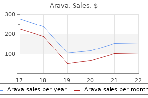
Order arava master card
Links between different health services medicine 72 hours proven arava 10mg, and between health and social care are essential. She sees a chiropodist separately, has a social worker, and her son recently arranged a visit to an osteopath. One healthcare professional should act as a keyworker for such patients and coordinate care. This section summarizes some evidence from the many studies, but it is worth noting that `[e]vidence-based advice depends on the existence of primary source evidence. This emerges only from clinical trial results in highly selected patients, using limited strategies. It does not address the range of choices available, or the order of use of additional therapies. Even if such evidence were available, the data would show median responses and not address the vital question of who responded to which therapy and why. Patient-centred care is defined as an approach to "providing care that is respectful of and responsive to individual patient preferences, need, and values and ensuring that patient values guide all clinical decisions". However, with care, patients can achieve considerable improvements in both without major physical or emotional side effects. The world of research is very different from the busy clinic with too many patients and too few staff. Focus on the key care issues for each patient and try to deliver them as efficiently and kindly as possible. Personalized goals the aim of diabetes care is to return the patient to as close a non-diabetic state as is safe and practical for that particular person. It is the combination of risk factors that cause diabetes complications, so treat them all. Children also need careful diabetes care, aiming for safe, near normalization of parameters, but this has particular risks. The goals cited here are supported by the literature and by the relevant specialist societies. They will not be achievable in some patients and care is needed in their application. There appears to be no threshold effect for cholesterol either, although research continues. Risk reduction will also be discussed in later chapters on complications of diabetes. These lipid values are not necessary to guide treatment as current evidence advocates statin treatment for most people with diabetes regardless of cholesterol levels (see Box 3. The insulin dose may need to be adjusted, as it may for those changing to e-cigarettes (safety evidence in diabetes awaited). First, vigorously encourage lifestyle measures with weight reduction as needed; a low sodium, high potassium diet; moderate alcohol only; and regular exercise. The tight control group showed a 24 % reduction in diabetes-related endpoints, 32 % reduction in deaths due to diabetes, 44 % reduction in strokes, and 37 % reduction in microvascular endpoints. Pursuing lower targets does not reduce coronary event rates, although stroke incidence may be modified. Amlodipine ± perindopril reduced cardiovascular endpoints more than atenolol ± thiazide, and was less diabetogenic in non-diabetics. Microalbuminuria warns that diabetes has damaged the kidneys and renal failure will follow. The rate of progression to renal failure can be slowed by prompt and vigorous treatment. Enalapril showed a reduction in development of microalbuminuria in normotensive diabetic patients (Ann Int Med 1998; 12:9828). Captopril (type 1 diabetes), lisinopril, and ramipril are licensed for use in diabetic nephropathy. Losartan is licensed in treatment of diabetic nephropathy in type 2 diabetes, as is telmisartan. Type 2 diabetes mellitus Statin* therapy is recommended for all patients with type 2 diabetes above age 40 irrespective of cholesterol level. There was a 22 % reduction vs placebo in cardiovascular events in those on simvastatin, with a 33 % reduction in those without overt vascular disease at outset, and a 27 % reduction if low-density lipoprotein-cholesterol < 3 mmol/l at the outset. The effect was less marked in type 1 diabetes, perhaps because of smaller numbers. Blood glucose control (see Chapters 713) Glucose control is a particularly challenging part of diabetes management. Intensive therapy reduced the risk of developing new retinopathy by 76 %, and slowed progression by 54 % in those with pre-existing retinopathy. Overall, intensive therapy reduced occurrence of microalbuminuria by 39 %, overt proteinuria by 54 %, and clinical neuropathy by 54 %. Post-trial, at 20 yrs, despite the HbA1c returning to that of the control group once the study ended, mortality was lower in the intensive glucose control groups than the control group (13 % less on sulfonylurea/ insulin, and 27 % less on metformin). A later analysis (2012) showed threshold effects-risks of macro- or microvascular disease or death were the same in the intensive vs control groups below HbA1c 53 mmol/mol (7. Above this for every ~ 11 mmol/l (1 %) higher HbA1c level the risk of events rose by 38 % for death, 38 % for macrovascular disease, and 40 % for microvascular disease. Quality of life was no different between intensively and conventionally treated patients. Thus reducing the HbA1c reduces the risk of diabetic tissue damage but potentially increases the risk of adverse effects. Aim to return the blood glucose towards that observed in non-diabetics but not under 4 mmol/l in patients on glucose-lowering medication. Consider aiming for a fasting blood glucose between 4 mmol/l and 7 mmol/l and a level up to 10 mmol/l after food, if safe for that individual patient. Teach all patients on glucose-lowering treatment how to recognize and treat hypoglycaemia. Teach them how to adjust their treatment to reduce the risk of further hypoglycaemia. Patients with varied timetables, meals, exercise, emotions, and varied compliance are particularly at risk of hypoglycaemia, as are children and the elderly. Educational background and ease of understanding medical advice may also be relevant. If this is the case, work together towards the best compromise between safety now and good health long term. Waist circumference provides a better estimate of adiposity and should be < 102 cm in men (< 92 cm in Asian men); and < 88 cm in women (78 cm in Asian women). In general, the most effective weight-reduction strategy combines dietary advice (see Chapter 5), regular exercise (see Chapter 13), and long-term help in changing everyday weight-gaining habits. Very-low-calorie diets are successful but these require expert supervision for safety (Diabetologia 2011; 54:250614; doi 10. It inhibits pancreatic lipase and therefore induces fat malabsorption (with frequent embarrassing gastrointestinal side effects if patients do not adhere to a low- fat diet). Bariatric surgery is effective in weight reduction and improving glucose control in obese diabetic patients. Identification and treatment of tissue damage the main thrust of diabetes care must be prevention of problems, but in reality much effort is needed to detect and slow progression of diabetic tissue damage. Up to half of all patients with type 2 diabetes have obvious tissue damage at the time of diagnosis. Many diabetic complications have specific treatments and their progress can be slowed by redoubled preventive care (see Chapters 14 and 15). This means rigorous checks by the patient (reporting visual change, foot problems, etc. Check tissue damage annually at present (note that there is no evidence to support a 12-month interval as being any better than a shorter or longer one). The main difficulty lies in the silence of much severe tissue damage until it is too late to prevent disability. If unhealthy or overweight, provide appropriate advice and dietetic referral as required. Symptoms of cardiovascular disease, eye problems, neuropathy, foot problems, sexual dysfunction, other complications.
Safe 20 mg arava
Pediatric There are no data related to the safety and efficacy of fosinopril in children younger than 5 years medicine januvia order generic arava on line. Geriatric Elderly patients with heart or kidney problems may require some adjustment in dosage. Category C means that the drug has been shown to harm the fetus in animal studies, but that no data from human studies are available. Category D means that there is positive evidence of harm to the fetus based on data from drug trials and marketing studies. In spite of the risks, drugs in both category C and category D may be prescribed for a pregnant 361 Recommended dosage Fosinopril is dispensed as 10 milligram (mg), 20 mg, or 40 mg tablets. Patients are advised to take these drugs with a glass of water at the same time each day, with or without meals as they prefer. However, taking the daily dose on an empty stomach an hour before a meal allows more efficient absorption of the drug. A missed dose should be taken as soon as it is remembered, unless it is almost time for the next dose. Patients should never take a double dose of fosinopril to make up for a missed dose. Diabetic nephropathy-A progressive kidney disease that develops as a complication of diabetes, in which the capillaries supplying the filtering units (glomeruli) of the kidneys lose their ability to filter body fluid. The diffuse form of scleroderma also affects the internal organs, most often the kidneys, esophagus, and lungs. Other conditions and allergies Patients with any of the following conditions must notify their doctor before taking fosinopril: · A history of angioedema, an allergic reaction to food or drugs characterized by the sudden and rapid swelling of subcutaneous tissues and mucous membranes. In addition, patients should alert their healthcare provider if they are receiving treatments to lower sensitivity to wasp or bee stings, receiving kidney dialysis, scheduled for surgery or any procedure requiring anesthesia, or presently taking any other medication for hypertension. The following side effects should be reported to a doctor immediately: · sudden swelling of the face, arms, legs, eyes, lips, or tongue, or problems with swallowing or breathing (symptoms of angioedema) · sudden and severe stomach pain (may indicate a condition called intestinal angioedema) · light-headedness severe enough to cause fainting · chills, fever, or other signs of infection (may indicate a drop in the number of white blood cells) · yellow discoloration of the skin or the whites of the eyes, dark urine, and pale stools (symptoms of jaundice and may indicate liver damage) · muscle pains or cramps, one-sided weakness, or shortness of breath · confusion, memory problems, depression, or other changes in mood or mental status · changes in vision · sped-up, slowed-down, or irregular heartbeat · unusual sweating Other conditions and allergies Fosinopril is reported to be less effective in African Americans than in members of other racial or ethnic groups. Fosinopril Interactions Fosinopril is known to interact with various drugs, supplements, and foods. Herbs and supplements Fosinopril is reported to interact with bitter melon (lowers blood potassium levels). Food and other substances Alcohol may increase such side effects of fosinopril as light-headedness or fainting. Patients should also avoid salt substitutes containing potassium, as they may interact with fosinopril to increase the levels of potassium in the blood. Furosemide is classified as a loop diuretic because its mechanism of action affects the loop of Henle in the nephrons of the kidneys. Furosemide acts to inhibit the reabsorption of sodium and chloride in this portion of the nephron during the process of urine formation. It is considered a short-acting diuretic because it begins to act within an hour and reaches peak effectiveness within two hours; its effect lasts about six to eight hours. Mayo Clinic: 5 Steps to Controlling High Blood Pressure: Your Personal Guide to Preventing and Managing Hypertension. Ascites-Accumulation of fluid in the peritoneal cavity, often associated with cirrhosis of the liver or congestive heart failure. Loop diuretic-A type of diuretic that acts on the loop of Henle in the nephrons (basic functional units) of the kidney to prevent reabsorption of sodium and chloride. Loop of Henle-The portion of a nephron that leads from the proximal convoluted tubule to the distal convoluted tubule. It is named for Friedrich Henle (18091885), the German anatomist who first identified it. Description Furosemide is available for oral administration in two forms, tablets and liquid. Furosemide tablets are white and round or oval in shape, and available in 20, 40, and 80 milligram (mg) strengths. Furosemide liquid solution is available only as a generic rather than a brandname formulation. The 10 mg per milliliter (mL) solution of the drug is flavored orange, and the 40 mg/5 mL solution is flavored pineapple/peach. The injectable form is administered intramuscularly or intravenously only by a physician or nurse in a hospital; patients are prescribed an oral form of furosemide for use at home. Canadian brand names Furosemide is marketed in Canada under the brand names Apo-Furosemide, Lasix, and Lasix Special. International brand names Furosemide is marketed by at least eight different international manufacturers under numerous brand names, including Beronald, Desdemin, Durafurid, Edemid, Fuluvamide, Furosedon, Lasilix, Mirfat, Nicorol, Odemase, Profemin, Rosemide, Salix, Teva-Furosemide, Trofurit, and Urex. Patients should not exceed their prescribed dose, as high doses of furosemide can cause irreversible hearing loss. Patients who are prescribed the liquid formulation of furosemide should use a marked measuring spoon, an oral syringe, or a medicine cup to measure the dose, as ordinary household tablespoons vary in the amount of liquid they hold and so may not provide an accurate dose. Furosemide solution should be stored tightly closed in a light-resistant container at room temperature, 59°F86°F (15°C30°C); the liquid formulation should be used within 60 to 90 days after opening the bottle. Furosemide should not be stored in the bathroom and should be kept out of the reach of children and pets. Pediatric To treat edema in infants and children, the initial dose is 2 mg/kg by mouth per day (about 0. To treat resistant hypertension in children and adolescents between 1 and 17 years of age, the dose is 0. Geriatric Elderly patients need more careful monitoring because of the increased risk of dehydration and electrolyte loss. The recommended dosage is an initial dose of 10 mg per day by mouth, gradually adjusted upward. The drug may cause loss of blood plasma volume in the mother and should be used during pregnancy only if the benefit clearly outweighs the risk. Furosemide is known to pass into breast milk and could potentially harm a nursing baby; therefore, nursing mothers should inform their doctor before taking furosemide. Other conditions and allergies Patients with any of the following conditions should not use furosemide: · severe electrolyte depletion · hepatic coma · severe allergy to sulfa drugs (sulfamethoxazole, sulfasalazine, sulfisoxazole) · allergy to furosemide itself or to any of the components in the tablets or solution · inability to urinate (anuria) Side effects Common side effects of furosemide include: · headache · muscle cramps · increased sensitivity to sunlight (photosensitivity) · skin rash · constipation or diarrhea · frequent urination · anemia Less common side effects include: · chills or fever · sore throat · indigestion · itchy skin · unusual bruising or bleeding · sores or white spots on the lips or in the mouth Patients should consult their doctor at once if they notice any of the following side effects of furosemide: · symptoms of angioedema (a severe allergic reaction), which include sudden swelling of the face, arms, legs, eyes, lips, or tongue and problems with swallowing or breathing · symptoms of jaundice, which include yellow discoloration of the skin or the whites of the eyes, dark urine, and pale stools · dizziness, fainting spells, or lightheadedness · signs of dehydration or electrolyte imbalance, which include dry mouth, thirst, weakness, drowsiness, nausea or vomiting, low blood pressure, decreased urination, and a rapid or irregular heartbeat · ringing in the ears or hearing problems · high blood sugar levels in patients diagnosed with diabetes · vertigo (sensation of spinning, or the feeling that objects and surroundings are spinning around the self) · signs of a furosemide overdose, which include irritability, mood changes, drowsiness, severe muscle cramps, numbness or tingling pains in the hands or feet, rapid breathing, seizures, weak pulse, and tremor · Aminoglycoside antibiotics. Other drugs that may interact with furosemide include aliskiren (an antihypertensive drug), chloral hydrate (a sedative), digoxin (used to treat congestive heart failure), and corticosteroids. Herbs and supplements Patients using furosemide should avoid dong quai, ephedra, yohimbe, and ginseng, as these herbs may worsen hypertension. They should also avoid garlic, which may increase the antihypertensive effect of furosemide. Patients should be warned against eating large amounts of licorice, as licorice interacts with furosemide to lower the level of potassium in the blood. Purpose Gabapentin is used in combination with other antiseizure (anticonvulsant) drugs to manage partial seizures with or without generalization in individuals over the age of 12. Gabapentin can also be used to treat partial seizures in children between the ages of 3 and 12. Gabapentin was previously studied in the treatment of bipolar disorder but did not show any effectiveness in clinical trials. Recommended dosage For epilepsy, people over the age of 12 can begin with an initial dose of 300 mg three times a day, which can be gradually increased as necessary, usually to no more than 1,800 mg daily. Pain dosing involves a gradual increase, with an initial does of 300 mg on the first day, followed by 300 mg twice on the second day, and 300 mg three times on day three. Pediatric For children ages 3 to 12, the dose is based on body weight, initially 1015 mg per kilogram (kg, or 2. For a child under the age of 3, the decision about use and dosage will be made by the doctor. Geriatric For older adults, the maximum daily dose does not usually exceed 600 mg three times a day. Description Brain cells normally transmit nerve impulses from one cell to another by secreting chemicals known as neurotransmitters. The actual mechanism of action by which gabapentin acts in the brain to control seizures and treat pain is not known, although it appears to alter the action of nerve cells. Gabapentin is available in 100, 300, and 400 milligram (mg) capsules; in 600 and 800 mg tablets; and as a liquid solution containing 250 mg per 5 milliliters (mL).
Diseases
- Coloboma of eye lens
- Formaldehyde poisoning
- Gonadal dysgenesis mixed
- Hyperkalemic periodic paralysis
- Dentin dysplasia, coronal
- Chromosome 20, deletion 20p
Cheap arava 10 mg online
The drug is a synthetic form of a metabolite of the antidepressant drug venlafaxine treatment quietus tinnitus discount generic arava canada. It was developed based on the theory that a significant portion of the therapeutic benefits of venlafaxine come from this metabolite. Desvenlafaxine is expected to have fewer interactions with other medications than venlafaxine due to differences in how it is processed in the body. Whether there is an advantage in taking desvenlafaxine instead of venlafaxine is debated. Recommended dosage Description Desvenlafaxine works by acting on the neurotransmitters serotonin and norepinephrine, which are chemicals in the body that help regulate normal brain and body functioning. It is believed that desvenlafaxine works by slowing the reabsorption of the neurotransmitters norepinephrine and serotonin, thus increasing their levels in the brain. The dosage varies depending on how an individual patient responds, and it is usually increased gradually. If side effects develop that are not tolerable, the patient may require a lower dose. Higher doses rarely result in better therapeutic effects, and they increase the risk of adverse side effects. Slowly increasing the dose may help minimize side effects, and some side effects may abate with continued use. Patients are periodically reassessed to determine whether there is a need for continued treatment with desvenlafaxine. Glaucoma-A condition involving increased pressure within the eye that may cause damage and blindness. Metabolite-A chemical compound that occurs as a result of a parent drug being broken down and metabolized in the body. A metabolite may be a medically active or inactive compound, depending on the drug in question. Norepinephrine-Also called noradrenaline, a chemical messenger in the brain that regulates attention and that powers the "fight-or-flight" stress response; precursor of epinephrine. Serotonin-A type of neurotransmitter involved in the regulation of the blood vessels, brain processes, and disease states such as depression. Geriatric Elderly patients tend to develop side effects more easily and may be more sensitive to the effects of the medication. Other conditions and allergies Patients with impaired liver or kidney function affecting drug metabolism require lower doses or less frequent dosing to avoid toxicity. Any patient taking an antidepressant drug should be monitored for changes in behavior and worsening depression. Such withdrawal symptoms include: · flulike symptoms · anxiety or agitation · vivid or bizarre dreams · insomnia, nausea · vomiting or diarrhea · sense of imbalance 225 Precautions Antidepressant drugs, including desvenlafaxine, have been associated with an increased risk of suicidal · chills · fatigue · dizziness · headache · numbness and tingling of the extremities · other sensory disturbances Discontinuation syndrome may be avoided if the dose is properly reduced over time. Serotonin overdose may also be caused by taking multiple drugs that increase the amount of serotonin signaling in the body. Symptoms of serotonin overdose range from mild to life threatening, depending on the individual situation. Symptoms may include: · high blood pressure · high fever · nausea or diarrhea · headache · sweating · increased heart rate · tremor · muscle twitching · delirium · shock · coma Serotonin overdose can also result in death. Kidney and liver function, as well as blood pressure and behavioral changes, may be monitored while patients are taking desvenlafaxine. The decision about whether to use category C drugs in pregnancy is generally based on weighing the critical needs of the mother against the risk to the fetus. The safety of desvenlafaxine use during breastfeeding is unknown; therefore, its use is not recommended. Common side effects of desvenlafaxine include: · nausea and vomiting · headache · dizziness · fatigue · constipation or diarrhea · sexual dysfunction · sweating · dry mouth · shakiness · tremor · palpitations · loss of appetite · weight changes · hot flashes or chills · elevated blood pressure · vision changes · ringing in the ears · anxiety · abnormal dreams · insomnia Rare but serious potential side effects include mania, worsened depression and suicidality, seizures, serotonin syndrome, discontinuation syndrome, electrolyte imbalances, lung damage or disease, urinary retention, skin reactions, abnormal bleeding, liver damage, and glaucoma. Interactions Patients should make their doctor aware of all drugs, herbs, and supplements they are taking before using desvenlafaxine. Drugs that affect the liver may alter the metabolism of desvenlafaxine, which can result in too much or too little of the drug in the body. Other drugs that increase serotonin in the body include sumatriptan (Imitrex), used to treat migraine headaches, and the antipsychotics chlorpromazine and fluphenazine. Diuretics (water pills) such as hydrochlorothiazide, diet pills such as sibutramine (Meridia), certain diabetes drugs such as sulfonylureas, mood stabilizers such as valproic acid, and antipsychotics such as haloperidol and clozapine should not be used with desvenlafaxine to avoid potentially toxic effects. There is increased risk of internal bleeding when desvenlafaxine is used with anticoagulant drugs such as aspirin and warfarin (Coumadin). Large doses of the herbal supplements red clover, dong quai, ginkgo biloba, feverfew, and concentrated green tea increase the risk of internal bleeding when used with desvenlafaxine. Food and other substances Using alcohol or caffeine while taking desvenlafaxine may create toxic reactions and should be avoided. Dexlansoprazole Dexedrine see Dextroamphetamine Dexilant see Dexlansoprazole Description Dexlansoprazole is marketed as a hard gelatin (twopiece) capsule. The 60 milligram (mg) capsules are solid blue and are imprinted with the number 60. The 30 mg capsules have a blue cap and a grey body and are imprinted with the number 30. The initial release produces peak blood levels in one to two hours after taking the capsule, and the secondary release peaks four to five hours after a dose. The name was changed because the old name could be confused with Casodex, which is used to block androgen, the male sex hormone. Canadian brand names In Canada, dexlansoprazole is also sold under the brand name Dexilant. Lansoprazole, like many other drugs, is a mixture of two molecules that are mirror images of each other, rather like the left and right hand are mirror images. In many cases, the R (right hand) and S (left hand) versions of the drug have differences in their actions or activity. Sometimes one version of the drug will have most of the desired activity, while the other may cause unwanted side effects. Dexlansoprazole has the same chemical composition as lansoprazole but is its "right-hand" version (r-enantiomer). Purpose Proton pump inhibitors reduce the level of stomach acid by blocking enzymes in the stomach wall that help create the acid. Recommended dosage the initial dose for treating erosive esophagitis is 60 mg taken once a day for up to eight weeks. After that, the dose may be reduced to 30 mg a day for maintenance and the prevention of heartburn. For patients who have difficulty swallowing capsules, dexlansoprazole capsules may be opened and sprinkled on applesauce. If this is done, the applesauce must be eaten immediately and the granules must not be chewed. The granules may also be administered through a nasogastric tube or an oral syringe. Other conditions and allergies No dose adjustment is required for patients with mild liver function problems. For patients with moderate liver function problems, the dose should be limited to 30 mg/day. The safety of dexlansoprazole has not been established in patients with severe liver function problems. If a single type of molecule binds with others of the same type to form a chain, it is called a polymer. Enteric-coated drug-A drug that has a coating that breaks apart only in the intestine, not the stomach. Enteric coatings may be used for drugs that would cause stomach irritation, as a way of delaying the release of the drug into the bloodstream, or for drugs that would be harmed by stomach acid. Esophagitis-Inflammation of the esophagus, the tube that connects the throat with the stomach. Dexlansoprazole is considered a pregnancy category B drug, which means that animal studies have not shown the drug to have adverse effects on a fetus, despite a lack of human studies.

Buy arava with amex
It aims to support clinical teams to deliver better care to women with diabetes who become pregnant medicine abbreviations arava 20mg amex. Imaging Approach to Abdominal Manifestations of Systemic Conditions Abdominal Manifestations of Systemic Conditions Organizational Approach to Abdominal Diseases Most information about imaging abdominal disorders, including the gastrointestinal and genitourinary systems, fits neatly into an organ-by-organ framework. However, this approach makes it difficult to discuss diseases or conditions with manifestations throughout the abdomen and beyond. Doing so provides a more accurate portrayal of these entities, and avoids unwanted redundancy. Because many systemic disorders affect lymph node groups, neural structures, or major vessels throughout the abdomen, medical illustrations provide a helpful reminder of important anatomical considerations. Degenerative conditions, such as sarcoidosis and vascular disorders, are rarely limited to a single organ. These are presented in all their guises, along with tips as to how to address differential diagnoses. Foreign bodies may be encountered throughout the gastrointestinal and genitourinary system and are well-known to be found repeatedly in certain individuals. Many malignant neoplasms are, by their very nature, systemic processes, such as lymphoma, leukemia, and malignant melanoma. Therefore, taking a systemic approach to such diagnoses gives us the opportunity to bring together some general principles about the presentation, diagnosis, and management of these important diseases. Finally, while some conditions, such as systemic hypotension or hypervolemia, do not represent disease per se, they can result in important clinical and imaging abnormalities that must be recognized to avoid misguided patient management. Imaging Modalities Plain radiography maintains an important role for surveillance of some generalized disease processes, such as the osseous and visceral manifestations of sickle cell anemia or cystic fibrosis. Ultrasound is an important imaging tool for the evaluation of biliary, vascular, gynecologic, and scrotal pathology, but it lacks both sensitivity and specificity in evaluating other processes, especially bowel pathology. In patients with cancer, for instance, the ability to quickly and accurately examine different anatomic areas (thorax, abdomen, and pelvis), organs, and structures of different composition. Catheter angiography remains the most accurate means of identifying certain vascular disorders and often results in catheter-based therapies in the same setting. For vasculitides, which routinely affect vessels throughout the body, angiography maintains an essential diagnostic and therapeutic role. The left gastric artery also has a separate origin from the aorta, though difficult to perceive on this image. The lumbar nodes are often referred to as para- or retroaortic (or -caval), indicating their position relative to the great vessels. Similar lesions were present in the liver, better seen on narrow window-width images (not shown). There are innumerable focal hypodense nodules in both kidneys, as well as upper abdominal lymphadenopathy. All lesions were found to represent sarcoidosis and responded to steroid medication. A digital radiograph shows a curvilinear radiopaque stripe within the right side of the abdomen. This is a classic gossypiboma, a retained surgical sponge that has resulted in a chronic abscess or foreign body reaction. This constellation of findings was found to represent disseminated mycobacterial infection. An unusual feature in this case is the mild obstruction of the intrahepatic bile ducts. Some of the enlarged nodes have a caseated or low-density centrally necrotic appearance characteristic of mycobacterial infection. The spleen was almost 20 cm in length, and a histologic exam showed that it was congested with activated T lymphocytes. Bone Infarcts From Other Causes · Trauma, steroid use, lupus erythematosus, Gaucher disease, hemophilia, thalassemia, etc. Note the presence of sclerosis in the femoral head, compatible with avascular necrosis. Affected areas show a characteristic double-line sign with adjacent hyperintense and hypointense rims around their margins, characteristic of a bone infarct. This is a very nonspecific finding, one that required biopsy for the diagnosis of amyloidosis. Subtle hypodense foci in the liver and spleen indicate the granulomatous nature of the pathology. Promteangtrong C et al: the role of positron emission tomographycomputed tomography/magnetic resonance imaging in the management of sarcoidosis patients. The renal involvement in this case is less typical and probably represents tubulointerstitial nephritis, a known but rare feature of sarcoid. A more common renal manifestation of sarcoid is nephrocalcinosis &/or nephrolithiasis. These features, along with lymphadenopathy (better shown on more caudal images), are very similar to those seen in primary biliary cirrhosis. Sarcoidosis must be considered in the differential diagnosis for widespread lymphadenopathy and splenomegaly. The diagnosis is based on compatible clinical and imaging findings and the presence of noncaseating granulomas on biopsy. This appearance is characteristic of shock bowel, seen during or following severe hypotension. The liver perfusion abnormality was not seen on portal venous phase images (not shown). Findings that suggest bowel or renal ischemia in a young patient, as in this case, should raise suspicion for vasculitis. This is a common appearance for a large vessel vasculitis (giant cell vasculitis) with active inflammation. The superior mesenteric artery is also completely occluded, with collaterals from an enlarged inferior mesenteric artery. This was found to represent Takayasu arteritis, which more often affects the aortic arch. Swallowing batteries is relatively common and can cause bowel perforation or obstruction as the acid in the battery may leak out. Some of the hyperechoic bands with posterior acoustic shadowing may reflect gas within the mass. All magnets should be retrieved immediately Foreign bodies vary in radiopacity and conspicuity on radiography vs. Guelfguat M et al: Clinical guidelines for imaging and reporting ingested foreign bodies. The patient had a long history of ingesting pins and had to be taken to surgery to remove the foreign bodies. The object turned out to be a penlight which was subsequently retrieved in the cystoscopy suite. Note that the stricture in the immediately distal small bowel is causing mild obstruction of the dilated proximal bowel. This radiopaque tag can be directly woven into the sponge or can be attached to it. Also note the presence of a plastic ring within the abdomen connected to the surgical sponge. At surgery, small bowel perforation was confirmed, and a plastic Bic pen was retrieved. It may be impossible to distinguish this from an abscess without the proper history, and in such cases, needle aspiration may be required. In some cases, gas can dissect into the bowel wall, simulating pneumatosis from bowel ischemia. The spleen is also involved, with splenomegaly and dozens of small hypodense nodules. Note the biliary stent placed to treat the extrinsic compression of the common bile duct by enlarged nodes and the liver mass. Notice the manner in which the lymph nodes surround the mesenteric vessels, often described as the sandwich sign. Note the lack of bowel obstruction despite significant bowel involvement, a characteristic feature of lymphoma.

10mg arava otc
Pediatric Escitalopram is not approved for use in children younger than 12 years of age medicine man lyrics discount arava 10 mg with amex. The dosage varies depending on the individual patient response to the medication regarding its effectiveness, and the individual patient response to the medication regarding side effects. Some people naturally require a higher dose of escitalopram in order to achieve the desired effect. Other patients require a lower dose either for therapeutic effect or to lessen adverse effects. Escitalopram used for major depressive disorder or generalized anxiety disorder is dosed at 1020 milligrams (mg) daily. Patients generally start at the 10 mg dose and gradually work their way up to the dose needed for effect after a week at the 10 mg dose. Slowly increasing the dose helps with minimizing side effects, and some side effects become lessened with continued use. Pediatric Children ages 12 to 17 may be given a trial of 10 mg per day of escitalopram under select circumstances with careful medical supervision. The dose may be increased to 20 mg after three weeks at a lower dose in some circumstances. Geriatric Elderly patients are usually kept at a lower dose of 10 mg, due to their increased sensitivity to these medications and their side effects. Bipolar disorder-Psychiatric mood disorder characterized by periods of manic behavior that may alternate with periods of depression; also known as manic depressive disorder. Depression-A mental state characterized by excessive sadness and loss of interest in life; other symptoms may include altered sleep or eating patterns, loss of concentration, agitation, lack of energy, and, in severe cases, attempts at self-harm or suicide. Neurotransmitter receptor-A physical recipient for chemicals called neurotransmitters. Social phobia-Psychiatric disorder involving excessive fear and anxiety due to social situations with other people. Potential effects on the newborn include respiratory problems, gastrointestinal problems, seizures, and feeding problems. Symptoms of neonatal abstinence syndrome in a newborn include irritability, physical agitation and restlessness, tremors, increased breathing rate, nasal congestion, nausea, vomiting, and diarrhea. Escitalopram may not be appropriate for use or may require careful monitoring in patients with liver or kidney impairment, electrolyte imbalances, or seizure disorders. Common side effects of escitalopram include nausea, headache, dizziness, constipation, sexual dysfunction, diarrhea, sweating, dry mouth, shakiness, loss of appetite, weight loss, rash, and insomnia. Rare but serious potential side effects include mania, worsened depression and suicidality (especially in the pediatric population), pediatric growth suppression in younger age groups, 293 Escitalopram seizures, serotonin syndrome, discontinuation syndrome, electrolyte imbalances, hypoglycemia, abnormal bleeding, priapism, and glaucoma. Food and other substances Using alcohol while taking escitalopram can create toxic reactions in the body, and alcohol should be avoided while taking this drug. Other drugs that cannot be combined with escitalopram due to risk of serotonin syndrome are the antibiotic linezolid and the drug methylene blue, used in some blood-related illnesses. Escitalopram may increase the potency and side effects of: · warfarin · digoxin · beta blocker cardiac drugs such as metoprolol · benzodiazepines such as diazepam (Valium) · antipsychotics such as haloperidol and clozapine · certain antiseizure drugs such as phenytoin Drugs that may cause toxicity with escitalopram include: · diuretics (water pills) · sleep aids like zolpidem (Ambien) · diet pills such as sibutramine (Meridia) · mood stabilizers such as lithium Escitalopram cannot be combined with the antipsychotic medications pimozide (Orap) or thioridazine (Mellaril) due to dangerous cardiac complications. Esomeprazole also gives the esophagus a chance to heal from the damage caused by the acid reflux. The drug is typically taken for a period of four weeks to relieve symptoms, but it may be taken longer if symptoms are not relieved and the doctor recommends another course, or for up to six months to maintain healing of the esophagus. When taking esomeprazole to begin healing of the esophagus, the doctor may recommend an adult dosage of 20 mg or 40 mg once a day for four to eight weeks. Some patients do not heal within this time and take the medication for an additional four to eight weeks. The capsule is usually taken whole by mouth, but its contents can be mixed with water or administered by caregivers through a feeding tube if needed. Neuroendocrine-The endocrine system involves cells that make hormones in the body. Neuroendocrine refers to cells that are found in organs that make hormones but also have nerve cells. Osteoporosis-A chronic and progressive disease that leads to bone weakening and brittleness. Pediatric Children ages 111 can take 1020 mg of esomeprazole once a day for up to eight weeks. Esomeprazole is a pregnancy category C drug, meaning that women who are pregnant should only take the medication if the potential benefits outweigh possible risks. It is not known whether the medicine is excreted in breast milk, so nursing mothers should decide whether to stop using esomeprazole while nursing or continue using the drug and choose not to breastfeed their infants. Other conditions and allergies People who have neuroendocrine tumors, which are growths that develop in hormone-producing cells of certain organs, may need to stop taking esomeprazole before having laboratory tests for the tumors because the medication can interfere with test results. People who have a history of liver disease or low magnesium levels in their blood should discuss these conditions with their doctor when considering taking esomeprazole. Side effects Some of the common side effects of esomeprazole include: · dry mouth · constipation · gas or nausea · headache Some side effects from taking esomeprazole can be severe and if they occur, the patient should contact a doctor immediately. Long-term use of esomeprazole can increase risk of osteoporosis-related bone fractures. Pediatric Esomeprazole should only be given to children younger than one year if they have erosion or damage to the esophagus. Geriatric Overall, research has shown no difference in safety or how well esomeprazole works in older adults compared with all adults. For instance, side effects such as diarrhea can cause more serious problems in older adults. It is important to tell the doctor about any medications, herbal therapies, or vitamins being used before taking esomeprazole. Drugs Some drugs can significantly decrease the amount of esomeprazole in the body. Among these are rifampin, a drug used to treat tuberculosis and some forms of meningitis. People who take clopidogrel (Plavix) to Estradiol, micronized prevent problems with the heart and blood vessels need to discuss the drug with their doctor because esomeprazole can affect how well the clopidogrel works. Esomeprazole may increase levels of methotrexate (Rheumatrex), which is used to treat severe psoriasis. Purpose Estradiol is prescribed primarily to relieve menopause-related symptoms, such as hot flashes, mood swings, tender breasts, vaginal dryness, lower sex drive, and sometimes migraine headaches. Nearly two-thirds of perimenopausal and menopausal women take oral estrogen at some point to relieve such symptoms. Women who have had a hysterectomy, which is considered surgical menopause, may also be advised to take estradiol. The use of estradiol to help correct estrogen hormone deficiency and to reduce related symptoms is called estrogen replacement therapy. Aside from relieving short-term symptoms, the benefits of hormone replacement therapy are also reported to include reduced incidence of osteoporosis-related fractures, especially hip fractures, and reduced incidence of diabetes among postmenopausal women. In addition, estrogen may be prescribed to treat endocrine disorders and reproductive problems in younger women. Local conditions such as atrophic vaginitis and atrophic urethritis may also be treated with the topical application of estradiol preparations, such as gels and creams containing the estrogen hormone. Estradiol is also prescribed for the palliative treatment of breast cancer and prostate cancer. International brand names Internationally, estradiol is sold under the brand names Alora, Climara, Delestrogen, Depo-Estradiol, Divigel, Elestrin, Estrasorb, Estrogel, Menostar, Minivelle, and Vivelle. Description Estradiol is the main form of the steroid hormone estrogen and the most abundant sex hormone in women of childbearing age. It is produced in the follicles of the ovaries, in the adrenal glands, and in fat tissue, and it is then released into the bloodstream. Estrogen circulating in the bloodstream attaches to estrogen receptors in the cells of certain tissues, including the breasts, uterus, brain, bones, liver, heart, and other body tissues. Estrogen is responsible for the development of female sexual characteristics and reproductive functioning. Estrogen production declines prior to menopause and gradually throughout the menopausal years. This natural transition of estrogen levels typically begins between the ages of 40 and 55 and may continue for several years, even up to 10 years in some women. Certain other conditions may cause estrogen deficiency, including failed ovary function (female hypogonadism), polycystic ovarian syndrome, pituitary gland dysfunction (hypopituitarism), abnormal kidney and liver function, extreme weight loss from illness or eating disorders, and certain drugs.
Syndromes
- Cesarean delivery or induction of labor before the is full-term
- An infection of the outer ear (otitis externa)
- Failure to use a condom each time you have intercourse
- Rapid breathing (respiratory) rate
- Muscle aches
- Cough that does not produce much sputum or mucus (dry cough)
- Non-alcoholic fatty liver disease
Buy online arava
The proximal end of the stricture is fairly abrupt symptoms stroke purchase arava with american express, while the distal end is more tapered. Cricopharyngeal achalasia is most often seen in patients with other esophageal disorders, such as reflux esophagitis or motility disorders. Note the absent gastric air bubble and the fluid-barium level within the esophagus. Esophageal Achalasia Esophagus (Left) Esophagram shows a typical appearance of achalasia, pre-Heller myotomy, with "bird beak" deformity of the distal esophagus, marked dilation of the proximal esophageal lumen, and absent peristalsis. The esophagus emptied readily in the upright position, due to gravity, while peristalsis was still absent. This is an example of vigorous achalasia, in which esophageal contractions are seen but are not effective. The abrupt narrowing of the lumen with overhanging edges and presence of nodular thickened folds suggests the true diagnosis of gastric carcinoma, causing this pseudoachalasia appearance. The esophageal dysmotility in this patient is probably a result of age as well as the effects of reflux esophagitis on motility. Multiple bariumfilled pulsion diverticula are evident, especially after the barium bolus has passed. Cфtй-Daigneault J et al: High prevalence of esophageal dysmotility in asymptomatic obese patients. Demographics · Age Achalasia and scleroderma: Young patients Presbyesophagus: Elderly · Gender Primary motility disorders: M = F Scleroderma Predominately women (M:F = 1:3) · Ethnicity Scleroderma: African Americans > Caucasians · Epidemiology 186 3. Esophageal Motility Disturbances Esophagus (Left) Spot film from an esophagram in a middle-aged man with presbyesophagus shows a small hiatal hernia with the gastroesophagealesophageal junction marked by a Schatzki ring. The Schatzki ring and dysmotility resulted in obstructed passage of a barium pill (not shown). The folds within the jejunum are thin and closely spaced, and the lumen is dilated with minimal peristalsis in this classic "hidebound" bowel. The esophagus was slow to empty, even in the upright position, with a fluid-barium level seen. These are relatively early signs of scleroderma, and no stricture or ulcerations have yet developed. Symptomatic narrowing of the B ring, which constitutes the Schatzki ring, probably results from reflux esophagitis. This lesion can be missed if it is tilted relative to the plane of imaging or if the esophagus is not optimally distended. Mьller M et al: Endoscopic findings in patients with Schatzki rings: evidence for an association with eosinophilic esophagitis. Intrathoracic stomach shows that while an air-fluid level is present within the stomach, there is no evidence of twisting or obstruction of the stomach; however, this patient is at risk for volvulus and strangulation. Note the position of a nasogastric tube within the herniated stomach and duodenum, which is within the abdomen. These are unusually well depicted, even with the esophageal lumen distended, suggesting that the varices may be thrombosed or sclerosed by endoscopic injection. The fixed nature of these mimics the appearance of the "varicoid" morphology of some esophageal carcinomas. Spontaneous or surgically created shunts help to decompress esophageal varices but increase the incidence of encephalopathy and portal vein thrombosis, making subsequent liver transplantation difficult or impossible. Large diverticula often compress the lumen of the esophagus and extend to 1 side of the midline due to mass effect. Decreased primary peristalsis and tertiary contractions were also evident on fluoroscopy. There are numerous intramural pseudodiverticula within the strictured segment as well as more proximally. Halm U et al: Esophageal intramural pseudodiverticulosis: endoscopic diagnosis and therapy. Calcified subcarinal lymph nodes were more evident on chest radiograph (not shown). Although this may represent a traction diverticulum, it may rather be a normal outpouching of the esophageal wall between the extrinsic indentations by the aortic arch and the left mainstem bronchus. Pulsion diverticula are almost always associated with esophageal motility disturbances and become more common with advanced age. Note the smooth outer contour that distinguishes it from the serrated surface of a coin. On first impression, this might be misinterpreted as an esophageal stent that had been placed across an obstructing esophageal lesion. Sahn B et al: Review of Foreign Body Ingestion and Esophageal Food Impaction Management in Adolescents. Due to concern for Barrett metaplasia or early cancer, an endoscopic biopsy of the lesion was performed following balloon dilation of the stricture. Pulsion Diverticulum (Epiphrenic) · Mucosa-lined pouch from distal esophagus · No free mediastinal gas or inflammation 3. The stomach is divided along its long axis, creating a gastric tube or conduit 5 or 6 cm in diameter, which is pulled up into the chest. This can be done through a right (Ivor Lewis) or left thoracotomy or even through laparoscopic ports. The conduit is not filled with retained fluid, and there is no evidence of lung injury from reflux. Alldinger I et al: Endoscopic treatment of anastomotic leakage after esophagectomy or gastrectomy for carcinoma with self-expanding removable stents. The conduit is probably twisted, as evidenced by the position of the gastric staple line. The conduit was pulled down into the abdomen at revision laparoscopy with resolution of symptoms. The horizontal portion of the conduit contributes to the impaired gastric emptying. Note that the point of narrowing is at the diaphragm, not the pylorus, which has been widened by pyloroplasty. The stomach is narrowed as it traverses the diaphragm, but the pyloroplasty is neither the site nor the cause of the delayed emptying. Note the position of the gastric staple line, which has rotated to the left anterolateral position. Note the smooth surface of the esophagus and the rugal fold pattern of the gastric conduit. There is a large soft tissue mass abutting the conduit and extending into the mediastinum in this patient with a recurrent tumor. The mass was resected and proved to be a benign leiomyoma arising from the esophageal wall. The mass is so long and bulky that it might be mistaken for an air bubble or debris within the esophagus. The mass is not readily seen within the proximal esophagus, but it distends the lumen. The polyp is continuous with thickened folds, which extend into the gastric fundus. However, a repeat film, with emphasis on suspended deep inspiration and Valsalva maneuver, demonstrates nodular thickened folds in the distal esophagus. Esophageal Carcinoma Esophagus (Left) Esophagram shows an "apple core" constricting lesion of the distal esophagus. There is an abrupt transition, or shoulder, at the proximal end of the tumor as it abuts the normal esophagus. There is also abnormal uptake within a left renal mass, which proved to be an unrelated primary renal cell carcinoma. Endoluminal sonography is the best method for determining the depth of tumor invasion. This adenocarcinoma was treated by esophagectomy with gastric interposition in the chest. However, other views (not shown) showed nodular thickened folds in the gastric fundus. It is divided into the cardia, fundus, body, antrum, and pylorus; each with its own specific function. The cardia is the portion of the stomach surrounding the esophageal orifice and the site where the lesser and greater curvatures meet.
Purchase generic arava
Intrathecal and extraforaminal enhancement can be seen in the acute and chronic polyneuropathies symptoms of diabetes order arava 10mg on-line. This patient had < 4 weeks of weakness, muscle pain, and areflexia involving her distal > proximal extremities. Axon loss associated with demyelination is the most important factor of disability, resistance to treatment. The intramedullary, intradural extramedullary, & extradural regions can be involved. Intradural extramedullary lesions are usually represented by leptomeningeal sarcoidosis infiltration, present in up to 60% of spinal cord lesions. Axial enhanced images can be used to confirm the intradural extramedullary nature of the distal cord and cauda involvement. Correlation between symptom resolution and resolution of imaging findings is poor, especially with spinal cord lesions. At clinical presentation, there may be faint parenchymal enhancement and diffuse swelling or less prominent swelling and possible normal-sized spinal cord with focal or multiple areas of enhancement. Osseous involvement is reported in 113% of patients, but the actual frequency may be higher as most of the osseous lesions are asymptomatic. These treated sclerotic lesions with sclerotic margins may extend into the pedicles and paraspinal region. Transbronchial biopsy of the lymph nodes revealed noncaseating granulomata consistent with sarcoidosis. Major clinical findings of dysesthesia, gait disturbance and impairment of position, and vibration sense in the lower ± upper limbs are 2° to dorsal column, lateral corticospinal tract, and sometimes lateral spinothalamic tract dysfunction. Lack of cobalamin (required as a cofactor for conversion of methylmalonylCoA to succinyl-CoA) leads to accumulation of methylmalonyl-CoA, causing in normal myelin synthesis and incorporation of abnormal fatty acids into neuronal lipids. The abnormal signal intensity in the dorsal columns is usually contiguous in length over several vertebral bodies. Immediate treatment with parenteral B may improve symptoms but imaging abnormalities may not completely resolve. Compression fractures are present at multiple levels, some with normal marrow due to old lesions. Grisel syndrome may occur with any stage of infection, from effusion to extensive abscess. Although the C1C2 articulation is not affected, one can appreciate how easily inflammatory changes can track to that articulation. Note the close anatomic relationship which would allow for C1-C2 joint involvement. Kerolus M et al: Atlantoaxial Instability of Inflammatory Origin in Adults: Case Reports, Literature Review and Rationale for Early Surgical Intervention. This edema pattern is nonspecific, and cord metastases and radiation change should also be considered. This symmetrical tract involvement would not be seen with metastases or radiation change. There can also be combinations of these pathways involved with the most typical being hematogenous metastatic dissemination to vertebral bodies with subsequent direct extension into the epidural space. Primary tumors located in soft tissues may extend into the vertebral column by direct extension. An example would include lung carcinoma extending into the chest wall and subsequently the paravertebral region and into the spinal column and epidural space. Prostate, bladder, or bowel carcinoma may extend into the presacral space and subsequently into the vertebral column and epidural space. Nasopharyngeal carcinoma extends into the clivus and skull base and may track along the cranial nerves. Findings of direct extension of neoplasm in the spine consists of a soft tissue mass with adjacent bone destruction and variable neural compromise. Direct extension of neoplasm into the epidural space is more likely from vertebral body through the posterior longitudinal ligament. The anterior longitudinal ligament and disc are relatively resistant to tumor invasion. The anterior longitudinal ligament is stronger than the posterior longitudinal ligament and has fewer perforating vessels. Once the tumor has access into the epidural space, it comes against the tough dura, which is an effective barrier to tumor penetration. These barriers result in the distinguishing features between a disc space infection with adjacent vertebral osteomyelitis (with the epicenter at the level of the disc space) and neoplastic involvement (with the epicenter involving the vertebral bodies with sparing of the disc space). Direct extension of tumor may also be seen with a primary cord tumor within the cervical spine, with extension into the infratentorial space. Rarely, brainstem or cerebellar neoplasms may extend into the upper cervical cord. Lymphatic spread is of limited importance in spine imaging relative to the more ubiquitous hematogenous spread. Local spread of pelvic tumors within the lumbar spine without pulmonary metastases would suggest venous or lymphatic route of extension. Hematogenous spread is the major pathway of extension of malignant tumors to the axial skeleton. Batson plexus is a longitudinal network of valveless veins running parallel to the spinal column. These veins lie outside of the thoracoabdominal cavity and communicate with multiple aspects of the venous system, including vena cava, spinal, portal, azygos, intercostal, pulmonary, and renal veins. Flow direction in Batson plexus is variable due to the variable intrathoracic and intraabdominal pressures. Tumors in multiple anatomic sites could cause metastatic lesions along the course of the venous plexus without lung or liver involvement. Prostate carcinoma cells could seed vertebral bodies via Batson plexus and not necessarily extend into the vena cava. Breast carcinoma might also seed the vertebral bodies via the azygos system into Batson plexus. For the vast majority of spinal metastases, there may be no clear answer to the precise route to the end target. Homing properties of the tumor cells and receptive properties of the implantation site may be more important than any particular vascular route. For example, this pattern of spread would be present in cord and leptomeningeal metastatic disease following the initial hematogenous dissemination in lung and breast metastatic disease. Pathologic Issues Forty percent of patients with cancer will develop visceral or bony metastases during their illness. Men are more frequently affected with vertebral metastases, with a male to female ratio of 3:2. Prostate, lung, and breast carcinomas account for the vast majority of spinal metastases. Primary tumors are generally composed of a variety of biologically different cells in regard to ultimate metastatic potential. Cells continually shed from the primary tumor and gain access to the circulatory system. Multiple anatomic pathways may be involved with varying flow patterns within the veins and arteries. Mechanisms of Tumorigenesis Tumorigenesis in humans appears to be multistep process. These steps reflect genetic alterations that drive progressive transformation of normal human cells into highly malignant versions. Cancers in humans have been shown to have an agedependent incidence which implicates 4-7 rate limiting, stochastic events. Tumor development proceeds via a succession of genetic changes, each conferring one or another type of growth advantage, which leads to the progressive conversion of normal human cells into cancer cells. By definition, cancer cells will have defects in regulatory circuits that govern cell proliferation and homeostasis. These defects have been categorized into 6 types of alterations in cell physiology: 1) Self-sufficiency in growth signals, 2) insensitivity to growth inhibitory signals, 3) evasion of programmed cell death (apoptosis), 4) limitless replicative potential, 5) sustained angiogenesis, and 6) tissue invasion and metastasis. Each of these 6 physiologic changes reflects the successful breaching of an anticancer defense mechanism that is hardwired into cells and tissues. Normal cells require growth signals before they can move into an active proliferative state.

Discount arava on line
There is irregular peripheral enhancement with some faintly enhancing central septations medicinenetcom medications quality 20mg arava. Expansion of the neural arch dorsally effaces the spinal canal, resulting in significant stenosis. In addition to extension into the paravertebral and paraspinous soft tissues, there is epidural extension with severe canal stenosis and compression of the spinal cord. Multiple deposits of amorphous bone matrix are visible in vertebral bodies, obliterating normal bony architecture. Fluid-filled nonenhancing regions are present, as well as enhancing tumor nodules. Although other neoplasms may develop reactive bone, it is rarely as extensive as in osteosarcoma. Prepontine cistern is effaced, and there is focal nodular excrescence deforming the surface of the pons. Hyperintense T2 signal, lobulated morphology, and fine hypointense internal septations are characteristic of chordoma. Epidural extension caudally along the dorsal aspect of the dens effaces the thecal sac and compresses the cervicomedullary junction. There is contiguous involvement of the anterior C3 vertebral body with a faint sclerotic margin. Soft tissue components are moderately and heterogeneously hyperintense with lobulated morphology. There is a pathologic compression fracture of the top vertebra, with ventral epidural tumor. Markedly heterogeneous enhancement mirrors the pattern of T2 hypointense septations. There is ventral paraspinal soft tissue involvement and epidural extension with severe compression of the thecal sac at L2. The signal is heterogeneous and only mildly to moderately hyperintense relative to the skeletal muscle. There is a cystic mass, an unusual variant appearance of sacrococcygeal chordoma, extending ventrally from this region, displacing the rectum. Appreciation of extension into the sacral canal and right S2 neural foramen is limited by lack of contrast. Tumor extends into adjacent soft tissues through small perforations in bone cortex. Demographics · Age 744 Ewing Sarcoma Neoplasms, Cysts, and Other Masses: Neoplasms (Left) Anteroposterior radiograph shows a Ewing tumor of L2. The bone destruction is difficult to see, but the left pedicle is destroyed and the vertebral body appears mottled. There is cortical destruction posteriorly, but the lateral vertebral body cortex is slightly thinned, with a small amount of periosteal new bone. The low signal intensity line is due to reactive sclerosis and is not the tumor margin. This is an example of osseous metastases from cervical Hodgkin lymphoma primary tumor. Lymphoma represents an increasingly important cause of morbidity and mortality in older patients. Despite this extensive paravertebral and epidural infiltration, there is bone destruction. The mass is typical for lymphoma given the infiltrative nature of the body involvement without bone destruction and the low T2 signal. Low signal T2 mass with diffuse enhancement is consistent with high cellularity tumor, such as lymphoma. The roots of the cauda equina are slightly prominent due to extensive leptomeningeal disease. The signal abnormality may be a combination of tumor marrow infiltration and hyperplastic marrow secondary to anemia. Bone scan may underestimate disease extent, especially in absence of significant cortical destruction. This represents a case of leukemic marrow infiltration precipitating initial leukemia presentation with compression fractures. Curvilinear low signal intensity structures extend partially through the vertebral body and resemble sulci seen in the brain. Epidural extension displaces the thecal sac posterolaterally with significant mass effect. However, there is posterior cortical breakthrough, which indicates intermediate lesion aggressivity. Advanced imaging is recommended in those with normal radiographs and monoclonal gammopathy or a solitary plasmacytoma. Radiographs allow identification of only those lesions with advanced destruction affecting at minimum 30% of trabecular bone. Bianchi G et al: Understanding biology to tackle the disease: Multiple myeloma from bench to bedside, and back. There may be abnormal patterns of marrow infiltration including focal, diffuse, and micronodular. The "salt and pepper" appearance of the marrow is suspicious for an infiltrative process. Pathologic compression fractures are seen in multiple midthoracic vertebral bodies. The spine has a background of mottled diffuse activity and a large hypermetabolic area at L3. The large focal lesions in T2 and the posterior T6 body cause slight bowing of the posterior convexity. Irtan S et al: Minimally invasive surgery of neuroblastic tumors in children: Indications depend on anatomical location and image-defined risk factors. Pathologic compression fractures and dorsal extradural heterogeneous enhancing mass are also present. Larger tumors show heterogeneous attenuation because of the presence of hemorrhage and necrosis, but they do not contain fat. Reconstitution of vertebral height results from endochondral ossification at the apophyseal plate. Demographics · Age Predominantly affects children, adolescents, or young adults, but may occur at any age 770 Langerhans Cell Histiocytosis Neoplasms, Cysts, and Other Masses: Neoplasms (Left) Lateral radiograph of the cervical spine reveals complete flattening of the C6 vertebra in a typical "vertebra plana" deformity. There is minimal enhancement of older lesion at C3 and moderate enhancement of the more active C5 lesion. Intermixed with the fatty component are islands of intermediate T1 signal (angiomatous tissue). Gelabert-Gonzбlez M et al: Spinal extradural angiolipoma: report of two cases and review of the literature. Hattori H: Epidural angiolipoma is histologically distinct from its cutaneous counterpart in the calibre and density of its vascular component; a case report with review of the literature. The extradural component within the spinal canal is causing significant canal stenosis. Meningioma would be an alternative diagnostic consideration, but the mass does not appear to be dural based and there is no dural tail. A small enhancing dural tail is seen, a finding suggestive of but not exclusive to meningioma. There is both an intraspinal and an intraforaminal component; therefore, this would be considered a dumbbell tumor, for which schwannoma would be a more likely diagnosis. Despite similarity to neurinoma, dorsal location favors meningioma, which was proven at surgery. An intraspinal component is not clearly in the epidural or intradural extramedullary compartments. Demographics · Age 20-70 years of age, median age in the 40s · Gender No preference · Epidemiology Rare 18. Natural History & Prognosis · Progressive growth · Local recurrence · Metastases Lung Liver Bone 22. Treatment · Options, risks, complications Surgical resection is mainstay of therapy Radiation therapy as adjuvant to surgery, primary treatment for unresectable tumor Limited benefit of chemotherapy, may be a role for antiangiogenic therapies Continued radiographic surveillance for late recurrence Fitzpatrick D et al: Intradural hemangiopericytoma of the lumbar spine: a rare entity.
Purchase arava 20 mg without a prescription
Thipphavong S et al: Nonneoplastic medicine used for anxiety generic arava 20mg line, benign, and malignant splenic diseases: cross-sectional imaging findings and rare disease entities. Note the lymphomatous infiltration in the adrenal gland and nodes throughout the abdomen. Melanoma is one of several tumors which can appear cystic, as in this case, and be misinterpreted as a splenic abscess. In most published reports, breast cancer is the most common primary source for splenic metastases. Depending on the stage of fetal development at which these defects occur, a variety of common and uncommon abnormalities of the liver and biliary tree may result, including congenital hepatic fibrosis, polycystic liver disease, Caroli disease, and biliary hamartomas. Segmental Anatomy of the Liver the Couinaud system of defining liver segmental anatomy divides the liver into 8 segments by vertical planes that extend through the course of the hepatic veins and by a horizontal plane that extends through the right and left portal veins. Understanding this anatomy is particularly important because of recent advances in surgical and interventional therapy for liver disease. Gadoxetate (Eovist) has a much greater degree of hepatobiliary excretion (50%) than any other agents. This phase, however, is usually not effective in detecting focal masses, even those that are hypervascular. Portal venous (parenchymal) phase (60-70 sec): this is usually the optimal phase for depiction of most focal hepatic masses and for visualization of the hepatic and portal veins. Conversely, cholangiocarcinoma and certain other lesions become more apparent due to delayed persistent enhancement on this phase. It is rarely necessary to obtain more than 3 phases of imaging for a particular patient, but the optimal combination of phases should be selected for maximum benefit for the disease process being evaluated. To render a clinically useful interpretation, it is necessary to characterize the lesion by imaging features and to then consider a list of differential diagnoses of lesions that may have these characteristics. First, define the liver itself and any lesions by their size, shape, vascularity, attenuation, or intensity on each phase of contrast enhancement. For example, hepatomegaly with diffuse low attenuation is usually due to steatosis or other infiltrative processes. A small liver with an enlarged caudate and small right lobe indicates cirrhosis or Budd-Chiari syndrome. Hepatic vein occlusion is usually the result of Budd-Chiari syndrome, hypercoagulable state, or tumor encasement. Portal vein occlusion is usually the result of portal hypertension, septic thrombophlebitis. Focal lesions that washout to become hypodense (hypointense) on parenchymal or delayed phase are usually malignant tumors, while lesions that become isodense (isointense) may be neoplastic or vascular in origin. A focal lesion that is hyperdense (hyperintense) only on delayed imaging has a very limited differential diagnosis, essentially limited to lesions with an extensive fibrous stroma such as cholangiocarcinoma, focal confluent fibrosis, treated tumors, and the central scar of a focal nodular hyperplasia or a fibrolamellar carcinoma. Imaging Approach to the Liver Liver Shape is another useful descriptor in determining the nature of a hepatic mass. Geographic focal steatosis can also have a wedge shape, often for the same reason, which is focally disordered hepatic metabolism due to altered vascularity. Other specific descriptors that help to characterize an hepatic lesion include the presence or absence of calcification, scar, hemorrhage, capsule, etc. Clinical correlation is always critical to the reasonable interpretation of imaging studies, and this should be incorporated into the analysis of hepatic imaging. The mass is nearly isoattenuating to normal liver on nonenhanced and portal venous phase images. In a young woman with no evidence of history of cirrhosis or extrahepatic malignancy, this likely represents a focal nodular hyperplasia. The falciform ligament plane separates the medial (segment 4) from the lateral (segments 2 and 3) left lobe. This coronal reformation shows both hepatic arteries arising from the proper hepatic artery, which in turn arises from the common hepatic artery. Some of the intravenously injected contrast medium is still circulating through the arteries, resulting in enhancement of the aorta. This, along with the presence of a capsule in a young, otherwise healthy woman is essentially diagnostic of hepatic adenoma. The hepatic vessels course through the low-density lesions without being displaced or occluded. While a neoplastic mass could not be excluded by imaging characteristics alone, the appearance is more suggestive of infection. Metastases could have an identical appearance but are rare within a cirrhotic liver. While cholangiocarcinoma could have a similar appearance, the diagnosis of focal confluent fibrosis is much more likely given the clinical and radiographic evidence of alcoholic cirrhosis. The lobular architecture in adjacent parenchyma is well maintained with normal central veins. This patient had biopsy-proven congenital hepatic fibrosis, which results in hepatic failure and accounts for splenomegaly. Demographics · Age Usually diagnosed in children (age 5-13 years) but rarely delayed until adulthood · Gender M=F · Epidemiology 592 4. Note the innumerable small hypodense lesions as well, probably representing biliary hamartomas. There are innumerable tiny bright cystic lesions in the liver, which are biliary hamartomas. The cysts ranged in size from microscopic to 5 cm in greatest dimension and contained clear fluid. This liver, which weighed 9 kg, was resected due to intractable patient discomfort and pressure on other organs. Note the compression of the stomach by a dominant cyst from the left hepatic lobe, which was subsequently marsupialized at surgery with resolution of symptoms. Abu-Wasel B et al: Pathophysiology, epidemiology, classification and treatment options for polycystic liver diseases. Venkatanarasimha N et al: Imaging features of ductal plate malformations in adults. The right kidney was displaced caudally and was hydronephrotic due to compression of the renal pelvis (not shown). Some cysts have higher than water density contents and others have peripheral calcification in cyst walls due to prior episodes of intracystic hemorrhage. Some are nearly black, as one would expect for simple fluid content, while others are of intermediate intensity. Note a clip from a prior cholecystectomy; the gallbladder normally lies in the interlobar plane. There is a collection of gas and fluid at the center of the mass that was aspirated and proved to be an infected, necrotic tumor. Two weeks later, the patient was clinically well and required no further treatment, though a residual liver mass was still present. On needle aspiration, blood-tinged purulent material was found and drained via pigtail catheter. Lymphomatous parenchymal involvement is rarely detected as such small discrete lesions. Hepatobiliary and pancreatic: Candida liver abscesses associated with endocarditis. Also seen are multiple small lesions with hypodense centers and hyperenhancing rims or capsules. As in this case, most (85%) amebic abscesses are solitary and in the right hepatic lobe. The contents are very heterogeneous and echogenic, with little apparent posterior acoustic enhancement. Most amebic abscesses are single or few in number, while pyogenic abscesses are clustered and multiple. Fungal abscesses are even more numerous and usually appear as innumerable "microabscesses" in the liver &/or spleen. At surgery, the appendix appeared to be thickened and chronically inflamed; an appendectomy was performed. Note the thick, fibrotic wall (pericyst) and the presence of peripheral "daughter" cysts within the larger cyst.
Buy 10mg arava amex
Impaired taste was found in 73 % of patients with type 1 diabetes vs 16 % of the non-diabetic controls in a French study (a tenth had no sense of taste at all) symptoms of anxiety buy discount arava online. Hypoguesia appeared linked with peripheral neuropathy and particularly applied to sucrose or similar tastes. Renal failure is the cause of death in 1020 % of people with type 1 diabetes, but only 12 % of those with type 2 diabetes. Near-normalization of the blood glucose slows the rate of deterioration of renal function. Care should be taken with non-insulin drugs, some of which are contraindicated in severe renal failure (E pp. Half of all people with diabetes who develop end-stage renal failure have type 2 diabetes. Low-salt diet Reduced sodium chloride intake may help control hypertension and reduce fluid retention. Once the patient has significant renal impairment, a low protein and/or low phosphate diet may help. Nowadays nephrologists are moving towards early kidney transplant as this produces better outcomes than dialysis. Diabetes specialist input to glucose management in diabetic patients on dialysis is essential. Patients with diabetic nephropathy always have retinopathy and often have foot problems, neuropathy, vascular disease, and ischaemic heart disease. Renal Registry data shows that, among people aged 65 yrs starting renal replacement treatment there is little difference in survival between those with or without diabetes. However 5-yr survival of people aged 1844 yrs was 70 % for those with diabetes vs 89 % of people without diabetes. Post transplant their renal function deteriorated more rapidly than in non-diabetic recipients. After 20 yrs of diabetes most people have evidence of impaired nerve function on detailed testing. About one in 12 people with newly diagnosed type 2 diabetes have clinically detectable neuropathy. One classification is: · Rapidly reversible hyperglycaemic neuropathy · Generalized symmetrical polyneuropathies (chronic sensorimotor, acute sensory, autonomic) · Focal and multifocal neuropathy (cranial, thoracolumbar radiculopathy, focal limb, proximal motor (amyotrophy)) · Superimposed chronic inflammatory demyelinating neuropathy (Diabetes Care 2004; 27:145886; doi:10. The commonest forms are chronic sensory distal peripheral neuropathy and autonomic neuropathy. Screening for neuropathy · Screen every patient at diagnosis of diabetes and annually thereafter · Symptoms: none, or numbness, tingling, weakness, postural dizziness · Examination: · Muscle wasting · Muscle weakness (if symptoms/wasting) · Feet: light touch-cotton wool or Ipswich touch test (E p. Remember to clean multiple-use equipment Check for non-diabetic causes of neuropathy Diabetic neuropathy is a diagnosis by exclusion. One in 10 diabetic patients with neuropathy has another cause which may be treatable. It usually comes on rapidly, produces peripheral symptoms, sometimes painful, with or without signs, and clears with good glucose control. Problems that arise are: · numbness so that rubbing or injury is not noticed · loss of temperature discrimination with a risk of burning · loss of position sense-walking or balance may be affected · loss of vibration sense · tingling or pain (E p. They include median (carpal tunnel syndrome), ulnar nerves, common peroneal nerve (foot drop), lateral femoral cutaneous nerve, those serving eye movement (E p. Several single nerves in different parts of the body may be affected in one patient. Truncal radicular neuropathy affects one or both sides of the thorax or abdomen, often with pain, and, rarely, muscle impairment. Neuropathic muscle weakness Muscle weakness may be secondary to an individual nerve lesion or more diffuse and symmetrical. Diabetic amyotrophy can affect the thighs with marked wasting, weakness, and pain. Symptoms of neuropathy (or not) Many patients are unaware of the extent of their neuropathy until they are affected by one of its consequences, such as a foot ulcer at the site of an unnoticed injury. If patients have severe pain, it limits them in any way, or their underlying condition has worsened, refer them to a pain clinic too (E p. This can be found on specific testing in most patients with long duration of diabetes. If the patient does have symptoms, they may be unpleasant and the prognosis is poor. Symptoms include: · postural dizziness or fainting · exercise intolerance · difficulty swallowing (E p. Investigation and treatment are complex, and these patients have multiple complications. Anaesthesia should be performed only in a hospital with full resuscitation and medical support. Altered sweating Patients may sweat less on the feet but more in the upper half of the body. This may cause dry skin and cracking-advise emollient cream but not between the toes. Gustatory sweating is facial sweating precipitated by spicy or highly flavoured foods. Urinary retention Symptoms of bladder dysfunction occur in up to half of all people with diabetes, and more may show this on testing. Preventing or treating neuropathy-general Glucose control People with persistently high blood glucose levels are more likely to have peripheral neuropathy than those with normal glucose levels. Near-normalization of the glucose concentration (some authorities believe insulin should be used) may relieve severe pain or abnormal feelings in neuropathy. Returning the glucose to normal slows worsening peripheral neuropathy, and may also improve it. Weight loss will help glucose control and may improve some improve entrapment syndromes. Medications and other treatments There is, as yet, no licensed treatment for loss of sensation or motor function. Aldose reductase inhibitors have shown promise (Diabetes Care 2006; 29:153844; doi:10. For general health reasons, it seems sensible to check vitamin D and replace it if low (E p. Painful neuropathy can be very distressing whether or not there is clinical evidence of nerve damage on examination. Capsaicin cream can be used for localized neuropathic pain or in patients for whom the other drugs are inappropriate or not wanted. Most of these conditions are more likely with smoking ± hyperglycaemia (Clin Diabetes 2005; 23:1718; doi:10. Treat with improved oral hygiene and nystatin or fluconazole (beware interactions). Some authors suggest that periodontal disease increases the risk of atherosclerosis. Practical and financial issues Every diabetic patient should have regular dental checks. Some patients with diabetes have severe oral and dental disease which worsens their glucose balance and can seriously risk their general health. Stomach and oesophagus Oesophageal dysmotility · Oesophageal dysmotility was reported in 63 % of Swedish patients · More likely with greater duration of diabetes · Retinopathy may be present · Abnormal oesophageal motility may impair swallowing · Other symptoms include meal-related cough and regurgitation · Must exclude other causes · Metoclopramide or proton pump inhibitors may help reflux symptoms (but the latter slow gastric emptying) · Erythomycin may help (J Diabetes Complications 2003; 17:1414; doi:10. Parietal cell antibodies are found in 1525 % type 1 diabetic patients (Diabetes Care, 2000; 23;13848; doi:10. More common in diabetic patients than in non-diabetics (Diabetes Res Clin Pract 2013; 99:2008; doi:10. Bowels Diarrhoea the most common causes of diarrhoea in diabetic patients are metformin or gastroenteritis. Diarrhoea and constipation due to bowel dysmotility may alternate in the same patient, making treatment hard. Erythromycin has been used to eradicate bacterial overgrowth and improve motility. Hepatocellular carcinoma is four times more common in people with diabetes than in non-diabetics.
