

Buy discount butenafine 15 gm online
If the average distance of each jump is (cm) and the frequency with which the jumps occur is (s-1) fungi definition yahoo answers purchase butenafine 15 gm free shipping, then the diffusion coefficient (D) is given by D= Osmolality and osmolarity the amount of osmotically active particles in a solution is sometimes expressed in terms of osmoles or milliosmoles. Osmole values depend on the number of particles dissolved in a solution, regardless of charge. For substances that dissociate when they dissolve, the osmolarity is the number of free particles times the molarity. Thus a 1 molar solution of pure NaCl would be 2 osmolar (1 osmolar for Na+ and 1 osmolar for Cl-). The concentration of a solution may therefore be expressed in terms of its osmolarity or its osmolality. Osmolarity is the number of osmoles per litre of solution and osmolality is the number of osmoles per kilogram of solvent. Isoosmotic solutions If two solutions are separated by a perfect semipermeable membrane, i. Isotonic solutions Biological membranes do not always function as perfect semipermeable membranes and some solute molecules in addition to water are able to pass through them. If two isoosmotic solutions remain in osmotic equilibrium when separated by a biological membrane, they may be described as being isotonic with respect to that particular membrane. Adjustment of isotonicity is particularly important for formulations intended for parenteral routes of administration (this is discussed in Chapter 36). This assumption is only strictly true at infinite dilution, and the value of D may therefore exhibit some concentration dependency. In a given solvent, the value of D decreases as the size of the diffusing solute molecule increases. Differences in the diffusion coefficient of a substance in solution in various solvents arise mainly from changes in jump frequency, which is determined, in turn, by the free volume or looseness of packing in the solvent. Since the mass (m) of a spherical particle is proportional to the cube of its radius (r), i. The appearance of a viscosity term in this type of equation is not unexpected because the reciprocal of viscosity, which is known as the fluidity of a liquid, is proportional to the free volume in a liquid. The experimental determination of diffusion coefficients of solutes in liquid solvents is not easy because the effects of other factors that may influence the movement of solute in the system. Summary this article has outlined the key fundamental issues relating to the properties of solutions. The issues discussed are of relevance both to dosage forms, which themselves comprise solutions, and to the fate of the drug molecule once it is in solution following administration. There cannot be vapour/vapour interfaces, as two vapours would mix, rather than form an interface. Pharmaceutically we often think of materials in terms of their bulk properties, such as solubility, particle size, density and melting point. However, surface material properties often bear little relationship to bulk properties; for example, materials can be readily wetted by a liquid but not dissolve in it, i. As contact between materials occurs at interfaces, knowledge of surface properties is necessary if interactions between two materials are to be understood (or predicted). Every process, reaction, interaction, whatever it may be, either starts or fails to start due to the extent of interfacial contact. As the molecule at the liquid surface has balanced forces pulling sideways, the imbalance is a net inward attraction in a line perpendicular to the interface. Because of the net inward force exerted on liquids, the liquid surface will tend to contract, and to form a sphere (the geometry with minimum surface area to volume ratio). The value of surface tension for a liquid will be related to the strength of the pull between the liquid molecules. The interfacial interactions are a consequence of long-range forces which are electrical in nature and consist of three types: dipole, induced dipole and dispersion forces. Dipole forces are due to an imbalance of charge across the structure of a molecule. This situation is quite common; most drugs are ionizable, and have such an asymmetric charge distribution, as do many macromolecules and proteins. Such materials are said to have permanent dipoles, and interactive forces are due to attraction between the negative pole of one molecule when it is in reasonably close contact with the positive pole of another. Hydrogen-bonding interactions are a specific sort of this type of bonding, occurring because hydrogen consists of only one proton and one electron, making it very strongly electronegative. This unique situation causes a strong attraction between the proton and an electronegative region from another atom. The strength of the hydrogen bond results in drastically different properties of interaction, exemplified by the fact that water has such a high surface tension, melting point and boiling point (in comparison with non-hydrogen-bonded materials). A bond between carbon and oxygen would be expected to be dipolar; however, if the molecule of carbon dioxide is considered (O=C=O), it can be seen that the molecule is in fact totally symmetrical, the dipole on each end of the linear molecule being in perfect balance with that on the other end. These dispersion forces occur between all materials, and thus even though the interaction forces are weak, they make a very significant contribution to the overall interaction between two molecules. Dispersion forces can be understood in a simplistic fashion by considering the fact that the electrons which spin around two neighbouring nonpolarized atoms will inevitably not remain equally spaced. This will result in local imbalances in charge that lead to transient induced dipoles. These induced dipoles, and the forces which result from them, will be constantly changing, and obviously the magnitude of these interactions is small compared with the permanent and induced dipole situations described previously. Dispersion forces are long range, of the order of 10 nm, which is significantly longer than a bond length. There are a number of methods by which surface tension can be measured, including the rise of a liquid in a capillary, but more usually the force experienced by the surface is measured using a microbalance. To do this, an object in the form of either a thin plate (Wilhelmy plate) or a ring (Du Nouy ring) is introduced to the surface and then pulled free, with the force at detachment being measured. For the Wilhelmy plate method, a plate (usually very clean glass or platinum) is positioned edge on in the surface whilst suspended from a microbalance arm; the force is then measured as the plate is pulled out of the liquid. The surface tension is obtained by dividing the measured force at the point of detachment by the perimeter of the plate. Water is the liquid with the highest value for its surface tension of all commonly used liquids in the pharmaceutical field (although metals have much higher surface tensions than water. Water is also of great pharmaceutical interest, being the vehicle used for the large majority of liquid formulations, and being the essential component of all biological fluids. In general, organic impurities are found to lower the surface tension of water significantly. On the basis of a linear reduction in surface tension in proportion to the concentration of methanol added, the surface tension of this mixture would be expected to be about 68. Methanol has been used as the example here, as it is one of the more polar organic liquids, containing just one carbon, attached to a polar hydroxyl group. However, it is its hydrophobicity that causes the significant reduction in surface tension. The methanol here is said to be surface active (surface-active agents are discussed elsewhere in this book; in particular in Chapters 4, 5 and 27). Inorganic additives also strengthen the bonding within water, so the surface tension is increased in their presence. Solid wettability the vast majority of pharmaceutically active compounds exist in the solid state at standard temperatures and pressures. Inevitably, the solid drug will come into contact with a liquid phase, either during processing, and/or in the formulation, and also ultimately during use in the body. Here the term wettability is used to assess the extent to which a solid will come into contact with a liquid. Obviously a material which is potentially soluble but which is not wetted by the liquid. When formulating an active pharmaceutical ingredient, it is important that the powder ultimately becomes wetted by body fluids so that it will dissolve. As with liquid surfaces, there is a net imbalance of forces in the surface of a solid, and so solids will have a surface energy. The surface energy of a solid is a reflection of the ease of making new surface, and in simple terms can be considered to be the same as surface tension for a liquid.
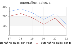
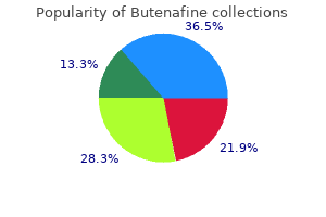
Rini (European Mistletoe). Butenafine.
- Dosing considerations for European Mistletoe.
- Pancreatic cancer.
- How does European Mistletoe work?
- Reducing the side effects of chemotherapy and radiation therapy, breast cancer, colorectal cancer, gastric cancer, bladder cancer, high blood pressure, internal bleeding, hemorrhoids, seizures, high cholesterol, gout, depression, sleep disorders, headache, menstrual disorders, and many other conditions.
- Are there safety concerns?
- Head and neck cancer.
- What is European Mistletoe?
- Are there any interactions with medications?
Source: http://www.rxlist.com/script/main/art.asp?articlekey=96882
Butenafine 15 gm overnight delivery
Other Points of Interest 1 the muscle is enclosed in a sheath formed mainly by the aponeuroses of the three flat muscles of the abdominal wall fungus youth proven butenafine 15 gm. One lies opposite the umbilicus, the second opposite the free end of the xiphoid process, and the third in between the two. The intersections are actually zigzag in course, traverse only the anterior half of the muscle, and are adherent to the anterior wall of rectus sheath. Embryologically, they may represent the segmental origin of muscle, but functionally they make the muscle more powerful by increasing the number of muscle fibres. It extends from the anterior superior iliac spine to the pubic tubercle, and lies beneath the fold of the groin. The upper surface of the ligament gives origin to the internal oblique from its lateral two-thirds, to the transversus abdominis from its lateral onethird, and to the cremaster muscle from its middle part. This is chiefly due to the tone of the oblique muscles, especially the internal oblique. The pectineal ligament or ligament of Cooper is an extension from the posterior part of the base of the lacunar ligament. The reflected part of the inguinal ligament consists of fibres that pass upwards and medially from the lateral crus of the superficial inguinal ring. The fasciculi form superficial loops from middle one-third of upper surface of inguinal ligament and deep loops from pubic tubercle, pubic crest and conjoint tendon. Here some fibres may be continuous with the internal oblique or transversus muscles. Along with the intervening connective tissue, the muscle loops to form a sac-like cremasteric fascia around this is a small triangular muscle. It is said to be tensor of the linea alba, but the need for such action is not clear. Section 2 Abdomen and Pelvis the conjoint tendon is formed by fusion of the lowest aponeurotic fibres of the internal oblique and of the transversus muscles, attached to the pubic crest and to the medial part of the pecten pubis. Sometimes it may be continuous with an inconstant ligamentous band, named the interfoveolar ligament, which connects the lower border of the transversus abdominis to the superior ramus of the pubis. The muscle also tends to close the superficial inguinal ring when the intra-abdominal pressure is raised. Cremasteric Reflex Upon stroking the skin of the upper part of the medial side of the thigh, there is reflex contraction of the cremaster muscle, as evidenced by elevation and retraction of the testis. Intercrural fibres of inguinal ligamnet pass towards the apex of the superficial inguinal ring. Infraumbilical median incisions are safer because the close approximation of recti prevents formation of any ventral hernia. Paramedian incisions through the rectus sheath are more sound than median incisions. The rectus muscle is retracted laterally to protect the nerves supplying it from any injury. In addition, these supply skin, muscles of the abdominal wall and parietal peritoneum. Tubercular infection of lung and pleura may cause radiating pain in the abdominal wall. Iliohypogastric and ilioinguinal nerves are anaesthetised by a needle above the anterior superior iliac spine on the spinoumbilical line. The superior epigastric artery is one of the two terminal branches of the internal thoracic artery. It begins in the sixth intercostal space, and enters the abdomen by passing behind the seventh costal cartilage between the costal and xiphoid origins of the diaphragm. It enters the rectus sheath and runs vertically downwards, supplies the rectus muscle, and ends by anastomosing with the inferior epigastric artery. In addition to muscular and cutaneous branches, it gives a hepatic branch which runs in the falciform ligament, and an anastomotic branch, at the level of the xiphoid process, which anastomoses with the artery of the opposite side. The musculophrenic artery is the other terminal branch of the internal thoracic artery. It runs downwards and laterally behind the seventh costal cartilage, and enters the abdomen by piercing the diaphragm between the seventh and eighth cartilages. It continues downwards and laterally along the deep surface of the diaphragm as far as the tenth intercostal space. Within the sheath, it supplies the rectus muscle and ends by anastomosing with the superior epigastric artery. A cremasteric branch to the spermatic cord in males or the artery of the round ligament in females. The pubic branch may replace the obturator artery, and is then known as the abnormal obturator artery. The deep circumflex iliac artery is the other branch of the external iliac artery, given off from its lateral side opposite the origin of the inferior epigastric artery. It runs laterally and upwards behind the inguinal ligament, pierces the fascia transversalis, and continues along the iliac crest, up to its middle where it pierces the transversus abdominis to enter the interval between the transversus and the internal oblique muscles. At the anterior superior iliac spine, it anastomoses with the superior gluteal, the lateral circumflex femoral and superficial circumflex iliac arteries. Just behind the anterior superior iliac spine, it gives off an ascending branch which runs upwards in the neurovascular plane. Posterior wall: It is deficient; the rectus muscle rests directly on the 5th, 6th and 7th costal cartilages. Between the costal margin and the arcuate line Anterior wall: External oblique aponeurosis and anterior lamina of the aponeurosis of the internal oblique. Midway between the umbilicus and the pubic symphysis, the posterior wall of the rectus sheath ends in the arcuate line or linea semicircularis or fold of Douglas. Below the arcuate line Anterior wall: Aponeuroses of all the three flat muscles of the abdomen. Abdomen and Pelvis Section Features Anterior Wall 1 It is complete, covering the muscle from end to end. Posterior Wall 1 It is incomplete, being deficient above the costal margin and below the arcuate line. Laterally, the anterior and posterior walls extend till linea semilunaris, which extends from tip of 9th costal cartilage to pubic tubercle. Contents Muscles Functions 1 It checks bowing of rectus muscle during its contraction and thus increases the efficiency of the muscle. Arteries Both leaves of external oblique aponeurosis and anterior leaf of internal oblique aponeurosis. Posterior Sheath 2 the inferior epigastric artery enters the sheath by passing in front of the arcuate line. Veins 1 the superior epigastric venae comitantes accompany its artery and join the vena comitantes of internal thoracic vein. Posterior leaf of aponeurosis of internal oblique and both leaves of aponeurosis of transversus abdominis. The three lateral abdominal muscles may be said to be digastric with a central tendon in the form of linea alba. Linea alba is a tendinous raphe between xiphoid process above to symphysis pubis and pubic crest below. Superficial fibres of linea alba are attached to symphysis pubis, while deep fibres are attached behind rectus abdominis to posterior surface of pubic crest. Section 2 Abdomen and Pelvis 1 the superior epigastric artery enters the sheath by passing between the costal and xiphoid origins of the diaphragm. It crosses the upper border of the transversus abdominis behind the seventh costal cartilage. Rectus sheath is formed by decussating fibres from three abdominal muscles of each side. Fibres from all three anterior leaves run obliquely upwards, while the posterior fibres run obliquely downwards at right angles to anterior leaves. Relation to Vessels and Nerves the inner surface of the abdominal muscles is lined by fascia which is separated from peritoneum by extraperitoneal connective tissue. At the lateral edge of the rectus abdominis, the aponeurosis of the internal oblique splits to pass partly posterior and partly anterior to the rectus abdominis; the anterior layer fusing with the aponeurosis of external oblique and the posterior layer with that of the transversus abdominis. Define the origins of the transversus and follow its aponeurosis to fuse with that of the internal oblique, posterior to the rectus abdominis above the arcuate line and anteriorly to the unsplit aponeurosis of internal oblique below the line. Inferiorly: It is attached to the inner lip of the iliac crest and to the lateral half of the inguinal ligament.
Generic butenafine 15gm amex
Anteriorly the midline incisive canal transmits the greater palatine artery and the nasopalatine nerve fungus quizlet purchase generic butenafine on line. It is largely formed, from anterior to posterior, by the septal cartilage, the perpendicular plate of the ethmoid and the vomer. Its surface area is greatly increased by three horizontal bony projections, the superior, middle and inferior nasal conchae (turbinates), and by diverticula, the paranasal air sinuses. The inferior concha is the largest and lies about 1 cm above the floor of the nose. Beneath each concha is a meatus, and above the superior concha the sphenoethmoidal recess. The sphenoid air sinus opens into the recess, the posterior ethmoidal air cells into the superior meatus and the nasolacrimal duct into the inferior meatus. The middle meatus possesses the bulla ethmoidalis, which contains the middle ethmoidal air cells. Such patients are identified by taking bacteriological swabs and treating those in whom the organisms are found. Wash your hands and put on non-sterile gloves and, because the patient may sneeze or cough, a face shield or eye protection. Obtain nasal, groin or perineal specimens as well as specimens from any devices penetrating the skin or mucosal membranes, such as catheters or tracheostomy tubes. Lymphatic drainage the anterior cavity drains to the submandibular nodes, the posterior part to the retropharyngeal nodes. They develop as diverticula from each nasal cavity and are lined by its mucoperiosteum. They are small at birth and enlarge during eruption of the second dentition, to reach adult size after puberty. The frontal sinus, situated above the medial end of the superciliary arch, is separated by a bony septum from its fellow. Its base forms part of the lateral wall of the nose, and its apex projects laterally into the zygomatic process of the maxilla. The roof separates the sinus from the orbit and conveys the infraorbital vessels and nerve in the infraorbital canal. The floor is the alveolar margin, containing molar teeth, the roots of which project into the cavity. The posterior wall contains the posterior superior alveolar nerve and lies in front of the infratemporal and pterygopalatine fossae. The anterior wall forms the facial surface of the maxilla and is covered in part by the mucous membrane of the vestibule of the mouth. The maxillary sinuses are the most commonly infected because their openings, well above the floor of the sinus, are not well positioned for natural drainage. Blood supply the ophthalmic, palatine and maxillary arteries are the main supply, but the anterior inferior part of the cavity receives additional branches from the facial artery. The lateral nasal wall is supplied by anterior ethmoidal branches superiorly and the superior alveolar nerve inferiorly. Surgeons have devised an approach to the pituitary, which lies immediately above the sphenoidal sinus, via the nose and sphenoidal sinus (trans-sphenoidal hypophysectomy). The most effective way to drain the maxillary sinus is to kneel in the Muslim praying position, which brings the opening of the sinus into a dependent position for drainage. Infection of a carcinoma of the maxillary sinus may, because of the close relation of the sinus floor to the upper teeth, produce pain referred to those teeth. Each ethmoidal labyrinth, lying between the orbit and the upper part of the nasal cavity, contains numerous air cells, the ethmoidal sinuses; these are divided into anterior, middle and posterior groups. An infection or abscess in these cells may readily invade the thin medial wall of the orbit and optic canal, causing optic neuritis and the risk of blindness. The sphenoidal sinuses are contained within the body of the sphenoid and usually communicate with each other through an incomplete bony septum. They lie below the sella turcica and pituitary gland, above the nasal cavity, and medial to the cavernous sinus and its contents. The alveolar arches, gums and teeth divide the cavity into an outer vestibule and an inner oral cavity. The vestibule, a slit-like cavity limited externally by the lips and cheeks, opens on to the face between the lips at the oral fissure; internally it is limited by the gums and teeth. It communicates with the mouth between the teeth or, when the teeth are occluded, by the retromolar space. The parotid duct opens into the vestibule just above and opposite to the upper second molar tooth. Paralysis of buccinator causes the most distressing symptom for sufferers of a facial nerve paralysis. They must keep a tissue in their hand to wipe the saliva that drools out from the corner of the mouth. The oral cavity is limited anteriorly and laterally by the maxilla and mandible and teeth; it possesses a roof, a floor and a posterior opening. The roof is formed by the palate, which separates the mouth from the nasal cavities. They are replaced by permanent teeth, which appear between the 6th and 24th years. Each half of each jaw contains five deciduous teeth (I2, C1, M2) and eight permanent teeth (I2, C1, P2, M3). The average time of eruption of the teeth in each half of the upper and lower jaws is shown in Table 19. The first deciduous tooth to appear is a lower central incisor, the first permanent tooth a first molar. On each side of the frenulum is a sublingual papilla, on to which opens the submandibular duct. They are composed of fibrous tissue covered by a vascular mucous membrane and are firmly attached to the alveolar margins. Nerve and blood supply the teeth of the upper jaw are innervated by numerous anterior and posterior superior alveolar branches of the maxillary nerve, the lower by the single inferior alveolar branch of the mandibular nerve. The labial surface of the upper gums is supplied by the infraorbital and posterior superior alveolar nerves, the lingual surface by the nasopalatine and greater palatine nerves. The labial surface of the lower gums is supplied by the mental and buccal nerves, the lingual surface by the lingual nerve. Imperfect oral hygiene leads to the development of cavities in the enamel and subsequent infection of the pulp cavity (dental caries). The cavity is inextensible and the pain (toothache) caused by the rise in pressure is severe. Inferior alveolar nerve block (usually known as an inferior dental block) is very commonly used in dentistry. All the teeth of that half of the mandible are anaesthetized, together with the lower lip, which is supplied by the mental branch of the nerve. Nerve supply Much of the inside of the cheeks and lips is supplied by buccal branches of the mandibular nerve with contributions from the mental branch of the inferior alveolar nerve and the infraorbital branch of the maxillary nerve. Its central cavity opens on to the apex of the root and is filled with pulp (loose connective tissue, vessels and nerves). The teeth are named, from the front and moving laterally: incisor (I), canine (C), premolar (P) and molar (M).
Purchase generic butenafine line
Initially antifungal honey purchase butenafine pills in toronto, the cephalic (mesencephalic) flexure forms in the midbrain region and the cervical flexure is at the junction of the brain and spinal cord. The ventral column forms the ventral horns of the spinal cord, containing motor neurons. Behind this is the lateral column and this, together with the ventral column, forms the basal plate. The cephalic flexure is in the region of the future midbrain and the cervical flexure at the junction of the brain and spinal cord. The alar plate consists of the dorsal column, which develops into the dorsal horn. The hindbrain the basal and alar plates of the brainstem are separated by a longitudinal groove, the sulcus limitans. In the brainstem, the columnar arrangement within the spinal cord is present alongside three additional columns: the special visceral afferent and efferent columns and the special somatic afferent column. This functional classification is simplistic but provides a mechanism for understanding the internal structure of the brainstem (see Table 24. The basal plate contains three columns: general somatic efferent, special visceral efferent and general visceral efferent. The general visceral efferent column provides parasympathetic innervation to parasympathetic ganglia of the head and controls pupillary constriction, lacrimation and the production of saliva. It is also the source of the vagus nerve, which provides the parasympathetic innervation of the thoracic and abdominal viscera. The alar plate contains four columns: general visceral afferent, special visceral afferent, general somatic afferent and special somatic afferent. The general visceral afferent column receives a wide range of information from the viscera and the carotid and aortic bodies. The general somatic afferent column receives the sensory modalities of pain, temperature, general sensation and proprioception of the face, nose, oral cavity, ears, pharynx and larynx. The special sensory column receives fibres responsible for the special senses of hearing and balance. The basal plates of the midbrain become the motor nuclei of the midbrain and form the cerebral peduncles that contain large descending motor tracts. The alar plates develop into specific structures that are discussed further throughout this chapter: the red nucleus, the substantia nigra and the superior and inferior colliculi. The cavity of the midbrain becomes increasingly narrowed during its development to form the cerebral aqueduct, joining the third ventricle to the fourth ventricle. As it proceeds through its development it forms the telencephalon and the diencephalon on each side. The cerebral hemispheres enlarge in a lateral, a caudal and then a ventral direction. As a result, the hemispheres, lateral ventricles and other structures, including the caudate nucleus and hippocampus, become elongated and C-shaped. The diencephalon can be considered to have dorsal and ventral portions and contains a central cavity that forms the third ventricle. The ventral posterior lateral nucleus of the thalamus: a receives sensory fibres from the face b is a relay station for auditory fibres c lies anterior to the pulvinar d receives sensory fibres from the upper limb e lies adjacent to the anterior limb of the internal capsule T/F ( ) ( ) ( ) ( ) ( ) Answers 1. The primary optic pathways pass through the: a anterior limb of the internal capsule b corpus callosum c genu of the internal capsule d posterior limb of the internal capsule e medial geniculate body Answer 1. The anterior limb of the internal capsule carries thalamocortical fibres to the frontal lobe and the posterior limb carries sensorimotor fibres. The corpus callosum is an interhemispheric commissure and the medical geniculate body is an auditory relay station. The superior temporal gyrus contains the auditory cortex and the cingulate gyrus is part of the limbic system. The superior temporal gyrus contains the auditory cortex, while the cingulate gyrus is part of the limbic system. The ventral posterolateral nucleus is part of the auditory and visual relay pathways and the ventral posteromedial nucleus is a relay station for trigeminal nerve fibres. The lateral nucleus relays fibres from the basal ganglion and cerebellum to the motor cortex. The primary motor cortex is situated in the: a precentral gyrus b superior frontal gyrus c postcentral gyrus d superior temporal gyrus e cingulate gyrus 3. The primary sensory cortex is situated in the: a precentral gyrus b superior frontal gyrus c postcentral gyrus d superior temporal gyrus e cingulate gyrus 4. Twelfth Match the following statement to the appropriate cranial nerve nuclei in the the above list. Vestibulocochlear Match the following statement to the appropriate cranial nerve nuclei in the above list. A 27-year-old woman attends her doctor with a 3-day history of a left facial weakness, with forehead sparing; she is otherwise well. A 60-year-old man presents with acute onset of weakness of his left arm and leg, and a left hemiparesis with a facial droop. A 36-year-old woman is thrown off her horse and presents with weakness of her legs and sensory loss below her umbilicus. The upper part of the face is not involved in a cortical (upper motor neuron) lesion as it is bilaterally innervated. The vertebral and associated spinal injury is at the level of the 10th thoracic vertebra. The epithelium has a rich blood supply and invaginates into the ventricular cavities. Lateral ventricle the lateral ventricles are C-shaped cavities located within the cerebral hemispheres. The frontal (anterior) horns extend anteriorly from the interventricular foramen and are separated by a thin translucent membrane known as the septum pellucidum. The body of the lateral ventricle extends from the interventricular foramen to the splenium of the corpus callosum. Interventricular foramen of Monro the lateral ventricles are connected to the third ventricle via the interventricular foramen of Monro. The choroid plexus from the lateral ventricle passes through the foramen and is continuous with the choroid plexus within the third ventricle. Third ventricle the third ventricle is a vertical slit-like cavity located in the midline of the diencephalon. The lateral walls are formed by the hypothalamus inferiorly and the thalamus superiorly, and are divided by a shallow groove (the hypothalamic sulcus) running from the interventricular foramen to the cerebral aqueduct. The ventricular surfaces of the thalami are interconnected by the massa intermedia in approximately 75% of people. The roof is formed by thin membranes and the body of the fornix, and is the location of the choroid plexus of the third ventricle. The floor is formed by multiple structures including the optic chiasm, infundibulum, tuber cinerum and mammillary bodies. Either side of the optic chiasm lie the supraoptic recess anteriorly and infundibular recess posteriorly. The lamina terminalis and anterior commissure form the anterior wall of the third ventricle. The posterior wall has multiple recesses (pineal and suprapineal recesses) and is formed from the pineal gland, habenular complex and commissure, the posterior commissure and the cerebral aqueduct posteroinferiorly. The roof of the cavity extends backwards into the cerebellum; it is formed superiorly by a glial sheet (the superior medullary velum) stretching between the superior cerebellar peduncles, and inferiorly by a thin ependymal sheet (the inferior medullary velum) between the inferior cerebellar peduncles. The inferior medullary velum is invaginated by blood vessels and pia mater and forms the choroid plexus of the ventricle.
Buy genuine butenafine line
It then courses through the cavernous sinus antifungal liver buy 15gm butenafine otc, pierces the dural roof of sinus and ends immediately lateral to optic chiasma and inferior to anterior perforated substance and divides into middle and anterior cerebral arteries. Deep branches of anterior cerebral artery supply part of internal capsule and the basal nuclei. It gives off deep or perforating branches which supply anterior limb of internal capsule and part of basal nuclei. The artery then passes out to the lateral surface of hemisphere at the insula of the lateral sulcus. The two anterior cerebral arteries are connected by anterior communicating artery. The internal carotids and posterior cerebral arteries of same side are united by the posterior communicating artery. Formation the branches of the circulus arteriosus are cortical, central and choroidal. Cortical or external branches run on the surface of the cerebrum, anastomose freely and if these get blocked, they give rise to small infarcts. The central branches perforate the white matter to supply the thalamus, the corpus striatum, and the internal capsule. These do not anastomose and if these get blocked, they give rise to large infarcts. Central Branches Anteriorly: Anterior communicating artery joining the two anterior cerebral arteries. Posterolaterally: Posterior communicating arteries Posteriorly: Posterior cerebral arteries the circulus arteriosus attempts to equalize the flow of blood to different parts of brain and provides a collateral circulation in the event of obstruction to one of its components. There is hardly any mixing of bloodstreams on right and left sides of the circulus arteriosus. These enter the medialmost part of anterior perforated substance to supply preoptic and supraoptic regions of anterior hypothalamus. These divide in two sets: Medial striate ascends through lentiform nucleus to supply this nucleus including caudate nucleus and internal capsule. The lateral striate ascends lateral to lentiform nucleus, turn medially, pass through the substance of this nucleus to enter internal capsule. These supply tuberoinfundibular and mammillary regions of hypothalamus, subthalamus, anterior and medial parts of thalamus, medial part of tegmentum and crus cerebri of midbrain. Inferior temporal gyrus excluding the part of the temporal pole is also supplied by posterior cerebral artery. Two arteries are connected by the anterior communicating artery Winds round cerebral peduncle to reach the tentorial surface of cerebrum 1. Cerebral Cortex Cerebral cortex is supplied by branches of all three cerebral arteries. Intimately applied to the capillaries, there are numerous processes of astrocytes and it has been estimated that these processes cover about 80% of the capillary surface. These include pineal body, hypophysis cerebri, choroid plexuses and area postrema in fourth ventricle of brain. The chief internal source of autoregulation is the adjustment of arterial muscle tone in response to intraluminal pressure changes. This is achieved by a direct myogenic response to distension produced by rising intraluminal pressure. The H+ ion concentration in the perivascular space is the chief external source of autoregulation of cerebral blood vessels. The subarachnoid and perivascular spaces are separated by a thin layer of pia mater. It is an upper motor neuron type of paralysis of one-half of the body, including the face. Through the superior and inferior anastomotic veins, it communicates with the superior sagittal and transverse sinuses. The orbital veins terminate in the superior cerebral veins or in the superior sagittal sinus. It is formed by the union of the thalamostriate and choroidal veins at the apex of the tela choroidea of the third ventricle. Its tributaries include the basal veins, and veins from the pineal body, the colliculi, the cerebellum and the adjoining part of the occipital lobes of the cerebrum. It is formed at the anterior perforated substance by the union of the deep middle cerebral vein, the anterior cerebral veins, and the striate veins. It runs posteriorly, winds round the cerebral peduncle, and terminates by joining the great cerebral vein. Its tributaries include (apart from the veins forming it) small veins from the cerebral peduncle, interpeduncular structures, the tectum of the midbrain, and the parahippocampal gyrus. Ultimately, all veins drain into the various cranial venous sinuses which, in turn, drain into the internal jugular vein. With occlusion of one internal carotid, the other internal carotid may perfuse both anterior cerebral arteries. With occlusion of basilar, each posterior cerebral artery may be perfused by the internal carotid of its own side. Further anastomosis occurs between cortical branches of cerebral arteries, prior to perforation of the branches into brain substance. Once the cortical and central branches perforate, they become end arteries hardly communicating at capillary level. It runs along the corpus callosum supplying maximum area on the medial surface of the cerebral hemisphere. These are superior cerebellar, anterior inferior cerebellar and posterior inferior cerebellar arteries. Ans: the symptoms in the present case are due to thrombosis of the largest branch of fourth part of vertebral artery, the posterior inferior cerebellar artery. The various nuclei and fibres involved are vestibular nuclei, inferior cerebellar peduncle, nucleus ambiguus and lateral spinothalamic tract of the opposite side. It supplies posterolateral part of medulla oblongata, lower part of pons, inferior surface of cerebellum including choroidal branches to 4th ventricle. Since the treatment was not done along the right lines, he suffered from haemorrhage of the left lateral striate arteries which supply the internal capsule. This is an upper motor neuron type of paralysis with exaggerated reflexes, increased tone of the muscles, etc. One night he felt severe headache and soon paralysis of both his right-sided limbs. Describe the intracranical course of vertebral, basilar and internal carotid arteries including the formation of circle of Willis. Besides detailed history and clinical examination, the following investigations may have to be done according to the need of each case. Collision of positrons with electrons of body tissues produces gamma rays, detected by gamma cameras, put around the patient. Angiography: the contrast medium is injected into the common carotid or vertebral arteries. Because of these modern and safe procedures, the older techniques-pneumoencephalography, ventriculography and myelography-have become obsolete. Nerve conduction studies done to estimate the rate of conduction through the nerve fibres. The superciliary border is marked by first joining points 2 and 3 by a line arching upwards just above the eyebrow, and then extending this line to point 4. The inferolateral border is marked by first joining points 4 and 5 by a line convex forwards (temporal pole), and by then joining points 5 and 1 by a line convex upwards, passing just above the external acoustic meatus. Lateral Sulcus Cerebellum It is marked behind the auricle, immediately below the marking for the transverse sinus lying between inion and base of mastoid process. The posterior ramus of the lateral sulcus is about 7 cm long and can be marked by joining points 4 and 8.
Cheap 15gm butenafine visa
The alterations in the face and head are byproducts of a change in posture from pronograde (four-footed) fungi fragmentation definition order butenafine once a day, through orthograde to a plantigrade (two-footed) one. An orthograde animal (ape or monkey) has smaller jaws and a larger head than in pronograde animals. Reduction in jaw size is attributable to the liberty of movements of the upper limbs, and also to changed habit of eating cooked food, both of which have greatly relieved the jaws of their diverse functions (tactile feeling, holding, sorting, breaking, biting, tearing, chewing, piercing, fighting, etc. With recession of the jaws, the oral aperture is reduced in size, and the lips are supported by a much better developed orbicularis oris. The distinctive external nose, with exuberant growth of cartilages forming the prominent dorsum, tip and alae is a characteristic human feature, although it appears to serve no special function. The palpebral fissures are larger in man than in any other primate, and the bony orbits are decidedly smaller than in the great apes. Further, the interorbital distance is greater in man than in apes in whom the nasal root is greatly constricted. The supraorbital margins of man are markedly reduced remnants of the highly developed brow ridges of other primates. The diminution in man is partly due to the receding jaws which relieve the ridges of their function as buttresses, and partly to the development of a prominent forehead because of increase in the size of the cranial cavity. The forehead protects the eyes from above, a similar function being performed by the brow ridges in apes. During any of these activities, the pupils dilate, skin gets pale, blood pressure rises, blood vessels of skeletal muscles, heart, lungs and brain dilate. There is hardly any activity in the digestive tract due to which the individual does not feel hungry. It consists of two ganglionated trunks, their branches, prevertebral ganglia, and plexuses. It supplies all the viscera of thorax, abdomen and pelvis, including the blood vessels of head and neck, brain, limbs, skin and the sweat glands as well as arrector pilorum muscle of skin. They leave spinal cord through their respective ventral roots, to reach sympathetic ganglia and beginning of ventral rami via white rami communicantes (wrc) [singular: ramus communicans]. Sympathetic trunks on either side of the body extend from cervical region to the coccygeal region where both trunks fuse to form a single ganglion impar. Thoracic Part There are usually 11 ganglia on the sympathetic trunk of thoracic part. Branches Abdominal/Lumbar Part It runs along the medial border of psoas major muscle. The postganglionic fibres pass along the spinal nerves to supply cutaneous blood vessels, sweat glands and arrector pili muscles. Aortic plexus: this plexus is formed by preganglionic sympathetic, postganglionic sympathetic, preganglionic parasympathetic and visceral afferent fibres around the abdominal aorta. This plexus is concentrated around the origin of ventral and lateral branches of abdominal aorta. These are known as coeliac plexus, superior mesenteric plexus, inferior mesenteric plexus and renal plexus. Pelvic/Sacral and Coccygeal Parts of Sympathetic Trunk Pelvic part runs in front of sacrum, medial to ventral sacral foramina. The ganglia receive the greater splanchnic nerves, lesser splanchnic nerves of both sides including some filaments of vagi and phrenic nerves. Secondary plexuses arising from coeliac and aorticorenal plexus are distributed along the branches of the aorta, namely phrenic, splenic, left gastric, hepatic, intermesenteric, suprarenal, renal, gonadal, superior and inferior mesenteric plexuses. Superior Hypogastric Plexus this plexus lies between the two common iliac arteries and is formed by: (i) aortic plexus, and (ii) branches from lumbar sympathetic ganglia. It divides into right and left inferior hypogastric plexuses (pelvic plexus); which runs on the medial side of internal iliac artery and is supplemented by pelvic splanchnic nerves (parasympathetic nerves). These fibres are relayed in the ciliary ganglion, and the postganglionic fibres pass via the short ciliary nerves to be distributed to ciliaris and sphincter pupillae muscles. These fibres leave the brain as nervus intermedius, and form part of the facial nerve and pass along its chorda tympani branch. Fibres of chorda tympani nerve relay in this ganglion from where postganglionic fibres supply the submandibular and sublingual salivary glands. Greater petrosal branch of facial nerve joins the deep petrosal (sympathetic fibres) to form the nerve of pterygoid canal. These postganglionic fibres supply lacrimal gland, glands of nose, pharynx and palate. The postganglionic fibres are given to the auriculotemporal nerve to reach the parotid salivary gland. They have their origin in the dorsal nucleus of vagus and pass along its pulmonary, cardiac, oesophageal, gastric and intestinal branches. Some of the parasympathetic fibres ascend from inferior hypogastric plexus to reach superior hypogastric plexus and finally the inferior mesenteric plexus. Thereafter, these fibres are distributed along the branches of inferior mesenteric artery to supply left onethird of transverse colon, descending colon and pelvic colon. These preganglionic fibers relay in the neurons situated in the wall of the viscera. Deep Cardiac Plexus It consists of two halves which are interconnected and lie anterior to bifurcation of trachea. Four ganglia, namely ciliary, pterygopalatine, submandibular and otic, are concerned with efferent parasympathetic fibres. These fibres pass along the respective ventral roots of thoracic nerves to synapse with the respective ganglia of the sympathetic trunk. After relay, the postganglionic fibres form thoracic branches which intermingle with the vagal fibers, to form cardiac plexus. These fibres then travel up the cervical part of the sympathetic chain and relay in superior, middle and inferior cervical ganglia. Thus, the pain may be referred to the area of skin supplied Branches from the plexus give extensive branches to pulmonary and right and left coronary plexuses. Small ganglia are found on these nerves for the relay of parasympathetic (brought via vagus neve) fibres. Parasympathetic system is bronchoconstrictor (motor) whereas sympathetic system is inhibitory. Sympathetic stimulation causes relaxation of smooth muscles of bronchial tubes (bronchodilator). The nerves form a plexus called myenteric plexus between two layers of the muscularis externa and another one in the submucous layer. Stomach and superior hypogastric plexus through the plexuses on the branches of inferior mesenteric artery.
Syndromes
- Problems keeping a steady gaze
- Accompanied by numbness or tingling at the joint or beyond it
- Women and girls need to wash the area between the lips of the vagina with soapy water and rinse well. Or if instructed, use a disposable towlette to wipe the genital area.
- Anti-inflammatory medicines to reduce the immune response
- To diagnose a urinary tract infection
- Tiredness
- Decreased ability to care for self
- Gums that bleed easily (blood on toothbrush even with gentle brushing of the teeth)
Buy butenafine with paypal
The mucous membrane is innervated by the greater and lesser palatine nerves and branches of the glossopharyngeal nerve antifungal kidney butenafine 15gm. Blood supply this is by palatine branches of the maxillary, facial and lingual arteries; venous drainage is to the pharyngeal venous plexus. In the centre of the sulcus is a pit, the foramen caecum, the origin of the thyroglossal duct. Extremely rarely thyroid glandular tissue can be found at the foramen caecum, as this is the embryological site of origin of the thyroid gland, which develops from the distal end of the thyroglossal duct. The mucous membrane of the oral anterior two-thirds of the dorsum of the tongue is covered with papillae. Taste buds are most numerous around the sides of the tongue and in front of the sulcus terminalis. The mucous membrane is raised posteriorly in a midline glossoepiglottic fold that separates two shallow fossae, the valleculae, limited laterally by the pharyngeal wall. Very occasionally the frenulum is short and restricts tongue movement (tongue tie). Within the posterior tongue are nodules of lymphatic tissue, the lingual tonsils, which give this part of the tongue a cobblestone appearance. It is the shape of an inverted shoe and is attached posteriorly mainly to the hyoid bone and the mandible, lying on the geniohyoid and mylohyoid muscles. The upper surface Muscles of the tongue Intrinsic and extrinsic muscles lie on each side of a midline fibrous septum. The intrinsic muscles, arranged in vertical, horizontal and transverse bundles, lie within the tongue and have no attachment to bone. If the genioglossus is paralysed or totally relaxed, for example as occurs in general anaesthesia or when the patient is in coma, there is a tendency for the tongue to fall back and obstruct the airway. Pulling the mandible forwards helps to protect the airway by pulling the tongue anteriorly, because of its attachment to the mental spine. On the upper medial surface of hyoglossus lie the tongue, lingual artery, middle constrictor muscle, stylohyoid ligament and glossopharyngeal nerve. Laterally and below lie styloglossus, the lingual nerve, the submandibular gland and duct, the sublingual gland and the hypoglossal nerve. Styloglossus passes between the superior and middle constrictors to the side of the tongue. Lymphatic drainage It should be noted that the lymph from any part of the tongue may drain to nodes on both sides of the neck. The tip of the tongue drains to the submental nodes or directly to the deep cervical nodes; the side of the tongue drains to the submandibular nodes and the dorsum to the submandibular and jugulodigastric nodes. With lingual cancer, cancers arising in the posterior third of the tongue spread to the superior group of deep cervical glands on both sides of the neck. However, cancers arising from the anterior tongue spread first to the submandibular and submental nodes and only later to the inferior deep cervical nodes. Venous drainage is by the lingual vein, which drains into the internal jugular vein. Nerve supply the lingual nerve supplies sensation to the anterior two-thirds of the tongue, the glossopharyngeal sensation to the posterior third. Taste fibres are conveyed by the facial nerve from the chorda tympani to the anterior two-thirds of the tongue and by the glossopharyngeal nerve to the posterior third. All muscles are supplied by the hypoglossal nerve, except palatoglossus, which is supplied by the pharyngeal plexus. The posterior belly is attached to the medial side of the mastoid process and the anterior belly to the digastric fossa on the mandible. The posterior belly is supplied by the facial nerve, the anterior by the inferior alveolar nerve. Their secretions clean and moisten the mouth, assisting in chewing, swallowing, phonation and digestion. It is irregular in shape and described as having anteromedial, posteromedial and superficial surfaces. The anterior border overlies masseter, and from it emerges the duct to pass forwards across masseter before turning medially around the anterior border of the muscle to pierce buccinator and open on to the mucous membrane of the cheek, opposite the second upper molar tooth. The posteromedial surface lies on the mastoid process, the sternocleidomastoid muscle and the posterior belly of digastric muscle. Medially the deep part of the gland is separated by the styloid process and its muscles from the carotid sheath and the pharynx. The upper part of the gland lies between the external auditory meatus and the temporomandibular joint. The gland is traversed by the external carotid artery deeply, the retromandibular vein and the facial nerve superficially. It is particularly painful because the parotid gland is encased in an inextensible envelope of the deep investing cervical fascia. The pain is worsened by chewing and opening the mouth, because of the attachments of this fascia to the jaw. Pain arising from a parotid tumour may be referred to the temporomandibular joint because of the common innervation of the gland and joint by the auriculotemporal nerve. Nerve supply Sympathetic fibres from the superior cervical ganglion pass with the external carotid artery; parasympathetic fibres are conveyed by the glossopharyngeal nerve via the otic ganglion and the auriculotemporal nerve. The parasympathetic nerves are secretomotor, producing saliva, and the sympathetic fibres vasoconstrictor, giving rise to a dry mouth. Lymphatic drainage the superficial part of the gland drains to the parotid nodes, the deep part to the retropharyngeal nodes. It has a fibrous capsule and is divided into superficial and deep parts by the posterior border of mylohyoid. The submandibular duct arises from the deep part and passes forward between mylohyoid and hyoglossus, medial to the sublingual gland, to open on the sublingual papilla in the floor of the mouth at the base of the frenulum. The lingual nerve crosses the submandibular duct laterally from above and then turns upwards medial to it. The facial artery grooves its posterior surface and passes lateral to the gland to reach the inferior border of the mandible. The deep part is wedged between mylohyoid and hyoglossus, separated from the latter by the lingual nerve and, below it, the hypoglossal nerve. Nerve supply Sympathetic fibres from the superior cervical ganglion are conveyed along arteries; parasympathetic fibres are carried in the facial nerve via the chorda tympani and hitch-hike on to the lingual nerve before synapsing in the submandibular ganglion. They are most common in the submandibular duct and cause painful swelling of the gland; this is exacerbated when salivary flow is increased, such as during eating. Submandibular duct stones may be removed surgically via the floor of the mouth, or, if this is impossible, the submandibular gland requires removal. If the incision for this operation is made about 3 cm below the angle of the mandible, such damage to the facial nerve should not occur. Each gland lies on mylohyoid, medial to the mandible and lateral to the submandibular duct, the lingual nerve and hyoglossus. Its blood and nerve supply and its lymphatic drainage are similar to those of the submandibular gland. These include the singular large midline frontonasal process and the left and right maxillary and mandibular processes, which derive from first pharyngeal arch tissues. Defects in the formation of the first pharyngeal arch therefore result in facial deformity that may involve the maxilla, zygoma and mandible. Defects can occur in the degree of closure of the stomodeum, resulting in either macrostomia (too little closure) or microstomia (too much closure). The left and right maxillary processes grow and fuse with the frontonasal process and form the upper cheek, palate and upper jaw.
Purchase 15 gm butenafine fast delivery
As the mantle layer forms the dorsal and ventral columns fungus gnats but no plants purchase butenafine toronto, the white matter becomes subdivided into dorsal, ventral and lateral white columns. Because of this recession of spinal cord, the intervertebral foramen no longer lie at the level at which corresponding nerves emerge from the spinal cord. One advantage of this recession of spinal cord is when cerebrospinal fluid is tapped for diagnostic purposes. During a lumbar puncture, the needle is inserted at the lower lumbar level, avoiding the lower end of the cord. The dilated part of the central canal of spinal cord within the conus medullaris is known as the terminal ventricle. Similarly, the cavity of septum pellucidum is sometimes called the fifth ventricle. Forebrain (prosencephalon) Subdivisions Cavity Lateral ventricle Third ventricle A. Telencephalon (cerebrum), made up of two cerebral hemispheres and the median part in front of the interventricular foramen B. Epithalamus, including the pineal body, habenular trigone and posterior commissure. Midbrain Crus cerebri, substantia nigra, tegmentum, and tectum, from before (mesencephalon) backwards 3. Clinical features: Flat face, small ears, slanting eyes, small mouth, short neck, short arms and legs, low muscles tone, loose joints, and below average intelligence. The brain damage is caused by brain injury or abnormal development of the brain that occurs- before birth, during birth or immediately after birth. Clinical features: Affected parts are body movements, muscles control, muscle coordination and muscle tone. Craniosynostosis (cranio, cranium; syn, together; ostosis relating to bone) is a condition in which one or more of the fibrous sutures in an infant (very young) skull prematurely fuses by turning into bone (ossification). Autism is a neurodevelopmental disorder characterized by impaired social interaction, impaired verbal and non-verbal communication, and restricted and repetitive behaviour. Autism affects information processing in the brain by altering how nerve cells and their synapses connect and organize. Symptoms: Impaired social interaction, impaired verbal and non-verbal communication, restricted and repetitive behaviour. Causes: Genetic and environmental factors Convulsion: A sudden, violent, irregular movement of the body, caused by involuntary contraction of muscles and associated especially with brain disorders such as epilepsy, the presence of certain toxins or other agents in the blood or fever in children. Epilepsy is a chronic disorder, the hallmark of which is recurrent, unprovoked seizures or convulsions. A person is diagnosed with epilepsy, if they have two unprovoked seizures (or one unprovoked seizure with the likelihood of more) that were not caused by some known and reversible medical condition like alcohol withdrawal or extremely low blood sugar. The narrowing of the spinal canal limits the amount of space for the spinal cord and nerves. Pressure on the spinal cord and nerves due to limited space can cause symptoms such as pain, numbness, and tingling. When this happens, the brain does not get enough oxygen or nutrients, and brain cells start to die. Ischemic strokes are caused by arteries being blocked or narrowed, and so treatment focuses on restoring an adequate flow of blood to the brain. Hemorrhagic strokes are caused by blood leaking into the brain, so treatment focuses on controlling the bleeding and reducing the pressure on the brain. Chiari malformation is a condition in which brain tissue extends into the spinal canal. It occurs when part of the skull is abnormally small or misshapen, pressing on the brain and forcing it downward. Chiari malformation is categorised into three types, depending on the anatomy of the brain tissue that is displayed into the spinal canal. There are three main types: Spina bifida occulta, meningocele, and myelomeningocele. The most common location is the lower back, but in rare cases it may be the middle back or neck. Signs of occulta may include a hairy patch, simple, dark spot, or swelling on the back at the site of the gap in the spine. The meningocele typically causes mild problems with a sac of fluid present at the gap in the spine. More effective regeneration will be again in the son as the injury is in a distal area. Oligodendrocytes and the control of myelination in vivo: New insights from the rat anterior medullary velum. Origin of microgila: Cell transformation from blood monocytes into macrophagic ameboid cells and microglia. A neuron with many dendrites arising from cell body and carrying impulses away from the neuron via the axon is: a. Ependymal Name the type of neuroglial cells that aid regeneration by forming a regeneration tube to help establish firm connection. Meninges of the Brain and Cerebrospinal Fluid the human brain contains more than 180 billion neurons. The neurons are interconnected through an amazing network of 100000 miles of nerve fibres. Subarachnoid 4 the cerebrospinal fluid fills the space between the arachnoid and the pia maters (subarachnoid space) and acts as a water cushion. The outermost meninx, the dura mater, not only separates the right and left cerebral hemisphere, but also partitions the cerebrum from cerebellum and hypophysis cerebri. Pull upwards the endosteum along with the fold of dura mater present between the adjacent medial surfaces of cerebral hemispheres, extending from the frontal lobe till the occipital lobe. Pull backwards a similar but much smaller fold between two adjacent lobes of cerebellum-the falx cerebelli. Separating the cerebrum and the cerebellum is another fold of dura mater called the tentorium cerebelli. Thus, the fused endosteum and dura mater get separated from the underlying subarachnoid mater, pia mater and the brain. Underneath the dura mater and separated by a flimsy subdural space is the cobweb-like arachnoid mater. It is separated from the underlying pia mater by the subarachnoid space, containing cerebrospinal fluid and blood vessels of the brain. The subarachnoid space is dilated around the brainstem and at the base of the brain forming the subarachnoid cisterns. Cerebrospinal fluid formed by choroid plexuses flows through the ventricles of the brain into the subarachnoid space to be absorbed via subarachnoid villi into the superior sagittal sinus. However, it may be recapitulated that it is 25 made up of two layers-an outer endosteal layer and an inner meningeal layer, enclosing the cranial venous sinuses between the two. With advancing age, the arachnoid villi enlarge in size to form pedunculated tufts, called arachnoid granulations. On the cerebellum pia mater dips and forms folds in relation to larger fissures of cerebellum. Prolongations 1 It provides sheaths for the cranial nerves merging with the epineurium around them. Vertebral epidural space is present between vertebral column and spinal dura matar. The subdural space is also a potential space between the dura and arachnoid maters. It surrounds the brain and spinal cord, and ends below at the lower border of the second sacral vertebra. Space between the nervous tissue and fold of pia mater with arterioles is known as Virchow-Robin perivascular space. Cisterns At the base of the brain and around the brainstem, the subarachnoid space forms intercommunicating pools, called cisterns (Latin reservoir). It is continuous with interpeduncular cistern cranially, with cerebellomedullary cistern behind and with spinal subarachnoid space caudally.
Order 15 gm butenafine free shipping
However fungus gnats lavender oil cheap butenafine 15gm with mastercard, in recent years, the inclusion of colours in formulations has become extremely complex because of the banning of many traditionally used colours in many countries. In addition to those properties previously discussed such as particle size and crystal form, other characteristics such as hygroscopicity, flowability and compactability are particularly important when solid dosage forms are being prepared where the drugs constitute a large percentage of the formulation. Hygroscopic drugs can require low moisture manufacturing environments and need to avoid water during preparation. Studies of the compactability of drug substances are frequently undertaken with use of instrumented tablet machines in formulation laboratories to examine the tableting potential of the material so as to foresee any potential problems during compaction, such as lamination or sticking, which may require modification of the formulation or processing conditions. Therapeutic considerations in dosage form design the nature of the clinical indication, disease or illness for which the drug is intended is an important factor when one is selecting the range of dosage forms to be prepared. Factors such as the need for systemic or local therapy, duration of action required, and whether the drug will be used in emergency situations need to be considered. In the vast majority of cases, a single drug substance is prepared in a number of dosage forms to satisfy both the particular preferences of the patient or physician and the specific needs of a certain clinical situation. For example, many asthmatic patients use inhalation aerosols, from which the drug is rapidly available to the constricted airways following deep inhalation for rapid emergency relief, and oral products for chronic therapy. Patients requiring urgent relief from angina pectoris, a coronary circulatory problem, place tablets of glyceryl trinitrate under their tongue (sublingual administration). This results in rapid drug absorption directly into the blood capillaries under the tongue. Thus, whilst systemic effects are generally obtained following oral and parenteral drug administration, other routes can be used as the drug and situation demand. Local effects are generally restricted to dosage forms applied directly, such as those applied to the skin, ear, eye, throat and lungs. Some drugs may be well absorbed by one route but not by another and must therefore be considered individually. Infants generally prefer liquid dosage forms, usually solutions and mixtures, given orally. Children can have difficulty in swallowing solid dosage forms, and for this reason many oral preparations are prepared as pleasantly flavoured syrups or mixtures. Adults generally prefer solid dosage forms, primarily because of their convenience. However, alternative liquid preparations are usually available for those unable to take tablets and capsules. Supercritical fluid processing using carbon dioxide as a solvent or antisolvent is one such method, allowing fine-tuning of crystal properties and particle design and fabrication. Undoubtedly, these new technologies and others, as well as sophisticated formulations, will be required to deal with the advent of gene therapy and the need to deliver such labile macromolecules to specific targets and cells in the body. Interest is also likely to be directed to individual patient requirements such as age, weight and physiological and metabolic factors, features which can influence drug absorption and bioavailability, and the increasing application of diagnostic agents will play a key role in this area. This topic incorporates (1) the use of in silico procedures to predict drug substance properties and (2) decision making and optimization tools, such as experimental design, artificial intelligence and neural computing. All these can facilitate faster and rational design of formulations and manufacturing processes. Summary this article has demonstrated that the formulation of drugs into dosage forms requires the interpretation and application of a wide range of information and knowledge from several study areas. Whilst the physical and chemical properties of drugs and additives need to be understood, the factors influencing drug absorption and the requirements of the disease to be treated also have to be taken into account when potential delivery routes are being identified. The formulation and associated preparation of dosage forms demand the highest standards, with careful examination, analysis and evaluation of wide-ranging information by pharmaceutical scientists to achieve the objective of creating high-quality, safe and efficacious dosage forms. Crystal engineering of active pharmaceutical ingredients to improve solubility and dissolution rate. Crystallisation processes in computing and formulation pharmaceutical technology and drug optimization. This article discusses the principles underlying the formation of solutions from a solute and a solvent and the factors that affect the rate and extent of the dissolution process. This process will be discussed particularly in the context of a solid dissolving in a liquid as this is the situation most likely to be encountered in the formation of a drug solution, either during manufacturing or during drug delivery. Because of the number of principles and properties that need to be considered, the contents of each of these chapters should only be regarded as introductions to the various topics. The student is encouraged, therefore, to refer to the bibliography at the end of each chapter to augment the present contents. The authors use a large number of pharmaceutical examples to aid the understanding of physicochemical principles. Definition of terms this article will begin by clarifying and defining some of the key terms relevant to solutions. Since the above definitions are general ones, they may be applied to all types of solution involving any of the three states of matter (gas, liquid, solid) dissolved in any of the three states of matter, i. However, when the two components forming a solution are either both gases or both liquids, then it is more usual to talk in terms of miscibility rather than solubility. One point to emphasize at this stage is that the rate of solution (dissolution rate) and amount which can be dissolved (solubility) are not the same and are not necessarily related. Solution, solubility and dissolution A solution may be defined as a mixture of two or more components that form a single phase which is homogeneous down to the molecular level. The component that determines the phase of the solution is termed the solvent; it usually (but not necessarily) constitutes the largest proportion of the system. The other components are termed solutes, and these are dispersed as molecules or ions throughout the solvent, i. The transfer of molecules or ions from a solid state into solution is known as dissolution. Fundamentally, this process is controlled by the relative affinity between the molecules of the solid substance and those of the solvent. The extent to which the dissolution proceeds under a given set of experimental conditions is referred to as the solubility of the solute in the solvent. The solubility of a substance is the amount of it that has passed into solution when equilibrium is established between the solute in solution and the excess (undissolved) substance. A solution with a concentration less than that at equilibrium is said to be subsaturated. Solutions with a concentration greater than that at equilibrium can be obtained in certain conditions; these are known as supersaturated solutions (see Chapter 8 for further information). Process of dissolution Dissolution mechanisms the majority of drugs are crystalline solids. Liquid, semisolid and amorphous solid drugs do exist but these are in the minority. For now, we will restrict our discussion to dissolution of crystalline solids in liquid solvents. In addition, to simplify the discussion, it will be assumed that the drug is molecular in nature. Similarly, to avoid undue complication in the explanations that follow, it can be assumed that most solid crystalline materials, whether drugs or excipients, will dissolve in a similar manner. The dissolution of a solid in a liquid may be regarded as being composed of two consecutive stages. First is an interfacial reaction that results in the liberation of solute molecules from the solid phase to the liquid phase. This involves a phase change so that molecules of the solid become molecules of the solute in the solvent in which the crystal is dissolving. After this, the solute molecules must migrate through the boundary layer surrounding the crystal to the bulk of solution. Boundary layers are static or slowmoving layers of liquid that surround all solid surfaces that are surrounded by liquid (discussed further later in this chapter and in Chapter 6). Mass transfer occurs more slowly (usually by diffusion; see Chapter 3) through these static or slow-moving layers. These layers inhibit the movement of solute molecules from the surface of the solid to the bulk of the solution. The solution adjacent to the solid will be saturated (because it is in direct contact with undissolved solid). The process of the removal of drug molecules from a solid, and their replacement by solvent molecules, is determined by the relative affinity of the various molecules involved.
Buy butenafine 15 gm free shipping
Parasympathetic fibres are derived from the ocu lomotor branch to the inferior oblique muscle anti fungal yeast order 15 gm butenafine with visa. Postganglionic fibres pass to sphincter pupillae and ciliary muscles via the short ciliary nerves; sympathetic fibres from the superior cer vical ganglion pass to the eyeball and sensory fibres from the eyeball are conveyed in the nasociliary nerve. The pterygopalatine ganglion is in the eponymous fossa suspended from the maxillary nerve by small branches. Para sympathetic fibres arrive from the facial nerve in the middle ear via its greater petrosal branch. Postganglionic fibres supply the lacrimal gland and glands of the nose, palate and naso pharynx; sympathetic fibres are derived from the superior cervical ganglion via the internal carotid plexus and supply the nose, palate and nasopharynx. Sensory fibres from the nose, palate and nasopharynx are conveyed via the ganglion to the maxillary nerve. The submandibular ganglion lies lateral to hyoglossus, suspended from the lingual nerve. Sympathetic fibres are derived from the superior cervical ganglion and are conveyed by the facial nerve to glands in the floor of the mouth; taste fibres may pass through the ganglion. Parasympathetic content is from the glossopharyngeal nerve via the tympanic plexus and the lesser petrosal nerve. Postganglionic fibres pass with the auriculo temporal nerve to the parotid gland; sympathetic fibres come from the superior cervical ganglion to the parotid gland. Parasympathetic fibres directly from the glossopharyngeal nerve supply the oropharyngeal mucosal glands and glands of the posterior third of the tongue; similar fibres directly from the vagus supply the glands of the larynx, laryngopharynx, oesophagus and trachea. The vertebral artery: a arises from the second part of the subclavian artery, from behind scalenus anterior b lies anterior to the inferior thyroid artery c passes through the foramen transversarium of C7 d lies anterior to the lateral mass of the atlas e meets its opposite artery to form the basilar artery, on the clivus T/F ( ) ( ) ( ) ( ) ( ) Answers 1. From there postganglionic fibres are carried by the auriculotemporal nerve to the parotid gland, where they are secretomotor. They accompany the several short ciliary nerves that carry the parasympathetic postganglionic fibres from the ciliary ganglion to the ciliary muscle and sphincter pupillae. Postganglionic fibres leave the ganglion via the zygomatic and palatine nerves to the lacrimal and palatine glands. In the carotid canal the nerve joins with the greater superficial petrosal nerve to form the nerve of the pterygoid canal. She had a distended abdomen, and peritoneal aspiration revealed a haemoperitoneum. She was also complaining of a persistent pain in her shoulders, which the physicians thought was likely to be referred pain. Further examination revealed miosis (constriction of the pupil) of his left eye and a partial ptosis of the left eyelid. The phrenic nerve supplies sensory fibres to the mediastinal and diaphragmatic pleura, the pericardium and, relevant to this case, the peritoneum underlying the diaphragm. Vertebral artery Match the following statements with the vessel(s) in the above list. Why is it dangerous to point the needle posteriorly when performing central venous catheterization into the internal jugular, subclavian or brachiocephalic veins The immediate posterior relationship of the great veins of the neck is the dome of the pleura and the apex of the lung. However, as the ribs slope downwards at a 45 degree angle, the pleura extends above the 1st rib anteriorly. Any catheterization that misses the veins and continues posteriorly may produce a pneumothorax. The inferior cervical and first thoracic sympathetic ganglia are often amalgamated to form the cervicothoracic or stellate ganglion just posterior to the origin of the vertebral artery on the neck of the 1st rib. A cervical or upper thoracic sympathectomy removes the sympathetic innervation to the whole of the head and neck on the same side. For this reason, when performing thoracic sympathectomies for vascular problems in the upper limb, it is very wise to avoid the stellate ganglion. The emerging cranial and spinal nerves pass through the meninges and form the peripheral nervous system. The autonomic nervous system overlaps both the central and peripheral nervous systems and is concerned with the innervation of smooth muscle, cardiac muscle and glands. The central nervous system consists of billions of neurons and their supporting cells, the neuroglia. The majority of neuronal cell bodies are found in the cortex of the cerebrum and cerebellum and form the grey matter. Deeply placed aggregations of neuronal cell bodies within the cerebrum and cerebellum are called nuclei, while those outside of the central nervous system are grouped into ganglia. Axons in the central nervous system are typically myelinated and, when massed together, form white matter tracts. Neuroglia are the supporting cells of the neurons and are important in maintaining an appropriate homeostatic internal milieu for optimal neuronal function. There are different populations of neuroglia including astrocytes, oligodendrocytes and microglia. The brain is situated within the cranium and comprises a forebrain (prosencephalon), a midbrain (mesencephalon) and a hindbrain (rhombencephalon). The cerebral hemispheres provide us with our higher cognitive (thinking) and executive functions (decision-making), alongside our ability to execute and control movement, interpret the various sensory modalities, learn and remember new tasks and apply an emotional and visceral response to these functions. The deeper placed basal ganglia (corpus striatum) aid higher motor structures in the initiation, planning and control of movements. The central part of the forebrain, consisting mainly of the thalamus and hypothalamus, provides an essential integrative function in providing sensory and motor information to the higher cortical centres. It also regulates important homeostatic and endocrine mechanisms to main- tain our internal milieu and control basic behavioural drives such as appetite, thirst, thermoregulation and reproduction. The brainstem forms a central axis connecting the forebrain with the spinal cord and cerebellum. It also houses the reticular activating system, which is vital for our consciousness, and contains important groups of nuclei that control the cranial nerves. The cerebellum is another motor structure that provides a mechanism of providing feedback to ongoing movements for their correction and for maintaining effective balance and posture. It sits inferiorly on the anterior and middle cranial fossae and the superior aspect of the tentorium cerebelli. The cerebral hemispheres are incompletely separated by the median longitudinal fissure, within which lies the falx cerebri, a fold of dura. The two cerebral hemispheres are joined by a broad band of white matter, the corpus callosum. Specific sulci and gyri can be clearly visualized on all three surfaces: the lateral surface, which lies against the calvarium; the medial surface, which is hidden away in the interhemispheric fissure; and the inferior surface, which sits on the base of the skull. The cortex is greatly convoluted, producing the ridges of the brain, the gyri, which are separated by sulci. These folds greatly increase the cortical surface area, with approximately two-thirds of the total surface area hidden from view in the sulci. The cerebral cortex can be classified macroscopically, based on its function and general topography, and microscopically, according to its degree of lamination on a histological level. On a topographical level, each hemisphere has a frontal, a parietal, a temporal and an occipital lobe. The frontal and occipital poles are the anterior and posterior extremities of the hemisphere. The anteroinferiorly placed temporal pole lies in the middle cranial fossa underneath the frontal lobe. The cingulate gyrus lies above the corpus callosum, bounded by the callosal sulcus below and the cingulate sulcus above. The central sulcus extends for a short distance on to the medial surface into the paracentral lobule.
