

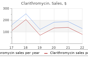
Generic clarithromycin 500 mg
However gastritis diet ���������� buy discount clarithromycin online, epirubicin was associated with less nausea, vomiting, neutropenia, and cardiotoxicity. Taxanes Until the development of taxanes in the 1990s, treatment options were much more limited. The conclusion was that taxanecontaining regimens were more effective than some, but not all nontaxane regimens. Taxanes have been studied in two main groups of patients; those who are anthracycline-naive and those who have been anthracycline pretreated. No significant differences in response rates or median survival were observed at equal doses of epirubicin and doxorubicin. Febrile neutropenia was more prevalent in the doxorubicin group, including cardiotoxicity, nausea, vomiting and stomatitis, whereas there was more diarrhea, neuropathy, fluid retention, skin and nail changes with docetaxel. At crossover to doxorubicin or paclitaxel during second-line therapy, response rates were 30% and 16%, respectively. The doxorubicin arm was more toxic than paclitaxel in terms of hematologic, gastrointestinal, and cardiac side effects, but counterbalanced by better symptom control. However, the dose of paclitaxel used in practice is usually 175 mg/m2 as higher doses have greater toxicities but have not demonstrated a better efficacy (70). Paclitaxel monotherapy is also active in those who have been exposed to anthracyclines. Paclitaxel 175 mg/m2 every 3 weeks was found to be inferior to 3-weekly cisplatin/oral etoposide in patients with advanced breast cancer pretreated with anthracyclines (73). Inference from the available data suggests that docetaxel may be superior to 3-weekly paclitaxel. However, docetaxel maintenance therapy is often limited by hematologic toxicities, peripheral neuropathy, fatigue, nail changes, and fluid retention. Notably, docetaxel has not been compared to the more commonly used weekly paclitaxel schedule, which has demonstrated a survival advantage over the 3-weekly regimen. Owing to a 30% incidence of grade 3 sensory neuropathy, the starting dose of weekly paclitaxel was amended from 100 mg/m2 to 80 mg/m2. Anthracycline Pretreated Patients Although grade 3 or more neutropenia was more frequent with the 3-weekly compared to weekly regimen (15% vs. Grade 3 neuropathy was a treatment-limiting toxicity more common with the weekly regimen (24% vs. On the contrary, nail changes and epiphora were significantly lower in the 3-weekly docetaxel schedule. Most hematologic and nonhematologic toxicities were related to increasing doses, including those of febrile neutropenia rates (4. Hence, lower doses of docetaxel must be considered for those who are more frail or who have tolerability issues. There is evidence of an incomplete cross-resistance between paclitaxel and docetaxel, since modest responses are still seen in those exposed to the alternate taxane (80,81). However, using a taxane after progression on the other may be best reserved for patients who relapse more than 12 months after adjuvant taxane-containing therapy or who had previous clinical response to taxanes with a reasonable time lapse of at least a year. The data suggest that treatment with an alternative taxane can result in objective responses. Studies support the notion that there is only partial cross-resistance between paclitaxel and docetaxel. However, it should be noted that there was wide variation in extent of prior anthracycline and/or taxane exposure in these studies, as well as the dose and schedule of taxanes used. Nab-paclitaxel is a Cremophor-free, albumin-bound formulation designed to distribute into tumor tissue more rapidly and at higher concentrations than conventional paclitaxel, thus possibly improving drug delivery and reducing toxicity. It allows higher doses of paclitaxel infusion, over a shorter duration of 30 minutes, with no need for antihistamine or corticosteroid premedications. Grade 4 neutropenia was significantly lower for nab-paclitaxel compared with standard paclitaxel (9% vs. No hypersensitivity reactions occurred with nab-paclitaxel despite the absence of premedication. Disease control rate (stable disease 16 weeks or confirmed overall complete or partial response) was significantly higher for patients receiving either dose of weekly nab-paclitaxel compared with docetaxel, but survival data were not mature at the point of this publication. Grade 3 or 4 fatigue, neutropenia, and febrile neutropenia were less frequent in the nab-paclitaxel arms, whereas the frequency and grade of peripheral neuropathy were similar in all arms. The higher cost of nab-paclitaxel may compare favorably to the cost of docetaxel (90). However, the lack of meaningful clinical efficacy when compared to conventional paclitaxel suggest that the extra cost associated with the use of nab-paclitaxel can only be justified in patients who cannot tolerate use of steroids needed in most patients treated with paclitaxel. Re-using anthracyclines in the metastatic setting after adjuvant exposure is not usually preferred due to the presence of dose-limiting cardiotoxicity and the availability of multiple other drug options. A taxane may also be used as firstline treatment for those who are taxane-naive or who have an adjuvant taxane-free interval of more than 12 months. In the latter scenario, an alternative taxane (docetaxel or paclitaxel) to that used in the adjuvant setting may be preferred. Both are reasonable options in the first-line setting with neither being definitively superior to the other. Resistance to anthracyclines and taxanes has been defined as disease recurrence occurring within 6 to 12 months of an adjuvant, neoadjuvant or first-line metastatic regimen or while on active treatment. Cardiac toxicity was equivalent in patients receiving single-agent doxorubicin and combination therapy perhaps due to the dose and administration schedule of the combination arm. Febrile neutropenia was significantly less common in the sequential compared to the combination arm (29. However, hematologic toxicity and asthenia were significantly increased in the docetaxel arm and neuropathy in the paclitaxel arm. The grade 4 neutropenia rate was significantly higher in the doxorubicin/paclitaxel arm (89% vs. Although febrile neutropenia rates were significantly higher in the doxorubicin/ docetaxel arm (33% vs. There was also a higher febrile neutropenia rate in the doxorubicin/docetaxel arm (33% vs. The neutropenia and febrile neutropenia rates were significantly higher in the anthracycline/taxane arms. This analysis has been hampered by incomplete and non-definitive abstract data, heterogeneity in median follow-up which could have affected survival analysis, and a lack of individualized patient data. On the basis of these results, anthracycline/taxane combinations should not routinely replace anthracycline-based regimens in clinical practice. The disappointing results of the anthracycline/taxane combinations are not completely unexpected as there is no preclinical evidence of synergy between them and both have overlapping and limiting hematological toxicities. These regimens should be reserved for only those patients with good performance status and life-threatening disease. Other Taxane Combinations After progression on anthracyclines, taxane combinations may sometimes be used if a higher response rate is required. Certain combinations have shown a survival benefit compared to single-agent taxanes such as docetaxel/capecitabine and paclitaxel/gemcitabine (32,33). However, there was no planned crossover in the studies, a third arm with singleagent capecitabine or gemcitabine were missing, and these combinations have been associated with increased toxicities. The capecitabine/docetaxel combination capitalizes on the synergistic antitumor activity of these two drugs observed in xenograft models (102). Docetaxel also causes upregulation of thymidine phosphorylase and Bcl-2 downregulation (105). Both drugs also capitalize on their nonoverlapping toxicities as docetaxel is myelosuppressive, but capecitabine has a low incidence of myelosuppression. The frequency of grade 3/4 neutropenia and neutropenic fever was 24% versus 28% in the combination versus docetaxel arms. Post-study docetaxel was administered in 20% and 7% of the combination and single-agent docetaxel arm respectively; and the use of post-study capecitabine was more common in the monotherapy compared to the combination arm (27% vs. As no crossover was planned and only a small proportion of patients on docetaxel subsequently received capecitabine, no definitive conclusions can be made regarding the relative merits of combination over sequential single-agent therapy (106). Lower doses of capecitabine and docetaxel may retain the efficacy while reducing the concomitant toxic effects as has been suggested by a retrospective analysis of this trial (107), which is important to consider when dealing with otherwise incurable disease and a primary goal of palliation.
Clarithromycin 500 mg without a prescription
Sexual dysfunction tends to worsen over the first several years of treatment gastritis ginger ale purchase discount clarithromycin, but improves with longer follow-up. While breast-conserving surgery has shown benefits over mastectomy with regard to body image, most studies find no difference between lumpectomy and mastectomy in regard to sexual functioning. A prospective evaluation of women during the first year after mastectomy or lumpectomy and studies of survivors evaluated 4 to 8 years after treatment failed to show a difference in sexual function or quality of life between groups (77). In these studies, however, the relationship of self-image to sexual function is not entirely clear. Younger women appear to be at increased risk of sexual dysfunction after receiving chemotherapy and the chemotherapy-induced sexual dysfunction appears to diminish over time such that, at more than 10 years out from treatment, sexual functioning is similar, regardless of whether they had received prior chemotherapy. Hormonal therapy with tamoxifen does not appear to cause sexual dysfunction, despite tamoxifen being associated with vaginal discharge. The women who had been treated with tamoxifen (n = 305), after controlling for adjuvant chemotherapy, did not report more sexual function troubles. Interestingly, in this same study, tamoxifen as a single agent did not produce sexual dysfunction. Although all these factors are important causes of sexual dysfunction, the most consistent predictor of sexual satisfaction in women with breast cancer is the quality of their relationships (81). Treatment of sexual dysfunction requires comprehensive assessment and intervention. If symptoms of sexual dysfunction were identified, recommendations for vaginal lubricants were provided along with individualized counseling and referral as indicated (82). Other data also suggest that vaginal dryness may play a significant, if not central, role in sexual dysfunction after chemotherapy. A prospective study in breast cancer survivors reported that women using Replens had decreased vaginal dryness equal to that of women using a water-soluble lubricating placebo; however, a decrease in dyspareunia was significantly better with Replens than with the placebo lubricant (83). Topical estrogen preparations appear to alleviate vaginal dryness more effectively than do nonestrogenic vaginal preparations. At least three formulations exist: Estring is an estrogen-impregnated ring that is inserted into the vagina, where it releases small amounts of estrogen over a 12-week period. Testosterone improves sexual desire and the frequency of sexual activity in women after surgical menopause or with sexual arousal disorders. Eligible women were randomly assigned to receive 2% testosterone in Vanicream, for a testosterone dose of 10 mg daily, or placebo Vanicream for 4 weeks and were then crossed over to the opposite treatment for an additional 4 weeks. Women who were on active testosterone cream had higher serum levels of bioavailable testosterone than women on placebo. However, the mean intrapatient libido change from baseline to weeks 4 and 8 was similar on both arms (84). The reason that this study was negative, while other similar studies have been positive, may be because all of the other positive studies involved women who were premenopausal or were receiving ongoing estrogen therapy, while the current study did not include women receiving concurrent estrogen therapy. Thus, it appears that testosterone, when used without concurrent estrogen, does not improve libido. In addition, a Southwest Oncology Group placebo-controlled trial has been developed to look at an omega-3-fatty acid preparation for alleviating this problem. Although there is some variability in the literature, in one study, the mean change in weight of 100 women treated with chemotherapy was +3. The majority of these patients (85%) received steroids as antiemetics, but no effect of steroid dose was seen on the level of weight change (91). Weight gain appears to be greater among premenopausal women; those who are node-positive; those receiving higher dose, longer duration, multiagent regimens; and those who enter into menopause. Psychosocial research suggests that weight gain has a profoundly negative impact on the quality of life for patients with breast cancer. Weight gain also leads to an increased risk of comorbid conditions such as cardiovascular disease, gallbladder disease, diabetes, and orthopedic complications and is associated with an increased mortality. However, other large studies did not show a correlation between posttreatment weigh gain and mortality (93). Limited research conducted in this area does not support overeating as a major cause of weight gain among breast cancer patients. Mean body weight increases significantly during chemotherapy, primarily due to an increase in mean total body and mean fat mass and a decrease in fat free mass and leg lean body mass. Both chemotherapy and radiation were associated with development of sarcopenic obesity (gain of fat mass without gain of lean tissue mass). Weight gain in the presence of lean tissue loss or the absence of lean tissue gain supports the need for interventions focused on exercise, especially resistance training in the lower body, to prevent undesirable weight gain (95). Premenopausal women starting adjuvant chemotherapy for breast cancer were randomized to a control group, or to receive monthly counseling by a dietitian aimed at weight maintenance. The median changes in average caloric consumption were reductions of 120 versus 46 cal/ day on weekdays and 196 versus 20 cal/day on weekends for the counseling and control groups, respectively. Routine prospective dietitian counseling aimed at weight maintenance thus appeared to produce small but statistically insignificant reductions in both caloric consumption and weight gain in this group of patients (96). Caloric restriction, either alone or in conjunction with other interventions such as exercise and psychological support or counseling through Weight Watchers, has led to successful weight loss in this population. A recent study of home-based diet and exercise intervention in older overweight or obese, breast, prostate, and colorectal cancer survivors showed decreased weight and increased functional capacity (97). Similarly, a home-based exercise intervention during the first 4 cycles of chemotherapy in 78 women with breast cancer demonstrated that women who adhered to the exercise program maintained their body weight, while nonexercisers steadily gained weight (p <. Finally, in a small randomized crossover study of a commercialbased exercise program (Curves), 6 months of the intervention resulted in moderate weight loss, but weight loss was not maintained postintervention (99). However, it is not clear whether promoting weight loss impacts the disease outcomes (100). Until more scientific evidence becomes available, women who are diagnosed with breast cancer should be advised to follow the general exercise recommendations of 30 minutes or more on most of the days of the week and follow a healthy diet, high in low-calorie density foods such as vegetable and fruits and low in fats and refined sugars and to maintain a healthy weight (100,101). Advances in this area may improve the quality of life of the almost 3 million breast cancer survivors in the United States alone. Impact of tamoxifen adjuvant therapy on symptoms, functioning, and quality of life. Safety and efficacy of tibolone in breast-cancer patients with vasomotor symptoms: a double-blind, randomised, non-inferiority trial. Estrogen and progestogen use in postmenopausal women: 2010 position statement of the North American Menopause Society. Intramuscular depot medroxyprogesterone versus oral megestrol for the control of postmenopausal hot flashes in breast cancer patients: a randomized study. Multicenter, randomized, cross-over clinical trial of venlafaxine versus gabapentin for the management of hot flashes in breast cancer survivors. Desvenlafaxine for the treatment of vasomotor symptoms associated with menopause: a doubleblind, randomized, placebo-controlled trial of efficacy and safety. Paroxetine is an effective therapy for hot flashes: results from a prospective randomized clinical trial. Paroxetine is an effective treatment for hot flashes: results from a prospective randomized clinical trial. Efficacy of escitalopram for hot flashes in healthy menopausal women: a randomized controlled trial. Citalopram and fluoxetine in the treatment of postmenopausal symptoms: a prospective, randomized, 9-month, placebo-controlled, double-blind study. Nonhormonal therapies for menopausal hot flashes: systematic review and meta-analysis. Newer Antidepressants and Gabapentin for Hot Flashes: An Individual Subject Pooled Analysis. Gabapentin for hot flashes in 420 women with breast cancer: a randomised double-blind placebo-controlled trial. Gabapentin, estrogen, and placebo for treating hot flushes: a randomized controlled trial. Gabapentin for the treatment of menopausal hot flashes: a randomized controlled trial.
Purchase generic clarithromycin on-line
Normal mammary epithelial development 10% Chromosomal instability and cancer-specific mutation signatures 20% 2 gastritis symptoms nz cheap clarithromycin 500 mg with amex. The cancer evolves through acquisitions of driver mutations (black stars), which produce clonal expansions. These driver mutations occur only infrequently in long-lived lineages of cells, which passively accumulate many mutations without expansion. One hypothesis is that chromosomes can be "pulverized" or undergo premature chromosome compaction (66), a phenomon observed during cell-fusion experiments, in which incompletely replicated chromosomes from the S phase nucleus shatter when induced to undergo chromosomal condensation by signals from the host cell in mitosis (67,68). But how this process involves only one or two chromosomes or a single chromosome arm remains to be explained. The end results of chromotripsis are the survival advantage that could be offered when tumor suppressors are lost and the generation of new fusion genes in the disrupted chromosome, as well as amplified oncogenes occurring on the derivative chromosomes. Of these, five mutations were prevalent in the primary cancer, six were present at lower frequency in the primary cancer (between 1% and 13%) and were more prevalent in the metastasis, and nineteen mutations could not be detected at all in the primary. The thickness of the branches reflects the proportion of tumor cells comprising that lineage. The length of the branches reflects the number of mutations specific to that lineage. The point estimates of timing for specific copy number gains are shown as arrows colored by the type of chromosomal aberration, with 95% confidence intervals generated by bootstrapping shown as horizontal lines. Molecular time is shown as an arrow, with the timing estimated as a fraction of point mutation time. These were interpreted to give estimated overall copy number (purple lines) and copy number of the minor allele (blue lines) across the genome (lower panel). The empiric histogram of mutations is shown in pale blue, with the fitted distribution as a dark green line. Also shown are the 95% posterior confidence intervals for the fitted distribution (pale green area). Chromosomes range around the outside of the circle, copy number changes are shown by the blue line in the inner ring, and somatically acquired genomic rearrangements are shown as arcs linking the two relevant genomic points. There were no new genomic rearrangements, suggesting that the process generating this complex regional remodeling had resolved before the patient was first diagnosed. Massive genomic rearrangement acquired in a single catastrophic event during cancer development. The samples were obtained from a 44-year-old African American woman with triple-negative breast cancer resistant to initial chemotherapy. The primary breast cancer contained 48 somatic, protein-coding mutations, which had a wide range of variant allele frequencies. The metastasis contained all 48 of these mutations, but about half of these mutations showed higher variant allele frequency in the metastasis, indicating enrichment or clonal selection in the metastasis. This enrichment was also seen in the xenograft that was derived from the primary cancer, and, because the sample to establish the xenograft was obtained prior to any cancer treatment, this argues that this enrichment or clonal selection is an intrinsic property of the cancer and not due to the effects of treatment. Studies of other cancer types also provide guidance about the type of genomic progression that can occur in breast cancer. Sequencing of a renal cell cancer that had metastasized to the lung and chest wall showed substantial intratumor genomic heterogenity (71). The researchers sampled 9 different areas within the primary tumor and 3 metastases (1 from the perinephric fat metastasis; 2 from the chest wall metastasis) and found that only 31% to 37% of the mutations were common to all samples. Based on these cases, a schema of clonal evolution in both the primary tumor and metastasis can be proposed. Because of genomic instability in the cancer cells, heterogenity and different subclones develop within the primary tumor. Metastases can develop either early or late in the cancer and are an opportunity for one or several subclones to grow at a distant site. The metastasis can derive from a dominant clone or a minor clone of the primary cancer, which will influence how similar the metastasis and primary cancer are in mutation pattern or even in response to treatment. The ability to sequence individual cancer cells (73) is providing further information about this clonal evolution process and will likely lead to future advances in this area. Additionally, a perrinephric metastasis (M1) and a chest wall metastasis (subdivided into two halves, M2a and M2b) were sequenced. Branch lengths are proportional to the number of nonsilent mutations separating the branching points. Potential driver mutations were acquired by the indicated genes in the branch (arrows). The cancer stem cell hypothesis proposes that there is a subpopulation of cells within the tumor that are capable of self-renewal and multi-lineage differentiation (74,75). In the cancer stem cell model, only mutations in these cells are propagated, and the clonal evolution in them gives rise to the genomic heterogenity in the cancer. They observed that all tumors contained a dominant subclone that accounted for more than 50% of cancer cells in the sample. They postulated that this expansion of a dominant clone is the final step in the development of a tumor that is responsible for triggering diagnosis, due to the emergence of a palpable mass. As there is minimal evidence of clonal expansion before the accumulation of all mutations in the dominant subclone, they suggest that the dominant clone becomes a cancer-initiating population, which is conceptually similar to a cancer stem cell (79). They observed that groups of mutations within individual cases have different clonal frequencies, indicative of distinct clonal genotypes. These triple-negative breast cancers had a wide range of clonal frequencies in the mutations sequenced, with some cases showing only one or two clonal populations (indicating a smaller number of clonal genotypes), whereas other tumours exhibited more extensive clonal evolution. The findings that many breast cancers have a dominant clone could be the result of this clone having a competitive advantage and taking over the tumor (the clonal evolution model) or could result from one or a few clones in the cancer stem cells, which then propagate and fill the tumor with their progeny. The cancer stem cell model has also been proposed to explain the existence of the intrinsic molecular subtypes of breast cancer defined by gene expression. This suggests that the molecular subtypes are mechanistically different and perhaps derived from progenitor cells (or stem cells) at different stages of differentiation (81,91,92). Cancer treatment imposes a selective pressure on a tumor that can create an evolutionary bottleneck. Drug resistant clones, which already existed as a minor population within the cancer, can be selected for and expand after cancer treatment (72,94). Genomic studies investigating the effects of treatment on breast cancer cells are still in progress, but evidence for this phenonemon comes from other cancer types. Two models of clonal evolution are diagrammed here, with either early or late dissemination of cancer cells. However, for many patients who are not cured, genomic heterogenity and Darwinian evolution at the cellular level is the root cause of their incurability. Columnar cell lesions of the breast: the missing link in breast cancer progression Genetic abnormalities in mammary ductal intraepithelial neoplasia-flat type ("clinging ductal carcinoma in situ"): a simulator of normal mammary epithelium. Comparative genomic hybridization and high-molecular-weight cytokeratin expression patterns. Identical allelic loss on chromosome 11q13 in microdissected in situ and invasive human breast cancer. Molecular differences between ductal carcinoma in situ and adjacent invasive breast carcinoma: a multiplex ligation-dependent probe amplification study. Accelerating mutagenesis will result in more clonal heterogenity in the cancer and has the potential to give rise to cancers that more difficult to treat. In 1976, Nowell proposed that more research should be directed toward understanding and controlling the evolutionary process in tumors (2). Similarly, at the other end of the scale, we still do not have a comprehensive catalog of the genomic landscape of advanced breast cancer. A comprehensive effort should be made to longitudinally sample the disease so that we can understand the genome dynamics of disease progression. It does appear that the process cannot be described as linear with a series of checkpoints, as Vogelstein imagined; this process is much more chaotic and complex than that, with any one tumor containing a spectrum of dominant and subdominant clones that constantly evolve in response to environmental and therapeutic stresses. Conservation of breast cancer molecular subtypes and transcriptional patterns of tumor progression across distinct ethnic populations. Comparative genomic hybridization of ductal carcinoma in situ of the breast-evidence of multiple genetic pathways. Relationship between hormone receptor status and tumour size, grade and comedo necrosis in ductal carcinoma in situ. Relationship of a new histological categorization of ductal carcinoma in situ of the breast with size and the immunohistochemical expression of p53, c-erb B2, bcl-2, and ki-67. Differentially expressed genes regulating the progression of ductal carcinoma in situ to invasive breast cancer.
Buy clarithromycin 250mg fast delivery
Cancer stem cells in solid tumors: accumulating evidence and unresolved questions uremic gastritis symptoms order clarithromycin 250 mg on line. The clonal and mutational evolution spectrum of primary triple-negative breast cancers. Transcriptome analysis of the normal human mammary cell commitment and differentiation process. How Darwinian models inform therapeutic failure initiated by clonal heterogeneity in cancer medicine. The most widely used classification of invasive breast cancers, and that used in this chapter (with minor modifications), is that of the World Health Organization (1). This classification scheme is based on the growth pattern and cytologic features of the invasive tumor cells and does not imply histogenesis or site of origin within the mammary duct system. For example, although the classification system recognizes invasive carcinomas designated "ductal" and "lobular," this is not meant to indicate that the former originates in extralobular ducts and the latter in lobules. In fact, subgross whole organ sectioning has demonstrated that most invasive breast cancers arise in the terminal duct lobular unit, regardless of histologic type (2). The most common histologic type of invasive breast cancer by far is invasive (infiltrating) ductal carcinoma. In fact, the diagnosis of invasive ductal carcinoma is a diagnosis by default, since this tumor type is defined as a type of cancer not classified into any of the other categories of invasive mammary carcinoma (1). In this chapter, the terms invasive ductal carcinoma, infiltrating ductal carcinoma, and infiltrating or invasive carcinoma of no special type are used interchangeably. The distribution of histologic types of invasive breast cancer has varied among published series (Table 25-1). These differences may be related to a number of factors including the nature of the patient population and variability in the confines of definition for the different histological types. In general, special type cancers comprise approximately 20% to 30% of invasive carcinomas, and at least 90% of a tumor should demonstrate the defining histological characteristics of a special type cancer to be designated as that histological type (6). The widespread use of screening mammography has had a dramatic impact on the nature of invasive breast cancers encountered in clinical practice. The value of mammography in detecting more cases of ductal carcinoma in situ, smaller invasive breast cancers, and fewer cancers with axillary lymph node involvement is well recognized. However, mammography has also resulted in a change in the distribution of the histological features of the invasive breast cancers detected. In particular, special type cancers (particularly tubular carcinomas) and cancers of lower histological grade are more frequently observed in mammographically screened populations than in patients who present with a palpable mass, particularly in the prevalent round of screening. Most invasive breast cancers have an associated component of in situ carcinoma, although the extent of the in situ component varies considerably. The prevailing view has long been that the invasive carcinomas derive from the in situ component. This is based not only on the frequent coexistence of the two lesions, but on the histological similarities between the invasive and in situ components within the same lesion. In addition, studies evaluating profiles of biological markers and genetic abnormalities have shown that coexisting invasive and in situ carcinomas often share the same immunophenotype and genetic alterations. The routine pathologic examination of invasive breast cancers has extended beyond simply determining and reporting the histologic type of the tumor. Although histologic typing provides important prognostic information in and of itself, other morphologic features that are evaluable on routine histologic sections are also of prognostic value. In this chapter, the various histologic types of invasive breast cancer will be discussed as will pathologic features important in the assessment of prognosis (prognostic factors) and response to therapy (predictive factors). Characteristic molecular and immunophenotypic features will also be noted where appropriate. An associated lymphocytic or lymphoplasmacytic infiltrate may or may not be present. Finally, the microscopic margins of the cancer may be infiltrating, pushing, circumscribed, or mixed. Recognizing that invasive ductal carcinomas are a histologically diverse group of lesions, many investigators have attempted to stratify them based upon certain microscopic features. The most common method to subclassify invasive ductal carcinomas is grading, which may be based solely on nuclear features (nuclear grading) or on a combination of architectural and nuclear characteristics (histologic grading). The histologic grading system currently in most widespread use is that of Elston and Ellis (reviewed in detail in reference 9). This system is a modification of the grading system proposed by Bloom and Richardson in 1957, but provides strictly defined criteria that are lacking in the original description. Tubule formation, nuclear pleomorphism, and mitotic activity are each scored on a 1 to 3 scale. The sum of the scores for these three parameters provides the overall histologic grade, such that tumors in which the sum of the scores is 3 to 5 are designated grade 1 (well differentiated), those with score sums of 6 and 7 are designated grade 2 (moderately differentiated), and those with score sums of 8 and 9 are designated grade 3 (poorly differentiated). The prognostic significance of histologic grading is discussed below (see section on prognostic factors). Although these tumors are most commonly encountered in pure form, a substantial minority exhibit admixed foci of other histologic types. The classification of tumors composed primarily of invasive ductal carcinoma with a minor component consisting of one or more other histological types is problematic. Some authorities categorize such lesions as invasive ductal carcinomas (or invasive carcinomas of no special type) and simply note the presence of the other types, whereas others classify them as "mixed. There are no clinical or mammographic characteristics that distinguish invasive ductal carcinomas from other histologic types of invasive cancer. Gross Pathology the classic macroscopic appearance of invasive ductal carcinoma is that of a scirrhous carcinoma, characterized by a firm, sometimes rock-hard, mass that on cut section has a gray-white gritty surface. This consistency and appearance is due to the desmoplastic tumor stroma and not the neoplastic cells themselves. Some invasive ductal carcinomas are composed primarily of tumor cells with little desmoplastic stromal reaction, and such lesions are grossly tan and soft. Although most invasive ductal cancers have a stellate or spiculated contour with irregular peripheral margins, some lesions have rounded, pushing margins, and still others are grossly well circumscribed. Invasive ductal carcinomas also display a wide variety of genetic and genomic alterations. In gene expression profiling studies, invasive ductal carcinomas may be found within all major molecular subtypes (24). However, even within this group prognostically favorable specialized tumor types can be identified, as discussed below. In particular, since the "classical" form of invasive lobular carcinoma was first described by Foote and Stewart (11), a variety of authors have described invasive breast cancers that they consider variants of invasive lobular carcinoma, thereby expanding the spectrum of this histologic type and accounting for a higher incidence of invasive lobular carcinoma in more recent series than in the past. In addition, recent studies have suggested that the increase in the frequency of infiltrating lobular carcinoma may be in part related to the use of postmenopausal hormone replacement therapy (14). Invasive lobular carcinomas are characterized by multifocality in the ipsilateral breast and appear to be more often bilateral than other types of invasive breast cancer. Lobular carcinoma in situ coexists with invasive lobular carcinoma in the majority of cases, with 70% to 80% of cases of invasive lobular carcinoma associated with foci of lobular carcinoma in situ. Histopathology Invasive lobular carcinomas as a group show distinctive cytologic features and patterns of tumor cell infiltration of the stroma. The classical form is characterized by small, relatively uniform neoplastic cells that invade the stroma singly and in a single-file pattern which results in the formation of linear strands. Furthermore, the tumor cells may infiltrate the breast stroma and adipose tissue in an insidious fashion, invoking little or no desmoplastic stromal reaction. This feature accounts for the difficulty in detecting some invasive lobular carcinomas on physical examination, mammography and gross pathologic examination. The nuclei of the neoplastic cells are usually small, show little variation in size, and are often eccentric. The cells may contain intracytoplasmic lumina which, in some, may be large enough to impart a signet ring cell appearance. However, in the classical form of invasive lobular carcinoma, cells with a signet ring configuration comprise only a small proportion of the tumor cell population. Many examples of invasive lobular carcinoma (as well as lobular carcinoma in situ) are characterized histologically by tumor cells that are loosely cohesive. This phenotype may be, at least in part, related to the fact that both in situ and invasive lobular carcinomas typically show loss of expression of the adhesion molecule E-cadherin. This is associated, in many cases, with mutations in the gene encoding this protein (17) or to loss of heterozygosity on chromosome 16q22. Although loss of E-cadherin expression characterizes lobular carcinomas and distinguishes them from ductal-type carcinomas, a subset of lobular carcinomas are reported to be E-cadherin positive (19).
Buy clarithromycin 250 mg cheap
Sequelae of axillary lymph node dissection in older women with stage 1 and 2 breast cancer gastritis symptoms for dogs cheap 250mg clarithromycin fast delivery. Long-term follow-up of elderly patients with operable breast cancer treated with surgery without axillary dissection plus adjuvant tamoxifen. Lumpectomy plus tamoxifen with or without irradiation in women age 70 or older with early breast cancer. Breast cancer in elderly women: a retrospective analysis of combined treatment with tamoxifen and onceweekly irradiation. Accelerated partial-breast irradiation using proton beams: initial clinical experience. Local control, toxicity, and cosmesis in women >70 years enrolled in the American Society of Breast Surgeons accelerated partial breast irradiation registry trial. Ten-year results of the treatment of earlystage breast carcinoma in elderly women using breast-conserving surgery and definitive breast irradiation. Radiotherapy after breast-preserving surgery in women with localized cancer of the breast. Management of breast cancer in the elderly by complete local excision and tamoxifen alone. Assessing the impact of a cooperative group trial on breast cancer care in the medicare population. Surgery versus primary endocrine therapy for operable primary breast cancer in elderly women (70 years plus). Long-term follow-up of elderly patients with locoregional breast cancer treated with tamoxifen only. American Society of Clinical Oncology technology assessment on the use of aromatase inhibitors as adjuvant therapy for postmenopausal women with hormone receptor-positive breast cancer: status report 2004. Adjuvant tamoxifen prescription in women 65 years and older with primary breast cancer. Predictors of tamoxifen discontinuation among older women with estrogen receptor-positive breast cancer. Older women with node positive breast cancer get similar benefits from adjuvant chemotherapy as younger patients: the Cancer and Leukemia Group B experience. Comparisons between different polychemotherapy regimens for early breast cancer: meta-analyses of longterm outcome among 100,000 women in 123 randomised trials. Pharmacokinetics and tolerance of vinorelbine in elderly patients with metastatic breast cancer. Adjuvant docetaxel/cyclophosphamide in breast cancer patients over the age of 70: Results of an observational study. Tumor characteristics and recurrence patterns in triple negative breast cancer: a comparison between younger (<65) and elderly (>/=65) patients. Administration of angiotensin-converting enzyme inhibitors and beta-blockers during adjuvant trastuzumab chemotherapy for nonmetastatic breast cancer: marker of risk or cardioprotection in the real world Although fewer than 7% of women diagnosed with breast cancer are younger than age 40, more than 13,000 young women are diagnosed annually with invasive or noninvasive breast cancer in the United States alone, with thousands more diagnosed worldwide (3). Incidence rates in young women appear to be fairly stable over the past several decades in young women in the Western world, despite increases in mammography and reproductive and lifestyle trends (4). A suggestion is that rates are increasing among young women, particularly in less-developed countries, but this may be owing to improvements in awareness, diagnosis, and reporting (5,6). Despite the relative rarity of breast cancer in young women, it is the leading cause of cancer-related deaths in women under age 40, and survival rates for young women with breast cancer are lower than for their older counterparts. The 5-year relative survival rate for women with breast cancer diagnosed before age 40 is 84% compared with 90% for women diagnosed at age 40 or older (3). The preponderance of evidence to date suggests that young age is an independent risk factor for disease recurrence and death, despite young women having conventionally received more intensive treatment than older women (7,8). Delays in diagnosis and the lack of effective screening in younger women may contribute to the poorer prognosis because they are more likely to present with larger tumors and more involved lymph nodes (9,10). However, survival differences also likely reflect biological differences in the type of breast cancer identified in young women. Young women are more likely to develop more aggressive subtypes of breast cancer with unfavorable prognostic features, and are less responsive to conventional therapy compared with disease arising in older premenopausal or postmenopausal women (11,12). Furthermore, studies suggest that the prognostic effect of age may vary by tumor phenotype, although additional research is clearly warranted (15,16). However, at the present time, it is uncertain whether young age will remain an independent prognostic factor as we continue to elucidate further the distinct molecular biologic features of breast cancers arising in both young and older women. Also, evidence suggests that biologic subtypes of breast cancer vary by race as a function of age (14,20). This higher prevalence of basal-like breast tumors and lower prevalence of luminal A tumors likely contributes to the poorer prognoses of young black women with breast cancer (20) (see Chapter 31, Prognostic and Predictive Factors: Molecular). In addition to being at higher risk of dying from breast cancer, despite conventionally receiving more aggressive therapy, young women face a variety of problems unique to , or accentuated by, their young age. They are more likely to be diagnosed at a life stage when role functioning in the home and work can be threatened or disrupted by the diagnosis and treatment of breast cancer. Young women are more likely to have young children for whom they are responsible, or desire to have biologic children following treatment. They also have an increased risk of harboring a genetic risk factor for breast cancer, and often suffer from a relative lack of information regarding treatment and survivorship issues compared with older patients. These concerns may contribute to the greater psychosocial distress seen in younger women at both diagnosis and in follow-up (21). Research to date on breast cancer in young women is limited by generally small sample sizes and heterogeneous cutoffs used to differentiate between young and old. Many investigators have chosen up to age 35 or 40 to define breast cancer in younger women, recognizing that previous work focusing on premenopausal women is composed primarily of women in their 40s, owing to the higher incidence of the disease in older premenopausal women. Consequently, younger women are at much lower risk even when compared with older premenopausal women. An average woman has a 1 in approximately 1,800 risk of developing breast cancer in her 20s, 1 in 230 in her 30s, and 1 in 70 in her 40s (3). Family history is the primary risk factor for developing breast cancer at a young age, particularly when breast cancer has occurred in a first-degree relative at a young age. Other factors, including a personal or family history of ovarian cancer, bilateral breast cancer, or Ashkenazi Jewish ancestry, may increase that risk. Some rare genetic disorders may predispose younger women to develop breast cancer. Young women exposed to ionizing radiation during childhood and the teenage years, such as survivors of pediatric Hodgkin disease treated with mantle field irradiation, are also at high risk of developing breast cancer (25). Despite preconceptions, most cases of breast cancers occurring in young women appear to be spontaneous and not clearly related to either carcinogens in the environment or family cancer syndromes (26). However, environmental and hormonal risk factors for breast cancer are not well characterized for younger women, but appear to be somewhat different than for older women. The excess transient early risk of breast cancer is most pronounced among women who are older at the time of their first delivery. Thus, pregnancy has a protective effect for postmenopausal breast cancer and is a risk factor for premenopausal breast cancer, particularly for older premenopausal women. Mammography is often of limited value in this population because of high breast tissue density, and targeted ultrasound or magnetic resonance imaging can provide additional discriminatory information in the workup of a breast abnormality (33,34). Breast cancers may be more extensive in younger patients, although it is not clear whether they are at higher risk of multicentricity or bilateral disease, in the absence of a hereditary predisposition, and no evidence indicates that multifocality affects survival in this population (35,36). Virtually no published clinical trials have focused on treatment issues for the youngest women. Trials reporting results of treatments for premenopausal women largely reflect outcomes for patients in their 40s. Thus, findings from studies that consider average results for premenopausal women may not be directly applicable to very young patients.
Order clarithromycin 250 mg otc
Although disease-free and overall survival were identical in the two arms gastritis long term discount 250 mg clarithromycin overnight delivery, this study closed early due to low accrual rate and underpowered analysis. In aggregate, neither the individual studies reported to date nor the completed meta-analyses provide us with the knowledge required to select with certainty premenopausal women who should receive ovarian suppression in addition to optimal chemotherapy, when appropriate, or to tamoxifen. It seems unlikely that women who receive modern chemotherapy and tamoxifen will obtain more than a modest benefit, if any, from the addition of ovarian ablation/suppression. One exception may be a group of women younger than age 40 whose menstrual cycles resume following adjuvant chemotherapy. Women older than age 40 are more likely to undergo chemotherapy-induced ovarian failure and may not obtain additional benefits from ovarian ablation/suppression. As reviewed in Table 45-1, the superiority of each generation of treatment regimens is independent of hormone receptor status. Other studies have not confirmed differences in outcomes by hormone report status. The benefit of paclitaxel was observed, regardless of hormone receptor status or tamoxifen administration. Newer studies comparing anthracycline- and taxanebased regimens tested different combination or sequences. The two regimens provided similar disease-free and overall survival benefits, regardless of hormone receptor status (84). The women were randomly assigned to either paclitaxel or docetaxel administered either every 3 weeks for four cycles or weekly for 12 cycles. Disease-free survival and overall survival were improved with weekly paclitaxel compared to paclitaxel every 3 weeks. Docetaxel administered every 3 weeks was also associated with improved disease-free survival compared to every 3-week paclitaxel, but not overall survival. Finally, newer studies compared outcomes of women treated with taxane-based therapy with or without an anthracycline. Although not completely consistent, the data presented to date support the use of the same chemotherapy regimens for women at high risk of recurrence based on tumor characteristics or nodal status, regardless of hormone receptor status. Thus decisions about type of chemotherapy should continue to be made based on estimates of risk of recurrence and death without consideration of hormone receptor status. Importantly, current and future studies will be likely enriched for women who are expected to benefit from chemo endocrine therapy compared to those included in older studies because of a more careful determination of tumor characteristics in addition to anatomical features. The role of hormone receptor status was also investigated in pivotal trials comparing outcomes following the administration of chemotherapy with or without trastuzumab. Women randomized to trastuzumab had a significant improvement in disease-free and overall survival. Disease-free survival and overall survival were improved in the trastuzumab-containing arms compared to the non-trastuzumab-containing arm. The improvement seen with trastuzumab was independent of the hormone-receptor status (92). Women receiving both treatments will likely receive chemotherapy followed by the endocrine agent and will be subjected to the possible side effects of each. Whether the sequential administration of the treatments will lead to a different or greater side effect profile has not been evaluated in detail. It is noteworthy that the concomitant administration of endocrine therapies and chemotherapy, in particular tamoxifen, can be associated with increased risk of thromboembolic events compared to each modality given alone-another argument against concurrent chemo endocrine therapy. Premenopausal women who receive chemotherapy who suffer treatment-related amenorrhea or ovarian failure may be more likely to report high prevalence and severity of menopausal symptoms compared to postmenopausal women or to their counterparts who have not received chemotherapy. Whether chemo endocrine therapy will have a detrimental effect on cognitive function compared to either approach alone is not known. One concern is that women treated with chemotherapy agents that are associated with specific toxicities will have a prolonged or worsening symptomatology once the endocrine treatment is initiated. Ovarian ablation/ suppression may be considered in select young women with high-risk tumors or in premenopausal women who cannot take tamoxifen. The value of a first-, second-, or thirdgeneration chemotherapy regimen should be discussed with an individual woman based on her tumor characteristics, estimates of recurrence, value added, and potential side effect profile with each type of regimen. Several studies investigated the question of sequential versus concurrent administration of chemo endocrine therapy, mainly using tamoxifen. While concurrent use of chemotherapy and tamoxifen appears to be slightly better than sequential administration, the comparisons are indirect and do not represent results from prospective data trials. This direct comparison generated sufficient concern about concurrent chemo endocrine therapy that most modern studies have required that tamoxifen, and by extrapolation other endocrine therapies, would be administered after completion of adjuvant chemotherapy. While it is possible that smaller studies in the neoadjuvant setting will address sequence questions prospectively, sequential administration appears to be the most cautious strategy in the absence of other randomized data. In summary, the selection of dose and duration of endocrine agents and chemotherapy regimens should be made independent of the decision to prescribe chemo endocrine therapy. Until other prospective data are available, chemotherapy should generally precede endocrine treatment. A greater understanding of tumor biology coupled with sophisticated assays allows us to make increasingly better informed recommendations to individual women. Likewise, a better understanding of host factors such as pharmacogenetics, and environmental factors such as diet or activity level, will lead to even better understanding of endocrine resistance and new methods to overcome it. Questions include not only whether specific chemotherapy regimens, dose, duration, or sequence are important, but also whether there is a group of women with tumors that are endocrine-resistant despite the expression of hormone receptors and who may not benefit from endocrine therapy either. Activation of tyrosine kinase receptors, for example, may predict for a more aggressive tumor phenotype and relative resistance to endocrine therapy. In these situations, although the estrogen-dependent growth is inhibited via endocrine manipulations, other growth signals stimulate proliferation and lead to relative endocrine resistance. Activation of other growth signals is usually associated with higher-grade tumors and those women have traditionally been offered cytotoxic-based therapy in addition to the endocrine manipulation. Emerging results in the advanced, acquired endocrine-resistance setting provide strong support for prospective studies of novel targeted agents in the adjuvant setting in tumors with de novo endocrine resistance. Agents that reverse epigenetic modification may also overcome endocrine resistance (100). Finally, studies evaluating the role of genetic variants in predicting differential response to endocrine therapies may also lead to identification of a subgroup of women that may benefit from modification of endocrine therapy (102). However, enhanced understanding of tumor biology and mechanisms of endocrine resistance has already led the way into new trial designs in which targeted treatments are investigated. Studies in the adjuvant or neoadjuvant settings will not only investigate early predictors of response and resistance, but will also attempt to integrate new targeted therapies or agents that enhance sensitivity or reverse resistance. Indeed, new generations of clinical trials will incorporate tumor-intrinsic subtype and host factors more commonly than ever before. Data today suggest that polychemotherapy with anthracycline and/or taxane-based regimen is superior to other therapies.
Purchase clarithromycin 500mg with visa
Nitrogen-containing bisphosphonates antral gastritis diet chart cheap clarithromycin master card, including pamidronate, ibandronate, and zoledronic acid, accumulate in the bone microenvironment and are released during bone resorption. They are then internalized by osteoclasts where they affect their activity and survival. Preclinical studies have shown that zoledronic acid inhibits angiogenesis in several in vitro and in vivo models (16). In addition, nitrogen-containing bisphosphonates prevent adhesion of cancer cells to bone, which may interfere with tumor seeding and invasion (13). Nitrogen-containing bisphosphonates may also induce cell death by activating cytotoxic T-cells in the host, though the clinical role of this effect awaits further clarification (19). Similar to zoledronic acid, the activity of denosumab is mediated by several mechanisms, including a potentially similar antiangiogenic effect (22). As a result of these trials, clodronate is approved outside of the United States for women with metastatic breast cancer and bone involvement. Despite this approval, the firstgeneration bisphosphonates are not optimal for long-term use. Etidronate adequately inhibits bone resorption, but also inhibits bone mineralization. Clodronate is poorly absorbed in the gastrointestinal tract and can cause local irritant toxicity, reducing the likelihood of long-term adherence. In light of the initial encouraging results of clodronate, multiple clinical trials were initiated with pamidronate for women with breast cancer metastatic to bone. Initial small studies sought to optimize the dose and schedule of intravenous pamidronate and had various endpoints including amelioration of bone pain and sclerosing of lytic bone metastases. Subsequently, two large placebo-controlled, randomized clinical trials (Aredia Protocols 18 and 19) were initiated in the early 1990s (25). Women enrolled in Protocol 18 received a stable endocrine regimen for their metastatic cancer, and those on Protocol 19 received cytotoxic chemotherapy. Patients were randomly assigned to receive either pamidronate 90 mg intravenously over 2 hours or an intravenous placebo, given every 3 to 4 weeks for 24 cycles. The primary efficacy endpoint was the skeletal morbidity rate, expressed as the number of events per year. Although the two trials were independently performed, their common time frame and eligibility criteria allowed the investigators to combine their efficacy data. In this trial, 367 women were randomized to pamidronate and 384 women were assigned to placebo. The addition of pamidronate to systemic therapy for advanced breast cancer resulted in a significant improvement in multiple parameters. Overall, skeletal events were seen in 51% and 64% of these same groups, respectively (p <. Despite the compelling skeletal benefits seen with pamidronate, there was no survival difference between the two arms. The potential benefits of increasing potency are to shorten the intravenous infusion times and allow for oral formulations with greater efficacy. Three of these potent heterocyclic, nitrogen-containing bisphosphonates began to enter clinical study: risedronate, ibandronate, and zoledronic acid (or zoledronate). Zoledronate is reported to be the most potent of these third-generation bisphosphonates with respect to bone resorption without affecting bone mineralization. Ibandronate, in both the intravenous and oral form, has been compared to placebo in the setting of bone metastases. In a second trial of similar patients, ibandronate either 2 or 6 mg intravenously was compared to placebo (27). The 2-mg dose was ineffective, but the 6 mg dose significantly reduced the skeletal morbidity rate (p <. However, ibandronate is not currently approved in the United States for the ancillary treatment of skeletal metastases. A large international, multicenter, randomized, doubleblind clinical trial was performed comparing pamidronate with zoledronate in patients with bone metastases from not only breast cancer but also multiple myeloma (4). Patients participated as one of three strata: patients with multiple myeloma; patients with breast cancer receiving chemotherapy; and patients with breast cancer receiving hormonal therapy. Patients were randomized to receive pamidronate 90 mg over 2 hours in 250 mL of 0. Because of the observation of creatinine elevation in the zoledronic acid arms, two protocol adjustments were made during the conduct of the trial: the infusion duration was increased from 5 to 15 minutes with the volume of hydration increased to 100 mL; and the 8 mg zoledronic acid dose was reduced to 4 mg. Although the original trial was designed as a 13-month efficacy and safety trial, a 25-month extension phase was subsequently performed and reported. This landmark trial included 1,130 women with metastatic breast cancer and 412 of these women entered the extension study. Adverse events that were reported as drug-related were infections, arthralgias and myalgias, cytopenias, fever, eye disorders, electrolyte abnormalities, and injection site reactions and were similar among the initial three groups. With regard to renal toxicity, clear differences among the groups were seen prior to protocol amendments that increased the zoledronate infusion time from 5 to 15 minutes and increased the infusion volume. As a result of this approval, patients with breast cancer and bone metastases were routinely placed on either pamidronate or zoledronate delivered every 3 or 4 weeks for a minimum of 1 year accompanied by systemic therapy for the cancer. Renal adverse events were stated to be more frequent in the zoledronic acid group, although the specific incidence and severity were not presented. Although ibandronate is not approved in the United States for treatment of bone metastases, it is widely used in the United Kingdom and Europe for this indication. This is the first randomized trial comparing oral ibandronate with any other bisphosphonate therapy and will serve to educate both physicians and their patients regarding the optimal option for systemic therapy for bone metastases from breast cancer. In addition, intravenous bisphosphonates carry the risk of inducing renal insufficiency as well as acute toxicities such as bone pain and fever. Patients were randomly assigned to receive denosumab subcutaneously every 4 weeks at a dose of 30, 120, or 180 mg; or denosumab every 12 weeks at a dose of 60 or 180 mg; or an open-label intravenous bisphosphonate every 4 weeks. Patients received 24 weeks of treatment and had a subsequent 32 weeks of follow-up. Based on data from this trial and pharmacokinetic and pharmacodynamic modeling, denosumab at a dose of 120 mg every 4 weeks was felt to be optimal. Throughout the study, the dose of zoledronate was modified based on the creatinine. Between April 2006 and December 2007, 2,049 patients with metastatic breast cancer and bone metastases were randomized. Acute-phase reactions and renal toxicity were more common in the zoledronic acid group and hypocalcemia was more common in the denosumab group. Although the trials had similar endpoints, no comparison should be made between the trials because of differing entry criteria and clinical factors for the women participating in the trial. The role of biochemical markers of bone turnover the following are the guideline recommendations based on these questions: 1. If using a bisphosphonate, recommendations were made regarding dose adjustments for renal function and monitoring of the creatinine. If using denosumab, no adjustments are needed for renal function, but for patients with poor renal function, close monitoring for hypocalcemia is recommended. The authors pointed out study design limitations, such as an open-label design and different clinic visit frequencies between the arms, among others. Regardless of the schedule used after the first year, there are even less data studying duration of therapy. Longer durations of bisphosphonate therapy may provide additional benefit, but this has not yet been tested in clinical trials. The Panel stresses that clinical judgment must guide what constitutes a substantial decline. Preventative recommendations that have been published include a thorough dental examination and completion of dental procedures including extractions prior to the initiation of intravenous bisphosphonates or denosumab. These three trials required on-study oral examinations at entry on the trial and every 6 months thereafter. Reported associated oral events included jaw pain in 80%, an associated tooth extraction in 61%, and a local infection in 48% of patients. Hypocalcemia Although both pamidronate and zoledronate are approved for the treatment of hypercalcemia of malignancy, their use in patients who are normocalcemic may carry the potential risk of inducing hypocalcemia. In light of this risk, pamidronate and zoledronate therapeutic clinical trials required that patients be given both oral calcium and vitamin D supplements. Although it is likely that adherence to these supplements was not optimal, the incidence of hypocalcemia was infrequently reported.
