

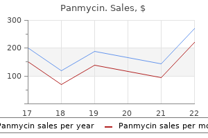
Buy cheap panmycin 500mg on line
According to the telephone theory antibiotics to treat cellulitis panmycin 500 mg free shipping, it remains for more central neural structures to "decode" these temporal patterns to deduce the features of the acoustic stimulus. In combination with these studies, other efforts have attempted to characterize the efferent neurotransmitter released on the hair cells and on afferent endings terminating on hair cells by efferent neurons originating in the brainstem. Evidence indicates that the afferent neurotransmitter is probably a single excitatory amino acid, or a structurally related compound, which is responsible for initiating auditory nerve action potentials. Besides this chemical transmitter substance, other chemicals, called neuromodulators,97 that influence the action of the transmitter are also believed to be released into the synaptic cleft. To date, concrete evidence for the auditory transmitter in the mammalian cochlea is scanty when compared with the findings of other studies of the central nervous system. That is to say, all of the criteria have yet to be met for any candidate afferent transmitter substance. However, based on our present ability to satisfy the above criteria, one of the most likely afferent transmitter substances is believed to be the excitatory amino acid glutamate. Each pure-tone cycle elicits a traveling wave that moves along the cochlear partition from base to apex. Microelectrode recordings from single hair cells and auditory nerve fibers yield an analog of the cochlear mechanical tuning curve. The four progressively darker lines show cochlear partition positions at three successive instants during 1 cycle of a 200 Hz stimulation tone. Scales at the bottom show linear distance along the cochlear partition measured from helicotrema (upper scale), from stapes (middle scale), and also in terms of one commonly used cochlear partition "frequency map" (bottom scale). Each envelope depicts a point on the partition approximately 30 mm from the stapes (vertical dashed line). The tuning curves of primary auditory nerve fibers have the same basic shape (ie, steep high-frequency slope, shallow low-frequency slope) as the mechanical tuning curves. It has now become clear that the sharpness of cochlear mechanical tuning is extremely vulnerable and that, even when great care is taken, the surgical and other manipulations necessary to obtain mechanical tuning curves in experimental preparations unavoidably cause broadening of the mechanical frequency response. Two characteristics of cochlear tuning are critical to the determination of its location and mechanism. First, the process by which filtering takes place is too rapid to permit a neural delay; thus, there is no possibility that tuning is sharpened by some sort of neural "lateral inhibition" analogous to that occurring at higher levels in the auditory pathway (see below). Almost all damaging agents, including hypoxia,118 ototoxic drugs,119 local mechanical damage,120 and acoustic trauma121 detune the neural tuning curves so that they closely approximate the broader mechanical tuning curves. In each panel, 2 fibers from the same animal, of similar characteristic frequency and threshold, are shown, indicating the constancy of tuning under such circumstances. The ordinate indicates the sound the contemporary at any particular in understandingpressure level requiredconcept of the frequency to elicit a given velocity amplitude cochlear amplifier is the calculation by Kim and (0. The tuning curves illustrated were obtained from humans (psychophysical) and from animals at the primary auditory neuron (neural), cochlear receptor potential (summating potential), and basilar membrane (mechanical) levels. The psychophysical tuning curves were obtained by a tone-ontone masking procedure. The red tuning curve is from a hearing-impaired listener; the green tuning curve is from a normal listener. The "notch" in the detuned hearingimpaired curve may be a technique-related artifact created by the detection of combination tones or beats made by combining masker and test tones. The in the course oftuning sitive" cell (presumably animals atpsychophysical expodamaged the primary auditory chophysical) and from curves the green tuningby a is from a "sensitive" cell. The redtuning curvesis from a hearing-impaired listuning curve illustrate the range of results mechanicaland basilar membrane (mechanical) levels. The obtained from tuning curve is were obtained by a tone-onpsychophysical tuning curves "notch" in the detuned measurements. The technique-related references 96 red tuning curve is from a permission from artifact created by97. The neural tuning curves were obtained fromhearingimpaired (green tuning a technique-related artifact crepigs beforecurve may be curve) and 20 minutes after (red ated by the detection trauma. The neural tuning 8/6/08 isoamplitude curves obtained from intracellular hair cell curves were obtained from guinea example of an "insenrecordings. The red tuning line is anpigs before (green tuning curve) and 20 minutes after (red tuning curve) expositive" cell (presumably damaged in the course of acoustic trauma. Basic information concerning the patient and stimulus mode is noted (above the patient and stimulus mode is noted (above center). However, because the physiologically noninvasively have some important implications vulnerable cochlear filter-sharpening process for our understanding of pathophysiologic proappears to involve basilar membrane vibrations, cesses in the cochlea. Elimination of the tip region raises threshold, but preservation of the tail region preserves neural responsiveness at high intensities. Thus, the loudness function (bottom right plot) is made abnormally steep because threshold is elevated. However, at high intensities, loudness is normal because a normal number of neurons are responding. The existence poorer as Clinical tuning becomes progressively of a vulfrequency is lowered, and processabout a100 Hz, nerable cochlear filtering below and related there are no cochlear partition amplitude maxima cochlear emission that can be recorded nonin152 and no tuned auditoryimportantThus, the physiovasively have some units. Another line inof evidence supporting the the tip links may be involved in importance of the encoding by openingor timing transduction of phase the transinformation comes from the outcome of studies ducer channels. The kinocilium is of the responses of auditory nerve fibers to cochlea, not present in the mature speech sounds. The workalthough it isaddressed the limiof Kiang present in vestibular hair cells. Solid diagrams at bottom depict increase in number of active auditory nerve fibers as tone level is increased in normal (left) and abnormal (right) ears. Solid diagrams at bottom depict increase in number of activeabnormal nerve fibers as tone level is increased number of active fibers responding becomes normal; hence, loudness and auditory growth of loudness) elevated detuning with (left) andwith permission frombottom depict increase (T) is indicated by auditory line fibers as tone level is in normal pathology. Because pathologic disease is to remove the sharply tuned "tips" but leave the broadly tuned "tails" relatively the threshold of the the effect ofcochlea is increased. However, as tone level is increased into the normal tail regions, the unaffected, the fibers responding becomes approximate loudnessfibers as tone level is level is increased would Intensity Coding. Loudness is the normal; hence, ing grows as stimulus increased into the normal tail number of activethreshold of the pathologic cochlea is increased. However, abnormally rapidly from the elevated threshregions, the number permission from reference becomes old. Loudness is the amount of nervous activity, with the physical dimension of in speech signals, of theamount of moderate to subjective correlateespecially thosefactor meaning the intensity. It is generally assumed delivered by a correlate of of Sachs and Young on the population studies nerve fi is the amount of time neuralpopulationof loudnessbers over a given nerperiod. Thus, to reconcile in ber population firing the number of fibers firing ratherthe effectsfall within relatively restricted neural refractoriness on thestimulus range, that ing rate afor an individual fiber. Thus, stimulus level is altered, is too narrow to account the dynamic simple terms) the extent can respond. Evans and Palmer over dynamic sounds in ear discriminates sound which the human communication is greatly discovered that approximately two-thirds of the aided by our keen sensitivity to the frequency levels. In addition, somewas a and frequency easily "code" loudness overaudithe tory nerve units have been found that do, major event in the evolution of the However, the necessary sound intensity range. In addition, some audimate subjective correlate of nerve fiber dimenremains toof the total auditorythe physical population. It also demonstrated all, 138 relative degrees of ranges, but these the only a that wide dynamic phase loudness are amount have the neural correlate of locking is of different frequency the total auditory nerve ber populaof nervous activity, with the amount sounds could minority of components in complex fifactor meancode the relative intensity levels of the delivered ing 137 total studies have also demonstrated that tion. However, because phase locking by a population of phase locking 138 a given time relative degrees of nerve fibers over of different does not components to 4 kHz, as mechanism period. Thus, loudness in complexthis a combinafrequency occur above 3 is encoded sounds could could relative intensity loudness coding rate at tion thetheaccount for the levels of the frequency code ofnot number of fibers firing and theof high frequencies. On closer auditory thethe input output auditory acceptable operating nerve firing encodeswithin ansystem.
Purchase 500 mg panmycin otc
Lacerations become even more problematic if portions of the pinna have become completely avulsed antibiotic resistance ks4 discount panmycin 250mg free shipping. If available, the avulsed portion should be reattached, essentially making it a full-thickness composite graft. Platelet inhibitors, anticoagulants, hyperbaric oxygen and surgical leeches have all been used to enhance the survival of these reattached pieces. If the avulsed portion is not available for re-implantation, the wound may be closed primarily or allowed to close by secondary intent, with formal reconstruction performed when the wound is stable. This is a not-uncommon injury in sports, particularly in wrestlers and boxers, and is the primary reason for use of head gear in these sports. Injury to a perichondrial blood vessel results in blood accumulation in the subperichondrial space, elevating the perichondrium off of the cartilage. If not drained, this separation of the cartilage from its blood supply may result in cartilage necrosis. The trapped blood and injured perichondrium will eventually organize into a fibro-cartilaginous mass, creating the deformity known as "cauliflower ear. Therefore, auricular hematomas must be evaluated and addressed as soon as possible following the injury, preferably within 72 hours. After drainage and removal of clot and fibroneocartilage, bolster dressings should be applied. This is usually done with dental roles applied to both sides of the auricle and secured with through and through permanent sutures. These bolsters are usually left in place for seven to 10 days before being removed. Some feel that incision and bolstering is unnecessary and an auricular hematoma can be managed by needle aspiration. Indeed, many published series have advocated needle aspiration, only using incision and bolstering for recurrent cases. A Cochrane systematic review of the literature was unable to find adequate data to demonstrate a best treatment method, so further study is needed. First-degree burns are essentially scald injuries and result in little necrosis but a great deal of inflammation and considerable pain. Treatment of a first-degree burn is usually minor; non-corticosteroidal medications for pain and emollient creams. Second-degree burns are partial thickness burns that will lead to epidermolysis and blistering. These, too, are often treated expectantly with non-corticosteroidal anti-inflammatory medications. These are treated the same way as second-degree burns, but are much more likely to result in significant tissue loss and require reconstructive intervention. In all types of burns to the auricle, pressure on the auricle should be avoided and the patient may need to wear a protective cup or bolster. It occurs in up to 25% of all second and third degree burns of the ear, and may result in significant loss of cartilage if not recognized and immediately addressed. With the greater awareness of this issue and the use of topical antibiotic ointments, the risk of this disorder has fallen to around 3%. It is usually caused by pseudomonas and often develops two to four weeks following the initial injury. In the initial phase, there is redness, pain and swelling are indicative of perichondritis. The later stage is characterized by abscess formation and will generally result in significant cartilage loss if not addressed. Because of the avascular nature of this stage, intravenous antibiotics alone are ineffective and surgical drainage is needed. With thawing, endothelial damage results in severe edema and sludging of blood, increasing the risk of necrosis. Noncorticalsteroidal antiinflammatory medications, corticosteroids, aloe vera and heparinization have all been recommended in the early stages to prevent necrosis, but no controlled studies have proven their roles. In the acute setting, it is recommended that the area be gently thawed by application of moist cotton pledgets slightly warmer than body temperature. Just as with burns, compressive dressings should be avoided and antibiotic creams should be applied. A rare late result of auricular frostbite is auricular ossificationwhich results from replacement of the elastic cartilage of the auricle with bone. A rigid, ossified auricle results that is uncomfortable to the patient and may prevent examination of the eardrum with a speculum. Inflammatory and Infectious Disorders the vast majority of patient complaints involving the external ear involve inflammatory or infectious disorders. The close proximity of the canal skin to the exquisitely sensitive periosteum can often make this a painful disorder. Failure to diagnose and treat adequately such problems can result in prolonged discomfort and potentially life-threatening spread of infection. In its later stages, it presents with severe pain in the affected ear and will frequently be associated with drainage and decreased hearing, but early infection may cause only itching and fullness. The clinician will usually elicit tenderness on manipulation of the pinna and observe erythema and swelling of the ear canal skin. Often the swelling of the canal skin prevents full evaluation of the eardrum, making it uncertain whether the eardrum is intact and the infection involves the middle ear. The infection may also cause excessive skin desquamation, resulting in the accumulation of a large amount of keratin debris in the canal. Not only can the debris prevent an adequate examination, but the debris in the canal will harbor microorganisms and prevent adequate penetration of drops. The importance of this often-overlooked step cannot be overstated as it is a frequent cause of therapy failures. After cleaning, the treatment usually involves the use of topical treatments in drop form. In situations where canal swelling prevents adequate penetration of the drops, a sponge wick can be used. Placed dry and compressed in the canal, the wick will swell in response to the instilled drops. A wick provides two important actions: first, it draws the antibiotic down into the canal to the site of infection; second, it puts pressure on the walls of the canal to decrease the swelling. A wick, like any other packing, can also turn into a nidus for infection, so should be removed or changed after a few days. The general treatment of acute external otitis is with the use of antibiotic otic drops. In ancient times, ear infections were treated by the instillation of vinegar into the ear. A number of commerciallyavailabe otic preparations are acids and are meant to work in this way, eg, Domeboro (boric acid) and Acetasol (acetic acid). For many years, the preferred otic drop preparation was a combination drop containing neomycin, polymyxin-B and hydrocortisone. Sold under a number of brand names most notably "Cortisporin," this otic drop was usually sold in two forms: a mineral-oil based liquid and an aqueous suspension. The mineral oil based drop had excellent penetration in small canals and those filled with debris, but could be quite painful if the drop entered the middle ear through an unknown perforation or tube. The aqueous suspension was better tolerated in open ears, but had a strong tendency to leave a white, filmy debris, the suspended solid crystals of hydrocortisone, which is insoluable in water. This filmy debris would often be mistaken for a fungal overgrowth, and the patient would be placed on an additional antibiotic. The neomycin, an aminoglycoside, had high activity against staphylococcus species and moderate activity against pseudomonas. The polymyxin B had strong activity against pseudomonas as well as staphylococcus. The hydrocortisone would lessen the inflammation, opening the ear and relieving the pain. The neomycin and polymyxin-B, however, both have a long-known toxicity to the inner ear when used systemically. This raised some concern about their extensive use in ears, often in situations where there is perforation or a ventilating tube. No cases of certain eardrop-related ototoxicity have ever been documented, and chronic infection itself is a risk to hearing, so it is difficult to make a case that these preparations are dangerous, especially considering their decades of popular use.
Purchase panmycin 250 mg without a prescription
Studies in animals have shown effective delivery into the scala tympani with rapid equilibration of drug virus 42 states purchase panmycin 250 mg fast delivery. Delivery of this type of system will probably be required to treat some of the ear diseases that evolve over long time periods such as presbyacusis. Important factors to consider are the time course of the injury and identification of the damage pathways that are activated. The time course of damage must be divided into periods in which damage that can be ameliorated from a rescue approach versus periods in which damage can only be prevented by treatment with a drug prior to the insult. Ten percent of Americans are exposed to pathogenic levels of noise in their work place. Higher intensities can cause damage with less exposure and lower intensities require longer exposure to cause damage. Depending on the severity of the exposure, individuals may experience either temporary or permanent threshold shifts, with temporary shifts likely caused by stereocilia/tectorial membrane damage followed by repair. The positioning of the ports along the length of the implant can be used to deliver drugs to different areas along the length of the cochlea. Interference with these pathways may protect or rescue hearing and balance function. Varying the vector and promoter used allows a wide variety of modification of inner-ear physiology. Acoustic trauma also causes an increased rate of free radical formation in the inner ear with resulting cellular damage. Increased cochlear oxidative stress is another mechanism by which crucial cell types of the inner ear and auditory pathway are pushed toward apoptotic pathways. Corticosteroids and antioxidants will likely prove protective to a variety of different diseases. Once cell death inducing pathways are activated, direct interference with apoptosis may preserve function until the acute event that initiated damage has passed. Chemotherapy for head and neck neoplasia as well as intracranial tumors in children frequently includes cisplatin. Toxicity in the inner ear involves the outer hair cells, stria vascularis, spiral ganglion and spiral ligament and is mediated by oxygen radicals. Corticosteroids, antioxidants, and capsase inhibitors are being studied as potential therapies. One important difference is between cisplatin and aminoglycoside ototoxicity is that cisplatin is mainly cochleotoxic whereas aminoglycosides can be toxic to both vestibular and cochlear organs. Each drug in the aminoglycoside family is different with respect to vestibular and cochlear toxicity profile. Whereas streptomycin, tobramycin, and gentamicin are more vestibulotoxic, neomycin, amakacin, and kanamycin are more cochleotoxic. Like cisplatin ototoxicity aminoglycoside ototoxicity is mediated by free radicals. Simultaneous administration of aminoglycosides with corticosteroids, discussed below, has been studied with regard to its mitigation of hearing loss. Experience gained from these studies has led to increasing use of intratympanic medications. With the recognition that this class of medications caused hair cell loss, they were applied to produce a chemical labyrinthectomy. This belief was based on the assumption of a selective vestibular ototoxicity with auditory hair cell resistance. Although this theory seemed to be in line with clinical findings, studies show equal toxicity to both hair cell types. Another problem with this initial theory is that many patients who experience relief of their vertigo maintain ipsilateral vestibular function. This issue led to a shift in the view that intratympanic aminoglycosides acted as a vestibular ablative to their role as a vestibular modulator. This stated that cells in the vestibular labyrinth that maintained endolymph ion concentrations were selectively affected by aminoglycosides. Studies do not show any evidence of secretory cell modulation by aminoglycosides; however, they do continue to show destruction of all hair cells types. Although the exact mechanism of action is not understood, intratympanic gentamicin has been shown to have favorable effects on refractory vertigo with maintenance of hearing in patients with residual auditory function. Clinically, the key points of treatment are that intratympanic aminoglycosides decrease aberrant unilateral vestibular output. This allows for central vestibular processing centers to calibrate a new set point and thus rids the patient of the sensation of vertigo. Like intratympanic corticosteroids, there is a basal-apical gradient and variability in the absorption. When viewed in context, hearing loss rates lower than one third represents an improvement rather than a complication. This is an expected outcome as this acute unilateral insult to the vestibular periphery results in vestibular asymmetry and typically resolves within 2 to 4 weeks. It is controversial whether vestibular suppressants may inhibit central compensation and some clinicians elect not to use vestibular suppressants; however, they are quite useful in minimizing the secondary autonomic dysfunction and hippocampus dysruption. It may be administered daily under fixed dosing or titrated dosing based on interval audiometry. Theoretically, titrated dosing protects more against cochlear ototoxicity although pooled studies show no difference in hearing between the two methods. Because successful therapy ultimately depends on central compensation, patients of advanced age may respond poorly. Corticosteroids Currently the most widely used medication type for intratympanic delivery is corticosteroids. Corticosteroids exert their effect by modulating proinflamatory pathways in the inner ear. Immunosuppression decreases damage to inner ear structures caused by inflammation. Inflammation may be brought upon by infectious, hypoxic, ischemic, mechanical and autoimmune stresses as discussed previously. Dexamethasone has relatively high anti-inflammatory effects and low sodium retention and hyperglycemic effects compared to other corticosteroids. Corticosteroids bind membrane receptors on inner ear cells and transverse the cell membrane and nuclear membrane to affect gene transcription. Additionally, they are protective against inflammatory damage mediated by cytokines such as tumor necrosis factor-alpha. In the case of dexamethasone, this results in an 88-fold higher concentration in perilymph. Basal concentrations are much higher than apical concentrations, and this distribution may account for differing effects of intratympanic corticosteroids on differing frequencies hearing loss. Systemic corticosteroid use can result in weight gain, abdominal stria, adrenal suppression, increased fat deposition, poor wound healing, thin skin, peptic ulcer disease, osteoporosis, cataract formation, hypertension, hyperglycemia, avascular necrosis, myopathy, psychosis, and immunosuppression leading to infections or malignancy. Rarely, administration results in chronic tympanic membrane perforation and/ or otitis media. The rate of perforation is higher for patients who use tympanostomy tube coupled to the microwick. Perspective studies show a 91% success rate in controlling vertigo using intratympanic dexamethasone, however, follow-up trials did not show any difference in improving hearing or tinnitus. Since distribution studies indicate poor distribution of dexamethasone through the inner ear, several attempts have been made to improve the availability of the drug at the round window membrane. These included the use of a microcatheter that pumps drug against the round window membrane as well as the use of a wick system that allows patients to self administer drug into the ear canal which is then drawn up to the round window via a porous carrier. Modification of corticosteroids has also been attempted to improve contact time with the round window. A temperature sensitive polymer in combination with corticosteroids has been developed. This polymer exists a liquid form at room temperature, and when heated to body temperature becomes a gel.
Discount 500mg panmycin with mastercard
Transgene correction maintains normal cochlear structure and function in 6-monthold Myo15a mutant mice antibiotic hair loss 250 mg panmycin amex. Cell role of the neurotrophins in maturation and maintenance of postnatal auditory innervation. Magnetic nanoparticles: inner ear targeted molecule delivery and middle ear implant. Identification of the protein product of the Coch gene (hereditary deafness gene) as the major component of bovine inner ear protein. This accumulated wisdom has this ral clinician and reduce redundancies within led volume, we present someabout the in great detail to a new understanding sections unique funcwhile of the inner haironly an overview. Traditionally, the the chapter information to enhance thefrom the transfer of in an effort about sounds learning experience provided. Finally, we must acknowlenvironment to the higher centers of analysis in edge the fine work of Brenda Lonsbury-Martin the central auditory nervous system was considand her be entirely aincludingprocess. According ered to colleagues, passive Glen Martin, who were the first to narrate this chapter and lent their to conventional thinking, the salient features collective expertise to its development over and of sound, including frequency, magnitude, the past 18attributes, were principally attempted by timing years. Although we have encoded to update this work so that it reflectssimply passed peripheral processes and then current understanding in the field, much of the original text relatively unaltered along the ascending system, remains. We have also gained a that central auditory centers also modify periphmuch better appreciation of the extensive subcorerally generated responses in an active manner. Traditionally, mental sounds, the modern view is to consider the transfer of information about sounds from the it as an active participant in controlling acoustic environmentso that the most meaningful features information to the higher centers of analysis in the registered. According to conventional thinking, the salient features Auditory Apparatus of sound, including frequency, magnitude, and A traditional approach principally encoded by timing attributes, weretoward understanding the primary sensitivity, and then tuning, and the peripheral processes frequency passed alongtiming functions of the peripheral auditory system ascending system, from one structure to another, is a divide the periphery into 3 discrete parts. Evidence for the sound-localizing an order of magnitude greater than other sensory function of the human external ear includes the systems and that a growing body of evidence indidemonstration of accurate sound localization in cates the temporal responsiveness of neurons at patients with monaural hearing and the loss of this subcortical and cortical levels can be modified by localization ability when the pinna of the hearing mechanisms involved in learning, it is clear that ear is strapped to the head. In addition, abnorrather than being a passive analyzer of environmally poor sound localization around an "impermental sounds, the modern view of the auditory fectly" remodeled pinna has been reported. In this manner, the in a significant increase the external ear, middle unique contributions that in sound pressure from approximately 2 to 7 kHz. Evidence from experimental studies suggests that the human pinna behind For example, when the sound source is serves two ear, interference of directly transmitted sound the functions: 1) it aids in sound localization, especially front-to-back and high-to-lowfldistincwith sound waves scattered off the pinna ange or tions, alters the responsetime differences provide helix where interaural in the 3 to 6 kHz range. The middle ear their neck and external ear by successively adding ossicles form a transmission model. In addition, abnormally poor sound localization around an "imperfectly" remodeled pinna has been reported. This localization ability suggests that the central part of the auditory system can use very subtle spectro-temporal cues in the analysis of environmental sounds. The middle ear ossicles form a transmission pathway that conducts sound energy from the tympanic membrane, at the interface of the external and middle ear, to the oval window of the cochlea. The discussion of middle-ear function is separated into two categories: 1) "impedance matching" between the air of the external environment and the fluids (perilymph and endolymph) of the cochlea and 2) the acoustic reflex of the middle-ear muscle system. A third set of functions is served by the eustachian tube (pharyngotympanic tube), a narrow, osseocartilaginous channel connecting the middle-ear space with the nasopharynx. A discussion of eustachian tube anatomy, physiology, dysfunction, and treatment appears in Chapter 15, "Eustachian Tube Dysfunction. Accordingly, the discussion of the middle ear begins by reviewing some basic principles of acoustic impedance. The of the middleear (labyrinth), which clear, and the series of tunnelsseries ofthe petrous portion of the Schematic drawing osseous labyrinthand the inner earis clear, (outer tunnel) (labyrinth), which consists oflabyrinth (inner tunnel) membranous a tion of the temporal the temporal bone. The osseous labyrinthis(outer tunnel) is clear, and the membranous tunnel) is tunnel) the petrous portion ofbone. The stippled parrest is illustrate the system when the tympanic membrane tition unstippled. The stippled ossicles and dashed cochtition illustrate the learpushed inward bysystem when thewhen the tympanic partition illustrate sound wave. This the apex of the the apex of the the cochlea this toward the apex ofcochlea (toward the helicotrema). These include ventilating the middle ear; equilibrating the air ventilating the middle ear; equilibrating the air pressure in the middle ear with that of atmospheric the manubrium or handle of the malleus. In turn, the long process of the incus and manubrium the long process of the incus and manubrium moves together because the malleoincudal joint is moves together because the malleoincudal joint is essentially fixed. Therefore, because the stapes is fixed at its posteroinferior border, movement of the tympanic membrane causes it to rock in and out of the oval window. The changes in acoustic pressure caused by the stapes moving in and out of the oval window are transmitted instantaneously by the perilymph through the cochlear partition and then to the round window, which acts as a pressure release port. This pressure transmission through the cochlear partition causes it to move either upward or downward, depending on the direction of the pressure change. As discussed below, this traveling wave disturbance causes the hair cells in the organ of Corti to stimulate the dendritic endings of the auditory nerve. The peripheral auditory system, thereby, translates sound energy into a neural code for use in the central auditory system. Interestingly, the change in vibration mode occurs at the threshold of feeling, thus suggesting that the somatic sensation caused by excessive sounds may result from the detection of the altered ossicular vibration by middle-ear bone and tendon receptors. Impedance Matching by the Middle Ear Hearing by terrestrial animals requires transmission of sound from an air to a fluid environment. A useful way to appreciate the problem in conducting sound effectively between two distinct media is to recall how difficult it is to listen to sounds produced even a few inches above the surface while swimming underwater. Thus, direct transmission of sound across an air/water boundary is extremely inefficient because the specific acoustic impedances of air and water differ greatly. Moreover, whenever energy is transmitted between media with different specific impedances, much of the energy is reflected back from the boundary between the two media. To help solve this problem, the middle ear matches reasonably well the impedances of the air with those of the cochlea and thereby greatly increases the efficiency of transmitting acoustic energy from the ambient environment into the cochlea. Thus, the more force required to move a mechanical system at a given speed, the greater its impedance. Together, mass, stiffness, and resistance, that is, friction, determine mechanical impedance. Friction, the resistive component of impedance, consumes energy and is independent of the driving frequency. Stiffness and mass store energy; thus, they comprise the reactive component of impedance. Acoustic impedance represents a special type of mechanical impedance in which force is replaced by pressure, that is, force per unit area, and the system is driven by sound. When air conducts sound, the stiffness component of its acoustic impedance is determined by the elastic coupling between air molecules, the mass component is determined by the mass of the air molecules, and the frictional component is determined by frictional resistance between the molecules. Because fluid is much denser and less compressible than air, it might seem at first that mass and stiffness create the principal difference between the acoustic impedance of the cochlea and that of air. Impedance matching by the middle ear is affected by the frequency of the sound being transmitted and is achieved by three factors: 1) the area of the tympanic membrane relative to the oval window, 2) the lever action of the middle-ear ossicles, and 3) the shape of the tympanic membrane. In the human, the area ratio of the tympanic membrane to the oval window is about 20 to 1. The ossicular chain lever ratio the impedance of a mechanical system involves is around 1. Friction, the resistive component dle earmotion of the tympanic membrane results in ofan increased consumes decreased velocity to proimpedance, force and energy and is independuce the driving frequency. Stiffness in the effecdent of approximately a fourfold increase and mass 9 tiveness of energy they comprise the reactive store energy; thus, transfer. Together, these actions of the middle-ear system result in anonce the component of impedance. Data on actual and, if the it tends to continue because of inertia; human middle ear pressure gain obtained using laser Doppler spring representing stiffness is compressed, it vibrometry in fresh temporal bones are generally tends to push backward. When air Muscles Mammals have two small Middle-Earconducts sound, the stiffness component of its acoustic impedance is determined skeletal muscles, the tensor tympani and the bystapedius, which are between air the ossicular the elastic coupling attached to molecules, the mass In primates, the stapedius muscle, which chain. Because water isto intense sound less reflexively in response much denser and stimcompressible than air, it might seem at first which uli. However, the tensor tympani muscle, that mass and stiffness create the principal difference attaches to the malleus and is innervated by the between the nerve, probably doesof the In laboratrigeminal acoustic impedance not.
Order 500 mg panmycin amex
The next most commonly affected canal is the horizontal semicircular canal antimicrobial cutting boards discount panmycin 250mg, which accounts for about 10% of cases, and the least affected is the anterior canal only involved in 2% of cases. These include looking up to reach an object on a top shelf, rinsing the hair, or turning in bed. However, patients may describe their episodes as lasting longer because they move their heads during the episode, prolonging their symptoms. Many patients, particularly those with motion sensitivity, have some residual imbalance for hours after each episode and others have lowlevel symptoms all the time. Some patients may have difficulty estimating the length of their acute symptoms and can report that spinning lasts for 5 to 10 minutes. In these patients, it is important to determine when attacks were prevalent, how long they lasted, if there were periods when they abated, when they recurred, and when the last attack occurred. The pathophysiology of this relationship is not known, but may involve direct injury to the otolith organs or an underlying vascular event with subsequent release. In this scenario, patients report constant, severe vertigo lasting hours to days and sometimes with evaluation in the emergency department. Vertigo is replaced by weeks of disequilibrium, which may eventually resolve completely. This can exacerbate anxiety and psychogenic-related dizziness, which can often be helped by explaining the difference between the two conditions to the patient. This places the posterior semicircular canal in the tested ear in the vertical plane, which facilitates otoconial movement in most cases of canalithiasis. This nystagmus is due to the ampullofugal motion of the particles in the posterior canal. The vertical nystagmus can be emphasized by looking in the plane of the affected canal (toward the ceiling) and torsional nystagmus can be emphasized by looking orthogonally (toward the wall). Nystagmus starts following a brief period of latency as the sinking otoconia reach a threshold for eliciting symptoms. Nystagmus will typically increase in intensity, then gradually decrease as the particles settle in the dependent portion of the canal. The intensity of symptoms may match the degree of nystagmus, but in some cases only typical eye movements are observed. If nystagmus persists after about a minute, another diagnosis such as central pathology may be the cause. With repeated tests, dispersion of the otoconia may cause symptoms to become less but this should be considered a diagnostic rather than therapeutic maneuver. Because the posterior canal and the superior canal are coplanar, a positive Dix-Hallpike maneuver may indicate pathology either in the posterior canal of the ear facing down or the superior canal in the ear facing up. This is likely to be due to the weight of the otoconia adherent to the cupula causing an ongoing effect rather than one that disappears as a mobile mass of otoconia sink into a new dependent position as is more typical for canalithiasis. Rarely, a labyrinthine fistula due to cholesteatoma may cause position-related symptoms due to gravitationally induced movement of the cholesteatoma. A unilateral sensorineural hearing loss may indicate a prior attack of labyrinthitis and, thus, unilateral vestibular hypofunction. Positional symptoms may also be caused by a Chiari malformation, vertebral artery insufficiency, or orthostatic hypotension. Canalith repositioning is the preferred first step in therapy and is often effective. Additional medical, pharmacological, and surgical options are reserved for the small percentage of patients that fail repeated attempts at canalith repositioning and in whom other vestibular abnormalities have been excluded. In canalithiasis, head rotation toward the affected canal leads to excitation of the canal and geotropic nystagmus (ie, toward the ground). Head rotation away from the affected ear causes inhibition of activity and nystagmus away from the affected ear, which will also appear as geotropic. The ear that is dependent during the maneuver, causing more intense symptoms or nystagmus, is considered the affected ear. In cupulolithiasis, the displaced otolith crystals are adherent to the end organ and cause gravity dependent deflection. Opposite of canalithiasis, the direction of nystagmus with the affected ear up or down will appear as apogeotropic (ie, away from the ground). This is consistent with the otoconial mass being located in the posterior portion of the horizontal canal. Thus, it is not possible to determine with certainty which is the affected ear simply by direction of nystagmus. The affected ear is typically toward the ground in the position that elicits the most pronounced nystagmus or worse vertigo. With the affected ear down, gravitational pull on the particles causes ampullofugal deflection of the cupula, which inhibits horizontal canal afferent activity. The resultant slow phase is toward the dependent ear, causing apogeotropic fast phases of nystagmus. When the affected ear is up, the sinking otoconia will cause ampullopetal deflection of the cupula and the nystagmus will be apogeotropic (ie, toward the affected ear). The inset shows the location of the debris near the ampulla of the posterior canal. The diagram of the head in each inset shows the orientation from which the labyrinth is viewed. In Panel 2, the patient is brought into the supine position with the head extended below the level of the table. Debris enters the common crus as the head is turned toward the contralateral side. Starting in the Dix-Hallpike position on the affected side, the patient is maintained in this position until nystagmus and subjective sensations of vertigo have fully passed. The rotation should be slow, and if the patient ever reports vertigo the examiner should turn the head back a little and wait until the symptoms subside. The patient must turn onto the shoulder and hip in order to rotate the head far enough toward the floor. The patient is then sat up, swinging the legs over the side of the examination table. It is critical to keep the nose toward the floor and looking over the shoulder while sitting up to prevent motion of the crystals out of the utricle. The Dix-Hallpike test may be repeated immediately after performance of the Epley maneuver to confirm satisfactory treatment, although the entire Epley maneuver should then be repeated to ensure that the otoconial debris does not escape the utricle again. The overall success of the canalith repositioning maneuver is greater than 75%, and over 90% of patients will respond well to repeated maneuvers. After waiting several minutes, the patient is then swung rapidly through the sitting position and onto the unaffected side, keeping the head stable on the body so the nose ends up pointing at a 45 degree angle toward the floor. The rapid swing through the sitting position is quick enough that the otoconia do not have time to change their position with respect to the posterior canal until the nose is facing down, at which point they sink into the utricle. This maneuver may be more time consuming and difficult to perform than the Epley maneuver, but has similar success rates with over 90% of patients responding after four sessions. It may also be easier to perform in some patients with limited mobility of the cervical spine. An alternative to repositioning maneuvers are the Brandt-Daroff exercises which the patient performs at home. The patient remains in this position for 30 seconds before sitting up for 30 seconds and repeating the maneuver to the opposite side for another 30 seconds.
Kaolin. Panmycin.
- Diarrhea, ulcers and inflammation in the colon (chronic ulcerative colitis), and other conditions.
- Are there safety concerns?
- Are there any interactions with medications?
- Dosing considerations for Kaolin.
- How does Kaolin work?
- What is Kaolin?
- Soreness and swelling inside the mouth, caused by radiation treatments.
Source: http://www.rxlist.com/script/main/art.asp?articlekey=96093
Order 250 mg panmycin visa
As an alternative infection attack 14 order cheap panmycin, many surgeons prefer to use a viscoelastic preparation of a non-inflammatory, high molecular weight fraction of sodium hyaluronate (eg, Healon, Abbott Laboratories Inc. Securing the Receiver Once the bone work has been completed and the cochleostomy opened, the internal device is secured in position with the retaining sutures. It is important during this process to be cognizant of the position of the electrode array so that excessive force or kinking is not encountered during the tightening of these sutures. It is also important that hemostats or other instruments not be used along any portion of the suture that will remain in the patient, as this weakens the material. Using a single throw in the first portion of the knot allows the second throw of the suture to slide along the monofilament nylon to achieve the appropriate level of tension and position of the internal device relative to the lateral aspect of the skull. It is also important that the knots be placed overlying the bone and not overlying the internal device. A total of eight knots are placed into each suture and a medium length tail to the suture is created when cutting the suture. After the retaining sutures are placed, the ground electrode is placed beneath the temporalis muscle for the N6 devices. To accomplish this, a Freer elevator is used to elevate the periosteum and temporalis muscle, and the ground electrode is placed medial to the muscle. Other means of fixation have been described99,100 and some have even advocated that no fixation is needed. Several different detailed descriptions of electrode insertion techniques have been published. This electrode design is a perimodiolar electrode and is preformed to conform to the modiolus. There is a stylet that is positioned within this electrode array that maintains the electrode in a straight configuration. In addition to the electrodes, there are 10 support bands that, together with the stylet, stiffen the electrode array. Of all available electrodes, this is the stiffest one and, consequently, is relatively easy to insert. The greatest disadvantage of this current electrode design is that once the stylet has been removed, it cannot be replaced. This is problematic should the electrode insertion be difficult because of anatomic variations. Manual positioning of the electrode tip within the opening of the cochleostomy is performed, and guiding the tip into this position is facilitated by the use of a claw-shaped instrument held in the dominant hand. Once the electrode tip is retained within the opening of the cochleostomy, bimanual advancement of the electrode array using two claw-shaped instruments held opposing each other, as close to the cochleostomy as possible, facilitates advancement of the electrode array within the scala tympani. The N6 with the Contour Advance electrode array has three silastic bands outside of the electrode array that represent the proximal limit, and these should remain outside of the cochleostomy. The electrode has a white marker that indicates that the electrode should not be advanced into the cochlea once this marker is positioned at the cochleostomy. After complete insertion has been achieved, fascia grafts are placed around the cochleostomy site to seal it, and fascia grafts are also placed between the electrode array and the facial nerve within the facial recess. In addition, fascia is placed between the electrode array and the tympanic annulus. For the 1j electrode, a Teflon (outer diameter of 2 mm) insertion tube is included. Should errors occur in electrode insertion, the electrode is easily reloaded into the insertion tube/insertion instrument and additional attempts at electrode insertion can be made. The major advantage of this method is that uniform pressure during insertion can be applied. The Helix electrode is a perimodiolar electrode; however, unlike the Nucleus Contour Advance perimodiolar electrode, the Helix has been designed so that it can be reloaded onto the stylet using a specially designed tool for that purpose. The MidScala electrode has a stylet for insertion and was designed to be their least traumatic electrode. Subsequent to insertion, fascia grafts are placed around the cochleostomy site to seal it. Fascia grafts are also placed between the electrode array and the facial nerve within the facial recess as well as between the electrode array and the tympanic annulus. The round window approach is gaining favor as the topic of discussion in the field has focused more in recent years on atraumatic insertion techniques. Once the internal receiver is secured and the cochleostomy is complete, the electrode array is held in the nondominant hand. The advancement is facilitated if small segments of the electrode array are inserted with each subsequent movement, as close to the edge of the cochleostomy as possible. Ideally the electrode should be inserted so that the single contacts are facing the modiolus, although the company does not recommend re-positioning the device in the event that the dot does not point in the direction of the modiolus after insertion is complete. For traditional electrode designs, there is a circumferential ring that represents the limit of the electrode array to be inserted; and, for the standard array, this ring is located 31. Once this is sealed at the cochleostomy, the manufacturer states that an adequate seal will be obtained if the cochleostomy is created at the optimal size. In addition, it is advisable to place strips of fascia around the electrode array within the cochleostomy site, and free fascia grafts are placed between the facial nerve and the electrode array as well as between the electrode array and the tympanic annulus. During the opening of the cochleostomy, if some ossification of the cochlea is encountered, and once drilling past 1 to 4 mm of the basal cochlea, a normal scala tympani is encountered, the compressed electrode array may be the appropriate array for insertion. This landmark is the limit of the dissection, thereby, avoiding injury to the facial nerve. If it is apparent that the standard electrode array would result in electrodes being extra-cochlear, the compressed electrode array should be used and fully inserted. Cochlear implantation with severe cochlear malformations, such as a common cavity, has been challenging. For children with this malformation, the surgeon provides the dimensions of the common cavity to the manufacturer, and an electrode is custom made so that the distal end is lengthened with a non-active segment of silicone ending with a small platinum ball at the tip. The small terminal ball is hooked through the inferior labyrinthotomy, and the terminal non-active part of the array is pulled out, leaving a loop within the common cavity. The surgical technique and early outcomes have been reported in a few small series of patients. The cochlea and facial recess are the same size at birth as they are throughout adulthood; and, therefore, it is important to secure the electrode array at the cochleostomy with the fascia graft, which will scar in place and bridge the surrounding promontory to the electrode array. Second sites of stabilization at the facial recess, with fascia grafts between the facial nerve and the electrode array as well as between the tympanic annulus and the electrode array, will further stabilize the relationship of the electrode array to the cochlea and facial recess. The sites of stabilization at the cochleostomy and facial recess will secure the distal-electrode array anteriorly while the sutures and fibrous capsule that will form around the internal receiver as well as the electrode within the trough created at the time of the operation will stabilize the proximal portion of the electrode array. This mechanism accommodates the natural growth and development while maintaining the integrity and position of the cochlear implant and its electrode array within the cochlea. Intra-operative Electrophysiologic Testing Intra-operative testing of the cochlear implant is a critical portion of the operation. First, impedance measurements are conducted to determine if the electrode array has been damaged during insertion and that all of the available electrodes are functional. We do this with distal, intermediate and proximal electrodes and determine thresholds and maximum amplitudes of wave V. Beginning with the advent of perimodiolar electrodes, we performed this before placing the electrode array in the perimodiolar position and after placing the electrode array in the perimodiolar position. While it is possible to determine behavioral thresholds in young children as young as six months old, we have found that it is valuable in the initial programming of these devices to have objective information regarding thresholds for three of the active electrodes. In addition, there is tremendous value to the families and to the patients receiving cochlear implants when we can state immediately postoperatively that not only is the electrode array fully inserted within the cochlea, but also that we have objective evidence of central auditory responses of the electrical stimulation delivered through these electrodes. This technique, together contacts (n = 12 pairs), but the total lengths of the electrode arrays are 13 or 21 mm, respectively, as compared to the standard electrode array of 31. During the surgical preparation of the patient, subdermal needle electrodes are placed on the forehead, nape of the neck, vertex, and ear contralateral to the implant. Markers that identify the electrode location are placed on the electrode leads, which are then draped under the operating table until they are needed. A subdermal needle electrode is placed on the sterile table and is inserted in the ipsilateral earlobe prior to the beginning of the surgical procedure. The electrode leads are attached to the amplifier of the evoked potential average that is externally triggered by the stimulus output of the software for the respective implant.
Syndromes
- Severe itching
- Effects on school performance and relationships
- Damage to the knee, foot, or ankle joint
- Blindness
- Pulmonary edema
- Is the child more active at school than at home?
- Congenital immunoglobulin deficiency disorders
- Venography
- Diabetes that have not been diagnosed
Discount panmycin 250mg free shipping
Innate immunity is a nonspecific antibiotic resistant ear infection 500mg panmycin overnight delivery, genetically derived system that is present in each individual at birth. Examples of tissues and substances involved in the innate immune system include the skin, mucous membranes, saliva, tears, perspiration, and gastric acid. Acquired immunity is specific and adaptive in that it reacts with antigens to produce a specific immune response to that particular antigen. Furthermore, the acquired immune system develops memory of the antigen to which it has been exposed so that future exposure to the same antigen produces responses that are almost immediate and more robust. Acquired immune responses can be divided into two subgroups: antibody-mediated and cellmediated responses. Antibodies, also known as immunoglobulins (Igs), are proteins that circulate in the blood and are produced by specialized cells named B-lymphocytes (B-cells). Antibodies bind to antigen in a specific manner and can initiate several types of responses such as binding to and killing bacterial cells or binding and inactivating a bacterial toxin. On the other hand, cell-mediated responses involve the production of specialized cells that react with antigens on the cell surface. An example of this is a virus-infected cell presenting foreign, viral antigens on its surface. The cell-mediated immune response would bind the foreign antigen and initiate a sequence of events that would result in the death of the infected cell before the virus could replicate. Lymphocytes are the specialized cells that react in a specific manner to antigens and subsequently elicit and propagate immune responses. B-cells are responsible for antibody-mediated immune response, and T-cells are responsible for cell-mediated immune responses. Pluripotent stem cells give rise to all blood cells including leukocytes, erythrocytes or red blood cells, and platelets. This maturation from pluripotent stem cells occurs in the bone marrow in adults and liver in fetuses. These organs, namely, adult bone marrow, thymus gland and fetal liver are referred as the primary lymphoid organs since they are involved in lymphocyte production. The first division in the schema of differentiation is between the myeloid cell line and the lymphoid cell line. The myeloid cell line gives rise to monocytes, neutrophils, eosinophils, basophils, erythrocytes, and platelets. Differentiation of a pluripotent stem cell into either the myeloid line or lymphoid line is mediated through receptors on the stem cell surface as well as interaction between various soluble chemical (cytokines) and these stem cell surface receptors. They are larger than B and T-cells and do not have antibody or T-cell receptors on their surface. Activation of a T-cell or B-cell requires binding its specific antigen to its receptor. The exact three-dimensional portion of an antigen molecule that reacts with a receptor is called the epitope. Analogous to one key may unlock multiple but identical locks, any one antigen may activate multiple lymphocytes. However, the number of lymphocytes is large enough that even activation of multiple lymphocytes will only represent a small portion of the overall lymphocyte pool. The lock and key hypothesis presented above leads us to the next logical question: How does the body respond to the seemingly infinite number of antigens if each antigen requires a separate receptor The method by which the immune system produces such a diversity of receptors is by a process called clonal selection. The immune system is therefore made up by numerous families of ancestral lymphocytes. All myeloid and lymphoid stem cells except for T-cell precursor cells mature in the bone marrow. Mature lymphocytes then enter the circulation and are exposed to potential antigens in the secondary lymphoid organs. Precursor T-cells leave the marrow and travel to the thymus gland, where the rest of their maturation occurs. Blood is being filtered as it travels through the secondary lymphoid organs where matured lymphocytes are exposed to antigens. Some of these lymphocytes become activated as they are exposed to their specific antigen. The other lymphocytes percolate through the lymph node and end up in the lymphatic system. The lymphatic system collects into various ducts and finally into the thoracic duct, which in turn empties into the subclavian vein. By using antibodies that bind specifically to these proteins, the cells can be sorted in the laboratory. In the circulation, they travel to various tissues, penetrate the tissue and become tissue macrophages. These lymphocytes are produced despite the fact that the host has not been exposed to that specific antigen. Each matured lymphocyte derived that from one particular ancestral lymphocyte is a clone with the exact same receptor configuration. Once the host organism is exposed to an antigen, those lymphocytes with receptors for that particular antigen are chosen to multiply and mature. Thus far, it has been established that lymphocytes start off in the primary lymphoid organs, circulate through the body to the secondary lymphoid organs, and return back to the circulatory system. Lymphocytes have certain homing receptors on their surface that guide them toward secondary lymphoid organs. Secondary lymphoid organs such as lymph nodes have special connecting venules to the lymph node called high endothelial venules. These high endothelial venules have specialized mucin-like glycoproteins expressed solely on their surface. This specialized glycoprotein binds to an E-selectin molecule that is expressed on the surface of most lymphocytes. As the lymphocyte approaches the lymph node, interactions between the E-selectin molecule on the lymphocyte and the specialized glycoprotein on the high endothelial venule surface cause the lymphocyte to slow down and start rolling along the surface of the high endothelial venule. Similar types of cell surface homing receptors and counter-molecules guide individual lymphocytes to their respective locations within a lymph node. In various disease states, the number of homing receptors may be up-regulated as in nasal polyposis. This characteristic is the reason we retain lifelong immunity to common viral pathogens encountered as children, and the reason why vaccinations work. The primary immune response is the resultant response when the immune system is exposed to an antigen for the first time. Adhesion, rolling, and transmigration are based on the linking of cell surface molecules on the surface of the lymphocyte and their corresponding protein on the surface of the endothelial cell. The secondary immune response results if the immune system is challenged with the same antigen in the following weeks, months, or even years. It differs from the primary response in that the initial lag period is essentially nonexistent and the height of the response is more robust than in the primary response. Immune Memory the theory of clonal selection presented earlier allows us to explain the immune memory concept. T-cells and B-cells in the secondary lymphoid organs can be characterized into three stages of maturation: virgin cells, activated cells, and memory cells. As virgin cells encounter antigens, some become activated cells and others become memory cells. Activated cells carry out immune responses; activated 100 B-cells secrete antibodies, and activated T cells carry out cell-mediated responses. If the immune system encounters the same antigen again, the memory cells are triggered. Memory cells bind antigens more tightly than virgin cells, have receptors of higher affinity, adhere more strongly to the other cells, and transduce intracellular messages more effectively. When the specific antigen for a particular antibody is encountered and binds to an antibody receptor on the surface of a virgin B-cell, the B-cell is either activated or becomes a memory B-cell. If the B-cell is activated, it becomes a plasma cell and secretes soluble, rather than membrane-bound, antibody with the same antigen specificity as the original virgin B-cell antibody receptor. Plasma cells have dedicated a great proportion of their intracellular machinery to the production and secretion of antibodies. Both antigen-binding sites on a particular antibody molecule are identical and are specific for one particular antigen. Because of the fact that each antibody molecule can bind two epitopes, they are said to be bivalent.
Generic panmycin 500mg with visa
In the human antibiotics tired purchase panmycin 250 mg, the dif- ing stimulus tensor tympani is time constant of 16 area ferentof the tympanic membrane to the oval of aboutof the stapedius value approximates the time ratio in that neurons of the ventral nucleus than that 200 ms. The abnormalities of the acoustic with neurons of the medial superior olive, nucleus, the clinical observation that the stapedius the effective area ratio reflex primarily implicate pathology at the level reflex motoneurons comprises the patients with ossicular chain also contributes to the transformer and facial exhibits "recruitment" in stapedius cochlear hearing loss,17 suggest that for the role of the lower brainstemaor cochlea. The reflexthe efficiency of energy by threshold curve evoked ventral nucleus of the lateral lemniscus muscles can Contraction of the middle-ear are also A final factor influencing pure tones parallels conical shape of the tym- involved. In summary, audibility threshold pure tones parallels the the transmission of low- tones thr frequency about 80 dB above it. These which acoustic percepts is known as immittance audiometry,findincludes with the clinical observation that the ings, along both impedance and its inverse, admit- applicatio tance. The latency or with acoustic reflex threshold correlates more time to changeab the first loudness change generally stimulus subjective detectablethan with absolutevaries from for the hu 10 to 30 intensity. The initial the first d Contraction of the of the ear muscles can reduction in impedance is frequency dependent. In addition, because loud that is, are attenuated begin prior to vocalization,9 sounds prevocalizaby the actions (4) movements reflex, muscles tion contractions, of the acoustic of facialit is likely that another function of tympani, is to stimulainvolving only the tensorthe reflex 9 (5) protect the inner ear against ear damage that can be caused tion of the external the canal,16 and (6) voluntary by overexposure to excessive sounds. Reflex is mainly a chain of four neurons with a able amplitude (measured with an immittance technique). The of red line of audibility frequency a small ipsilateral = facial nerve motoneuron. The the handle the tympan arrow) atta posteroinfe the oval wi les can cluding moveactions ocalizamuscles timulaluntary immittance change method. However, they sounds that might otherwise interfere with audi- are not specific enough to represent independent tory function. Contractions during chewing and diagnostic tests mainly because of limitations in other facial and body movements would attenu- the frequency range over which impedance is easily ate the resultant internal body sounds, which are measured at the tympanic membrane and the wide largely low frequency, while preserving sensi- variations in their normal values. The middle middle-ear (scala scala media and stria va in specific indicators of compartment dysfunction. Thesegaining in popularity in vessels, all enclosed middle-ear function are compartments sent blood uli. Bottom tracenormal threshold and were delivered tostimuli filled with Na+-rich perilymph. The are separate them were 90 dB abovethe delayed feedback test for malinterms of their ability to describe better the contri- from the were 90ear. The tensor tympani muscle (top arrow) attaches to the handle of the malleus and pulls it medially, tensing the handle of the malleus and pulls it medially, tensing couldinbe to attenuate low-frequency masking even normal individuals. The stapedius muscle (bottom sounds that might otherwise interfere with audi- the Cochlea. The cochlea, performs two basic arrow) attaches to the neck of of the stapes and pullsposarrow) attaches to the neck the stapes and pulls the the Cochlea. Both of these funcear after exposures to low-frequency noise than role in vocalization and in speech discrimination tions are considered below. The middle tube However, whether this protective effect is a "true" significant deficits when administered the delayed function of the stapedius reflex has been criti- feedback test for malingering,24 2) stutterers have or scala media is the cochlear extension of the cized on the basis that the continuous loud sounds a deficit in prevocalization middle ear muscle membranous labyrinth and+is filled with a potas+ against which the reflex is supposed to protect do contraction,25 and 3) absence of the stapedius sium (K)-rich, sodium (Na)-poor electrolyte fluid called endolymph. Together, the + media and filled with reticular lamina provideK+rods of Corti and the perilymph, a Na -rich, the poor electrolyte fluid. Although the effect of voluntary contraction may differ from the effect of lamina and the tectorial membrane are two"tight" the cells of Reissner membrane) are not as memtary contraction may differ from the effect of normal involuntary contraction, the general branes in the as the junctions that are particularly normal involuntary contraction, the general (dotted line) organ of Corti between the sensory observation that middle ear muscle contraccritical in stimulus transduction. When a disease in activated bystapedius museach class of recep 23 cle can be and its contents, bounded these patients scala mediacompletely paralyzed. The dendrites of the auditory nerve enter e lar lamina and the tectorial membrane are two is the in the scala media through the habenulae perforatae, shown surfaces of the the organ of Corti that are parmembranes inlose their myelin sheaths. Together, the rods of yrinth three tubesticularly critical in stimulus transduction. The Corti and the reticular lamina provide the skeletal lar lamina and the tectorial membrane are two odium the edia is reticular lamina, supported by the rods are parmembranes in the organ of support of the organ of Corti. The ch, sodium nd the reticular lamina, supported by the rods of Corti, 30 dolymph. The flexible membranous band that allows it to move tectorial membrane resembles a rather stiff, oval, 35 up and down like theattached to the limbus by a gelatinous tube. It is attached to the limbus by a flexible membranous band that allows it to move up and down like the cover of a book. The fine fingerlike projections present on the apical surface of the hair cells are called stereocilia. Considerable research efforts have been directed toward understanding the physiologic, morphologic and molecular properties of hair cells and their stereocilia because these are the essential elements of mechanoelectrical transduction. The the two compartments (scala tympani and scala vestibuli) are re-tone ne stim-stim-compartments (scala tympani and scala vestibuli) are two compartments (scala tympani and scala vestibuli) are enclosed in compartments. The circles represolidblood vessels, all of which have tight junctions thatrepline represents "very tight" junctions. The circles sent sent blood vessels, all of which have tight junctions that resent blood vessels, allintracochlear spaces. The kinocilium is not present in the mature cochlea, although it is present in vestibular hair cells. The aforemenbased Telephone biomechanical functioning of there is also considerable evidence for a teletionedthe organ of Corti. For example, Hz, there are no cochlear partition amplitude cochlear tuning becomes progressively poorer conthat is, mechanoelectrical transduction, and maxima and no tuned auditory units. Principal fiber 1 the 2, are are shown inspiral bundles (fibers the (fibers labeled "1" are with on the organ of Corti. Analyzing Basilar membrane deformation permission from permission from reference 131. One proposition is that this process may involve fluid streaming between the sliding parallel plates formed by the reticular and tectorial membranes. With continued high-level sound exposure, the stereocilia fuse and then degenerate, resulting in permanent hearing loss. Inner attached to thedeflection may be longitudinal, that is, perhair cell cilia tectorial membrane. This results in a reduction of potencally important in cochlear Depolarization tial difference or depolarization. Ca2+ ions (3) cause transmitter vesicles to fuse with the basal part of the cell membrane. Fusing vesicles release Cochlear Microphonics and Summating transmitter substance into the synaptic cleft. In this formulation, displacement of the hair cell bundle opens the transduction channels located close to the tips of the stereocilia to allow positively charged ions to flow into the cell. The influx of K+ depolarizes the cell, causing calcium (Ca2+) channels at the base of the hair cell to open, thus admitting Ca2+ into the base of the cell. The Ca2+ ions, in turn, stimulate the transmitter vesicles to fuse with the hair cell membrane and release transmitter into the synaptic cleft. Transmitter substance then diffuses across the synaptic space to initiate action potentials in the adjacent auditory nerve fibers. Recent results obtained using high speed Ca2+ imaging showed that compared with the tallest first row of stereocilia, Ca2+ signals were up to tenfold larger and were faster in the middle and shortest rows of stereocilia, suggesting that transduction channels are located at the tip link insertion point on the shorter of two adjacent stereocilia. Another more rapid adaptation process that results in channel closure occurs when Ca2+ enters into and binds to the tip link complex. Little was known about the molecular constituants of tip links until recently when it was discovered that each tip link is comprised of two cadherin proteins, protocadherin 15 and cadherin 23, which have unusually long extracellular domains. When the appropriate stimulus is applied, most sensory end-organs generate bioelectric events called receptor potentials. These potentials differ from action potentials in that (1) they are graded rather than all or none, (2) they have no latency, (3) they are not propagated, and (4) they have no apparent postresponse refractoriness. These potentials differ from action potentials in that (1) they are graded rather than all or none, (2) the name "microphonic"86). The traveling wave displaces the basilar follows the waveform of the input stimulus. The chemical synapse-like of chemical led to the hypothesis that auditory structures found thereare stimulated electrically.
Discount 500mg panmycin otc
The proton stays in the nucleus virus 58 symptoms order discount panmycin on-line, but the electron, which is of low energy, is emitted, but is of such low energy that it has no biologic effect. Through this process it is transformed into a new element, 60 Ni, the nucleus of which instantly emits two gamma photons. The gamma rays are of high energy and are highly penetrating; this energy is absorbed in tissue and can cause a biologic effect. In practice, this means that after little more than five years, the treatment time for the same prescribed dose is doubled because there are only half as many atoms decaying now; and, therefore, half as many gamma rays are being given off in a given period of time. Types of nonionizing radiation include heat, microwaves, visible light, and radio waves. These types of energy have longer wavelengths than X- and gamma rays and therefore are of lower energy; usually the nonionizing types of energy are absorbed more uniformly and evenly and much greater amounts of energy in these forms are needed to produce damage in living things. On the other hand, ionizing radiation, such as X- and gamma rays, are of high energy; the important characteristic of ionizing radiation is the localized release of large amounts of energy, for example, enough energy to break chemical bonds and produce biologic effects. In this process, the gamma-ray photon interacts with a loosely bound electron of an atom in the tissue. The electron, now called a "fast" electron, is freed from its bond to the atom (therefore, the atom has been "ionized") and can go on to ionize other atoms of the tissue or break chemical bonds. The net result is the production of a large number of fast electrons in the tissue. These fast electrons, then, can cause damage to tissue in two ways: indirect action and direct action. Eighty percent of a cell is composed of water; and, for gamma rays, indirect action dominates. The most common form of cell death is mitotic death: cells die attempting to divide because of damage to chromosomes. Apoptosis is programmed cell death and is common in embryonic development as certain cells become obsolete, for example, when tadpoles lose their tails. However, radiation can induce apoptosis in certain cell types, such as lymphoid cells. Therefore, although mitotic death is the predominant mechanism of cell killing, apoptosis can also contribute, especially in lymphoid and hematopoietic cells. Absorbed dose, or "dose," of radiation is the energy imparted per unit mass by ionizing radiation to matter at a specific point. The previously used special unit of absorbed dose, the rad, was defined to be an energy absorption of 100 erg/g. Rad, however, is an older, outdated term and has been replaced by Gy; 1 Gy is equivalent to 100 rad. For example, it is frequently prescribed to a point, for example, "isocenter," the point at which the gantry of a linear accelerator rotates around, which ideally corresponds to somewhere inside the target, and sometimes to an "isodose line. In the case of Gamma Knife surgery treatments, the dose is frequently prescribed to the 50% isodose line, which ideally conforms tightly around the tumor. Therefore, this ensures that the periphery of the tumor will receive at least the prescribed dose, but the dose will be higher than the prescribed dose inside the tumor as discussed below. The first reason is that for stereotactic treatments, one of the goals is to minimize the dose to the normal, surrounding tissue. If the dose is prescribed to the 50% isodose line, the fall-off of dose outside the target area is the most rapid. Therefore, the surrounding tissues are spared the most, when the treatment is prescribed in this way. The second reason is that there are many clinicians who believe that a large hot spot of dose in the center of the tumor is critical for successfully treating with Gamma Knife surgery. When one prescribes to the 50% isodose line, the maximum dose in the tumor is actually double the prescribed dose; for instance, if one prescribes 12 Gy to the 50% isodose line, there is a hot spot of 24 Gy inside the tumor. Care is also taken in the treatment-planning phase to avoid overlap of the hot spot with the course of the facial nerve or the cochlea, as will be discussed later in detail. Traditionally, this is the way Gamma Knife surgery treatments have been prescribed with good results. With linear accelerator stereotactic treatments, a hot spot in the center of the tumor is not necessarily obtained, and good results have also occurred with this type of treatment. Therefore, whether or not the hot spot in the center of the tumor is critical to the treatment is unknown, but there are many ardent believers that this is the reason why gamma knife is so effective. First, the number of energy transporting photons decreases with depth as they are absorbed by the tissue. Second, the dose decreases with depth due to the inverse square law, which states that when the distance to the radiation source is doubled, the dose is decreased by a factor of four. This effect dominates in the case of Gamma Knife surgery as the distance from source to target is short; however, a single beam delivers most of its radiation dose to the region of its entrance. In the following section, the strategy developed by the Gamma Knife system to overcome this issue is outlined. Irradiation Technique To overcome the problem that single-photon beams deliver most of their energy at the beam entrance, we need to cross fire the target with radiation from many directions. A large number of beams can be directed so that they converge toward one single region where the target resides during treatment. Outside that region, the radiation dose decreases rapidly so that tissue between the beam entrance and target is spared of significant amounts of radiation. There are several technical solutions for achieving a convergent beam irradiation technique. Four overlapping beams of two different widths demonstrate the difference in additive radiation effects around a central target. The safest and most reliable converging irradiation technique is with irradiation emitting sources moving and the patient remaining stationary during treatment. Radiophysical and technical tolerances of all components that affect dose delivery are so narrow that all beams of the unit are identical from a radiophysical perspective. This fact holds true not only for each unit but also for all units of the same design, facilitating comparison of clinical results published by different Leksell Gamma Knife centers. As the beam characteristics are identical, they do not need to be measured at each individual Leksell Gamma Knife installation. They are prestored in the treatment planning software, Leksell GammaPlan, thus greatly simplifying commissioning of a new Leksell Gamma Knife. Once the couch is docked in its treatment position, the helmet collimator and corresponding collimators in the unit form a beam channel, allowing the radiation that is continuously emitted by the sources to reach the patient. At the end of each irradiation "shot," the couch is automatically withdrawn, either to its idle position or to a position outside the radiation focus for the unit to reposition the patient within the head-frame for the next irradiation "shot. The axis of all beams must intercept at one single point, and its exact location and space must be known precisely. The radiophysical character of the radiation beams must be well known, be stable in time, and optimize to the sharp beam edges. Any intersection of the beam axes that deviates from the ideal will affect the dose distribution. Imperfections inherent in all equipment, regardless of manufacturer, must be kept small enough not to have any clinical significance during the lifetime of the treatment unit. Consequently, technical and radiophysical characteristics of the system and their consistency limit the smallest collimator that safely and reproducibly can be used. This also illustrates the beam size effect and the penumbra (the region near the edge of the field where the dose falls rapidly) associated with each of the four collimator helmet sizes. The distance at which the beams begin to overlap depends on beam size for a given irradiation technique, meaning that the dose outside the target periphery depends on the beam size. This theoretically means that a relatively high dose can be delivered outside the target to a large volume of normal tissue. Thus, as the beam size increases, at some point one can no longer claim that the treatment is selective, and one must find an alternative irradiation technique. The dose absorbed in normal tissue adjacent to the target periphery is the most significant factor limiting the largest volume that can be treated radiosurgically. Biologic Effects of Radiation Radiation dose is the single most important factor that determines the outcome of a radiosurgical procedure, but it is far from being the only factor. Earlier, the treatment with gamma (photon) irradiation was described; however, protons, neutrons, and electrons are also used to treat certain tumors. In stereotactic radiosurgery, there is typically one fraction; however, with traditional external beam radiation therapy, the fraction size (number of treatments) can be anywhere from two to 40 or more. Fractionation is important in terms of reducing normal tissue complications; and, in certain situations (such as optic nerve tumors), fractionated stereotactic radiotherapy (rather than radiosurgery) is preferred. Therefore, by breaking up the radiation into relatively small dose fractions (usually 1.
