

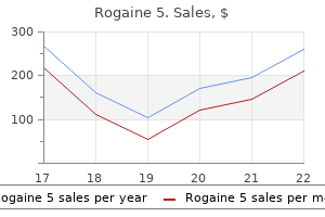
Buy rogaine 5 overnight delivery
However prostate formula buy rogaine 5 60ml with mastercard, even mild events may increase the risk of secondary infections, and patients must cope with chronic discomfort, itching, and the disagreeable appearance of the rash. The rash predominantly affects visible areas of the body, which can cause distress, anxiety, negative self-image, and low self-esteem in some patients. Furthermore, high-grade (grade 3 or higher) skin reactions may lead to morbidity, treatment interruption, or dose modifications. Among patients treated with erlotinib, rash is most likely to develop in nonsmokers, individuals with fair skin, and patients older than 70 years. Strategies include use of topical moisturizers or corticosteroids, administration of systemic steroidal medications or antihistamine drugs to palliate pruritus and inflammation, and dose delay or reduction in the case of severe reactions. Although several guidelines for managing cutaneous side effects have been published, they are based mainly on anecdotal evidence and clinical experience. Topical antibiotics include clindamycin 1-2%, erythromycin 1-2%, metronidazole 1%, or fusidic acid 2%. Topical corticosteroids, ammonium lactate, and moisturizing creams are recommended for xerosis (Table 49. For paronychia, topical antibiotics or antiseptics and silver nitrate applications can be beneficial. Dermatologic adverse events associated with afatinib: an oral ErbB family blocker. Papulopustular (acneiform) rash (left) may substantially improve after treatment with topical corticosteroids for 2 weeks (right). Paronychia with granulation tissues improves after weekly applications of silver nitrate for 4 weeks. This reaction is characterized by well-defined, tender palmoplantar hyperkeratotic or blistering lesions, especially in areas of trauma or friction. Although skin rash (type unspecified) has been reported for some patients after infusion of bevacizumab, it is not a common toxicity of bevacizumab. The incidence of high-grade (grades 3 to 5) diarrhea was greater with gefitinib (3% to 3. Diarrheal episodes are usually moderate and are generally well controlled with dose reduction and administration of loperamide. However, the most common reason to discontinue afatinib was diarrhea in five patients (3. In a series of Japanese patients, chest computed tomography images showed diffuse ground-glass opacities and evaluation of tissue samples indicated that there was diffuse alveolar damage with hyaline membrane formation. Other than Japanese ethnicity, risk factors for the development of interstitial lung disease include male gender, a history of smoking, and a presence of interstitial pneumonia (odds ratios, 3. Approximately 90% of patients in whom gefitinib-induced interstitial lung disease develops have received prior radiotherapy or chemotherapy. Serious interstitial lung disease, including fatal cases, can occur with erlotinib treatment. Two milligrams of loperamide can be taken every 4 hours to a maximum of 20 mg/ day until diarrhea improves to grade 1. Patients should consume enough water and electrolytes to prevent dehydration and renal damage. Because diarrhea is a common side effect of many cancer treatment regimens, guidelines for its management are well established. Patients with frequent diarrhea should consume a light diet without dairy products. A noteworthy concern with this class of agents is the potential for vessel injury and bleeding, which has been seen in patients with squamous cell lung cancer. In a pooled analysis of two large clinical trials in patients with colon cancer, individuals who needed surgery while being treated with bevacizumab had a higher frequency of serious wound healing complications than individuals treated with placebo (13% vs. A majority of patients receiving bevacizumab require antihypertensive therapy, particularly patients receiving higher doses and more prolonged treatment. In some studies, reversible posterior leukoencephalopathy developed during bevacizumab treatment in patients with poorly controlled hypertension. Gastrointestinal perforation, a potentially life-threatening complication of bevacizumab, has been reported in up to 11% of patients with ovarian cancer, perhaps related to the presence of peritoneal carcinomatosis and to prior abdominal surgery. The rate of colonic perforation is less than 1% in patients with breast or lung cancer who receive the antibody. However, special caution should be taken for patients with lung cancer and peritoneal metastasis who are receiving bevacizumab. Incidence of febrile neutropenia was higher in patients treated with ramucirumab than in controls (grade 3: 10% vs. Incidence of epistaxis of any grade was significantly higher in the ramucirumab group than in the control group, but few grade 3 or worse events occurred. Of note, this trial enrolled patients with both squamous cell and nonsquamous cell carcinoma excluding major blood vessel involvement and intratumor cavitation. Hypertension occurred more frequently in the ramucirumab group than in the control group, with one grade 4 hypertension event occurring in the ramucirumab group. Studies in rats have demonstrated that crizotinib causes reductions in the rate of retinal dark adaptation but not the ability to achieve full dark adaptation, offering a partial explanation for these clinical findings. Drug holidays followed by rechallenge at a lower dose have allowed ongoing treatment in some cases of severe neutropenia or transaminitis, but permanent drug discontinuation is occasionally required. Biweekly monitoring of transaminases for the first 2 months of treatment is recommended. Skin tumors, especially keratoacanthoma and cutaneous squamous cell carcinoma, have developed in a high percentage of patients in clinical trials of vemurafenib or dabrafenib. Cutaneous toxicities such as rash, hyperkeratosis, cutaneous squamous cell carcinoma, and keratoacanthoma occur with both vemurafenib and dabrafenib, but have been reported to occur to a lesser degree with dabrafenib. Of note, cutaneous squamous cell carcinoma has occurred in 19% of patients with vemurafenib and in only 5% of patients with dabrafenib. Other common cutaneous toxic effects were a diffuse hyperkeratotic perifollicular rash (55%), photosensitivity (52%), and alopecia (45%). Keratoacanthoma and cutaneous squamous cell carcinoma occurred in 14% and 26% of the patients, respectively. For small and superficial lesions, destructive modalities such as curettage and electrodessication or cryosurgery may be sufficient. When surgical treatment is either impractical or undesirable, other strategies such as topical 5-fluorouracil may be used. Bexarotene and other systemic retinoids may be helpful for vemurafenib-associated cutaneous squamous cell carcinoma and keratoacanthoma. Among patients in whom a cutaneous squamous cell carcinoma developed, at least one additional cutaneous squamous cell carcinoma developed in approximately 33% with continued dabrafenib. The median time between diagnosis of the first and second cutaneous squamous cell carcinoma was 6 weeks. In a melanoma trial, serious febrile drug reactions- defined as serious cases of fever or fever of any severity accompanied by hypotension, rigors or chills, dehydration, or renal failure in the absence of another identifiable cause. The incidence of fever (serious and nonserious) was 28% in patients treated with dabrafenib and 10% in patients treated with dacarbazine. In a melanoma trial, ophthalmologic examinations including retinal evaluation were performed at baseline and at regular intervals during treatment; central serous retinopathy developed in one patient (less than 1%) who received trametinib; however, no cases of central serous retinopathy were identified in chemotherapy-treated patients. In addition, no cases of retinal vein occlusion had been reported at the time of analysis. If repeat ophthalmologic evaluation indicates resolution of the central serous retinopathy within 3 weeks, the patient may resume trametinib at a reduced dose. As many as 14% of patients had grade 3 or grade 4 treatment-related adverse events, the most common of which was fatigue. Immune-related adverse events of all grades, including rash (12%), pruritus (9%), diarrhea (11%), transaminitis (3% or less), thyroid abnormalities (3% or less), and infusionrelated reaction (3% or less), occurred in 41% of patients. Grade 3 or grade 4 immune-related adverse events occurred in 6% of patients, which mainly included diarrhea, rash, transaminitis, and thyroid abnormalities. Pneumonitis (any grade) developed in nine patients (3%); grade 3 or grade 4 pneumonitis developed in three (1%).
60 ml rogaine 5 with amex
Use of Mass Media mens health workouts buy discount rogaine 5 60 ml online, Including New Social Media Advocacy groups are increasingly using mass media in facilitating community education and building awareness about lung cancer and its impact. Social media outlets such as Facebook allow a cost-effective broader reach into the community and for those who have been touched by lung cancer to share the messages within their networks. Common use of lung cancer-themed hashtags and approaches are becoming more visible. New technologies within social media platforms such as Thunderclaps allow advocates to amplify the message with the power of crowdsourcing and help people be heard by saying something together. A cross-country initiative, "Shine a Light on Lung Cancer," is the largest coordinated awareness event in the world, with more than 125 vigils in the United States, 30 events in Australia, and additional vigils held in Brazil, Egypt, New Zealand, and Poland The purpose of each vigil is to build the lung cancer community, and each year more voices are added to this movement. There are many methods of raising awareness of lung cancer, including public meetings and petitions, position statements, advertising, and mass publication of informational leaflets. The West Japan Oncology Group, for example, has hosted 25 public lectures in 11 cities across Japan between 2001 and 2012. These lectures on lung cancer have attracted more than 11,000 attendees, consisting of people with lung cancer (29%), their families (34%), and others. The format of the meetings has included a lecture from a key specialist on a topic. The lectures have also gained public awareness through articles in local and national newspapers, with articles published in 89 million printed newspapers. A key product of this initiative is the creation of a guidebook for people with lung cancer. Advocacy groups have much to offer in changing this picture, particularly in light of recent advances in screening for the early detection of lung cancer. There is increasing hope that more survivors will produce more advocates and a stronger global voice in this disease. With the current research focus on biomarkers and immune therapy in lung cancer, it is clear that, over the next few years, lung cancer will be a disease of interest in the development of targeted therapy and immunotherapy treatments. In addition to the direct benefits of new therapy options, this will likely mean more people with lung cancer in clinical trials, more clinicians engaged in lung cancer research, and a higher profile for the disease in general. The challenge for lung cancer advocates will be to ensure that these benefits are realized. Working with scientists and health-care professionals at the local, national, and international level is much more effective and will result in a focus on local priorities. At a global level, the International Association for the Study of Lung Cancer has engaged with the advocacy movement through representation on key committees and at professional meetings. Closer collaboration can only be beneficial when working toward better future outcomes for those affected by this disease. Political Lobbying In the United States, political lobbying by the Lung Cancer Alliance, including campaigning for legislative change and print/ Web efforts to harness public opinion, has led to a $68. Assessment of guilt and shame in patients with non-small-cell lung cancer compared with patients with breast and prostate cancer. Informing and Influencing Health Services Providers Advocacy groups can play a large part in influencing health services workers to provide better care to their patients with lung cancer. Lung Foundation Australia has campaigned to support lung cancer specialist nurses in Australia, and in the United Kingdom, the Roy Castle Lung Cancer Foundation and the National Forum for Lung Cancer Nurses have developed a report on the value of lung cancer nurses. Advocacy groups lead in the formation and conduct of lung cancer patient support groups, which include face-to-face and telephone formats. Other programs may include webinars, mindfulness classes, make-up sessions for women receiving treatment, relaxation technique sessions, cooking classes, and art and music therapy. Stigma, shame, and blame experienced by patients with lung cancer: qualitative study. Setting standards and streamlining referral processes may reduce waiting times, but these strategies only work if there are adequate resources available to provide the necessary care. Centralizing treatment services may improve overall outcomes, if this can be achieved without compromising access to care. At any point in time, the state of knowledge and the state of technology set an upper limit on what is achievable for patients with cancer. However, what is actually achieved depends not only on what would be achievable with optimal care but also on how close we come to delivering optimal care, a quantity that has been termed the attainment factor. The goal of biomedical and clinical research is to improve outcomes through innovation, whereas the goal of health services research is to improve outcomes through the optimization of health system performance. Innovative biomedical and clinical research both have the potential to improve outcomes greatly in the long term, but health services research may offer the best opportunity of improving outcomes in the short term. However, in the case of diseases such as lung cancer, for which innovative biomedical and clinical research has been slow to yield real improvements in outcome, it is important to put a high priority on health services research in order to achieve the maximum societal benefits from existing treatments. Quality describes the extent to which the right care is delivered in the right way. Efficiency describes the extent to which accessibility and effectiveness are optimized in relation to the resources expended. Each of these dimensions of health system performance must be optimized in order to achieve optimal outcomes. Health services research is concerned with measuring access, quality, and efficiency; understanding the factors that influence them; and discovering ways to enhance them. The three dimensions of health system performance are clearly not independent of one another. For example, interventions aimed at enhancing quality have the potential to adversely affect accessibility and/ or efficiency. It is therefore unwise to focus on one dimension of health system performance without at least keeping an eye on what is happening in the other two dimensions. Health research may be considered as a continuum of four overlapping domains: basic or biomedical research, clinical research, health services research, and population health research. Health services research is defined as a "multidisciplinary field of scientific investigation that studies how social factors, financing systems, organizational structures and processes, health technologies, and personal behaviors affect access to health care, the quality and cost of health care, and ultimately, our health and well-being. Clinical research is primarily intended to guide decisions of physicians about the care of individual patients, whereas health services research is intended to guide the decisions of managers and policymakers about the design and implementation of healthcare programs. Spatial accessibility describes the relationship between the location of supply of service and the location of the patients who need the service, taking into account travel times and costs. It also encompasses indirect costs, for example, loss of earnings during treatment that may deter use of the service. The term structure is defined broadly to include facilities, equipment, personnel, and organizational structures. The term process includes both the type of care that is delivered and the way in which it is delivered. Donabedian reasoned that optimal process is necessary for optimal outcome; that adequate structure is necessary, although not sufficient, for optimal process; and that outcomes may be enhanced by identifying and correcting deficiencies in structure and/or process. In this chapter, we will review the results of research studies that have sought to optimize the accessibility and quality of programs of care for patients with lung cancer. This work involves identifying barriers to optimal care as well as designing and evaluating interventions to overcome those barriers. However, before one can identify deviations from optimal health system performance, one must first identify appropriate indicators of performance and establish standards of performance with respect to those indicators. We will therefore begin by reviewing the prescriptive research that has been undertaken to establish standards of care for patients with lung cancer. We will distinguish between standards of care for the individual patient and standards for the operation of the health programs that are required to deliver care to a population of patients. Standards of care for the individual patient should be based, whenever possible, on the results of randomized clinical trials that directly compare the outcomes of alternative forms of treatment. Likewise, standards for the operation of health programs should be based on the results of randomized trials that directly compare the effectiveness of alternative approaches to health-care delivery or at least on the results of well-controlled observational studies. However, we will show that the empirical evidence that underpins program standards today is usually much weaker than the evidence that supports guidelines for the care of the individual patient. This concept is the essence of evidence-based medical practice, defined by Sackett et al. The Cochrane Collaboration has been instrumental in promoting systematic reviews of the medical literature. The Collaboration provides guidance for undertaking the systematic reviews necessary to identify all of the relevant evidence, for evaluating the quality of the evidence, and for synthesizing the evidence through meta-analysis. What is achievable in terms of cancer control depends on the total health-care budget, on how much of that total budget is directed to cancer care, and on how efficiently the available resources are used in providing cancer care. Efficiency measures whether we are getting the best value for money from the available health-care resources.
| Comparative prices of Rogaine 5 | ||
| # | Retailer | Average price |
| 1 | Bed Bath & Beyond | 461 |
| 2 | Hy-Vee | 681 |
| 3 | GameStop | 587 |
| 4 | Darden Restaurants | 716 |
| 5 | Macy's | 487 |
| 6 | Defense Commissary Agy. | 948 |
| 7 | Meijer | 565 |
| 8 | Lowe's | 771 |
| 9 | Albertsons | 300 |
Buy discount rogaine 5 60 ml line
Thymic carcinoma prostate histology buy online rogaine 5, part 1: a clinicopathologic and immunohistochemical study of 65 cases. Early Masaoka stage and complete resection is important for prognosis of thymic carcinoma: a 20-year experience at a single institution. Postoperative radiotherapy in thymic carcinoma: treatment results and prognostic factors. Thymic epithelial tumours: from basic principles to individualized treatment strategies. Thymic epithelial tumours: a population-based study of the incidence, diagnostic procedures and therapy. Thymic carcinoma: a multivariate analysis of factors predictive of survival in 290 patients. Masaoka stage and histologic grade predict prognosis in patients with thymic carcinoma. Nomenclature and classification of neuroendocrine neoplasms of the digestive system. Thymic neuroendocrine tumor (thymic carcinoid): a clinicopathologic study in 15 patients. Rapid, efficient, and expert management and treatment are therefore of paramount importance. The oncologic emergencies encountered by patients with lung cancer are not exclusive to lung cancer and, for the most part, may affect all cancer patients. Nevertheless, some circumstances and aspects of lung cancer emergencies are unique. Worldwide, lung cancer ranks first in cancer incidence and mortality,1 but in the United States, it ranks third in incidence behind prostate and breast cancer. Not surprisingly, respiratory problems are the most common chief complaint for patients with lung cancer on presentation to the emergency department. This chapter offers practical insights and perspectives into the etiology, evaluation, and management of these three complex, controversial, and dreaded complications of lung cancer. Central airway obstruction is a complex and frequent sign of progressive disease in patients with lung cancer and in patients with malignancies that metastasize to the lungs and airways. Lung cancer-related central airway obstruction often requires emergent evaluation and treatment to prevent hospitalization and admission to the intensive care unit, to control progression of disease, to palliate and treat other lifethreatening diseases, and to avoid immediate death. Central airway obstruction is associated with many presenting signs and symptoms, a handful of diagnostic modalities used in its evaluation, a multitude of available interventional therapies, and, most importantly, a number of patient-related issues relevant to the diagnostic and therapeutic management of this emergency. In this chapter, we briefly address some of these issues as they relate to the oncologist, radiologist, cytopathologist, interventional pulmonologist, critical care specialist, thoracic surgeon, medical ethicist, and radiation oncologist operating as members of a multidisciplinary team for lung cancer management. Types of Central Airway Obstruction Presenting as Emergencies Traditionally, central airway obstruction is classified as exophytic (intraluminal), extrinsic. This classification is enhanced by specifying the location and extent of the airway abnormality; describing whether the obstruction is focal, multifocal, or diffuse; and indicating whether associated abnormalities are present, such as edema, bronchitis, airway necrosis, purulent secretions, obvious infection (which may be primary or secondary), bleeding, perforation or fistula, dehiscence, or airway distortion. It is also helpful to ascertain whether the abnormality is a primary or secondary disorder. For example, central airway obstruction may be a result of new, progressive, or recurrent disease, or it may be an iatrogenic complication after a procedure, such as airway intubation, mechanical ventilation, stent insertion, brachytherapy or laser resection, other bronchoscopic airway manipulation, externalbeam radiotherapy, or thoracic surgical intervention. Bronchoscopic images of four types of central airway obstruction prompting emergency intervention. When the airway obstruction has caused an emergency, one must determine whether the emergency is immediately life-threatening. This last point has important implications for diagnosis, treatment, and ethical aspects of care. Symptoms of Lung Cancer-Related Central Airway Obstruction the symptoms of central airway obstruction associated with lung cancer are similar to those found in other instances of central airway obstruction and include dyspnea, cough, hemoptysis, hoarseness, and respiratory failure. They can be signs of progressive although manageable disease or represent an immediate precursor or cause of death. Central airway obstruction should be suspected in all cases of new onset or increasing symptoms in any patient with known or suspected lung cancer and in patients who have recently undergone palliative or curative therapeutic interventions for their lung cancer. The medical evaluation, therefore, must ascertain the presence, severity, and contributing roles of possible heart failure; esophageal extension; pleural disease; other malignancies extending to the lung, mediastinum, and airways; emphysema and chronic bronchitis; pneumonia; radiation-induced lung or airway injury; clinical depression; malnutrition; and failure to thrive. Chest radiograph showing resolved right atelectasis after emergency intervention with flexible bronchoscopy in a patient with a known right lower-lobe tumor. In this case, respiratory insufficiency and radiographic abnormalities (ipsilateral mediastinal shift and atelectasis) were due to mucus plugging seen and removed at the time of emergent inspection bronchoscopy. On some occasions, the obstruction is discovered only after a patient has emergency intubation and is placed on mechanical ventilation. In other cases, symptoms of significant obstruction may warrant intubation, raising issues about life-sustaining treatment, appropriate use of medical resources, costs, and roles of palliative care and procedures. A third scenario may involve a patient with dyspnea or other complications who is denied admission to the intensive care unit and further diagnostic evaluation, either because the diagnosis of central airway obstruction is not considered or because the condition is diagnosed but may be considered irreversible. This last scenario raises issues of professionalism, competency, and resource allocation because levels and quality of care depend, in part, on physician expertise, team experience, institutional biases, finances, and societal philosophies regarding extent of care for patients with life-threatening illnesses. The setting of emergency central airway obstruction is often complex and stressful for health-care providers, patients, and their families. Patients with malignant central airway obstruction may have a median survival as short as 3 months. Mortality increases with the number of failed organs, severity of comorbidities, and presence of airway obstruction. In one study, the hospital mortality rate was 83% for patients with lung cancer and central airway obstruction who were receiving mechanical ventilation, compared with 62% in patients without an obstructed airway. In some life-threatening situations, flexible bronchoscopic inspection is performed to provide immediate information to assist in establishing indications for or against therapeutic interventions to restore airway patency, alleviate dyspnea, postpone or prevent the onset of respiratory failure requiring intubation, or palliate other symptoms (such as hemoptysis). Clinical Findings Findings related to central airway obstruction may include decreased breath sounds on chest auscultation, prolonged expiration, and unilateral wheezing. Patients may lose the ability to phonate in cases where an airway stent has migrated proximally to impinge on the vocal cords from below. Vocal cord paralysis may be suggested by cough, hoarseness, change in voice, or episodes of recurrent aspiration and may be related to a primary lung mass or enlarged mediastinal lymph node impinging on the recurrent laryngeal nerve. Hemoptysis may suggest central airway obstruction in patients with known lung cancer or cancers that are known to metastasize or otherwise spread into the airways (such as colon cancer, malignant melanoma, renal cell carcinoma, thyroid cancer, esophageal cancer, adenoid cystic carcinomas, sarcomas, and some lymphomas). Chest Radiographs Often Aid in Diagnosis Chest radiographs may show atelectasis, ipsilateral mediastinal shift, lobar consolidation, stent migration, or a mass impinging on the central airway. Patients with a history of radiotherapy may have signs of fibrosis or radiation pneumonitis. It also provides information pertaining to associated airway abnormalities that may affect management decisions and indications for or against palliative or curative interventions to restore airway patency. In experienced hands, airway inspection is performed very quickly with minimal risk to the patient. Depending on the setting, bronchoscopy can be performed through the nares or the mouth (using a bite block), from behind the head of the patient or standing in front of and to the side of the patient, always in conjunction with supplemental oxygen and with or without sedation. For example, in a patient with impending respiratory failure, bronchoscopy can be performed with the patient receiving high-flow oxygen and/or through a continuous positive airway pressure mask, without sedation (to avoid risks of iatrogenic respiratory suppression), and with the patient in the seated position (to avoid aspiration or respiratory suppression related to the supine position). If absolutely necessary, the patient can be intubated temporarily with an endotracheal tube over the flexible bronchoscope, or, after appropriate sedation and airway management, intubation can be performed via laryngoscopy before transport to the operating room or interventional bronchoscopy suite. Many decision points must be considered, some of which are disease related, whereas others are lesion related, patient related. To be certain that information is obtained to address each of these aspects of care, a four-step approach can be used that includes an initial evaluation, a review of procedural strategies, procedural techniques and expected/known results, and a long-term management plan (Table 57. In recent years, a commonly used therapeutic palliative approach is the combination of endobronchial debulking (using thermal, nonthermal, or mechanical techniques) with or without stent insertion, followed by external-beam radiotherapy and/or systemic therapy, if indicated or possible. According to numerous studies, laser resection is an effective palliative procedure for central airway obstruction. Complications are uncommon in well-trained hands, but physicians should always consider the possibility of failure to control the airway, airway fires (especially with indwelling airway stents or endotracheal tubes), failure to control bleeding, and airway necrosis. Data from numerous studies confirm the efficacy of silicone stents to restore and maintain airway patency, although complications such as stent migration, kinking, obstruction by tumor or mucus, and even infection have been reported. Models are available in all shapes and sizes, including stents that fit onto the carina or secondary bifurcations. By improving functional status, stent insertion allows the medical team to proceed with palliative chemotherapy or radiotherapy if indicated.

Cheap rogaine 5 online
The optimal port placement for a completely portal robotic lobectomy using all four arms prostate 180 at walgreens order 60ml rogaine 5 visa. The four ports are placed over the same rib: over the top of the ninth rib for lower lobectomy and over the top of the eighth rib for upper lobectomy. The 12-mm access port (A) is placed halfway between the camera port (C) and robotic arm 1 (1) for upper and lower lobes and between the camera and robotic arm 2 (2) for middle lobectomy. The port is placed as low as possible staying just above the diaphragm as carbon dioxide is insufflated to help push the diaphragm down. Port Placement/Docking the ports are inserted in the seventh intercostal space over the top of the eighth rib for upper/middle lobectomy and in the eighth intercostal space over the top of the ninth rib for lower lobectomy. The ports are marked as follows: robotic arm 3, a 5-mm port is located 1 cm to 2 cm lateral from the spinous process of the vertebral body; robotic arm 2, an 8-mm port is located 10 cm medial to robotic arm 3; the camera port (we prefer a 12-mm camera) is located 9 cm medial to robotic arm 2; and robotic arm 1 (a 12-mm port) is placed directly above the diaphragm anteriorly. The assistant port (12 mm) is placed as low as possible in the chest, triangulated exactly halfway between the most anterior robotic port (which is robotic arm 1 in the right chest and robotic arm 2 in the left chest) and the camera port, and as low as possible to remain just above the diaphragm, which is being pushed downward by the insufflating humidified carbon dioxide gas. Sequence of Port Placement A 5-mm port is placed first in the camera port position, and carbon dioxide insufflation is initiated at a pressure of 10 mmHg. Then the 5-mm thoracoscope is used to help assist the placement of all other ports, which are placed under direct vision. The camera port is placed first, robotic arm 3 is placed second, and 7t 9t and chest), with a monitor on the opposite side. We perform mediastinal lymph node dissection before lobectomy not only to evaluate the lymph nodes but also to access arterial and venous branches and the bronchus. Right Side the inferior pulmonary ligament is divided to gain access to station 9 lymph nodes, which are removed along with station 8 lymph nodes. Robotic arm 3 is used to retract the lower lobe medially and anteriorly to remove lymph nodes from station 7. Care is taken to control the two feeding arteries that make the subcarinal lymph node bloody. Robotic arm 3 is used to retract the upper lobe inferiorly, whereas robotic arms 1 and 2 are used to dissect lymph nodes at stations 2R and 4R, clearing the space between the superior vena cava anteriorly, the esophagus posteriorly, and the azygos vein inferiorly. Avoiding dissection too far superiorly can prevent injury to the right recurrent laryngeal nerve that wraps around the subclavian artery. The 5-mm camera is then moved to the port for robotic arm 2, and the two most anterior ports (robotic arm 1 in the right chest and robotic arm 2 in the left) and the access port are placed under direct vision using a seeking needle. We use a zerodegree scope for the entire procedure to help prevent torquing of the intercostal nerve. The port placement for left-sided lobectomy is a mirror image to that previously described. The difference is that robotic arm 3 is next to robotic arm 1, rather than next to robotic arm 2. The robotic arms are docked to the ports, maximizing the amount of space between the arms to avoid collisions. The instruments used to start the surgery are an 8-mm Cadiere forceps in the left robotic arm, an 8-mm bipolar curved thoracic dissector in the right robotic arm, and a 5-mm thoracic grasper in robotic arm 3. For their initial placement, robotic instruments should be inserted under direct vision during thoracic surgery. Once instruments are safely positioned, they can be quickly and safely inserted or changed for other instruments by properly using the memory feature of the robot, which automatically inserts any new instrument to a position that is exactly 1 cm proximal to the latest position. However, when this memory feature is used, the surgeon must ensure that no vital structures have moved into the path of the newly placed instrument. The insertion of robotic instruments deserves special attention, as does the passing of vascular staplers around fragile structures such as the pulmonary artery and vein. We have developed our own communication system between the bedside assistant and the surgeon to prevent iatrogenic injuries. This communication system uses the anvil of the stapler as the hour hand of a clock so that the degree of articulation can be quantified and communicated. Left Side the inferior pulmonary ligament is divided to facilitate removal of lymph nodes at station 9. Station 7 is accessed in the space between the inferior pulmonary vein and lower lobe bronchus, lateral to the esophagus. The lower lobe is retracted medially/anteriorly with robotic arm 3 during this process. Absence of the lower lobe facilitates dissection of lymph nodes at station 7 from the left. Lastly, robotic arm 3 is used to wrap around the left upper lobe and press the lobe inferiorly to allow dissection of stations 5 and 6 lymph nodes. Care should be taken while working in the aortopulmonary window to avoid injury to the left recurrent laryngeal nerve. Station 2L cannot typically be accessed during left-sided mediastinal lymph node dissection because of the presence of the aortic arch, but the 4L node is commonly removed. General Concepts In general, for a right-handed surgeon, a blunt instrument such as a Cadiere forceps is placed in robotic arm 2, which is always the left hand, and the right hand, which controls robotic arm 1, uses a thoracic dissector. The stapler may be placed through one of three ports: the access port, robotic arm 1, or robotic arm 2. The current design of commercially available white or gray vascular staplers requires a 12-mm port; the green-loaded stapler commonly used for the bronchus requires a 15-mm port. We prefer to remove the trocar and leave it docked to the robotic arm and then place the stapler through the skin incision. We prefer to place a vessel loop under a vessel to be stapled to help elevate it while the stapler is passed beneath it. We commonly use a prerolled sponge to absorb blood from the operative field or facilitate blunt dissection to improve visibility. Removal of lymph nodes from surrounding structures should be done before stapling them in the interests of both ensuring an oncologically sound operation and facilitating isolation and division of structures. The order in which the structures are isolated and divided during lobectomy varies somewhat, depending on patient anatomy. The use of robotic technology for the performance of anatomic lung resection is increasing. To maintain safe and effective robotic surgery, surgeons must continue to design evidence-based pathways to the credentialing of robotic surgical teams. Despite the small number of studies reported in the literature from several single centers and a handful of surgeons, the results show good intraoperative results with anatomic pulmonary resection and promising longterm survival rates. Further studies on the true cost to society (not solely to the hospital or patient) and the actual 5-year to 10-year survival rates for people with cancer treated robotically are needed. In addition, literature that evaluates the reproducibility of this type of robotic surgery across centers and its feasibility in impoverished or Third-World countries is also needed. Short-Term Results Two studies showed that short operative times (132 min and 175 min, respectively) are possible as the experience of each surgeon grows. An updated series of 282 patients undergoing robotic lobectomy demonstrated an average blood loss of 20 mL, 0. Because robotic anatomic pulmonary resection is relatively new, few studies have reported an actuarial 5-year survival rate. Initial consecutive experience of completely portal robotic pulmonary resection with 4 arms. Open, video-assisted thoracic surgery, and robotic lobectomy: review of a national database. Nodal upstaging in robotic and video assisted thoracic surgery lobectomy for clinical N0 lung cancer. Robotic assistance for video-assisted thoracic surgical lobectomy: technique and initial results. Robot-assisted lobectomy for early-stage lung cancer: report of 100 consecutive cases. Initial experience with robotic lung lobectomy: report of two different approaches. The prevalence of nodal upstaging during robotic lung resection in early stage non-small cell lung cancer. Performing robotic lobectomy and segmentectomy: cost, profitability, and outcomes. Initial multicenter community robotic lobectomy experience: comparisons to a national database. Surgeons have been trying to achieve an optimal balance between radical surgery and surgery that preserves postoperative lung function.
Order rogaine 5 60 ml line
Long-term survival after lung-sparing total pleurectomy for locally advanced (International Mesothelioma Interest Group Stage T3-T4) nonsarcomatoid malignant pleural mesothelioma prostate zones buy discount rogaine 5 60ml on-line. Adenovirus-mediated herpes simplex virus thymidine kinase/ganciclovir gene therapy in patients with localized malignancy: results of a phase I clinical trial in malignant mesothelioma. A phase I trial of repeated intrapleural adenoviral-mediated interferon-beta gene transfer for mesothelioma and metastatic pleural effusions. Immuno-gene therapy with interferon-beta before surgical debulking delays recurrence and improves survival in a murine model of malignant mesothelioma. Re-challenge with pemetrexed in advanced mesothelioma: a multi-institutional experience. A phase 2 study of gemcitabine and epirubicin for the treatment of pleural mesothelioma: a North Central Cancer Treatment Study, N0021. In the visceral compartment, the most common mass is lymphadenopathy associated with metastatic disease. These tumors are most commonly found in the mediastinum, where they account for 15% of anterior mediastinal masses in adults. These teratomas may contain any type of tissue, including teeth, hair, bone, cartilage, and occasionally higher order structures. Although many patients will be asymptomatic, benign teratomas have the capacity to compress, erode, rupture, and fistulize into surrounding structures. The treatment approach for benign teratomas can vary and has included median sternotomy and lateral thoracotomy. For large masses with bilateral extension, a clamshell incision (a combined upper median sternotomy and anterior thoracotomy) provides excellent exposure. Benign teratomas are often adjacently adherent, which can make dissection challenging, and total resection is not always possible. The mediastinum is the region of the thorax between the pleural cavities, commonly described in three compartments: anterior, visceral or middle, and posterior. The anterior mediastinum contains the thymus, lymph nodes, connective tissue, and fat. The visceral mediastinum contains the heart and its vasculature within the pericardium, trachea and proximal bronchi, esophagus, thoracic duct, lymph nodes, and the vagus, phrenic, and recurrent laryngeal nerves. The posterior mediastinum contains the sympathetic chain and proximal intercostal arteries, veins, and nerves. The differential diagnosis for a mediastinal mass is influenced by its anatomic position (Table 54. Systemic symptoms may occur because of endocrine tumor activity or fever, chills, and weight loss associated with malignancy (Table 54. When active germ cell cancer is present in the surgical specimen, two additional courses of chemotherapy should be given. Patients who have disease recurrence have a particularly poor prognosis, although a small number of patients may benefit from salvage chemotherapy. Approximately 15% of mediastinal masses are lymphomas, typically in the anterior or middle compartment. About 90% of mediastinal lymphomas represent disseminated disease, and one-third are Hodgkin lymphomas. Most patients present with some combination of systemic B symptoms, and symptoms associated with local compression. Diagnosis typically requires tissue quantities only obtainable by surgical biopsy. However, lymphoblastic lymphoma is usually first identified in bone marrow and peripheral blood; thus a mediastinal biopsy is not necessary. Lymphoblastic lymphoma is particularly aggressive, and treatment with chemotherapy and possible bone marrow transplant should not be delayed by staging procedures. Large B-cell lymphoma is treated with chemotherapy, and some centers also use radiotherapy for the treatment of B-cell lymphoma and lymphoblastic lymphoma. The majority of bronchogenic cysts are discovered before the onset of symptoms, but most do eventually cause symptoms. Bronchogenic cysts can cause local compression and erosion and may become infected. Like bronchogenic cysts, esophageal cysts can also cause local compression and may become infected. These neurogenic tumors arise from the sympathetic chain or intercostal rami, and they most commonly take the forms of nerve sheath tumors (schwannoma and neurofibroma). Nearly all neurogenic tumors are benign, and approximately 30% of neurofibromas are associated with von Recklinghausen disease. Neurogenic tumors may erode osseous structures; this contributes to the development of an intraspinal (dumbbell) extension. Relative contraindications for complete resection include a mass greater than 6 cm and spinal artery involvement. Chemotherapy and radiotherapy may be used when complete resection is not possible. However, a growing pericardial cyst can cause hemodynamic compromise, and excision should be performed for symptomatic patients. Substernal goiters are the most common superior mediastinal tumors, usually found in the anterior mediastinum; however, 10% to 15% of substernal goiters are found in the posterior compartment. A palpable thyroid is present in 88% of patients, and 16% of patients have hyperthyroidism. Although malignancy is possible, needle biopsy is not indicated, as the malignant focus may be missed and its presence does not alter the operative indication. When the extent of thoracic involvement does not permit resection by a collar incision, possible approaches include manubrial split, sternotomy, and thoracotomy. Postoperative complications are infrequent and include hypoparathyroidism and recurrent laryngeal nerves paralysis. More aggressive exposure is used for large and/or malignant tumors, for which complete resection is crucial and dissection is potentially challenging. Access to small anterior midline tumors is commonly approached by a median sternotomy, with the patient in the supine position with the arms tucked to the side. In this position, the mediastinal vasculature, left and right hemithoraces, and lung hila are well exposed; however, exposure of the left lower lobe and posterior aspect of the lungs is poor. In some cases, a partial sternum-sparing approach will allow for sufficient exposure, such as a manubrial split for superior mediastinal mass. A collar incision may also provide sufficient access to the superior mediastinum if the mass can be accessed through the neck. For large tumors extending into either of the hemithoraces, a hemiclamshell incision provides good exposure. Approximately 22% of parathyroid adenomas are found in a mediastinal parathyroid gland. Localization has no diagnostic role and is used only for preoperative planning in biochemically proven cases. Most mediastinal parathyroid tumors can be resected transcervically, and videomediastinoscopy may be useful in difficult cases. Intraoperative monitoring of the parathyroid hormone level is used to confirm systemic cure. Primary mediastinal leiomyoma not associated with adjacent organs is exceedingly rare. The ipsilateral lung may be collapsed to allow for anatomic resection of involved lobes, if necessary, and access to the posterolateral aspect of the tumor. If the tumor has substantial cervical extension, the upper sternotomy may be extended superiorly along the anterior border of the sternocleidomastoid. In these cases, a so-called trap door incision, created by extending the upper sternotomy along the superior margin of the clavicle, will allow for appropriate exposure. Excision of the medial third of the clavicle may also assist in exposure of the tumor. A large tumor that extends into both right and left hemithoraces may be resected through a bilateral clamshell incision, with the patient in the supine position and the arms abducted or flexed over the forehead. A curvilinear incision should be made along the inframammary crease from the right to left anterior axillary lines for access to the fourth intercostal space; the mammary vessels are then ligated, and the sternum is divided transversally. An upper sternal split may be made in the event that an initial clamshell approach provides inadequate exposure of the superior mediastinum. A collar incision is often sufficient, but more involved goiters can require a sternal split. Parathyroid adenomas causing primary hyperparathyroidism should be resected with a minimally invasive approach.
Syndromes
- Six-minute walk test
- Low blood pressure that develops rapidly
- Name of the product (ingredients and strengths, if known)
- Sleepiness
- Your symptoms are very bad.
- Rash with red or purple spots (petechiae)
- Mitral stenosis
Purchase rogaine 5 60ml with amex
Ninety-six percent of patients received the planned preoperative chemotherapy androgen hormone replacement buy rogaine 5 mastercard, and 45% received postoperative chemotherapy. At the time of study closure, 336 of the planned 600 eligible patients had been enrolled. Neoadjuvant chemotherapy was well tolerated, with 79% of patients receiving all three cycles. Seven patients in the chemotherapy arm died postoperatively, compared with four patients in the surgery alone arm. With a median follow-up of 53 months, the median survival was 75 months for the chemotherapy arm compared with 46 months for the surgery alone arm; the 5-year survival rates were 50% and 43%, respectively. Progression-free survival trended in favor of perioperative chemotherapy (median, 33 vs. In the preoperative arm, 97% of patients started the planned chemotherapy, and the radiographic response rate was 53. Surgery was performed in 94% of patients; surgical procedures and postoperative mortality were similar across the three arms. In this trial, in which the treatment allocation was made before surgery, more patients were able to receive preoperative than adjuvant treatment. The rate of postoperative complications was not higher in the combinedmodality arm and no impairment of quality of life was noted. The study was closed prematurely after the random assignment of 270 patients, 129 to combined therapy and 141 to surgery alone. These findings are not consistent with the results of S9900, which found no difference in treatment effect by stage,107 and the results of the trial by Depierre et al. The preoperative group received two preoperative cycles followed by two additional preoperative cycles, while the postoperative group underwent two preoperative cycles followed by two postoperative cycles, the third and fourth cycles being given only to responders in both cases. Tumor response occurs in 40% to 50% of patients, with treatment compliance that is generally better than that for adjuvant chemotherapy. However, the tumor is downstaged in fewer than 20% of cases and the rate of complete response is low. The size of the square is directly proportional to the amount of information contributed by the trial. Preoperative chemotherapy for non-small cell lung cancer: a systematic review and meta-analysis of individual participant data. Thus, there is a strong possibility that sample collection methods, processing protocols, single-institution patient cohorts, small sample sizes, and peculiarities of the different microarray platforms are contributing substantially to the results. To address these issues, a multiinstitutional collaborative study was conducted to generate gene expression profiles from a large number of samples with a priori determined clinical features, useful to evaluate proposed prognostic models for potential clinical implementation. The risk scores produced correlated strongly with actual outcomes, especially when clinical and molecular information were combined to build prognostic models for early-stage lung cancer. Multivariate analysis in both cohorts indicated that no standard clinical risk factors could account for or provide the prognostic information derived from tumor gene expression. Another approach that has been evaluated as a method for selecting patients for adjuvant chemotherapy is to identify a predictive molecular determinant for cisplatin. The trial sought to demonstrate the feasibility of customized adjuvant chemotherapy based on timely biomarker analysis within a 2-month postsurgery delay. The study failed to show a significant benefit from treatment customization, but a longer follow-up time is needed to conclude definitively in favor of a completely negative study. A precise quantitative estimate of the survival gain is not known, but meta-analysis suggests it is approximately 5% at 5 years. A new meta-analysis that includes the most recent generation of these positive and negative randomized clinical studies will substantially contribute to determining the role of adjuvant chemotherapy. Meta-analysis of postoperative adjuvant chemotherapy with tegafur-uracil in non-small-cell lung cancer. A practical molecular assay to predict survival in resected non-squamous, non-small-cell lung cancer: development and international validation studies. There is a need, however, for reliable predictive and prognostic factors that stratify patients who do or do not need adjuvant therapy in order to avoid the exposure of most patients to unnecessary treatments. In the near future, genomic (or pharmacogenomic) and proteomic assays may drive the identification of patients who are ideal candidates for adjuvant therapy. Neoadjuvant chemotherapy may be better suited than adjuvant therapy for evaluating novel agents, as the effect of the drug on the target can be assessed by pretreatment biopsy (at diagnosis) and after chemotherapy (at surgery). However, targeted agents matched to specific mutations may need to be administered for long periods of time, which is better accomplished in the adjuvant setting, where a curative resection option is not jeopardized in patients who fail to respond preoperatively. The duration of the administration of novel agents postoperatively, for example, in patients who have an initial response, will need careful evaluation in randomized trials. It is hoped that better patient selection and better matching of individual patients to a specific treatment regimen based on molecular profiling can lead to more effective treatment. It is hoped that newer radiographic methodologies will allow for better characterization and even earlier detection of malignancies to decrease the number of diagnostic procedures performed to remove small lesions that are not malignant. In the long run, improved molecular technologies are likely to also allow for earlier detection by nonradiographic methods. Sites of recurrence in resected stage I non-small cell lung cancer: a guide for future studies. Postsurgical stage I bronchogenic carcinoma: morbid implications of recurrent disease. Timing of local and distant failure in resected lung cancer: implications for reported rates of local failure. Preoperative staging of non-small-cell lung cancer with positron-emission tomography. Detection of extrathoracic metastases by positron emission tomography in lung cancer. Detection of disseminated lung cancer cells in lymph nodes: impact on staging and prognosis. Detection of micrometastatic tumor cells in pN0 lymph nodes of patients with completely resected non-small cell lung cancer: impact on recurrence and survival. Molecular staging of lung cancer: real-time polymerase chain reaction estimation of lymph node micrometastatic tumor cell burden in stage I non-small cell lung cancer-preliminary results of Cancer and Leukemia Group B Trial 9761. Postoperative adjuvant therapy for non-small cell lung cancer: a consensus report. Postoperative radiotherapy in non-small-cell lung cancer: systematic review and meta-analysis of individual patient data from nine randomized controlled trials. Risk of death from intercurrent disease is not excessively increased by modern postoperative radiotherapy for high-risk resected non-small-cell lung carcinoma. Role of postoperative radiotherapy in resected nonsmall cell lung cancer: a reassessment based on new data. The benefit of adjuvant treatment for resected locally advanced non-small cell lung cancer. Adjuvant chemotherapy after radical surgery for non-small-cell lung cancer: a randomized study. Adjuvant chemotherapy with cyclophosphamide, doxorubicin, and cisplatin in patients with completely resected stage I non-small cell lung cancer. Adjuvant radiotherapy versus combined sequential chemotherapy followed by radiotherapy in the treatment of resected non-small cell lung carcinoma. Lack of prognostic significance of p53 and K-ras mutations in primary resected non-small cell lung cancer on E4592: a laboratory ancillary study on an Eastern Cooperative Oncology Group prospective randomized trial of postoperative adjuvant therapy. Cisplatin-based adjuvant chemotherapy in patients with completely resected non-small cell lung cancer. Chemotherapy for patients with non-small cell lung cancer: the surgical setting of the Big Lung Trial. Long-term results of the international adjuvant lung cancer trial evaluating adjuvant cisplatin-based chemotherapy in resected lung cancer. A pooled-analysis of 6494 patients in 12 studies, examining survival and magnitude of benefit. Survival benefit of neoadjuvant chemotherapy in non-small cell lung cancer: an updated meta-analysis of 13 randomized control trials. Adoption of adjuvant chemotherapy for non-small-cell lung cancer: a population-based outcomes study. Adjuvant chemotherapy for non-small-cell lung cancer: it does not always fade with time. Interpreting trial results in light of conflicting evidence: a Bayesian analysis of adjuvant chemotherapy for non-small-cell lung cancer.
Cheap rogaine 5 60 ml online
If the prognosis for patients who have segmentectomy is not significantly inferior to that for patients who have lobectomy and if the postoperative pulmonary function is significantly better for patients who have segmentectomy prostate problems treatment purchase rogaine 5 with amex, we can definitively conclude that the standard surgical modality for these early tumors should be segmentectomy. It is reasonable to perform sublobar resection, such as segmentectomy and wedge resection, for patients with limited cardiopulmonary reserve. The use of sublobar resection may be justified for most early lung cancers with minimal or no invasive features located in the outer region of the lung parenchyma. The feasibility of sublobar resection for lung cancer with overt invasive features is under investigation, with particular focus on tumors 2 cm or less in diameter. Lobectomy should be recognized as the standard mode of resection for appropriate patients. Japan Clinical Oncology Group Lung Cancer Surgical Study Group radiographically determined noninvasive adenocarcinoma of the lung: survival outcomes of Japan Clinical Oncology Group 0201. Radical sublobar resection for small-sized non-small cell lung cancer: a multicenter study. Outcomes of sublobar resection versus lobectomy for stage I non-small cell lung cancer: a 13-year analysis. Anatomical segmentectomy and wedge resections are associated with comparable outcomes for small cT1N0 non-small cell lung cancer. Recurrence and survival outcomes after anatomic segmentectomy versus lobectomy for clinical stage I non-small-cell lung cancer: a propensity-matched analysis. Comparison of thoracoscopic segmentectomy and thoracoscopic lobectomy on the patients with non-small cell lung cancer: a propensity score matching study. Intrapericardial approach to the lung root in treatment of bronchial carcinoma by dissection pneumonectomy. The surgical management of carcinoma of the lung: a study of the cases treated at the Massachusetts General Hospital from 1930 to 1950. Segmental pneumonectomy in bronchiectasis: lingula segment of the left upper lobe. Randomized trial of mediastinal lymph node sampling versus complete lymphadenectomy during pulmonary resection in the patient with N0 or N1 (less than hilar) non-small cell carcinoma: results of the American College of Surgery Oncology Group Z0030 Trial. International Association for the Study of Lung Cancer International Staging Committee; Participating Institutions. Adjuvant cisplatin and vinorelbine for completely resected non-small cell lung cancer: subgroup analysis of the Lung Adjuvant Cisplatin Evaluation. International Association for the Study of Lung Cancer International Staging Committee and Participating Institutions. Prognosis of 6644 resected non-small cell lung cancers in Japan: a Japanese lung cancer registry study. Japanese lung cancer registry study of 11,663 surgical cases in 2004: demographic and prognosis changes over decade. A new method of segmental resection for primary lung cancer: intermediate results. Is segmentectomy with lymph node assessment an alternative to lobectomy for non-small cell lung cancer of 2 cm or smaller Tumor dimension and prognosis in surgically treated lung cancer: for intentional limited resection. Prospective study of extended segmentectomy for small lung tumors: the final report. A casematched study of anatomical segmentectomy versus lobectomy for stage I lung cancer in high-risk patients. Oncologic outcomes of segmentectomy versus lobectomy for clinical T1a N0 M0 non-small cell lung cancer. Video-assisted thoracoscopic surgery is a safe and effective alternative to thoracotomy for anatomical segmentectomy in patients with clinical stage I non-small cell lung cancer. Intraoperative lavage cytologic analysis of surgical margins in patients undergoing limited surgery for lung cancer. Differences in the prognosis of resected lung adenocarcinoma according to the histological subtype: a retrospective analysis of Japanese lung cancer registry data. Limited resection versus lobectomy for older patients with early-stage lung cancer: impact of histology. A systematic review and meta-analysis of sublobar resections versus lobectomy for non-small cell lung cancer according to patient selection. Survival after sublobar resection for early-stage lung cancer: methodological obstacles in comparing the efficacy to lobectomy. In addition, a thorough lymph node evaluation by means of either systematic node dissection or at least a lobe-specific node dissection related to the location of the primary tumor is required. A minimum of six lymph node stations must be removed, of which three must be located in the mediastinum and must include the subcarinal station. Complete (R0) resection can be challenging in cases of posterior tumors located near the costovertebral angle or involving vertebral bodies, and analysis of frozen sections is not feasible for tumors with osseous margins. Staging the goal of surgical treatment in cases of T3 and T4 lung cancers is to obtain an R0 resection. Surgical treatment may be part of a multimodality approach that includes induction chemotherapy or chemoradiation therapy to reduce the tumor volume and to optimize resection margins. Nevertheless, as it is clear that a chest wall resection may induce additional respiratory compromise, clinical judgment by an experienced thoracic surgeon and discussion of each individual case by a multidisciplinary tumor board are necessary when this procedure is anticipated. Published series report that mediastinal nodal involvement is a poor prognostic factor and that extensive surgery is not warranted when mediastinal lymph node metastases are present. Tumors infiltrating the second or first rib and surrounding structures usually are considered to be superior sulcus or Pancoast tumors when neurologic symptoms are present. In selected cases, these procedures are supplemented with mediastinoscopy to reduce the false-negative rate as much as possible. Surgical Resection Depending on the location of the primary tumor and its extension into the chest wall, the incision is carefully chosen and may be centered on the anterior, lateral, or posterior chest wall. The thoracic cavity is entered away from the primary tumor, as every attempt should be made to obtain an en bloc resection with complete removal of the primary tumor together with the invaded chest wall to avoid any spillage of tumor cells in the pleural cavity. Stoelben and Ludwig1 described four categories of chest wall involvement for determining the subsequent resection (Table 30. When the tumor easily detaches from the chest wall in the extrapleural plane by finger dissection, this usually indicates only inflammatory adhesions that do not require chest wall resection. Frozen section analysis of suspicious areas of the parietal pleura may clarify this. When the tumor is densely adherent or frankly invades the chest wall, rib resection is indicated. For posterior tumors, resection of transverse processes or vertebral bodies may be needed, and a spine surgeon (either an orthopedic surgeon or a neurosurgeon) should work collaboratively with the thoracic surgeon. To reduce trauma to the uninvolved extrathoracic muscles, rib resection can be performed from inside the chest with use of the technique described by Cerfolio et al. A pneumonectomy is necessary in some cases, but it is a high-risk procedure when combined with extensive chest wall resection and should be performed only in centers with extensive experience. No reconstruction is required for defects of 3 cm or less that are covered by the scapula. However, when the defect is located at the tip of the scapula, chest wall reconstruction is required to prevent entrapment of the scapula, which is a highly symptomatic complication that is associated with poor cosmetic results. Synthetic material should be covered by viable muscle or musculocutaneous flaps to reduce the risk of infection. Results and Long-Term Survival At experienced centers, the mortality and morbidity rates associated with these procedures have decreased. The mean postoperative mortality rate is about 6%, with the majority of deaths being caused by pulmonary complications and respiratory failure. In addition to standard complications after thoracotomy, specific complications related to the chest wall resection include infection necessitating removal of the prosthetic material, herniation of the scapula, paradoxical respiration (flail chest), and, in cases involving dissection close to the spinal cord, paraplegia and leakage of cerebrospinal fluid. Coronal computed tomography image demonstrating large tumor invading the chest wall in a 58-year-old patient. Adjuvant chemotherapy or radiotherapy also remains controversial, and no specific guidelines exist for patients who have chest wall resection. Radiotherapy may be indicated for patients with close or positive surgical margins, but no randomized evidence is available to support its routine use.
Order rogaine 5 line
Many of these round or oval masses still represent fibroadenomas that have enlarged because of hormonal fluctuation or menopausal hormone therapy prostate cancer 185 buy cheap rogaine 5 60 ml online. Screening mammogram on a 52-year-old woman shows a new obscured mass in her inferior central right breast (arrow). Their suspicious features include spiculated margin (A), ill-defined margin (B), and high density (C). Their suspicious features include nonparallel orientation (A), irregular shape (B and C), indistinct margin (C and D), echogenic rim (E, blue arrows), posterior acoustic shadowing (B, arrow), calcifications (A, arrow), and duct extension (C, arrow). E, Did you notice that the Cooper ligament (yellow arrow) terminates at the echogenic margin Analysis of enhancement curves is only useful when the morphologic features are benign. Persistent enhancement in the setting of a mass with suspicious morphologic appearance is irrelevant. The morphologic appearance of the mass always trumps the enhancement pattern in deciding the level of suspicion. There are three variations of complex mass: cyst with thick walls or septations, intracystic mass, and mixed cystic and solid components. Clustered microcysts that have ill-defined margins, thick walls or septations, or intracystic masses are also included in this category. All subtypes of complex masses are associated with a similar incidence of breast cancer. Rim or heterogeneous enhancement or washout kinetics may be suspicious despite a round or oval shape with well-defined margins. Some patients with high parenchymal enhancement may have enhancement of many benign masses, some with washout. Final Comments Comparison with the prior mammograms is very helpful in determining whether a round or oval mass requires recall from screening or, if the mass is solid, biopsy (Table 7-1). This small round mass in the lateral right breast (blue arrow) has welldefined margins but a washout enhancement curve. Note that the enhancing area in the transverse plane (yellow arrow) is due to the biopsy track. Accuracy of short-interval follow-up mammograms by patient and radiologist characteristics. Short-term follow-up of palpable breast lesions with benign imaging features: Evaluation of 375 lesions in 320 women. Probably benign breast masses diagnosed by sonography: Is there a difference in the cancer rate according to palpability Periodic mammographic follow-up of probably benign lesions: Results in 3184 consecutive cases. Nonpalpable, circumscribed, noncalcified solid breast masses: Likelihood of malignancy based on lesion size and age of patient. Probably benign breast lesions: When should follow-up be recommended and what is the optimal follow-up protocol Frequency and predictive value of a mammographic recommendation for short-interval follow-up. A 40-year-old woman recalled from baseline screening mammogram for a round massintherightbreast. A 69-year-old woman is recalled from screening due to interval enlargement of a massintherightbreast. A 72-year-old woman is recalled from screening to evaluate a mass in her right breast. A 58-year-old woman is recalled from screening for evaluation of a mass in her right breast (arrow). The spot compression views show a lobular, circumscribed mass, possibly fatcontaining. Before we use the card though, we need to carefully look at each one to ensure that a cancer is not masquerading as a benign mass. The rule of multiplicity in mammography is that multiple and bilateral similar-appearing findings with lowsuspicion features have a very high probability of being benign. The basis for this rule is that the genetic, hormonal, and other influences that produce benign findings in one breast tend to produce similar findings in the other, and it is very uncommon for malignant lesions to present simultaneously as similar-appearing findings in multiple regions of both breasts. When multiple bilateral masses with circumscribed margins are detected by mammography, the rule of multiplicity can be applied and the findings are considered benign. However, with multiple masses, it is important to avoid the pitfall of using the rule of multiplicity too soon. Benign masses may obscure other lesions or distract the radiologist from findings that need further evaluation. Etiology of Multiple Bilateral Masses Multiple bilateral masses may represent one type of lesion or more than one type. Milk of calcium may be seen mammographically, providing clues to the cystic nature of one or more of the masses. Fibroadenomas are the second most common lesion causing multiple bilateral masses. When coarse calcifications are seen within one or more of the masses, it suggests that other, noncalcified masses may represent fibroadenomas as well. Multiple papillomas are usually round or oval masses with circumscribed margins and are associated with an increased risk for breast cancer. Papillary carcinoma commonly presents as multiple circumscribed masses with segmental distributions on mammography. Nevertheless, metastatic disease should still cross your mind when evaluating multiple bilateral breast masses. Unfortunately, breast involvement may be the first sign of recurrent disease in a woman thought cured of a malignant disease. Melanoma is the most common nonbreast primary neoplasm to metastasize to the breast. The appearance of the mammogram is usually different from that seen in the benign causes of multiple masses. The masses are typically denser than cysts or fibroadenomas, and the margins are often ill defined. Metastatic lesions of the breast are typically located throughout the organ, including the fatty tissue in the inframammary fold and axilla, whereas cysts and fibroadenomas are located within the breast tissue. Metastatic disease to the breast is often associated with axillary adenopathy, and some of the breast masses may represent metastatic intramammary lymph nodes. Lymphoma, leukemia, and rhabdomyosarcoma are rare in the breast and will be covered in more detail in Chapter 11, Expanding the Differential Diagnosis. Intramammary lymph nodes typically demonstrate a lucent notch representing the fatty hilum. However, if the lucent notch is not visible, lymph nodes may contribute to the pattern of multiple bilateral masses. When lymph nodes have a characteristically benign appearance, we do not consider them one of the multiple bilateral masses. For example, if there are two indeterminate circumscribed masses in one breast and a typical intramammary lymph node in the other, the findings are not considered as multiple bilateral masses. Skin lesions, such as nevi or neurofibromas, can mimic multiple bilateral breast masses. Ideally, your technologists will help you by noting them on the clinical history sheet (Box 8-1). Screening mammogram on a 48-year-old woman showing multiple bilateral similar-appearing masses with partially circumscribed margins. A and B, There are multiple bilateral similar-appearing masses with circumscribed margins. When compared with previous mammograms, some masses had decreased in size while others showed enlargement. There are multiple bilateral masses, most with coarse calcifications typical of fibroadenomas. In women with fibrocystic changes, complicated cysts or complex masses are frequently detected. Benign solid masses, which also frequently occur in these patients, often have indeterminate sonographic features. There is strong support in the literature, when certain criteria are met, to consider multiple bilateral masses benign, even when there are no previous mammograms for comparison. Leung and Sickles (2000) studied 1440 women with multiple bilateral masses, with circumscribed margins, whose mammograms were prospectively interpreted as benign, irrespective of the availability of prior mammograms. In an earlier study (Sickles, 1991), 1 cancer was found at follow-up of 253 women (0. Management recommendations for multiple bilateral masses with benign features differ among radiologists.
Generic rogaine 5 60 ml
Bile is secreted by the hepatocytes into the Hepatic vein Liver intercellular canaliculi androgen hormone vaginal dryness buy cheap rogaine 5 on line, and drains via the ductules into the tributaries of the bile duct. They are also important in the phagocytosis of bacteria entering the liver via the portal vein. Stellate cells are subsinusoidal cells which are important in retinoid storage, including vitamin A, and play a central role in fibrosis of the damaged liver. Parasites such as the larvae of Ascaris lumbricoides, and Entamoeba histolytica enter the systemic circulation via this route as well, and Entamoeba can form liver abscesses. Bacteria, including Staphylococcus aureus, yeasts, and the malaria parasites are examples. Any compromise of anatomical or physiological integrity will allow bacteria, or yeasts, to establish themselves. Stones or sludge within the system give rise to inflammation in the gallbladder (cholecystitis) and common bile duct (cholangitis). Adult Ascaris lumbricoides can enter the common bile duct and cause obstruction to bile flow; this is a consideration in the patient who is from tropical regions of the world where this parasite is endemic. Acute pancreatitis is o en drug induced, but a wide range of organisms are associated with the condition too. Bacteria and yeasts can take advantage of this inflammatory situation if it is prolonged. While there is no strict rule, single abscesses can be polymicrobial, with multiple abscesses caused by a single bacterium. Klebsiella pneumoniae is recognized for its ability to cause abscesses on its own. Salmonella Typhi is an example of exogenous bacteria that will reach the systemic circulation via the portal vein. Necrosis in the pancreas can result in development of a pseudocyst in the adjacent lesser sac, which contains pancreatic enzymes and necrotic material. Organisms in the duodenum, including Pseudomonas aeruginosa and Candida, can cross the inflamed barrier into the pseudocyst. Key to management is consideration of which organism(s) are likely to be involved, and what microbiological investigations need to be done. Prompt collection of blood cultures, preferably before antibiotics are started, is o en the first step. This is important as a necrosis score of 50% or more directs the appropriate consideration of prophylactic antibiotics. Imaging is also necessary to guide the insertion of drains and obtain fluid for microbiological diagnosis. Fluid specimens need to be sent immediately to the laboratory for microscopical examination and culture. The microbiologist will discuss positive results with the clinical team, and discuss the appropriate antibiotic regime. Di cult infections involving the biliary system, pancreas and adjacent zones of the peritoneum can occur a er surgery involving these organs, with the development of fistulas further complicating the situation. It is important that the clinical team involves the microbiologist in these cases. In particular, there needs to be ongoing dialogue to review the relevance of new organisms appearing in specimens, as well as changing antibiotic profiles. In addition to Candida, the environmental bacterium Stenotrophomonas maltophilia is an example, accessing the bowel in the same way as Pseudomonas aeruginosa. Although it is considered a relatively less pathogenic organism, its presence in these situations complicates the antibiotic decision process. However, sporadic cases are identified locally, and so lack of a travel history should not exclude testing. Transmission occurs via contaminated water or food, including fresh fruit and vegetables and undercooked molluscs. Individuals are infectious 2 weeks before symptoms and signs appear, but then rapidly become non-infectious. If seroconversion is shown, the virus infection is considered as being cleared from the liver. It is the age at which the acute infection occurs that determines the likelihood of chronic carriage. This reflects the problems the immature immune system has in dealing with this virus. The source of these infections is not clear, but may be from pork or wild animal meat. It is an enveloped virus with two glycosylated proteins, E1 and E2, and the core protein forms the capsid. Most clinical infections are mild, but severe infections occur in pregnancy, with a mortality rate of up to 25%. There are complex treatment protocols, with drug interactions, side-e ects and resistance creating challenging management situations, and recommendations are therefore regularly updated. Nucleoside and nucleotide analogues are second-line agents that can be continued long term. The aim of treatment is to suppress the viral load in blood below detectable levels, and achieve biochemical and cellular remission in the liver. Due to cytokine-associated side-e ects with interferon, combinations of drugs that target the specific enzymatic functions of the virus are now being increasingly employed. Many infections arise following a breach of the skin, emphasizing the importance of this natural barrier. The bite of a dog or cat introduces members of their oral flora, including Pasteurella multocida and Capnocytophaga canimorsus into the tissues, and can cause serious local and systemic infection. Penicillin sensitive Pasteurella multocida is an organism to consider Sacral bed sores can erode down to the bone. Haemophilus influenzae b was a common cause of arthritis and osteomyelitis in children under 5 years. However, such invasive disease is now rare in countries where Hib vaccination is practised. In the individual with a chronic joint infection, it is always important to consider a wider range of organisms, and this includes mycobacteria. Joint replacement is common in orthopaedic surgery, and when infection occurs, this can be particularly challenging to treat. In addition to local infections, the skin is an important site for the manifestation of systemic disease. Small, non-blanching lesions progressing to larger petechial and then ecchymotic lesions prompt the consideration of meningococcal sepsis. In the immunosuppressed patient, new skin lesions should be seen by the dermatologist and biopsied for histological and microbiological investigation. Cryptococcus neoformans is one example, as is Fusarium, which is also an environmental fungus. Spirochaetes such as Treponema pallidum, the causative agent of syphilis, and Borrelia burgdorferi, the causative agent of Lyme disease, produce skin rashes. Lyme disease is transmitted from animals such as deer by ticks (a zoonosis), and the rash migrates out from the site of the tick bite and is termed erythema migrans. Osteomyelitis can follow a compound fracture of the skull Osteomyelitis of the sternum may be a complication following cardiac surgery Acute osteomyelitis in patients with sickle cell disease can be caused by Salmonella spp. Infected prosthetic joint replacements can result in adjacent osteomyelitis and joint failure. Coagulase-negative staphylococci are common here Staphylococcus aureus is the commonest cause of osteomyelitis Trauma, rheumatoid arthritis and osteoarthritis predispose to acute infectious arthritis Neisseria gonorrhoeae is a cause of bacterial arthritis in adults A patient with a septic arthritis of the fingers should be asked if he/she keeps tropical fish; Mycobacterium marinum is always a consideration here In the unvaccinated child Haemophilus influenzae b can cause osteomyelitis Lyme disease (Borrelia burgdorferi) and viruses. Organisms 197 Skin surface Macules are flat, non-palpable lesions Skin surface Papules are palpable lesions Skin surface Vesicles are palpable, fluid-filled lesions.
Purchase rogaine 5 60ml with visa
This analysis also further highlighted the importance of smoking cessation influencing cost-effectiveness prostate cancer news 2016 purchase rogaine 5 pills in toronto. In addition, in advanced lung cancer, targeted therapies and personalized approaches to treatment cost more than chemotherapy, further highlighting the potential importance of early detection and treatment. The challenge for policy makers is assessing unique federal or national conditions, health-care systems, and costs to judge if a lung cancer screening program will be cost-effective, and thus appropriate for local populations. It is widely anticipated that personalized care will increasingly influence clinical decision making in the future. As such, the drive to identify and develop biomarkers that can assist in discrimination of high or low risk of lung cancer, or benign or malignant tumor will continue. A future biomarker will need to gain sufficiently high sensitivity (to rule out disease), or high specificity (to rule in disease), or alter clinical decision making, as well as being acceptable to patients, and contribute to the cost-effectiveness of any lung cancer early detection program. In the interim, we anticipate continued efforts to help improve the selection of individuals who will benefit most from screening by using risk prediction models to characterize the highest risk groups in our communities. The future implementation of lung cancer screening will require coordinated programs with multidisciplinary team management, integrated smoking cessation, quality assurance/accreditation, and standardized algorithms to maximize the benefits and minimize the harms of screening. It is paramount that these endeavors continue in parallel with efforts to reduce smoking prevalence. A molecular biomarker could come from a variety of sources including sources that are tissue based (dependent on accessibility) or biofluid based (including peripheral blood, urine, sputum, and breath). International Association for the study of lung cancer computed tomography screening workshop 2011 report. Lyon, France: International Agency for Research on Cancer (World Health Organization); 2014. Clinical characteristics and survival of patients with surgically resected, incidentally detected lung cancer. Early Lung Cancer Action Project: overall design and findings from baseline screening. Results of three-year mass screening programme for lung cancer using mobile low-dose spiral computed tomography scanner. Screening for lung cancer with low-dose helical computed tomography: anti-lung cancer association project. Lung cancer screening using multi-slice thin-section computed tomography and autofluorescence bronchoscopy. Diagnostic performance of low-dose computed tomography screening for lung cancer over five years. Screening for lung cancer with low-dose computed tomography: a systematic review to update the U. Baseline characteristics of participants in the randomized national lung screening trial. Attitudes towards screening for lung cancer among smokers and their nonsmoking counterparts. British Thoracic Society guidelines for the investigation and management of pulmonary nodules. Risk of malignancy in pulmonary nodules: a validation study of four prediction models. Cancer, concepts, cohorts and complexity: avoiding oversimplification of overdiagnosis. Attitudes towards lung cancer screening in socioeconomically deprived and heavy smoking communities: informing screening communication. Informed participation in a randomised controlled trial of computed tomography screening for lung cancer. Contributions of the European trials (European randomized screening group) in computed tomography lung cancer screening. The National Lung Screening Trial: results stratified by demographics, smoking history and lung cancer histology. Survival of lung cancer patients treated at a referral hospital in Zaragoza (Spain). Overdiagnosis during lung cancer screening: is it an overemphasised, underappreciated, or tangential issue Longterm outcomes of patients with ground-glass opacities detected using computed tomography. Smoking and all-cause mortality in older people: systematic review and meta-analysis. Influence of smoking cessation after diagnosis of early stage lung cancer on prognosis: systematic review of observational studies with meta-analysis. Lung cancer screening and smoking abstinence: 2 year follow-up data from the Dutch-Belgian randomised controlled lung cancer screening trial. The impact of a lung cancer computed tomography screening result on smoking abstinence. Brief tailored smoking cessation counseling in a lung cancer screening population is feasible: a pilot randomized controlled trial. The effectiveness of a computer-tailored smoking cessation intervention for participants in lung cancer screening: a randomised controlled trial. Economic evidence for the use of risk-selection and risk-stratification for lung cancer screening programs. Stopping Smoking Reduces Mortality in Low-Dose Computed TomographyScreening Participants. Lung cancer screening with helical computed tomography in older adult smokers: a decision and cost-effectiveness analysis. Methods and biomarkers for the diagnosis and prognosis of cancer and other diseases: towards personalized medicine. Autoantibodies in lung cancer: possibilities for early detection and subsequent cure. Current guidelines recommend a study design to include prospective collections of specimens and retrospective blinded evaluation. In this chapter, we focus on current and potential biomarkers for the early detection of lung cancer. First, it has poor specificity because of the high prevalence of nonspecific benign pulmonary nodules. The ultimate goal of lung cancer early detection biomarker research is to develop a marker that identifies early stage lung cancer (or even preneoplasia) and prompts a change in clinical practice that saves lives. Of nodules that are ultimately surgically resected, up to 30% are found to have benign pathology. Depending on their assay performance characteristics, biomarkers could guide the clinician toward reassurance, watchful waiting, or immediate biopsy or resection, and thus decrease the anxiety, cost, and uncertainty of lung cancer screening. Lung cancer biomarkers may also reduce the problem of overdiagnosis in lung cancer screening. At the New York University screening program, one-third of the cancers diagnosed were indolent adenocarcinomas, which were followed for a prolonged period before resection and were still stage I at the time of surgery. Lung cancer is the leading cause of cancer deaths in the United States and worldwide. Biomarkers for lung cancer have several potential clinical uses in addition to early detection. They may be used for risk stratification, optimal treatment selection, prognostication, and monitoring for recurrence. At this preclinical stage, the marker identifies individuals without disease but with factors that may predispose them to lung cancer. Markers are currently used for treatment selection, prognostication, and monitoring for recurrence in patients with known disease. Lung cancer biomarkers have many potential clinical uses, depending on the marker and the clinical stage. Four clinical contexts for biomarker use include the following: (1) During the period before lung cancer is detectable, markers may be used for risk assessment and to identify populations that may benefit from lung cancer screening or chemopreventive measures. Biomarkers that identify preneoplasia would lead clinicians to recommend close monitoring and chemoprevention if available.
