

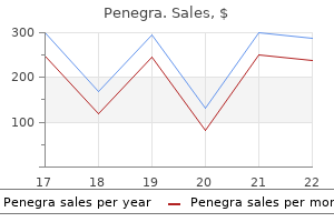
Cheap penegra 100mg visa
Lesions (especially lacunar infarction) involving the corticospinal tracts in the basis pontis may produce a pure motor hemiplegia with or without facial involvement androgen hormone jacksonville order 50mg penegra with amex. A combination of dysarthria and a history of previous transient gait abnormality or vertigo favor a pontine lesion as a cause of pure motor hemiparesis rather than a more common internal capsular lesion. In this syndrome, facial weakness and severe dysarthria and dysphagia occur together with clumsiness and paresis of the hand. Hyperreflexia and a Babinski sign may occur on the same side as the arm paresis, but sensation is spared. Vascular Supply of the Pons the blood supply to the pons derives from three groups. First, paramedian vessels (four to six) arise from the basilar artery and penetrate perpendicularly into the pontine parenchyma. They supply the medial basal pons, including the pontine nuclei, corticospinal fibers, and medial lemniscus. Second, short circumferential arteries also arise from the basilar artery and enter the brachium pontis. In this syndrome, hemiparesis, which is more severe in the lower extremity, is associated with ipsilateral hemiataxia and occasionally dysarthria, nystagmus, and paresthesias. The ataxia is unilateral, probably because transverse 512 Brainstem Syndromes fibers originating from the contralateral pontine nuclei (and projecting to the contralateral cerebellum) are spared. Locked-in syndrome Bilateral ventral pontine lesions (infarction, tumor, hemorrhage, trauma, or central pontine myelinolysis) may result in the locked-in syndrome (deefferented state). It consists of ipsilateral cerebellar ataxia due to involvement of cerebellar connections, contralateral hemiparesis due to involvement of the corticospinal tract, and variable contralateral hemihypesthesia for pain and temperature due to involvement of the spinothalamic tract. Pontine hemorrhage Pontine hemorrhage usually arises from paramedian arterioles and often begins in the basis pontis. Signs and symptoms of pontine hematoma depend on size, location, and the presence or absence of ventricular rupture or hydrocephalus. Massive (classic) pontine hemorrhages cause coma, decerebrate rigidity, quadriparesis, hyperthermia, absent horizontal eye movements, and miotic but reactive pupils. An acute locked-in syndrome may occur, but often these lesions symmetrically dissect the pons, destroying the more dorsal structures. With ventral extension, there may be contralateral hemiparesis (due to corticospinal tract involvement) or paralysis of conjugate gaze toward the side of the lesion (due to involvement of the paramedian pontine reticular formation). The paramedian vessels (the retromammillary trunk) arise from the origins of the posterior cerebral arteries and include the thalamoperforating arteries (supplying the thalamus) and the peduncular arteries (supplying the medial peduncles and the midbrain tegmentum, including the oculomotor nucleus, the red nucleus, and the substantia nigra). Convergence-retraction nystagmus on upward gaze (especially elicited by inducing upward saccades by a downmoving optokinetic target). During horizontal refixations, the abducting eye may move more slowly than the adducting eye (pseudoabducens palsy), perhaps reflecting excess convergence tone. Mesencephalic Hemorrhage Hemorrhage within the mesencephalon often presents with headache and vomiting followed by loss of consciousness. Unequal pupils, which are unreactive to light but retain the near reflex, are common, as is impairment of conjugate upward gaze. When supranuclear fibers for horizontal gaze are interrupted in the medial peduncle, a supranuclear-type conjugate gaze palsy to the opposite side may occur (the midbrain syndrome of Foville). Top of the Basilar Syndrome Occlusive vascular disease of the rostral basilar artery, usually emboli, frequently results in the top of the basilar syndrome due to infarction of the midbrain, thalamus, and portions of the temporal and occipital lobes. Upper Motor Neuron Lesions Dorsal Mesencephalic Syndromes Dorsal rostral mesencephalic lesions produce mainly neuroophthalmological abnormalities. This syndrome includes all or some of the following signs: Further Reading Bassetti C, Bogousslavsky J, Barth A, and Regli F (1996) Isolated infarcts of the pons. Bassetti C, Bogousslavsky J, Mattle H, and Bernasconi A (1997) Medial medullary stroke: Report of seven patients and review of the literature. Kataoka S, Hori A, Shirakawa T, and Hirose G (1997) Paramedian pontine infarction. Tatu L, Moulin T, Bogousslavsky J, and Duvernoy H (1996) Arterial territories of human brain: Brainstem and cerebellum. Terao S, Izumi M, Takatsu S, Takagi J, and Mitsuma T (1998) Serial magnetic resonance imaging shows separate medial and lateral medullary infarctions resulting in the hemimedullary syndrome. Vaudens P and Bogousslavsky J (1998) Face-arm-trunk-leg sensory loss limited to the contralateral side in lateral medullary infarction: A new variant. Vuilleumier P, Bogousslavsky J, and Regli F (1995) Infarction of the lower brainstem. It involves the administration of an electrical stimulus to the scalp via two electrodes, passage of the circuit through the brain, and with consequent generalized seizures. In most countries, the procedure is performed under general anesthesia in a hospital setting, with an attendant psychiatrist, anesthesiologist, and the nursing staff. Adjustments in the number and timing of treatments, stimulation parameters, and electrode placement are made according to the observed therapeutic effects. Ladislas von Meduna had observed earlier that recurrent seizures were associated with psychiatric improvement in patients with cooccurring epilepsy and psychosis, and began the practice of chemical seizure induction in nonepileptic psychotic patients. Earlier to the routine use of neuromuscular blocking agents, bone fractures occurred, and the early technique and dosing is said to have caused a more cognitive impairment than today. It is distinctive among psychiatric treatments for both its speed and degree of effectiveness. It has been shown to be particularly effective in cases of depression with psychotic features, and is also highly effective for bipolar mania, with or without psychotic features. It is also useful but less commonly used for acute schizophrenic psychosis, particularly in schizoaffective disorders or other conditions in which there is a prominent mood component, and for catatonia. Notably, response rates are higher among the elderly, and among patients with psychotic depression and catatonic features. Advances in clinical technique and research methodology have led to a greater understanding of the relationship among electrode placement, electrical dosage relative to seizure threshold, therapeutic efficacy, and side effects. Although study results vary and consensus is elusive, data from welldesigned randomized trials suggest that bilateral electrode placement with a stimulus energy of 1. It is a very safe procedure, with the risk of serious procedural complications or death paralleling those of anesthesia itself. Postictal confusion is typical, and usually lasts for less than 45 min, while the patient is in recovery. Anterograde amnesia (impaired encoding of new memory) is common, occurring during the treatment period and resolving soon after. Retrograde amnesia (impaired retrieval of established memories) can be especially troubling to patients. The two forms of memory disturbance are generally transient, with a return to normal functioning in most patients within several weeks to months. Memory disturbances increase with the number of treatments administered, and are more prominent with bilateral electrode placement. Attention should be given to control unusually high or sustained hypertension, while being careful to avoid bradycardia or hypotension. Patients with recent strokes or cerebral aneurysms are at a higher risk due to the possibility of recurrent infarction or hemorrhage. High-frequency stimulation causes an excitatory neuronal response, while low-frequency stimulation causes an inhibitory one. These effects have been observed to influence the neuronal activity in areas connected to the stimulated region. Once the treatment parameters are established, the daily treatments may be administered by a nurse or technician. However, patients with epilepsy may be at an increased risk for prolonged or tardive seizures. Consultation with the treating neurologist and careful informed consent on these points are recommended. Although no systematic research on safety and efficacy has been conducted, there are a number of published case reports that describe good outcomes. This is the energy required to cause involuntary movement in the contralateral thumb when the coil is held over the motor cortex. However, positive results have been obtained with both longer and shorter courses. The results of controlled trials, in general, have shown clinically modest but statistically significant rates of response in patient populations exhibiting mild or moderate levels of prior treatment resistance. The effects of stimulation can be either excitatory or inhibitory, depending on the energies used. Postoperative confusion is more common, especially in the elderly, but is usually transient.
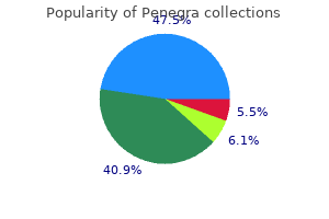
Penegra 100 mg generic
Clinical Symptoms and Signs Most patients with atherosclerosis and basilar artery occlusion have transient ischemic attacks that precede strokes prostate 75 cheap penegra 50 mg overnight delivery. The most common symptoms and signs in patients with basilar artery thrombosis involve the motor and ocular motor systems. The corticospinal tracts in the basis pontis are the most frequently involved structures. Most patients with symptomatic basilar artery occlusive disease and pontine ischemia have some degree of paresis and corticospinal tract abnormalities either as part of their stroke or as a component of transient ischemic attacks. The abnormality might consist of slight weakness, hyperreflexia, or an extensor plantar reflex. The weakness is often asymmetric, but usually there are abnormalities on the nonparetic side such as abnormal spontaneous movements, shivering, twitching, shaking, or jerking. Toe-to-object and heel-to-shin testing usually shows that there is a rhythmic cerebellar-type component to the dysfunction. The ataxia is almost always bilateral but may be asymmetric and more severe on the weaker side. Weakness of muscles innervated by lower cranial nerve motor nuclei is also very common and an important cause of morbidity and mortality. The bilateral involvement is usually due to involvement of upper motor neuron fibers in the dorsal part of the basis pontis near the central tegmental tracts. Symptoms include facial weakness, dysphonia, dysarthria, dysphagia, and limited jaw movements. Some patients become unable to speak, open their mouth, protrude their tongue, swallow, or move parts of their face at will or on command. Secretions pool in the pharynx and aspiration is an important and serious complication. Ocular motor abnormalities are also prominent and are due to involvement of the oculomotor nuclei and their fascicles, abducens nuclei, medial longitudinal fasciculi, and the paramedian pontine reticular formation (often called the pontine gaze center) structures located in the medial pontine Pathology the most common and important disease that affects the basilar artery is atherosclerosis. In some necropsy studies, the basilar artery is the earliest and most severely affected intracranial artery. The earliest atherosclerotic change in the basilar artery is an increase in connective tissue, especially in the intima and media. Sometimes the predominant increase in connective tissue is in the adventitia, with strands of collagen spreading into the media causing medial fibrosis. Thickening of the internal elastica and splitting of the elastic membrane are common. The intimal connective tissue increases in size and may replace the internal elastic elements and become hyalinized and fragmented, especially in its deeper layers. Later, fibrous plaques that are grossly elevated form and thickened regions that are grossly visible along the basilar artery form and gradually lead to stenosis of the artery. As the basilar artery lumen becomes progressively stenosed, cracks in plaques and mural thrombi may appear. In most autopsied patients with fatal basilar artery-related brainstem infarction, superimposed thrombosis of the vessel has developed. The atherosclerotic changes are relatively evenly distributed in the proximal, middle, and distal portions of the artery. Some thrombi originate in an intracranial vertebral artery and propagate into the basilar artery. Emboli most often arise from plaques and clots from the heart, aorta, and the extracranial and intracranial vertebral arteries. Emboli that reach the basilar artery Encyclopedia of the Neurological Sciences, Volume 1 doi:10. Double vision, internuclear ophthalmoplegia (loss of adduction of the ipsilateral eye and abducting nystagmus of the contralateral eye), sixth nerve palsy, and conjugate lateral gaze palsy are the clinical findings. Coma and a reduced level of consciousness are due to involvement of the reticular formation nuclei in the medial tegmentum of the pons. Although some patients with basilar artery occlusion report paresthesias, sensory abnormalities are usually not a prominent part of the clinical picture. Maintenance of blood pressure and blood volume helps to maintain blood flow to the brainstem. Cerebral Venous Thrombosis Further Reading Outcome and Treatment When the rostral part of the basilar artery becomes occluded causing bilateral rostral pontine and midbrain ischemia, the prognosis is very poor. In other patients, basilar artery occlusion can cause only minor transient neurological abnormalities. Although there have been no randomized trials of treatment in patients with basilar artery occlusion, anticoagulants are often given in an attempt to diminish propagation and embolization of the basilar artery thrombus. He championed the concept that afferent muscular impulses play a part in coordinated movements. He made a celebrated study of nematodes, and later was a forceful proponent of spontaneous generation. He was a house physician in neurology at the State Asylum for Criminal Lunatics, Broadmoor, in 1865, and was then appointed assistant physician and lecturer in Pathology at St. In 1868 he was appointed physician to the Hospital for the Epileptic and Paralysed, later the National Hospital, Queen Square, London. He was professor of the Principles and Practice of Medicine in the University of London from 1887 to 1898. He was a fellow of the Royal Society and of the Royal College of Physicians of London. This work earned him the fellowship of the Royal Society at the age of 31, though he gave up his study of nematology when he developed an allergic reaction to them. He began his long and productive neurological career with a series of articles on the controversial topic of aphasia. He described patients with focal brain pathology who had circumscribed inability to read, to write, or to understand spoken language, leading him to propose discrete brain centers for each of these functions. Bastian was a key participant in the debate on the concept that sensory organs in muscles send information about movement to the brain. He coined the term kinesthesia to describe sensory information derived from movement, which he thought could be conscious or unconscious. He thought that this muscular sense projects to the prerolandic cortex, which is therefore intrinsically sensory rather than motor, and that some type of cortical sensory event initiates all movement. Bastian was an astute neurologist, one of the first to practice scientific bedside localization. His books on the localization of paralysis, and his clinical lectures on the same subject, established his reputation as a teacher of scientific neurology. He also wrote a treatise on hysterical and functional paralysis, in which he firmly distinguished between the two. He was a lecturer on nervous diseases in New York, a physician of nervous disorders to the Demilt Dispensary, and a fellow of the New York Academy of Medicine and the New York Academy of Sciences. When civilization, plus these five factors, invades any nation, it must carry nervousness and nervous disease along with it. Regarding the pathogenesis and treatment of neurasthenia, Beard, Michell, and other neurologists promoted an organic paradigm of nervous exhaustion, in which stimulating and restorative treatments were purported to replenish depleted neural energy stores. Electrical treatments and the rest cure were increasingly seen to be effective (if they were at all) more for psychological reasons than for any physical change in the brain. In addition, the medical and cultural view of the disease shifted so that neurasthenia was seen more as a problem of lack of work among the lower classes, rather than a problem of overwork of the upper classes. Ultimately, progressive American psychiatrists rejected the strict somaticism of the organic neurologists and instead adopted the European view of a nonorganic origin for such complaints, particularly with the development of the psychodynamic formulation of the neuroses. Thus, after its period of phenomenal popularity between 1890 and 1910, neurasthenia was increasingly discounted and ultimately abandoned as a diagnostic entity. Beard was to be accepted, he should feel like throwing his diploma away and joining the theologians. Putnam, for example, reported that he had never seen instances of cure when an actual disease existed. However, in the year that followed the general tone of the discussion favored Beard. Seguin, for example, argued in essence that the end justified the therapeutic means. Jumping was rarely discussed in the medical literature after 1912, until further cases were described in the mid-1960s.
Syndromes
- Difficulty breathing because the lungs are "wet," congested, or filled with fluid (heart failure).
- Gordofilm
- Damage to the nerve that moves the face on the side of the operation
- Flank pain
- Pain during intercourse
- Fever
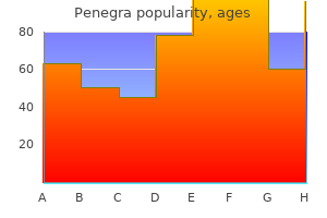
Discount 100 mg penegra with amex
The only way to be sure is to avoid the allergen for a week (which will result in improvement in the eczema) and then to reintroduce it again to establish a rapid relapse in the eczema mens health vitamins order penegra cheap online. Some children under age 3 also have gastro-intestinal symptoms with a good history of a dietary trigger. This food intolerance may be temporary, and reintroducing the item several months later may be possible. Environmental allergens (cats, house dust mite) are difficult to avoid, and a positive reaction is generally unhelpful. Complete emollient therapy the use of emollients on dry and eczematous skin is an important part of the treatment and prevention of atopic eczema. Emollients should be used liberally and frequently for moisturising, washing and bathing, even if the skin is clear. Everything that goes on the skin should be emollient based so that the skin barrier is repaired and maintained. Lighter (cream) emollients can be used for daytime or summer, and heavier (ointment based) emollients used at night and in winter. Patients should be offered a leave-on emollient, a product to wash with (soap must be avoided) and a bath emollient. Aqueous cream is best avoided as it contains sodium lauryl sulphate, which is a skin irritant. Moisturisers should also be applied to the skin after getting out the bath, while the skin is still warm and moist. Apply the moisturiser starting at the neck, and smoothing (not rubbing) it in downwards on the trunk, arms and legs. The moisturiser should be applied first, and left on for 20 minutes before applying a topical steroid. Some products such as Doublebase Dayleve and Oilatum cream contain film-forming agents that form an additional protective layer over the surface of the skin, helping to reduce water loss and penetration of irritants and allergens. Emollients which contain antiseptic products such as chlorhexidine and benzalkonium chloride (Dermol cream, Eczmol cream) may help to reduce bacterial colonisation and infection, which can exacerbate eczema. Antipruritics such as lauromacrogols have properties of a mild topical anaesthetic and have an antipruritic effect (Balneum Plus cream, E45 Itch Relief cream). Allergen-free products such as ointments or products containing no preservative Emollient sprays are useful in elderly patients who have difficulty in reaching areas of skin (back, lower legs). Topical steroids Topical steroid ointments are still the mainstay of treatment for atopic eczema. Patients or parents are often afraid of using topical steroids because of the widespread publicity about side effects. Failure to treat active eczema will cause more harm to the patient by allowing allergens to penetrate the defective cutaneous barrier. By using a steroid with the appropriate potency to quickly control the eczema and continuing treatment until completely clear will reduce the likelihood of a rebound flare. Then taper therapy to alternate days before reducing to proactive treatment: twice weekly applications of a topical steroid to previously affected skin for up to a month. Continuous long term use of potent steroids on the face and body folds must be avoided, and a topical calcineurin inhibitor may be used instead (see p. Ointments work much better than creams, since the grease forms an occlusive barrier preventing evaporation of water and delivers the steroid more effectively to the skin. First apply a moisturiser all over, and after 20 minutes the topical steroid can be rubbed into the eczematous areas. It is important to give the patient enough ointment, and to monitor the amount being used (too little may be as bad as too much). A record of the amount of steroid prescribed and used (ask the patient or parent to bring back used tubes) will be helpful in monitoring this. Soak the affected areas in one of these solutions by applying moistened gauze swabs or by emersion in a bath or hand basin. Topical immune-modulators these drugs work by blocking the molecular mechanisms of inflammation in the skin (see p. Use in the following situations: proactive (preventative) treatment twice weekly to previously affected areas patients with facial, periocular or neck eczema patients who have not responded, are using too much or too strong a steroid, or who have developed steroid side effects patients with poor compliance with topical steroids because they are afraid of the side effects patients with perioral dermatitis (see p. Treatment of secondary infection If the eczema becomes suddenly worse, infection with S. Wrapping techniques Localised wrapping or occlusion will cool, soothe and protect from scratching inflamed, excoriated or lichenified skin. Because the zinc paste is sticky, it will need to be covered by a tubular bandage or secondary dressing. Generalised wrapping with a suit consisting of long sleeved vest, leggings, socks and mittens. It prevents scratching and the occlusion provided by the garment allows better penetration of the moisturiser. The affected areas of skin are treated with a topical steroid ointment, and then the whole body surface is covered with a suitable emollient (see p. They are available made of viscose (Acti-Fast, Comfifast, Easifast, Skinnies, Tubifast) or silk (Dermasilk, Dreamskin, Skinnies). The advantage of silk is that it is cooler, requires less moisturiser on the skin, and does not wear out, but it is more expensive. Wet wrapping is very effective in bringing a generalised flare of eczema in children and adults under control quickly. It cools the skin, improves moisturisation, prevents scratching and reduces the need for topical steroids. Two layers of the therapeutic suits (as above) are put on following application of moisturiser and topical steroid. The layer adjacent to the skin is moistened with luke-warm water, squeezed dry and then a second dry layer is put on over this. Many children do not like it initially, but once put on the skin feels so much better that they can feel the benefit. On removal, if the garment sticks to the open eczematous areas, it should be soaked off in a bath. Antihistamines Sedative antihistamines are useful if the child is not sleeping at night and is keeping the family awake. They will not stop the itching, but if given in adequate dosage the child will sleep through the night. In adults, start with 10 mg of promethazine, hydroxyzine or alimemazine and double the dose as necessary until the patient sleeps through the night. They can be tried if sedative antihistamines produce unacceptable drowsiness, or during the day. Then apply alternate days for 2 weeks, and thereafter only twice weekly up to a month. On left as removed from packaging which can be applied directly to skin (right leg), and then covered with blue line tubifast (left leg). Either apply single layer dry, Or moisten first layer with luke warm water, squeeze dry, and put on a second dry layer over it. It is suggested that this table can be reproduced and given to the patient as instructions of what to use where. Other considerations Cotton clothing worn next to the skin is comfortable, while wool is irritating. Vacuum all household carpets (especially around skirting boards), fabrics, and sofas daily. Laminated floor coverings are preferable to carpet in the bedroom as they can be damp mopped. Keep pets out of the bedroom particularly at night and do not allow them to sleep on the bed. Only avoid contact with animals if definite improvement away from them (and relapse on re-exposure) can be demonstrated. Patient and parent support can be provided by specialised dermatological nurses, the local dermatology department or the National Eczema Society. Creams containing parabens, antibiotics, antihistamines or even a topical steroid may also cause an allergic dermatitis (see also pp. In theory avoidance results in a cure, although in practice this is not always the case. The wet type consists of plaques made up of numerous vesicles that break to produce exudate Some patients with atopic eczema, allergic contact dermatitis or unclassifiable eczema also have discoid plaques of eczema. Red macules appear around follicles and gradually coalesce to areas of widespread erythema.
Discount penegra 50mg without a prescription
Selective attention can occur overtly in response to a task demand and the requirement to execute a response prostate oncology 77058 order penegra online now, or covertly in an automatic reflexive manner, such as when we respond to a loud noise in the environment. Other elements of attention are evident in the context of particular behavioral and cognitive demands. The nature and degree of attentional inconsistency are indicative of sustained attention. Accordingly, attention can be distilled into several key elements or component processes. A number of neuropsychological theories and models have been developed to delineate and operationalize the primary component processes underlying attention. Before considering some of these models, the authors will briefly review past cognitive and neuropsychological approaches to the study of attention in order to illustrate how current knowledge about the cognitive and neural substrates of attention evolved. Early Cognitive Approaches As cognitive psychology began to coalesce as a field in the middle of the twentieth century, attention became a major subject of investigation for researchers working from the perspective of information theory. Attentional processes facilitate cognitive and behavioral performance in several ways. Attention serves to reduce the amount of information (the number of stimuli) in order to receive additional cognitive processing, whereby particularly salient information could be selected from the large quantity of stimuli initially processed in parallel for more intensive serial processing. Given that people are constantly flooded with an almost infinite number of signals from both the outside world and within, an attentional process was considered to be necessary so as to restrict the information to be processed in accordance with the available capacity of the individual. Accordingly, selection was considered to be a key element in most cognitive theories of attention. Selective attention was conceptualized as a filter (Broadbent) or an attenuation (Treisman) process. These theories tended to view attention as a unitary operation occurring at some point in the stream of information processing. Considerable research and debate ensued as to whether this selection process occurred at an early stage of processing soon after sensory registration or at a later stage based on prevailing response demands (Deutsch and Deutsch). Studies employing dichotic listening paradigms without preexisting demands to attend to a particular type of information demonstrated that selective attention does occur at a very early stage of sensory processing, leading many cognitive psychologists to conclude that late-stage models of selection were not correct. Yet, cognitive research also showed that it was possible to bias attentional selection by creating particular cognitive or behavioral response demands. As Schneider and Shiffrin demonstrated in a series of seminal experiments, both the accuracy and speed with which people could perform visual selective attention varied as a function of the memory demands implicit in a task. When there was little demand for working memory, selection could proceed relatively automatically. However, when the objects of attention were not consistent or familiar, greater attentional control was required. This eventually led to what has become a well-established distinction between automatic and controlled forms of attentional processing. A related, although somewhat separate, line of research evolved, which focused on delineating factors that influence Attention 305 the consistency of attentional performance over time. Clearly, attention is temporally distributed, as performance is never entirely consistent over time even among people who are attending optimally. Accordingly, vigilance and sustained performance were important elements of attention that needed to be considered in addition to selective attention. Besides being selective and having a temporal dynamic (sustained attention), it was also apparent to many cognitive scientists that attention also had an intensity that varied in accordance with the task, situation, and the biological state of the person who is attending. People have a limited capacity to selectively engage and focus their attention in an effective manner. Kahneman was among the first cognitive scientists to link attentional capacity to intrinsic and extrinsic biological factors, such as arousal, motivational state, and reward. Subsequent psychophysiological research grounded attention in a biobehavioral context. Pavlov had explicitly conceptualized that orienting to a potential conditioned stimulus was necessary before classical conditioning took place. The neuronal substrates of habituation and sensitization have been established for simple organisms Attention and memory encoding are highly interdependent even at the most elementary levels of neuronal activity. Cognitive scientists in the middle of the twentieth century tended to focus primarily on selective and sustained attention. Yet, neuropsychological studies found that patients with brain dysfunction had problems with other aspects of attention, such as responding behaviorally in a consistent manner in the context of changing task demands. Patients with executive dysfunction also frequently have problems directing attention to available response alternatives, along with impairments of response intention and selection. Heilman and colleagues conducted a series of seminal studies showing that sensory attention and response intention can be dissociated. Clearly, attention and executive functions were strongly associated with one another. Shalice proposed one of the first cognitive models to formally link attention and executive functions through a supervisory attention frontal system. Stuss elaborated on this relationship and concluded that processes involved in executive attention represented distinct attentional components. Posner and other cognitive researchers reached similar conclusions and as their theories of attention evolved, executive attention has been dissociated from sensory selective attention. This dissociation is reflected in a distinction that is now made between anterior and posterior brain attentional systems. Attention is clearly not a unitary process occurring at a single stage of cognitive processing as proposed in early cognitive theories. Instead, it refers to a set of cognitive processes that enable the selection of stimuli and response and focus over time. For example, memory is not a unitary process, but rather involves encoding, storage, and retrieval. In fact, short-term memory and working memory have attentional underpinnings, as they are highly dependent on the quality of attentional focus. Elements of Attention There is now considerable confluence among current neuropsychological models of attention. Most distinguish between attention involved in sensory and perceptual selection and attention linked to the intention, selection, and control of behavioral responding. Furthermore, most current models now recognize the influence of level of consciousness (alertness), arousal, and other organismic factors on attention. Pribram and McGuiness proposed one of the first comprehensive neuropsychological models of attention based on neurophysiological evidence from animal studies in which attentional control occurred through the integration of arousal, activation, and effort. Heilman and colleagues proposed models to account for hemiinattention and intention disturbances associated with neglect syndrome that were driven by evidence regarding the functional neuroanatomy underlying these conditions based on findings from patients with stroke and other neurological brain disturbances. These models provided a comprehensive description of the multitude of brain systems involved in these syndromes and therefore were extremely important in delineating the neural systems responsible for attention. The neural systems now known to govern attention will be discussed in greater detail in the section Sensory Selection. These three networks map on closely to the functional organization of attentional processes described in the section Elements of Attention. Orienting involves the allocation of attention toward exogenous cues and most closely relates to sensory selective attention. The three neural systems proposed by Posner are relatively broad in scope and really are comprised of a large number of interrelated brain subsystems as described in earlier models of neglect syndrome. Mirsky took an alternative approach, employing factor analytic statistical methods to examine neuropsychological performance on tests thought to be sensitive to attention. Five factors were originally identified: (1) encoding, (2) focusing, (3) executive, (4) sustaining, and (5) shifting. Validation studies provided some support for this taxonomy of attention, although there was also some evidence that these attentional elements reflect processes occurring at different levels. Cohen proposed an alternative, although somewhat similar, framework in Neuropsychology of Attention based on factor analytic analyses. These components reflect attentional processes that had been identified and well studied in the cognitive sciences and neuropsychology validated by neuropsychological performance data. Sensory Selection Early in the cognitive stream of processing, initial attentional selection occurs, such that attention is oriented to and engaged in particular sensory stimuli, whereas others are ignored. This process involves active facilitation of attention response to the selected object, whereas ignoring or even actively inhibiting attention to other stimuli. However, unlike attention occurring at subsequent stages of cognitive processing, selective attention can occur covertly and automatically, especially when task demands are not excessive. In reality, there are multiple processes that underlie sensory selection, and these processes vary according to the sensory modality that is considered.
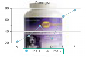
Order cheap penegra
Although the size of the vessels is increased androgen hormone pdf 100mg penegra sale, their number is thought to be relatively normal. Capillary telangiectases are the second most common cerebral vascular malformation. They are believed to be congenital lesions, arising from a localized failure of the involution of brain capillaries that normally occurs during the second month of gestation. Most capillary telangiectases are an incidental finding at autopsy and have a benign clinical course. However, clinically symptomatic lesions have occasionally been reported, usually in association with other cerebral vascular lesions. Combined lesions with elements of both cavernous malformations and capillary telangiectases are well documented. The occurrence of combined lesions, containing elements of both cavernous malformations and capillary telangiectases as well as transitional forms that are difficult to classify into either category, has led several authors to propose that capillary telangiectases are in fact the precursors of cavernous malformations. The stereotypical lesions are absent at birth, developing on a delayed basis during the second and third decades of life. Radiographically, capillary telangiectasias are not visualized by cerebral angiography. Papilledema from raised intracranial pressure to vasodilatation is variably present. Investigations the diagnosis is made on the basis of the clinical picture, exclusion of confounders and mimics, and the performance of arterial or capillary blood gases. Anything that can cause hypoventilation can cause hypercapnia: starting at the brainstem and spinal cord level, infarcts, demyelination, tumor (either intrinsic or extrinsic and compressive), sedative drugs and opiates, and syringobulbia may be responsible. High spinal cord lesions can also produce hypercapnia if lesions involve the medullary respiratory center or its outflow through the reticulospinal tracts in the anterolateral funiculi of the spinal cord. Other spinal cord problems, such as motor neuron disease, can directly affect the outflow from the spinal cord. Myopathies, especially those affecting the diaphragm, are other neurological causes. Nonneurological causes of hypoventilation are actually more common than neurological causes. Chronic obstructive, restrictive, and destructive (affecting alveoli) lung disease and chest deformities are common, but these often require a precipitating cause such as a chest infection or the use of sedative drugs to cause decompensation and severe hypercapnea. Sleep-related breathing disorders, including central and obstructive sleep apnea, can also lead to hypercapnia, but seldom is this severe. Management Underlying causes and precipitants should be sought and treated if possible. If the patient is obtunded, an endotracheal tube is inserted to protect the airway. With chronic conditions in which the diagnosis is known and the cause is not reversible, for example, motor neuron disease, it is best to discuss the management of respiratory failure with the patient and close family members before it occurs. Many patients with such a prognosis do not wish to be placed on a ventilator, but rather elect for conservative management with emphasis on palliation of distress. Ortiz Vasquez J (1979) Neurological manifestations of chronic respiratory disease. Piper A (2010) Obesity hypoventilation syndrome: Therapeutic implications for treatment. Clinical Features Patients with symptomatic hypercapnia of a chronic or recurring nature are often worse in the morning because of reduced respiratory drive during sleep and periods of hypoxia. Introduction Cardiac arrest is characterized by cessation of effective mechanical contraction of the heart resulting in complete cessation of blood flow and thus oxygen delivery to the entire body. Complex and coordinated postcardiac arrest care is required to both prevent rearrest and to provide optimal conditions for neurological recovery and return of the individual to prearrest function. Why short periods of global brain ischemia are less well tolerated than longer periods of stroke is not entirely clear but may be due to the involvement of brain areas called the hippocampus (responsible for many aspects of memory and learning), which are not commonly injured during stroke. Electrical therapy in the form of defibrillation is used to treat certain heart rhythms that occur during cardiac arrest (ventricular fibrillation and ventricular tachycardia). Patients exhibiting either of these rhythms as their initial rhythm and who are treated rapidly with defibrillation have the best chance of survival and favorable neurological outcome. Pharmacological therapy is aimed at enhancing blood flow to the heart and brain by chest compressions (using vasopressors such as epinephrine and vasopressin) and making defibrillation of the heart easier (using antiarrhythmics such as amiodarone). Although seemingly heart specific, these therapies contribute to producing the best neurological outcome possible by ensuring optimal postcardiac arrest blood Epidemiology of Cardiac Arrest the epidemiology of cardiac arrest is complicated by the fact that it is a symptom of numerous disease states and it occurs at least once in every human. Worldwide, it is estimated that well over 2 million people are treated for unexpected cardiac arrest every year. Pathophysiology of Cardiac Arrest the brain and heart are the organs most susceptible to damage from the lack of oxygen delivery that occurs during cardiac arrest mainly due to their high metabolic rates and limited ability for cell division and repair. Furthermore, myocardial infarction is a major cause of sudden death, and the combination of focal and global myocardial ischemia makes resuscitation all the more difficult. In the brain, energy stores are depleted within 2 min of cardiac arrest onset and irreversible ischemic brain damage begins to occur after as few as 5 min. Restoration of prior neurological function rarely occurs after durations of untreated cardiac arrest greater than 10 min. The majority of these deleterious molecular mechanisms that occur during and after cardiac arrest are shared with other types of brain insults, such as stroke or traumatic brain injury, and include equalization of membrane gradients and activation of proteases, phosphatases, and lipases. In general, the poor neurological outcomes associated with survival from cardiac Encyclopedia of the Neurological Sciences, Volume 1 doi:10. Unfortunately, no specific postcardiac arrest pharmacological therapy aimed at combating the molecular pathophysiology of cardiac arrest-induced brain injury has proven successful in clinical trials (oxygen-free radical scavenging, excitatory amino acid blockade, etc. The mechanisms by which hypothermia improves neurological outcome are not clearly known but are likely to be pleiotrophic. Despite its clear ability to improve outcome, great debate still surrounds therapeutic hypothermia in terms of methods of cooling (endovascular, surface, and intranasal), rapidity and degree of cooling, and length of cooling needed (hours to days). Implementation of therapeutic hypothermia requires a significant institutional and multidisciplinary commitment. Therapeutic hypothermia can also be associated with an increase in risk of infection, seizures, electrolyte abnormalities, and coagulopathies, thus requiring close monitoring to mitigate such complications. Much more research is needed to derive and validate prognosticators of both good and poor outcome in the new era of improved postcardiac arrest care. However, much remains to be discovered and clinically translated to further improve neurological outcomes after cardiac arrest. Bass E (1985) Cardiopulmonary arrest: Pathophysiology and neurologic complications. Hypothermia after Cardiac Arrest Study Group (2002) Mild therapeutic hypothermia to improve the neurologic outcome after cardiac arrest. In patients who awaken early after cardiac arrest, prognosis is dependent primarily on cardiovascular function and any underlying disease. Neurological complications of cardiac arrest include seizures, postanoxic myoclonus, amnestic syndrome, persistent or transient vegetative states, cortical blindness, cognitive impairment, cortical infarcts, secondary parkinsonism, hypoxic ischemic leukoencephalopathy, spinal stroke, and brain death. In general, headache, fatigue, malaise, drowsiness, and generalized muscle weakness are common neurological features of cardiac glycoside toxicity, but dizziness, vertigo, syncope, apathy, lethargy, excitement, euphoria, insomnia, irritability, agitation, hiccups, restlessness, nervousness, seizures, opisthotonos, stupor, and coma may also occur. Neuropsychiatric disturbances are especially likely to develop in geriatric patients with atherosclerotic disease, and are easily overlooked in patients receiving chronic cardiac glycoside therapy. These effects include disorientation, confusion, depression, memory impairment, amnesia, aphasia, bad dreams, delirium, delusions, illusions, and hallucinations. Visual disturbances induced by toxic doses of cardiac glycosides probably result from a direct effect on the retina (cones are affected more than rods). Color vision is commonly affected and objects may appear yellow or green or, less commonly, brown, red, blue, or white. Blurred vision flashes or flickering of light, photophobia, halos or borders on objects (often are white and appear on dark objects), diplopia, macropsia, and micropsia may occur. Transient or permanent amblyopia and scotoma, including teichopsia, have also occurred. The incidence and severity of digoxin toxicity Antihypertensives Several drugs used to treat hypertension may adversely affect the nervous system. It is important to keep in mind that hypertension itself is a risk factor for vascular dementia and that aggressive lowering of blood pressure may also have a deleterious effect on cognition.
Cheap penegra online master card
It can occur on the feet from tight shoes man health magazine men health purchase penegra with amex, on soles after climbing a ladder, around the waist, on palms from gripping, and so forth. Occasionally the skin lesions may be widespread and associated with blisters or erosions in the mouth. Unfortunately there is no simple test to identify the cause of a drug rash, and if patients are taking numerous drugs it is often not possible to be sure of the exact cause or even whether a drug is responsible (a viral exanthem may look identical). This leaves the doctor with a problem, not only during the current episode but also in the future, because he or she needs to know whether it is safe to prescribe the drug again. A complete list of all drugs the patient is taking (prescribed, over-the-counter or borrowed). It is more likely that a rash is from a recently started drug than one that has been taken for years. A drug rash is unlikely to have developed in less than 4 days if the drug has not been taken before. In the United Kingdom, antibiotics, sleeping tablets and tranquillisers are the most frequent causative agents. The rash has the typical mauve hue of lichen planus but the lesions are larger and more confluent. Drugs that can cause it include: -blockers chloroquine chlorpropamide ethambutol gold mepacrine methyldopa penicillamine quinine thiazide diuretics. Single or multiple tender, red nodules with a central punctum can occur anywhere on the body except the palms or soles. Without treatment the abscess will eventually point on the surface, discharge and heal leaving a scar. Epidermoid cysts have a microscopic opening and through this, staphylococci can enter. Sudden painful enlargement of a previous cyst is indicative of secondary infection or rupture of the cyst and a subsequent foreign body reaction (see also p. Otherwise, treat with oral flucloxacillin (or erythromycin) 500 mg qid for 7 days. The infection usually comes from the patient him- or herself, so carriage sites should be treated with topical mupirocin, fucidic acid or neomycin ointment twice daily for 2 weeks. If recurrent boils occur even if carriage sites have been treated, take swabs from other members of the family and treat them if infected. Long-term low-dose antibiotics (flucloxacillin or erythromycin 250 mg bid) may be necessary if the boils reoccur or persist. For cysts, excise after the infection has settled down, not at the time the cyst is red and painful. The virus, varicella zoster, lies dormant in the dorsal root ganglion following chickenpox and later travels down the cutaneous nerves to infect the epidermal cells. For several days before the rash appears there is pain or an abnormal sensation in the skin. The rash is unilateral and confined to one or two adjacent dermatomes with a sharp cut-off at or near the midline. The pain may continue until healing occurs, but in the elderly may go on for months or even years. These drugs are competitive inhibitors of guanosine and because they are converted to the triphosphate by viral thymidine kinase, they are effective only in the presence of actively replicating virus. They are all very expensive so should only be given in the early phase of the disease. The redness and swelling disappears after about 10 days to leave a dark-brown patch that remains for several months (see p. You can confirm which drug is the cause, as giving it again will produce the same reaction at the same site within 2 hours. The patient gives a history of having been in the garden clearing weeds, often using a strimmer, or walking in the countryside on a sunny day. The rash is characteristically linear, made up of blisters where the plants have touched the skin, usually on the lower legs or arms. Sometimes the distribution affects the exposed or light-aggravated sites of face, neck, hands and arms with a chronic lichenified dermatitis (chrysanthemums and other compositae). Compositae dermatitis occurs in florists and gardeners, and if suspected it should be patch tested for. Poison ivy, oak or sumac dermatitis is very common in North America and the Far East. If the rash is due to strimming, the patient should wear trousers tucked inside his or her socks or boots in future. Bullous pemphigoid can on occasions be induced by: clonidine diclofenac furosemide ibuprofen. A pemphigus type of drug reaction is seen with: captopril penicillamine rifampicin. The prodromal illness is usually mild so that the rash is the first evidence of illness. The lesions start off as pink macules, which develop quickly into papules Crops of lesions occur over a few days so that there are always lesions at different stages of development present. The spots are very itchy and secondary infection may lead to pock-like scarring A small bead of pus sits around a protruding hair and there may be slight erythema at the base. One or several follicles may be involved, but there is no tenderness or involvement of the deep part of the follicle. It can be caused by, or made worse by, the application of greasy ointments to the skin, tar preparations or plasters. The wearing of oily overalls may precipitate folliculitis of the thighs (oil acne). In adults and immunocompromised patients, treatment with famciclovir or valaciclovir will reduce the severity of the disease (see p. Treat any infected carriage sites with topical mupiricin, fucidin or neomycin cream bid for 2 weeks. Persistent folliculitis may require long-term suppressive treatment with low-dose oral antibiotics (250 mg bid for up to 6 months). In the newborn you may see intact blisters with pus in them (bullous impetigo, see. Folliculitis is a superficial infection of the hair follicle with a pustule at the opening of the follicle (see p. Deeper infection results in either a boil, if the whole follicle is involved, or a carbuncle, if multiple adjacent follicles are involved (see pp. It causes recurrent skin abscesses and cellulitis, which do not respond to routine doses of flucloxacillin or erythromycin. Do not treat with topical antibiotics, as the infection is deep and they will not work. The ulcer itself will take at least 4 weeks to heal, but antibiotics do not need to be continued for this long. A common mistake is to assume that weeping eczema is infected and treat it with antibiotics rather than topical steroids. Either put the solution in a bath or bowl and soak the affected area or soak a towel or flannel in the solution and apply it to the affected area for 10 minutes four times a day. If a contact allergy is suspected, refer the patient for patch testing once the rash has settled. The source of infection is often an older sibling with impetigo, infected eczema or scabies. The pain will stop almost immediately but the skin peeling takes longer to stop due to persistence of the toxin. Other children in the family may require treatment for impetigo or infected eczema at the same time. The prognosis of toxic epidermal necrolysis can be very poor, especially if large amounts of skin are lost. The extensive skin loss will need to be treated just like a burn and admission to a burns unit or intensive care unit is essential. The immunoglobulins inhibit Fas-mediated epidermal cell death and allow the epidermis to recover.
Paeonia daurica (Peony). Penegra.
- Muscle cramps, gout, osteoarthritis, breathing problems, cough, skin diseases, hemorrhoids, heart trouble, stomach upset, spasms, nerve problems, migraine headache, chronic fatigue syndrome (CFS), and other conditions.
- What is Peony?
- Dosing considerations for Peony.
- Are there safety concerns?
- How does Peony work?
- Are there any interactions with medications?
Source: http://www.rxlist.com/script/main/art.asp?articlekey=96082
Generic 50 mg penegra overnight delivery
Although most of the spiny neurons in the centromedial amygdala are probably projection neurons that send axons to other brain regions mens health big black book of secrets purchase penegra 50mg amex, other aspiny neurons have been seen that may function as interneurons. In general, it appeared that these amygdalectomized monkeys exhibited a specific type of visual agnosia characterized by the inability to recognize the emotional or behavioral significance of sensory stimuli. Subsequent studies revealed that animals with amygdalar lesions also did not respond appropriately to auditory, somatosensory, and olfactory cues. Thus, it appears that the amygdala is critical for producing appropriate behavioral responses to biologically relevant sensory stimuli and events in the external world. In fact, the amygdala is thought to constitute an essential link between brain regions that process sensory information The cortical and medial nuclei receive olfactory information from the olfactory cortex and the main and accessory olfactory bulbs. The latter structure is part of the vomeronasal system, which is involved in detecting special odors (pheromones) that are produced by individuals of the same species. Pheromones elicit hormonal and behavioral responses involved in species-specific reproductive and social activities. The amygdala receives visual and auditory information from the temporal lobe, somatosensory and viscerosensory (including gustatory) information from the insular lobe, and polysensory information from the prefrontal cortex and hippocampal region (including the subiculum, cornu Functional Anatomy of the Amygdala In a classic study performed in 1939, Kluver and Bucy found that bilateral lesions of the amygdalar region rendered monkeys remarkably tame and hypoemotional. These nonolfactory inputs primarily target the basolateral and, to a lesser extent, centromedial amygdala. The basolateral, but not the centromedial amygdalar nuclei, have reciprocal projections back to these same cortical regions. It has been suggested that these amygdalocortical projections may be important for attention to emotionally and behaviorally significant stimuli and for the storage of emotional memories. Connections with Subcortical Brain Regions the amygdala has connections with several subcortical regions, including the basal forebrain, diencephalon, and brainstem. Others course in a thin fiber bundle termed the stria terminalis, which takes a more circuitous route dorsal to the internal capsule. Projections from the dorsal thalamus to the amygdala arise mainly from the midline thalamic nuclei and medial part of the medial geniculate nucleus and adjacent posterior thalamic nuclei. These projections, which terminate primarily in the basolateral and central amygdalar nuclei, convey auditory, somatosensory, viscerosensory, and visual information to the amygdala. Amygdalothalamic projections are more limited and consist of projections from the central nucleus to the midline thalamic nuclei and from the basolateral amygdala to the mediodorsal thalamic nucleus. Because the latter nucleus has extensive reciprocal connections with the prefrontal cortex, it provides an indirect link by which the amygdala can influence the activity of the prefrontal region. In each of these affective states, the amygdala appears to elicit a coordinated response consisting of autonomic, endocrine, and behavioral components by way of its projections to various subcortical regions, especially the hypothalamus. Interestingly, many of the hormones secreted by the glands targeted by pituitary hormones can affect the activity of the amygdala via receptors expressed by amygdalar neurons. Thus, there is a very high density of estrogen and androgen receptors in the medial and cortical nuclei. Glucocorticoid receptors are located in all portions of the amygdala, but particularly high levels are found in the centromedial nuclear group. All connections are reciprocal except those to the caudate and nucleus accumbens, which do not have projections back to the amygdala. Lesion studies indicate that the projections of the basolateral amygdala to the striatum are important for controlling behavior related to the reinforcing properties of sensory stimuli. The central nucleus is the main amygdalar region exhibiting connections with the brainstem and basal forebrain. Among these targets are several brainstem areas involved in visceral function, including the parabrachial nucleus, dorsal vagal nucleus, and nucleus solitarius. It also has projections to the periaqueductal gray and reticular formation, which are important for pain modulation and behavioral responses to stress. In addition, the central nucleus innervates several brain regions that give rise to neurotransmitter-specific fiber systems that target the amygdala and other forebrain areas. These regions include the locus ceruleus (which provides a noradrenergic innervation of the amygdala and cortex), substantia nigra and ventral tegmental area (which provide a dopaminergic innervation of the amygdala and striatum), raphe nuclei (which provide a serotonergic innervation of the amygdala and cortex), and nucleus basalis (which provides a cholinergic innervation of the amygdala and cortex). The latter region is also innervated by portions of the basolateral nuclear group. These transmitter-specific systems are activated in certain behavioral states, particularly during stress, and can modulate amygdalar activities related to emotion, attention, and memory. Functional and Clinical Significance of the Human Amygdala Consistent with the results of animal experiments, recent investigations of the human amygdala have shown that it is critical for the recognition of the emotional significance of auditory, visual, and olfactory stimuli, including facial expressions, vocal intonation, and expressive body movements. It has also been demonstrated that electrical stimulation of the human amygdala elicits fear, rage, or other emotions. Investigations in humans have shown that the amygdala is important for learning conditioned emotional responses (usually fear) to sensory stimuli and events. These findings are in agreement with results of numerous animal studies showing that the amygdala is essential for Pavlovian fear conditioning to simple sensory cues and complex sensory representations, such as the context in which an emotional event has occurred. Additional investigations in humans and animals have demonstrated that the release of noradrenaline in the amygdala is essential for the formation and recall of memories involving emotional events. The amygdala exhibits degeneration in schizophrenia, and recording studies have detected abnormal activity in the amygdala in this condition. There is evidence that dopamine levels are increased in the amygdala in schizophrenia and that this brain region may be one of the main sites of action of atypical antipsychotic drugs Consistent with numerous rodent studies implicating the amygdala in fear and anxiety, there is evidence that anxiety disorders in humans, such as posttraumatic stress disorder, are associated with excessive activity in the amygdala. Moreover, studies in animals and humans have shown that the amygdala has very high levels of benzodiazepine receptors, which is critical for the anxiolytic actions of these drugs. Recent positron emission tomography investigations have demonstrated that there is increased activity in the human amygdala in major depression and that administration of antidepressive medication, which modulates levels of noradrenaline and serotonin in the amygdala, causes a decrease in amygdalar activity that is associated with amelioration of depressive symptoms. This article is a revision of the previous edition article by Ming Cheng, Kris A Smith, volume 1, p 128, r 2003, Elsevier Inc. Amygdalohippocampectomy is a cranial surgery for seizure control that selectively removes the mesial temporal lobe structures with minimal resection of the lateral temporal cortex. Unlike the standard temporal lobectomy, amygdalohippocampectomy is designed to remove only the portion of the temporal lobe that is generating the seizures. The hippocampus contains neurons with specific properties that create an especially low threshold for neuronal damage. When damaged, these cells and cells in the nearby amygdala become the focus of temporal lobe seizures. This condition is known as mesial temporal sclerosis, even when the original damaging events to the hippocampus may have occurred many years previously. By removing the mesial structures only, the surgeon decreases the risk of injury to areas involved with language, cognition, and vision. Failure of seizure control after an amygdalohippocampectomy may be caused by bilateral seizure foci, misdiagnosed temporal neocortical epilepsy, or an extratemporal seizure focus. An approach to remove the amygdala and anterior 3 cm of the hippocampus alone was described as early as 1958 with good seizure control and neuropsychological outcomes. However, the technical difficulty of the procedure led to its abandonment, especially in the setting of successful outcomes of the anterior temporal lobectomy. As microneurosurgical techniques advanced, surgeons developed new methods to perform the amygdalohippocampectomy. Yasargil and Wieser introduced the transsylvian technique in which dissection of the sylvian fissure and a small incision in the temporal stem at the limen insula spared the optic pathways. However, this incision at the temporal stem disconnected tracts between the frontal lobe and the lateral temporal structure. Niemeyer, Bello, and Olivier described a lateral transtemporal corticectomy to reach the temporal horn and mesial temporal structures. Although this approach was less technically demanding, it requires transection of functional temporal cortex. The subtemporal approach allows resection of the mesial temporal lobe structures while minimizing transection of normal temporal lobe and disconnection via the temporal stem. Over the past 10 years at Barrow Neurological Institute, Phoenix, Arizona, a minimal access subtemporal amygdalo- hippocampectomy to treat patients with mesial temporal sclerosis has been used. The use of frameless stereotactic magnetic resonance imaging guidance allows identification of the trajectory to the tip of the temporal horn of the lateral ventricle and a cortical incision into the fusiform gyrus or collateral sulcus. Entry into the temporal horn allows identification of anatomical landmarks of the posterior inferior surface of the amygdala as well as the choroidal fissure overlying the hippocampus. Ultrasonic aspiration is used to resect the uncus, amygdala, hippocampus, and parahippocampal gyrus posteriorly to the plane of the collicular plate. Based on neuropsychological testing, most patients who underwent this surgery demonstrated either stable or improved verbal fluency and visual memory scores.
Discount penegra online mastercard
Proximal median neuropathy around the elbow causes paresthesias and numbness of the thenar eminence prostate cancer yellow skin purchase 100mg penegra with mastercard, and affects the muscles in the forearm responsible for thumb flexion (flexor pollicis longus), arm pronation (pronator teres and quadratus), and wrist flexion (flexor carpi radialis). Patients with mild symptoms may be relieved by the use of a neutral wrist splint during sleep. Addition of a nonsteroidal antiinflammatory drug for 2 or 3 weeks can also decrease pain in the wrist. If symptoms recur or persist after a trial of splinting for a few weeks, the next step in treatment may be steroids, either orally or by direct injection into the carpal tunnel. Local steroid injections provide better symptom relief than a short course of oral steroids. A subgroup of patients in whom local steroid injection are indicated includes the elderly and poor surgical candidates with complaints of pain. Steroid injections may relieve pain within a few days and benefit may last from a few weeks to 6 months. Disadvantages of steroid injections are that the effects are temporary and more than two or three injections are not advised due to the danger of focal tendon damage and rupture. Both open and endoscopic techniques have been shown to have similar longterm results in relieving median nerve compression, as represented by subjective pain relief in the patients as well as functional status. Postoperative care in carpal tunnel release includes wrist splinting in a neutral position for several weeks. However, a prospective study found that patients who did not receive wrist splinting postoperatively had earlier functional recovery than those splinted for 2 weeks, without an increase in complications. Many patients present early in the disorder due to pain and paresthesias before any muscle atrophy or axonal loss has occurred. Patients whose main complaints are intermittent pain and paresthesias without any fixed motor or sensory deficits usually respond well to conservative treatment. If persistent sensory or motor deficits are present at the time of surgery, recovery will depend on whether the deficits are caused by demyelination at the site of compression, leading to conduction block, secondary axonal loss, or a combination of the two. If the cause is conduction block, then remyelination after decompression is usually complete within a few weeks. If the deficits are secondary to axonal loss, then recovery is expected to be slow over several months. In advanced cases in which thenar atrophy is prominent, motor and sensory recovery is usually incomplete, although pain and paresthesias often improve. In the past, elderly patients as well as patients with diabetes were considered to have poorer outcomes from carpal Carpal Tunnel Syndrome 605 tunnel release surgeries. However, recent prospective studies have shown that elderly patients (470 years old) and diabetic patients have similar symptom improvement and satisfaction to matched controls. Marshall S, Tardif G, and Ashworth N (2007) Local corticosteroid injection for carpal tunnel syndrome. Mondelli M, Giannini F, and Giacchi M (2002) Carpal tunnel syndrome incidence in a general population. Quality Standards Subcommittee of the American Academy of Neurology (1993) Practice parameter for carpal tunnel syndrome. Tapadia M, Mozaffar T, and Gupta R (2010) Compressive neuropathies of the upper extremity: Update on pathophysiology, classification, and electrodiagnostic findings. Pellagra was apparently unknown before the introduction of maize into Europe from the New World. He began the practice of medicine in Madrid and in 1720 was appointed physician to the city of Oviedo in the Asturias, now part of northern Spain. His fame increased and he became known as the Asturian Hippocrates and later somewhat more broadly as the Spanish Hippocrates. Thiery considered the disorder to be a form of leprosy, but this conclusion was discounted by other observers. In 1771, the Italian Francesco Frapolli noted that, in Italy, the disease was associated with poverty and a diet largely restricted to maize-based polenta, exacerbated by sun exposure, and known locally as pellagra (pelle, skin and agra, rough). Castle was then an assistant resident at the Thorndike Memorial Laboratory of Boston City Hospital, which had recently come under the direction of Minot. Considering why normal individuals do not have to eat large amounts of liver every day to maintain a normal blood count, Castle made two critical observations. First, gastric achlorhydria precedes the other clinical manifestations of pernicious anemia, including the hematological and neurological manifestations. To test this idea and its subsequent elaborations, Castle devised and implemented an ingenious series of experiments. Castle then fed himself raw beef patties daily instead of breakfast and an hour later regurgitated his semiliquid stomach contents using pharyngeal stimulation. They also established that extrinsic and intrinsic factors have to interact for effective erythropoiesis in patients with pernicious anemia, and that nutritional deficiencies can result from malabsorption or impaired metabolism in addition to inadequate intake. Over two decades, from the late 1920s until the late 1940s, increasingly potent liver extracts were manufactured that could be given parenterally. Progress was slow in isolating the active substance in these factors because of the need for bioassays using untreated cases of pernicious anemia and because of inadequate separation methods. Shortly thereafter, Castle and colleagues identified vitamin B12 as extrinsic factor. Intrinsic factor was determined to be an intestinal transport vehicle for extrinsic factor. The effect of the administration to patients with pernicious anemia of the contents of the normal human stomach recovered after the ingestion of beef muscle. The effect of the administration to patients with pernicious anemia of beef muscle after incubation with normal human gastric juice. Narcolepsy in humans is typically caused by dysfunction of a neuropeptide called hypocretin. This same neuropeptide is alternatively sometimes called orexin in the literature. The cardinal features of narcolepsy are daytime somnolence, hypnagogic hallucinations, sleep paralysis, and cataplexy. Cataplexy is characterized by the sudden loss of muscle tone while awake, typically triggered by a strong positive emotion such as laughter or surprise. When an experienced clinician witnesses a cataplectic attack, confirmatory sleep laboratory testing for narcolepsy may not be necessary. Narcoleptic patients remain conscious during the attack and are able to remember the details of the event afterwards. Some patients can have other narcoleptic symptoms manifest during an episode of cataplexy, such as hypnagogic hallucinations and sleep paralysis, or they may simply fall asleep. Typically, the jaw sags, the head falls forward, the arms drop to the side, and the knees buckle. The severity and extent of cataplectic attacks can range from a state of absolute powerlessness, which seems to involve the entire body, to no more than a fleeting sensation of weakness. Respiration may become irregular during an attack, which may be related to weakness of the abdominal or intercostal muscles. Complete loss of muscle tone, which results in a fall with risk of serious injuries, including skull and other bone fractures, may be noted during a cataplectic attack. Patients may perceive this abrupt and short-lasting weakness and may simply sit or stand against a wall. Speech may be slurred owing to intermittent weakness affecting the arytenoid muscles. If the weakness involves only the jaw or speech, the subject may present with wide masticatory movement or odd attacks of stuttering. A patient, particularly a child, may present with repetitive falls that cannot be easily explained. A clinical suspicion of atonic seizures or drop attacks may lead to a misdiagnosis. The duration of each cataplectic attack, partial or total, is highly variable and usually ranges from a fleeting few seconds to minutes. Laughter and anger seem to be the most common triggers, but a feeling of elation while listening to music, reading a book, or watching a movie can also induce the attacks. Merely remembering a funny situation may induce cataplexy, and it may also occur without obvious precipitating acts or emotions. Animal models for narcolepsy have helped in understanding the pathophysiology of cataplexy.
Purchase 100mg penegra with mastercard
One can discern the severity of discomfort by observing subtle findings prostate cancer hip pain purchase penegra now, such as the intensity of cry and the extent of facial grimacing. By the age of 26 gestational weeks, the infant will blink to a light stimulus, and a pupillary response to light can be demonstrated at approximately 29 weeks. From 32 to 36 weeks, the infant will turn toward a diffuse light, and at 32 weeks exposure to light will provoke lid closure as long as the light source is present. By 32 weeks of gestation, there is some visual fixation that improves thereafter, and at 34 weeks most infants will track in a small arc or a red object such as a 4 in. Normal visual function continues to improve throughout infancy and early childhood. Vibroacoustic stimulation has been monitored by ultrasound in utero and responses could be elicited as early as 24 or 25 weeks of gestation. A preterm infant of 28 weeks of 480 Brain Development, Normal Postnatal gestation startles or blinks to a loud clap or noise and a normal term infant may respond more quickly to a similar stimulus. By the age of 3 or 4 months, the normal infant will turn toward the source of the sound. Later, the infant communicates by laughing, screaming, or having a temper tantrum. Soon after the infant begins to smile, he or she begins to vocalize and by 3 or 4 months can make some consistent sounds, sometimes squealing with delight. With maturation, the infant learns what effect his or her sounds or words have on other people. The sounds become more meaningful and the infant has an understanding of many words before he or she can articulate them. Innumerable developmental processes occur during the early postnatal period, some of which occur simultaneously and others at different times. There is an inexorable progression and an elegant integration of development that continues until maturation. Neligan G and Prudham D (1969) Norms for four developmental milestones by sex, social class and place in family. Saint-Anne Dargassies S (1977) Neurological Development in the Full Term and Preterm Neonate. Introduction Fossil evidence of soft structures such as the brain is generally nonexistent or incomplete so that our concept of brain evolution is largely inferred. It is based on comparisons of similarities and differences in the organization of neuronal systems in different living vertebrates, and these animals can no longer be considered to represent one linear series of ever-increasing complexity (the eighteenth century scala naturae). Rather, living vertebrates are the result of various distinct radiations that have evolved largely independently and at different rates for more than 400 million years. Indeed, early in their evolution, the stem reptiles split into two major groups: the sauropsid reptiles, which gave rise to all modern reptiles and birds, and the therapsid reptiles, which, through a series of now-extinct intermediate forms, evolved into mammals. Thus, the study of living reptiles is of utmost interest to comparative neurobiology. Such a theory is impossible to test empirically because living reptiles, birds, and mammals cannot be considered ancestral to one another. However, detailed comparative developmental studies of the cerebral hemispheres in reptiles, birds, and mammals, with new molecular markers for mitotically active and migrating neurons, have largely confirmed this view of the evolution of the cerebral hemispheres in vertebrates. Brain Evolution in Primates From Reptiles to Mammals the cerebral hemispheres in reptiles possess a well-characterized cortex and markedly developed basal ganglia. However, studies undertaken with histochemical methods to visualize neurotransmitters, particularly catecholamines, have led to a revision of this concept of brain evolution. However, the filling of fossilized crania with latex or other flexible materials produces endocranial casts or endocasts that can serve to determine the total volume of the brain, the relative importance of each hemispheric lobe, and the location of the large veins or arteries associated with the envelops that surround and protect the brain (meninges). During their relatively short evolutionary history, the hominids appear to have been under a strong and constant selection pressure for brain enlargement. The brain:body weight ratio is approximately 1:45 in modern human and even higher (1:12) in the squirrel monkey. For example, the French writer and Nobel-prize winner Anatole France had a brain of approximately 1000 g compared to 2200 g for that of the British poet Lord Byron. It must also be recalled that the Neanderthal man disappeared from the surface of the earth approximately 50 000 years ago despite the fact that it had a brain slightly larger than ours (Table 1). More than information on brain weight variations, knowledge of the way the brain has developed and matured its wiring during evolution would be of utmost importance to understand how evolving primates have successfully adapted to their changing environment. Although this type of data is not available, significant information about primate brain evolution can be derived from the observations made on the brain of living primates. However, these values do not take into account the enormous difference in body weight between insectivores and humans. When the data for striatal volume are correlated with those for the body weight, they indicate a clear increase in striatal volume from insectivores to humans. For instance, in animals weighing approximately 1 kg, the striatal volume would be 126 mm3 in insectivores, 500 mm3 in prosimians, and 1000 mm3 in simians. Together, these findings indicate that primate evolution is characterized by a massive increase in brain size. However, the various components of the brain have evolved at very different rates. For example, the brainstem and cerebellum remain relatively unchanged, whereas the cerebral hemispheres underwent an unprecedented enlargement. Among the various thalamic nuclei, those termed associative nuclei underwent the most massive increase. This includes the pulvinar, which represents approximately one-third of the Brain Evolution 483 total thalamic volume in humans. This augmentation most likely reflects the functional importance of these fiber systems, which are involved in crucial state-dependent activities. The major regression that occurs at the forebrain level during primate evolution concerns the olfactory system, which is rudimentary in humans and even completely absent in certain cetaceans (dolphins). However, like the various portions of the brain, the different cortical regions appear to have evolved at different rates. For example, the occipital or visual cortex is markedly increased in some New World monkeys, but it is the frontal cortex that is most enlarged in Old World monkeys. The latter tendency reaches its peak in humans, in which the frontal lobe occupies approximately one-third of the total hemispheric surface. The rostral portion of the frontal lobe (prefrontal cortex), which is particularly well-developed in humans, consists of association areas whose major function is related to the capacity of the organism to weigh the consequences of future actions and to plan accordingly. The development of this unique cerebral capacity has allowed humans not only to adapt perfectly well to their environment but also to literally take over the environment, for the best and the worst. The anatomical constraints created by the tentorial edge and its surrounding structures lead to several clinical syndromes. Uncal or lateral herniation consists of compression of the midbrain by the medial portion of the temporal lobe, hippocampal gyrus, and uncus from laterally located mass lesions. The herniating brain compresses the ipsilateral cerebral peduncle, oculomotor nerve, and posterior cerebral artery. The associated clinical signs are dilation of the pupil ipsilateral to the lesion from interruption of sympathetic fibers, inability to move the eye in any direction except abduction (sparing of the abducens nerve), and loss of direct and consensual response to light in the affected eye. In most cases, hemiparesis contralateral to the lesion from compression of the ipsilateral cerebral peduncle progresses to abnormal extensor posturing. Some mass lesions compress and shift the midbrain to the contralateral side, thereby compressing the contralateral cerebral peduncle against the contralateral tentorial edge. Even relatively brief compression of the posterior cerebral arteries can lead to infarction in the occipital lobes and to cortical blindness. Central herniation results from a downward shift of the diencephalon through the tentorium toward the foramen magnum with displacement, shearing, and compression of perforating arteries to the brainstem that arise from the basilar artery. In both uncal and central herniation syndromes, alterations of consciousness and ocular findings precede the latter and often terminal consequences of respiratory changes and cardiovascular instability. Subfalcine herniation is often noted radiographically as a shift of the cingulate gyrus under the falx from an ipsilateral mass lesion. Although rare, mass lesions in the posterior fossa theoretically can generate enough force to lead to upward herniation of the brainstem through the tentorium.
Discount penegra 100mg on-line
A decade later prostate cancer 35 discount penegra 100mg without a prescription, Carl Wernicke, a 26-year-old German physician, put forth a more extensive model of language processing and localization. His model suggested that the comprehension and formulation of language takes place in the posterior superior temporal gyrus, after which the information is transmitted to the inferior frontal gyrus, where it is prepared for articulation. These syndromes are typically defined by deficits in fluency, auditory comprehension, repetition, and naming Table 1 Aphasia types and criteria for classification Fluency Nonfluent Nonfluent Nonfluent Fluent Fluent Fluent Fluent (Table 1). The most common form of aphasia, anomic aphasia, is characterized by word-finding problems, with relatively intact fluency, comprehension, and repetition. Mild, anomic aphasia can be very frustrating to patients, who find that their slowed ability to retrieve words disrupts the natural flow of conversation. Interestingly, word-finding problems exist across all forms of aphasia, although in anomic aphasia, they are the predominant problem. Anomic patients may present with mispronunciations, misnamings, or a complete block in retrieving the target word. Some patients will produce mostly phonemic paraphasias, substituting the target words with words that sound similar Others will produce more semantic paraphasias with related words substituted for target words When the desired word cannot be retrieved at all, cueing the patient with the first sound will often facilitate word finding. In these cases, recognition of the target word is also immediate when alternatives are provided. Some of these patients may perseverate on an incorrect choice and be unable to select another one unless provided with the correct word. Impairments in the comprehension of grammatically complex structures, however, may result in difficulty following complex commands. More severe forms result in such profound language production deficits that speech is restricted to recurring utterances, such as a single word or phrase. Speech is rife with semantic paraphasias and neologisms, and nonsense words are also common. Although some global aphasics may appear to comprehend simple conversation, thorough psycholinguistic testing generally reveals severe impairments in all modalities. To communicate, global aphasics must rely heavily on a combination of facial cues, gestures, and drawing. Initially, some patients may be reluctant to initiate speech and are erroneously diagnosed as demented or depressed. As spontaneous speech improves, it is often contaminated by perseverations and echolalia. In conversation, patients may talk rapidly, but the meaning of what they say is lost due to paraphasic errors. Although they may do poorly on confrontation naming tasks, patients with transcortical sensory aphasia have an uncanny ability to repeat even long, complex sentences. Preserved oral reading with impaired reading comprehension also occurs in these rare cases. Patients often demonstrate an unawareness of the severity of their communication deficit. Writing key words, drawing simple pictures, or using gestures are vital to communicative success. Conduction aphasia is a fluent form of aphasia marked by a striking inability to repeat phrases and sentences, whereas auditory comprehension remains relatively intact. Highfrequency words, such as common nouns, are apt to be repeated accurately, whereas low-frequency words or phrases Patients present with speech that is understandable but contains frequent phonemic paraphasias. Global aphasia, the most severe of the aphasic syndromes, is characterized by profound impairments in all aspects of language. Patients with global aphasia typically present with little or no productive output, a near total lack of auditory comprehension, and an inability to repeat. Recent work with modern neuroimaging techniques and more sophisticated behavioral testing has modified this localization model. Attempts to localize aphasia syndromes have probably failed because these syndromes are collections of many individual language deficits or symptoms. Instead of localizing entire syndromes, it has been more fruitful to study the relationship of lesions to the individual symptoms that exist across aphasia types. For example, any lesion that involves the superior portion of the precentral gyrus of the insula will result in apraxia of speech, a disorder of articulatory planning. Patients with such lesions have difficulty in coordinating articulatory movements so that the tongue, lips, and larynx make the correct movements at the appropriate time. Regardless of the aphasia type, the lesion always involves this part of the insula. Whether or not apraxia of speech is part of the deficit depends on whether this area has been injured. Because the most apparent deficit in conduction aphasia is the repetition disorder, it is not surprising that these patients all have lesions involving these two regions. In these cases, the repetition deficit occurs in addition to their other deficits in fluency, auditory comprehension, and naming that arise from the extension of their lesion into other regions of the brain. Although anomia is common in all types of aphasia, lesions to different brain areas lead to different patterns of symptoms. Lesions to dorsolateral frontal regions can lead to difficulty in retrieving words from the lexicon, as evide nced in confrontation naming tasks as well as in free conversation. This pattern is seen with chronic anomic aphasics with frontal lesions as well as acute transcortical motor aphasic patients, who also typically resolve into anomic aphasics. Frontal anomic patients are likely to improve their naming skills relatively quickly, suggesting that frontal brain regions may assist in word retrieval but not in lexical storage, per se. However, posterior lesions, particularly those involving the middle temporal gyrus and underlying white matter tend to result in misnaming of objects with less chance of recognizing the correct name when it is offered. These patients have a more central deficit in the language system than their frontal counterparts. When the lesion is small and involves only part of the middle temporal gyrus, a transcortical sensory aphasia may result with temporary auditory comprehension problems and intact repetition skills. However, as the comprehension problems resolve, the classification changes to anomic aphasia. Lesions resulting in symptoms of conduction aphasia were thought to involve the arcuate fasciculus. It is now known that damage to this fiber tract can cause a much more severe production deficit. It appears that information processed in posterior language areas cannot pass through to motor speech mechanisms in the frontal lobe. When the arcuate fasciculus is bisected, the result is repetitive, automatic words or phrases with little or no productive output. When many or all of the aforementioned speech and language areas are injured, a global aphasia results. This is usually due to a large middle cerebral artery infarction that affects most of the left hemisphere. When certain of the above areas are spared, an atypical pattern of performance may result with preservation of certain speech or language functions. In general, all aphasic patients will present with more profound language deficits acutely that, given time, resolve into a less severe form of aphasia. Knowledge of the relationship between lesion site and specific behavioral deficits can assist in more accurate prognoses. It is also important to note that although the most dramatic recovery occurs in the first year, aphasic patients continue to make language gains far beyond the first 12 months. Age, etiology, general health, motivation, and other patient variables are also prognostic indicators. Each variant presents with characteristic behavioral symptomatology, associated neuroimaging findings, and neuropathology. Often, motor speech deficits, such as apraxia of speech or dysarthria, are the initial presenting symptom. Frank agrammatism in production and comprehension may not be seen until the later stages of the disease. Neuroimaging studies have indicated atrophy in areas underlying these functions, specifically, the left inferior and middle frontal gyri, motor and premotor cortex, and the anterior insula.
