

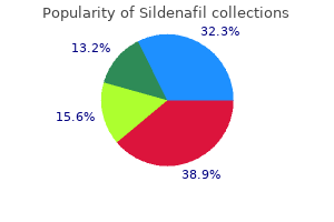
Cheapest generic sildenafil uk
As a result smoking erectile dysfunction statistics cheap sildenafil generic, attempts to evaluate urinary concentrations of practically anything can be very misleading. Postmortem Procedures Postmortem procedures, literally those procedures performed after the death of the animal, include confirmation of the identification number and sex of the animal, an external examination, examination of the significant internal organs in place prior to removal, and then removal, weighing of appropriate organs, and collection of tissue specimens for histological processing and microscopic examination. The microscopic examination of tissue specimens by a qualified veterinary pathologist may be the single most important source of information in understanding the toxicity of a test article. Ordinarily, the list of tissues to be routinely weighed, collected, and processed for histological examination will be specified in the study protocol. In addition to the tissues specified in the protocol, specimens are usually collected of all lesions or target organs that have been identified during the course of the study or at gross necropsy. It is important to provide the necropsy staff with a current list of abnormal clinical observations, especially any evidence of visible or palpable masses, as this is the time when the visible and palpable lesions can be linked to the histopathological evaluation of those lesions. Every effort should be made to locate all lesions described and collect representative tissue from those sites. A detailed description of necropsy procedures is beyond the scope of this discussion. It should be emphasized that the necropsy process, particularly when conducted on a large number of animals at the scheduled termination of a study, is a process in which a large number of tissue samples may be collected and a similarly large quantity of data may be gathered during a short period of time. As such, this process presents many opportunities for loss or misidentification of samples and data. A rigorous system of accounting for which tissues have been collected from each animal, and for tracking the samples and data collected is critical to the accurate interpretation of the toxicity study. It is often possible to select a strain for testing that is particularly vulnerable or resistant to either the test article or a particular type of lesion that might be expected to be associated with that test article. The small size of the mouse confers economy in acquisition, husbandry, handling, and test article consumption. The relatively short gestation period and life span of the mouse are useful in conducting reproductive studies, or studies in which the test article will be administered for a high percentage of the lifetime of the animal. The small size of the mouse is responsible for most of the disadvantages of the species as well. Small size and blood volume makes it difficult or impossible to collect multiple samples of blood and urine over short periods of time. Certain physiological evaluations, such as electrocardiograms, are difficult owing to the small size and high activity level of the species. The Hamster the hamster is the third most frequently used laboratory animal following the rat and mouse at a level of w146,000/year (Renshaw et al. Although historically the hamster saw extensive use in carcinogenesis testing, as will be overviewed, this has changed. It has many attractive features as a laboratory animal because of its reproduction ease, unique anatomical and physical features, rapid physiological development, short life span, low incidence of spontaneous diseases, and a high susceptibility to induced pathological agents. Hamsters historically have been used in several fields, especially in carcinogenesis because of its low incidence of spontaneous tumors, but currently most of their use is seen in testing associated with buccal delivery of drugs (Gad, 2016). Hamsters have also played an important role in blood vessel physiology because their cheek pouches with thin vascularized walls are very accessible. The remaining 20% are primarily Chinese, followed distantly by European, Armenian, Rumanian, Turkish, South African, and Dzungarian hamsters. The Syrian was originally native to the arid, temperate regions of Southeast Europe and Asia Minor. The only proof that the species existed was the preservation in alcohol of two hamsters, one in London and the other in Beirut. Specimens of the species were finally obtained from the wild starting in the 1900s and have since been bred in multitudes in captivity. The ears are pointed with dark coloration; and the eyes are small, dark, and bright. The Syrian hamster was introduced into the laboratory in 1930 to study the Mediterranean disease kalaazar. Israel Aharoni (Hebrew University, Jerusalem, Israel) collected 11 young golden hamsters from Syria in 1930 while on a zoological expedition. Aharoni and his wife kept the hamsters in their house until one night when they all escaped. Nine hamsters were recovered and given to the animal facilities supervisor of the Weizmann & Seiff Institute, Jerusalem, Israel. Of the nine, five escaped the first night in the new facility, leaving only one female. The Syrian has been involved in oncology, virology, endocrinology, physiology, parasitology, genetics, and pharmacology research. The cheek pouch of the Syrian hamster has provided the technology for studying microcirculation and the growth of human tumors. Though the Chinese hamster is smaller than the Syrian, its testicles, spleen, and brain are larger. Mice were extremely scarce at the time so hamsters were used to determine the best therapy for the patients with pneumonia. Watson, in December 1948 (right before the Communist takeover of China), was given 10 female and 10 male hamsters from C. Watson placed the hamsters on what he believes was one of the last Pan Am flights out of China to San Francisco. Schwenter of the Harvard Medical School obtained the hamsters and eventually successfully bred them in the laboratory. Of the original 20 hamsters, 4 of the females and 3 of the males produced offspring that gave rise to the present Chinese hamster population. The Harvard colony has since become extinct; however, colonies were established at the Upjohn Company (which became Pharmacia and most recently was acquired by Pfizer), Kalamazoo, Michigan, and the C. Common and Species Names and Chromosome Number Species Name Mesocricetus auratus Cricetus griseus or barabensis Cricetus cricetus Mystromys albicaudatus Mesocritceus newtoni Mesocricetus auratus Cricetulus migratorius Phodopus sungorus Chromosome Number 44 22 22 32 38 42/44 22 28 I. These hamsters interbreed readily and produce offspring with a diploid number of 44. Besides hibernation research, Turkish hamsters have been used in immunology, genetics, and reproductive behavior research (Yerganian, 1972; Cantrell and Padovan, 1987). European Hamster the European hamster was first found in a West Germany industrial area. The European hamster is a very aggressive animal, and in the wild each adult lives in its own burrow. It has a white face and feet, bodies are dorsally reddish brown and ventrally black with white patches laterally. They are about the size of a guinea pig, averaging 27e32 and 22e25 cm in length and weighing 450 and 350 g for males and females, respectively. Males reach sexual maturity at 60 days of age, whereas females at 80e90 days of age, and they are mainly seedeaters. In their natural habitat, European hamsters can live up to 8 years, whereas under laboratory conditions, the average life span is 5 years. This reduction is believed to be due to the lack of hibernation afforded to a laboratory-raised European hamster (Mohr and Ernst, 1987). The European hamster has been used only in hibernation studies and in inhalation studies because its tidal volumes are the largest of any laboratory rodent species. Rumanian Hamster the Rumanian hamster was initially trapped and described in 1965. It is native to the Bucharest area and is used in the laboratories surrounding that area. Its care, size, and management are similar to that of the Syrian hamster, although it does not reproduce as well as the Syrian. Its face is more pointed and ratlike than the Syrian hamster, but it is similar in appearance to the Turkish hamster. The males are 11 cm long and 40e50 g in weight; the females are 9 cm long and weigh 30 g at maturity. The Dzungarian hamster has a short tail about 1 cm in length, which is usually hidden by the body fur.
Generic sildenafil 75mg without prescription
Particle size dictates the location where the test article will be deposited and absorbed in the respiratory tract erectile dysfunction doctor in bhopal purchase sildenafil 100mg overnight delivery. Larger particles are deposited in the nasopharyngeal region, with successively smaller particles deposited in the trachea, bronchial, bronchiolar, and finally the alveolar region for particles of about 1 m or less. The technology of particle generation and uniform distribution through the exposure apparatus is complex in itself. In addition to generating and uniformly distributing the test atmosphere, care must be exercised to capture the exhaust from the exposure apparatus, such that the test article can be contained without contamination of the laboratory or environment. Exposure periods can range from a few minutes, appropriate for test articles that may pose only an acute exposure risk to continuous exposure over a prolonged period, appropriate for test articles that may pose a risk of long-term environmental or occupational exposure. Exposure apparatus generally takes the form of a chamber that contains the whole animal or groups of animals, or a device that exposes only the head or nose of the animal(s) to the experimental atmosphere. Chamber (Whole Body) Inhalation chambers allow relatively large numbers of mice to be exposed simultaneously without restraint. The aerodynamic considerations are complex, but simpler than for a head-only or nose-only exposure system. The flow rate through a chamber must be adequate to provide temperature and humidity control. Disadvantages of whole body chambers include the tendency for test article to accumulate in the fur, from which it can be ingested; on the skin and eyes, which may interfere with the intended route of exposure and the difficulty in monitoring respiratory volume and rate of individual animals. Head/Nose Exposure (Head Only/Nose Only) Head- or nose-only exposure apparatus limits exposure of the mouse to the test article by routes, other than inhalation, as only a small amount of skin and fur are exposed to the test environment. In addition, it is possible to monitor respiratory volume and rate of individual animals with some of the head-or-nose-only equipment. Disadvantages to this equipment include the fact that only a relatively small number of animals can be simultaneously exposed, and those animals must be restrained in a position that keeps their heads or noses in close contact with the exposure apparatus. This restraint imposes stress on the animals and virtually precludes continuous exposure, as the processes of eating and drinking are not possible with most of this equipment. Heart rates in awake mice have been measured in the range of 300 to more than 800 beats/min (Hoyt et al. Reliable blood pressure measurements are best made by cannulation of a major artery, such as the carotid. Such procedures require anesthesia and surgery, neither of which is especially desirable during the course of a study that may be of long duration and involve many animals. Clinical Observations and Physical Examinations Clinical observations entail the recording of effects that can be detected by direct observation, such as abnormal gait and body weight. For the sake of this discussion, a variety of parameters that can be observed or measured directly will be discussed in this section. Clinical observations often provide the first indication of which physiological systems are being affected by the test article. Mice should be observed regularly throughout the in-life portion of a toxicity study. The type and frequency of these observations should be tailored to meet the scientific objectives of the specific study. Most effects observed following administration of acute (single) doses occur within a relatively short time after dosing. As acute iv doses are often associated with almost immediate effects, it might be appropriate to observe treated mice within 5 min, at about 15, 30, and 60 min, and again at 2 and 4 h after dosing. Observations should be repeated at least once daily on all subsequent study days throughout the postdosing I. This schedule should provide information on the times of onset, peak activity, and remission from toxic effects as well as information on the sequence and severity of effects observed. The high intensity of data collection on the day of dosing in acute studies requires that the system for conducting and recording observations be simple and time efficient. Typically, a system of "exception reporting" is used, in which observations of exceptions from the norm are recorded, and the absence of comment on a system. Clinical observations in repeated dose studies should be conducted at approximately the same time each day to assure that changes in findings over the course of the study can be attributed to the accumulation of or adaptation to toxic effects rather than incidental changes attributable to circadian rhythm or time after dosing. Minimally, all animals should be observed early in the day, prior to daily dosing, and it is highly desirable to conduct at least one additional daily observation at 2e4 h after dosing (or late in the day) to be aware of effects that may be associated with higher blood levels of test article usually found from a few minutes to a few hours after dosing. The simplest form of clinical observation is an observation for survival and moribundity. This or a higher level of observation must be conducted at least once daily in all toxicity studies. The next level of observation is an observation for clinical signs of toxicity, such as abnormal level of spontaneous motor activity, abnormal gait, abnormal respiration, and abnormal quantity or quality of fecal output. During the conduct of a physical examination, specific parameters are evaluated, such as quality of coat, body orifices (for excessive or unusual discharges), eyes, respiratory sounds, and in studies longer than about 26 weeks, animals are examined carefully for evidence of visible or palpable masses. Body weight and feed consumption are typically monitored in studies longer than a few days. An appropriate interval for measuring body weight and feed consumption is about a week. These two parameters should be measured concurrently, such that changes in one can be compared directly to changes in the other. In longer studies, in which the mice have reached maturity and body weight gain has approached zero, the frequency at which body weight and feed consumption are measured can be reduced to as infrequently as once per month. Clinical Laboratory Evaluations Clinical laboratory evaluations of mice refer to evaluations of blood and urine. Blood is routinely collected at sacrifice in repeated dose studies, and small quantities. Interim (nonterminal) blood samples can be collected by retro-orbital venous plexus puncture, cardiac puncture, and tail snip, among other techniques. Retro-orbital puncture is technically difficult and may require anesthesia or immobilization of the animal. Cardiac puncture typically requires anesthesia, and cardiac injury may compromise the histological evaluation of cardiac tissue. Tail snip often yields samples that are contaminated with extravascular, extracellular fluids. Any administration of anesthetic agents during the study of a test article that is not thoroughly understood engenders some risk to the interpretation of the study, as potential interactions of the anesthetic with the metabolism or direct effect of the test article are nearly impossible to predict. Blood collected at the time of sacrifice is typically drawn from the inferior vena cava or the abdominal aorta while the mouse is under anesthesia. In the case of terminal blood collection, potential interaction of the anesthetic agent with the test article, induction of liver enzymes, etc. In addition, bone marrow smears may be prepared but are usually only prepared at sacrifice in mice. Caution should be exercised in comparing experimental data with results obtained from the literature or with results obtained on different instrumentation or by different procedures. For greatest utility, a set of normal values should be compiled for the laboratory procedures and equipment used to produce the data in the toxicity study. As a practical matter, urine is not usually collected in routine toxicity studies. The primary difficulty in conducting urinalysis is that the mouse produces a very small volume of urine during a reasonable collection period. The fur on the dorsal side is gray with a dark-brown or black stripe from the nape of the neck to the base of the tail. The present Dzungarian hamster population is the result of the mating of one female to two males who were domesticated in 1965. The Dzungarian has been used in research involving photoperiodism, the pineal gland, and thermoregulation. Its body size, weight, care, and maintenance are similar to that of the Chinese hamster. Scientists in the United States wanted to find more species of the dwarf hamster (like the Chinese), and the Armenian species has been the only species found. Although the Armenian hamster has been used on a limited basis, its research use has been in cytogenetics and oncology. South African Hamster the South African is the only member of its genus and the only hamster native to Africa. In its natural habitat, South African hamster is a nocturnal, solitary burrowing rodent.

Buy sildenafil 25mg on line
Systems responses of rats to aflatoxin B1 exposure revealed with metabonomic changes in multiple biological matrices erectile dysfunction radiation treatment buy generic sildenafil from india. Ford Department of Pharmaceutical Sciences, College of Pharmacy & Health Sciences, St. Tremendous variability in etiology and severity of the underlying abnormalities can confound detection and diagnosis. The traditional clinical tools for assessing renal function are often inadequate for exposing early or mild impairment and lack specificity for evaluating injury, and the search for sensitive and specific clinical biomarkers of renal injury has increased dramatically in the past 20 years. Improved biomarkers are also needed for field screening and studies of populations at risk from exposure to environmental contaminants or toxins. Drug development is another area in which biomarker exploration has been extensive. Early recognition of nephrotoxic liability in preclinical studies allows pharmaceutical companies to eliminate less-promising drug candidates at the earliest stage possible. In addition, researchers studying the underlying mechanisms of nephrotoxic agents need reliable and expedient ways to assess kidney function in laboratory animals. The complexity of the kidneys presents numerous challenges in assessing renal status. The kidneys are sensitive to events such as exposure to toxic chemicals, low perfusion, immune system activity, and prerenal pathological conditions such as diabetes, each of which may affect renal structure and function in distinct ways. However, two developments have provided structure to classification of renal impairment. The other significant development was a change in terminology from acute renal failure to acute kidney injury. Generally the traditional markers are indices of function, whereas newer biomarkers have been developed to detect injury, serving as signals for specific damage such as tubular necrosis or damage to the glomerulus. Renal biomarkers may be used to evaluate current status, to monitor function over time, or to predict the risk for decline in renal function (Table 15. However, some of the most common are affected by extrarenal events and should be interpreted with consideration of the individual patient. The traditional biomarkers have several disadvantages for clinical assessment, most notably that they are able to reveal changes in function or structure only after significant damage has occurred. The model compounds should be freely filtered at the glomerulus and neither reabsorbed nor secreted in the tubule. Urea is reabsorbed in the nephron back into the bloodstream, which compromises its value as a biomarker; nonetheless, it is easy to measure and well established. Creatinine has the advantages of being produced at a constant rate, freely filtered, and not being reabsorbed. There are several problems with creatinine including that its production and plasma concentration can be increased during excessive muscle breakdown or decreased following prolonged illness and loss of muscle mass. Nonetheless, serum creatinine (sCr) is routinely measured as a renal function test and can be a valuable index when renal dysfunction is anticipated, such as monitoring kidney function when nephrotoxic drugs are used (Caires et al. For example, increases in sCr that were smaller than recommended clinical guidelines were shown to be correlated with poor renal outcomes after surgery (Machado et al. Proteinuria the glomerular capillaries are the barrier to distribution of large plasma proteins into urine. In normal kidneys, only small amounts of large proteins such as albumin and IgM are filtered, most of which is degraded by the proximal tubule epithelium. Smaller proteins and peptides are filtered across the glomerular barrier depending on size, charge, and configuration and then degraded in the tubules. Consequently, there is generally little protein detectable in the urine of humans with healthy kidneys. Proteinuria may occur due to pathology of the glomerular filtration barrier, as in the case of the nephrotic syndrome, as well as damage to the proximal tubule. Glomerular damage may result in large proteins appearing in the urine, which overwhelms the modest ability of the proximal tubule to remove them; in contrast, injury to the proximal tubule will impair the ability to remove smaller proteins such as b2-microglobulin or retinal binding protein from the tubular fluid. Thus, if glomerular filtration is reduced, b2-microglobulin will increase in the blood, whereas if the proximal tubule cells are damaged, b2microglobulin will be found in the urine. Several specific proteins are currently being used as indices of renal dysfunction either individually (see following sections) or as part of biomarker panels (Chen et al. Its presence in urine of the patients was clearly associated with renal tubular dysfunction (Butler and Flynn, 1961). It is produced by all nucleated cells at a constant rate and is cleared only by the kidneys. It is not secreted, and although it is reabsorbed by proximal tubule epithelia, it is completely catabolized within the cells and does not re-enter the plasma (Filler et al. The production of cystatin C should be constant, but on a population basis, serum values have been correlated with physiological factors such as diabetes, body size, and inflammation (Grubb et al. In addition, there are reports of large intraindividual variability compared to creatinine, which would limit its usefulness in monitoring individuals over time (Filler et al. As with other clinical indices Enzymuria Enzymes released from damaged cells of the tubule have been used as markers of injury inasmuch as appearance of these enzymes in the urine is specific to kidney and they have been used in research with laboratory animals for several decades. Although their use for human studies has been minimal, in the future they may be valuable as part of a biomarker panel. The utility of cystatin C to evaluate the renal function of neonates shows promise (Askenazi et al. It was discovered as a protein secreted by neutrophils and bound to gelatinase (Kjeldsen et al. It has been shown to have protective functions against infection and ischemic kidney injury (Ma et al. There are a number of validated clinical analytical platforms that allow results in 15e30 min (Hassanzadeh et al. For example, nonrenal sources such as neutrophils or urinary tract infections with leukocyturia may contribute to its concentration in some cases. In rat kidneys subjected to reperfusion or toxicant injury, Kim-1 expression was found to be specific to proliferating cells in the S3 segment of the proximal tubule, with little response in the convoluted section (Ichimura et al. It exists in a precursor form until cleaved by caspase-1 after which it has proinflammatory actions. In the normal human kidney it is found primarily in the distal segments of the nephron (Gauer et al. They are expressed in several tissues besides the liver, including the renal proximal tubules. In the kidneys it binds the breakdown products of lipid peroxidation facilitating their excretion into the urine, preventing damage by reactive oxygen species (Xu et al. The serum concentrations of these proteins increase in renal injury, which is convenient for cases where urine volume is drastically reduced. Genomics, Proteomics, Metabolomics the complexity of the kidney structure and function in health and disease makes it difficult to conceive of and develop a single biomarker that can give an adequate snapshot of renal status in an individual at a specific time, particularly when the need for information is critical. A panel of biomarkers is more likely to detect important changes and give a richer description of renal impairment. In fact, several companies are marketing immunology-based biomarker panels for assessment of renal function. The eomic analyses take this further in describing a pattern of gene, protein, or metabolite changes that can be analyzed and compared with fingerprints of various pathologies. Genomic analysis has the ability to reveal increased expression of proteins and signaling molecules related to kidney injury, elucidating mechanistic information that can identify potential biomarkers and therapies (Devarajan et al. They observed changes in gene expression in pathways related to creatinine biosynthesis, kinase signaling, cell cycle, renal transporters, renal injury, regenerative responses, drug metabolism, and resistance. An important issue with using genomic analysis for human studies is obtaining tissue samples (Ju et al. Characterization of protein expression has been productively exploited in studying renal biomarkers (Devarajan, 2007; 2008; Slocum et al. Proteomic analysis is amenable to biofluid analysis, particularly urine, serum, and cell culture media. The ability to detect multiple peptides and proteins in a sample will facilitate the discovery of new biomarkers and unraveling of mechanisms. Commercial renal protein array kits with antibodies to cytokines and renal injury biomarkers offer a useful alternative. Metabolomics is the outgrowth of the concept of metabolic profiling or metabolic pattern analysis of metabolites in biofluids, initiated in the 1940s and further pursued in the 70s (Gates and Sweeley, 1978).
| Comparative prices of Sildenafil | ||
| # | Retailer | Average price |
| 1 | O'Reilly Automotive | 775 |
| 2 | J.C. Penney | 756 |
| 3 | BJ'S Wholesale Club | 455 |
| 4 | Aldi | 369 |
| 5 | AT&T Wireless | 460 |

Generic 50mg sildenafil otc
The objective is to block venous return impotence journal best 25mg sildenafil, but not arterial supply, thus dilating the veins. As an alternative to a tourniquet, some toxicologists prefer to warm the tail with a gauze sponge wetted in warm (not hot) water to enhance vasodilation. The needle should be inserted with the bevel up to minimize the chance of puncturing through both sides of the vein. Successful venipuncture will result in the reflux of a small amount of blood into the hub of the needle. Owing to the small volume of blood that usually refluxes, this phenomenon will be most easily visualized if needles that have transparent "flashback" hubs are used. The initial attempt at venipuncture should be made toward the tip of the tail, such that if the vein is missed, a subsequent attempt can be made closer to the base of the tail without risk that the dose will leak out of the initial hole. When the needle is securely in the vein, it can be held with the "tail-holding hand" while the plunger of the tourniquet is depressed to open the vein with the "dosing hand. One convenient method to assure even dose administration is to divide the dosing period and the dose volume into a convenient number of parts. A 2-min dosing period might be divided into eight 15-s intervals, and the dose volume divided by eight. The doser can then administer one-eighth of the total dose volume over each 15-s interval for 2 min to assure a relatively even rate of injection. When the full dose has been administered, a clean dry gauze sponge should be pinched over the injection site and the needle should be withdrawn. Maintaining pressure on the site of the injection for 10e30 s after withdrawal of the needle is usually adequate to prevent bleeding. As iv injections typically result in a rapid onset of activity, it is often appropriate to observe a mouse for the first few minutes after dosing for clinical signs of toxicity. Intraperitoneal administration leads to absorption primarily through the portal circulation. As a result, test articles that are metabolized by the liver are subjected to extensive (or even complete) metabolism prior to reaching systemic circulation and target organs, unless, of course, the target organ is the liver, in which case toxicity may even be amplified. Test articles that are excreted in the bile are similarly subject to elimination prior to reaching the systemic circulation and target organs. Water-insoluble mixture aqueous suspensions, for example, can be administered by the ip route. This may provide the opportunity for rapid systemic absorption of lipid-soluble or certain other test articles. Solutions or suspensions for ip injection should be adjusted to a pH in the range of about 5e9 to reduce the potential for irritation. Dose volumes for ip administration are in the range of 5e10 mL/kg/day, but volumes as high as 20 mL/kg/day are acceptable, particularly if the study is of limited duration, or if it is known that the test article will be absorbed by the ip route. One of the most significant disadvantages of ip administration is the risk of peritonitis. Peritonitis can result from any of three primary causes: physical irritation caused by accumulation of a truly insoluble or irritating test article in the peritoneal cavity, introduction of exogenous microbiological contamination, or microbiological contamination resulting from injury to the gastrointestinal tract or urinary bladder. The potential for a test article to produce physical irritation or chemical peritonitis can be assessed in studies of one to a few days in duration. Although physical or chemical peritonitis is the most frequently seen form of peritonitis in toxicity studies, it is still found with only a small percentage of test articles. Mice are relatively resistant to microbiological infection, so microbiological peritonitis is even less common than physical or chemical peritonitis. Peritonitis resulting from injury during the injection process is extremely rare when injections are administered by qualified toxicologists. There is a slight risk to the animals of physical injury to a major organ or vessel during the injection process, but again this is extremely rare in the hands of qualified dosers. Description of Technique Intraperitoneal injections are administered into the peritoneal cavity using a hypodermic needle attached to a graduated syringe. Each mouse receives a single daily dose, administered as a bolus, for the duration of the toxicity study. For initial training purposes in dosing by the ip route, it is useful to sacrifice a mouse, in which the Intraperitoneal Injection the ip (intraperitoneal) route of administration generally offers the second most rapid absorption of a test article among the parenteral routes, with systemic availability second only to iv injection. Rapid absorption is conferred by the large surface area of the lining of the peritoneal cavity, and by the rich blood supply to that area. This will allow a novice to hold the animal in a dosing position and clearly visualize where the lobes of the liver, the spleen, and the urinary bladder will be, and the area of less vulnerability between these organs. Hypodermic needles used for ip injections to mice need be no longer than about 5/8 in and should be the smallest diameter that will allow easy injection of the dose volume to minimize the trauma to the abdominal wall with commensurate potential for leakage. Needles in the range of 23e25 gauge are appropriate for use with solutions and suspensions of low viscosity. Needles as large as 19e20 gauge can be used, but they require great care to avoid injury and leakage of the test article from the injection site. Prior to initiation of a toxicity study, dosing formulations should be prepared, and samples are analyzed for concentration and homogeneity of suspensions, if appropriate. The mouse is picked up with one hand, and held with the ventral surface toward the doser. The needle should be inserted at an angle of about 15e30 degrees into the abdominal cavity to facilitate penetration of the abdominal wall. The location should be to the right of the midline (to avoid the spleen) at a position about midway between the lower edge of the liver and the urinary bladder to a depth of about 1 cm (3/8 in. Following insertion, the needle is withdrawn slightly, moved about, and the angle of insertion is reduced to assure that the tip has not penetrated or snagged any internal organs. If a large-bore needle has been used, it may be necessary to apply gentle finger pressure over the injection site for a few seconds to prevent leakage of the dose. The ability to control the rate of absorption can be a significant advantage in some cases, as it allows the toxicologist to administer a dose of a test article that may be absorbed over a period of many hours or even days. This can be especially useful in the case of test articles that have short half-lives after absorption, as a result of rapid metabolism and/or elimination. Limitations to im dosing include the limited number of muscle groups in the mouse that are large enough to accept dosing. If possible, the same injection sites should not be treated every day to allow time for absorption and recovery from the trauma of dosing. This means that while a single acute dose might be divided into the hind limbs, repeated daily doses should be administered into alternate limbs. An acute study in which each animal is dosed once would allow 1 mL/kg to be administered into each hind limb, for a total dose volume of 2 mL/kg. This dose volume coupled with the limit of solubility or suspendability of the test article in the vehicle selected may restrict the maximum dose of test article below toxic levels. A further limitation on toxicity testing by the im route is that the formulation to be injected must not cause significant local irritation, particularly if repeated doses will be administered. This limitation may require that a separate study be conducted to assess im irritation potential prior to initiation of a repeated dose study by this route. Intramuscular injection is more labor-intensive than most other routes with the exception of iv injection. Description of Technique Intramuscular injections are administered into the large muscle groups of the posterior aspect of the femoral region using a hypodermic needle attached to a graduated syringe. Each mouse receives a single daily dose, administered as a bolus into alternate hind limbs for the duration of the study. Hypodermic needles used for im injection should be the smallest diameter that will allow injection, but in the range of 27 gauge up to a maximum of about 23 gauge. The mouse Intramuscular Injection the im route of administration is less commonly used in toxicity testing, but it may be appropriate if the test article is intended for im administration to humans. The im route generally results in slower absorption of a test article, with lower peak plasma levels, but more sustained effects than iv or ip injection. The rate of absorption can be influenced by the amount of vascular perfusion of the tissue surrounding the injection, the I. The muscle may be massaged gently to distribute the dose prior to returning the mouse to its cage.
Order sildenafil online now
Chloridazon caused only resorptions in the rat but caused rib and tail anomalies in hamster fetuses of several litters erectile dysfunction drug samples purchase sildenafil 50mg mastercard. Prometryn produced head, limb, and tail defects in rat fetuses following daily administration during gestation (Schardein, 2000). The compound when given by gavage to rats at maternally toxic doses reduced fetal body weight and increased the frequency of extra ribs. In rabbits, after dermal exposure, eye defects and neural malformations accompanied by maternal toxicity have been reported. Dinoterb, another chemical of the same group, induced skeletal malformations by both oral and dermal administration in the rat, and skeletal, jaw, head, and visceral malformations in the rabbit (Schardein, 2000). Compound such as astridiphane, a dinitroaniline compound, was a potent developmental toxicant in the mouse, and induced cleft palate and other toxicity at maternally toxic doses, but under the same conditions in the rat the compound increased the frequency of minor skeletal variations (Hanley et al. Fungicides Fungicides are agents that are used to prevent or eradicate fungal infections from plants or seeds. In agriculture, they are used to protect tubers, fruits, and vegetables during storage or are applied directly to ornamental plants, trees, field crops, cereals, and turf grasses. Numerous substances having widely varying chemical constituents are used as fungicides. According to the mode of application, fungicides are grouped as foliar, soil, and dressing fungicides. Foliar fungicides are applied as liquids or powders to the aerial green parts of plants, producing a protective barrier on the cuticular surface and systemic toxicity in the developing fungus. Soil fungicides are applied as liquids, dry powders, or granules, acting either through the vapor phase or by systemic properties. Dressing fungicides are applied to the postharvest crop as liquids or dry powders to prevent fungal infestation, particularly if stored under less than optimum conditions of temperature and humidity. With a few exceptions, most of the newly developed chemicals have a low order of toxicity to mammals. Public concern has focused on the positive mutagenicity tests obtained with some fungicides and the predictive possibility of both teratogenic and carcinogenic potential. Use of inorganics such as sulfur, lime, copper, and mercury compounds has declined since the 1960s, but captan, chlorothalonils, and other organic materials account for 90% of fungicide use. Newer groups, such as benzimidazoles, conazoles, dicarboximides, and metal organic compounds, account for w10% of fungicide use (Osteen and Padgitt, 2002). All members have an ethylenebisdithiocarbamate backbone, with different metals associated with the individual compounds. Mancozeb, maneb, and metiram are unlikely to present an acute exposure hazard under conditions of normal use (Freudenthal et al. Most of the administered dose is excreted within 24 h, with about half eliminated in the urine and half in the feces. Biliary excretion is minimal, indicating that only w50% of oral doses are absorbed. In another study with a comparatively larger group of workers, the authors concluded that, altogether, the differences between exposed and controls were not consistently correlated to any clinical impairment and suggested that the seasonal application of mancozeb does not pose a significant health risk to exposed subjects (Colosio et al. Anilinopyrimidines Anilinopyrimidines are a new chemical class of fungicides that are highly active against a broad range of fungi. The anilinopyrimidine class of fungicides includes cyprodinil, mepanipyrim, and pyrimethanil. The compounds have low toxicity and are unlikely to present acute hazards in normal use. Mepanipyrim causes hepatocellular fatty vacuolation and lipofuscin deposition in Kupffer cells and hepatocytes of dogs, whereas such changes are not observed in cyprodinil-treated rats (Terada et al. Pyrimethanil produces thyroid follicular cell tumors in rats and enhancement of hepatic thyroid hormone metabolism, which may be responsible for thyroid tumorigenesis (Hurlety, 1998). Enhancement of hepatic thyroid hormone metabolism and excretion are considered to be the mode of action of thyroid tumorigenesis. Cyprodinil induced microsomal protein and cytochrome P450 contents along with ethoxyresorufin O-deethylase, pentoxyresorufin O-depentylase, and lauric acid 11- and 12-hydroxylase, and cytosolic glutathione S-transferase activities in rats. Cyprodinil and mepanipyrim induce the opposite effects on liver and blood lipid parameters in rats. In general, anilinopyrimidines do not have adverse effects on developmental toxicity. No unchanged parent molecule could be found in urine, whereas minor amounts of unchanged cyprodinil were found in feces. Most of the administered cyprodinil was metabolized by sequential oxidation of the phenyl and pyrimidine ring. The major phase 1 metabolite was identified as 4-cyclopropyl-5-hydroxy6-methyl-N-(4-hydroxy)-phenyl-2-pyrimidinamine (metabolite 2). This metabolite was excreted in the urine as b-glucuronic acid conjugate, as well as monoand disulfuric acid conjugates. Although female rats formed the monosulfate almost exclusively, the males excreted equal amounts of the mono- and disulfate. Further oxidation of the methyl group led to the formation of 4-cyclopropyl-5-hydroxy-6-hydroxymethyl-N(4-hydroxy)-phenyl-2-pyrimidinamine (metabolite 3), which was excreted in the urine in unconjugated form. Urinary and biliary metabolites were found to be conjugated with b-glucuronic acid and sulfuric acid. The major metabolites identified in feces were the 5-hydroxypyrimidine derivative of cyprodinil (metabolite 1) and metabolite 4. Two additional metabolites were found in liver and/ or kidney tissue but not in excreta. Metabolite 7 was identified as ring-hydroxylated N-phenyl-guanidine, a breakdown product of the pyrimidine ring moiety. Metabolite 7 was found exclusively in the liver, where it represented the major metabolite. It seems the metabolism of cyprodinil in humans has not been documented in the scientific literature. Chloroalkylthiodicarboximides (Phthalimides) this class of chemicals contains broad-spectrum fungicides (captan, folpet, captafol, etc. Some compounds of this class cause developmental effects, whereas others do not, perhaps because of, and/or masked by, maternal toxicity and possible nutritional deficits (Costa, 1997). Captafol differs from captan and folpet in a number of ways, including structure and chemical activity. They are irritant to mucus membranes, especially of skin after repeated exposures (Gordon, 2010). It is therefore unlikely that these compounds or even thiophosgene would survive long enough to reach systemic targets such as the liver, uterus, or testes. Because of rapid elimination, meat, milk, or eggs from livestock/poultry would be devoid of the parent materials. Humans appear to metabolize captan in a similar manner to other mammals (Krieger and Thongsinthusak, 1993). Thiophosgene, the reactive degradate that is formed from the trichloromethylthio side chain, reacts not only with thiols but also with other functional groups and its t1/2 is less than 0. Due to rapid degradation systemic exposure to captan, folpet, or their common degradate, thiophosgene, is absent. Chlorothalonil is a nontoxic halogenated benzonitrile fungicide with broad spectrum activity against vegetable, ornamental, orchard, and turf diseases. In dogs, there is no evidence of either neoplastic development or the occurrence of preneoplastic lesions in the kidney or stomach. The absence of stomach lesions in dogs is attributable to the anatomical differences between rodents and dogs (dogs do not possess a forestomach). Continued administration of chlorothalonil leads to the development of a regenerative hyperplasia within the renal proximal tubular epithelium. Continued regenerative hyperplasia ultimately results in progression of the kidney lesion to tubular adenoma and carcinoma. Initial cytotoxicity and regenerative hyperplasia within the proximal tubular epithelium are essential prerequisites for subsequent tumor development.
Order sildenafil 25 mg online
Toxic potential of municipal solid waste leachates in transgenic Drosophila melanogaster (hsp70-lacZ): hsp70 as a marker of cellular damage erectile dysfunction test yourself purchase 25mg sildenafil amex. Immunity in Drosophila melanogaster-from microbial recognition to whole-organism physiology. Early-life xposure to widespread environmental toxicants and health risk: a Focus on the immune and respiratory systems. Effects of methylmercury and alcohol exposure in Drosophila melanogaster: potential risks in neurodevelopmental disorders. Methylmercury exposure induces sexual dysfunction in male and female Drosophila Melanogaster. Effects of carbaryl, chlorpyrifos and endosulfan on growth, reproduction and respiration of tropical epigeic earthworm, Perionyx excavatus (Perrier). Influence of temperature and soil type on the toxicity of three pesticides to Eisenia andrei. Environmental levels of atrazine and its degradation products impair survival skills and growth of red drum larvae. Stress protein response (Hsp70) and avoidance behaviour in Eisenia fetida, Aporrectodea caliginosa and Lumbricus terrestris when exposed to imidacloprid. Effects of charge and surface ligand properties of nanoparticles on oxidative stress and gene expression within the gut of Daphnia magna. Pesticide and pathogen: heat shock protein expression and acetylcholinesterase inhibition in juvenile Chinook salmon in response to multiple stressors. Inhalation toxicity of indoor air pollutants in Drosophila melanogaster using integrated transcriptomics and computational behavior analyses. Effect of malathion on the male reproductive organs of earthworms, Eisenia foetida. Linking subindividual and supra-individual effects in Daphnia magna exposed to sub-lethal concentration of chlorpyrifos. Reducing the use of laboratory animals in biomedical research: problems and possible solutions. Exposure to atrazine alters behaviour and disrupts the dopaminergic system in Drosophila melanogaster. Antioxidants maintain Ecadherin levels to limit Drosophila prohemocyte differentiation. Studies on the toxicity of the mercurial fungicide Agallol 3 in Drosophila melanogaster. Effects of chronic dietary and waterborne cadmium exposures on the contamination level and reproduction of Daphnia magna. The influence of soil characteristics on the toxicity of four chemicals to the earthworm Eisenia fetida andrei (Oligochaeta). Developmental, behavioral, and reproductive effects experienced by Japanese medaka (Oryzias latipes) in response to short-term exposure to endosulfan. Long-term exposure to gaseous formaldehyde promotes allergen-specific IgE-mediated immune responses in a murine model. Acute toxicity test with Daphnia magna: an alternative to mammals in the prescreening of chemical toxicity The environmental pollutant, polychlorinated biphenyls, and cardiovascular disease: a potential target for antioxidant nanotherapeutics. Adverse effect of organophosphate compounds, dichlorvos and chlorpyrifos in the reproductive tissues of transgenic Drosophila melanogaster: 70kDa heat shock protein as a marker of cellular damage. Induction of hsp70, alterations in oxidative stress markers and apoptosis against dichlorvos exposure in transgenic Drosophila melanogaster: modulation by reactive oxygen species. Hazardous effect of organophosphate compound, dichlorvos in transgenic Drosophila melanogaster (hsp70-lacZ): induction of hsp70, anti-oxidant enzymes and inhibition of acetylcholinesterase. Metallothionein and Hsp70 trade-off against one another in Daphnia magna cross-tolerance to cadmium and heat stress. Behavioral responses of juvenile Daphnia magna after exposure to glyphosate and glyphosate-copper complexes. Assessment of the reproductive and developmental toxicity of pesticide/fertilizer mixtures based on confirmed pesticide contamination in California and Iowa groundwater. Behavioral effects of chronic exposure to low levels of lead in Drosophila melanogaster. Dermal exposure of Eisenia andrei earthworms: effects of heavy metals on metallothionein and phytochelatin synthase gene expressions in coelomocytes. Organochlorine pesticides, their toxic effects on living organisms and their fate in the environment. Comparative toxicity of imidacloprid, of its commercial liquid formulation and of diazinon to a non-target arthropod, the microcrustacean Daphnia magna. Sodium fluoride adversely affects ovarian development and reproduction in Drosophila melanogaster. Heavy metals induce oxidative stress and trigger oxidative stress-mediated heat shock protein (hsp) modulation in the intertidal copepod Tigriopus japonicus. Effect of methyl methanesulfonate on hsp70 expression and tissue damage in the third instar larvae of transgenic Drosophila melanogaster (hsp70-lacZ). Effects of perchlorate on earthworm (Eisenia fetida) survival and reproductive success. The potential acute and chronic toxicity of cyfluthrin on the soil model organism, Eisenia fetida. Toxicity effects of di-(2ethylhexyl) phthalate to Eisenia fetida at enzyme, cellular and genetic levels. Properties of silver nanoparticles influencing their uptake in and toxicity to the earthworm Lumbricus rubellus following exposure in soil. Atrazine exposure affects longevity, development time and body size in Drosophila melanogaster. The comet assay in environmental risk assessment of marine pollutants: applications, assets and handicaps of surveying genotoxicity in non-model organisms. Effects of diamond nanoparticle exposure on the internal structure and reproduction of Daphnia magna. Effects of three pestic c cides on the earthworm Eisenia fetida (Savigny 1826) under laboratory conditions: assessment of mortality, biomass and growth inhibition. Tracing the tracks of genotoxicity by trivalent and hexavalent chromium in Drosophila melanogaster. Identification of Drosophila-based endpoints for the assessment and understanding of xenobiotic-mediated male reproductive adversities. Long-Term resistance of Drosophila melanogaster to the mushroom toxin alphaamanitin. Endosulfan impacts on the developing chick embryos: morphological, morphometric and skeletal changes. A comparison of the transcriptome of Drosophila melanogaster in response to entomopathogenic fungus, ionizing radiation, starvation and cold shock. Hazardous effects of effluent from the chrome plating industry: 70 kDa heat shock protein expression as a marker of cellular damage in transgenic Drosophila melanogaster (hsp70-lacZ). Synthetic pyrethroid cypermethrin induced cellular damage in reproductive tissues of Drosophila melanogaster: Hsp70 as a marker of cellular damage. Biomarkers indicate mixture toxicities of fluorene and phenanthrene with endosulfan toward earthworm (Eisenia fetida). Chlorpyrifosinduced hsp70 expression and effect on reproductive performance in transgenic Drosophila melanogaster (hsp70-lacZ) Bg9. Evaluation of toxic potential of captan: induction of hsp70 and tissue damage in transgenic Drosophila melanogaster (hsp70-lacZ) Bg9. Toxicity assessment of polluted sediments using swimming behavior alteration test with Daphnia magna. Three-dimensional analysis of the swimming behavior of Daphnia magna exposed to nanosized titanium dioxide. Endocrine disruptors in soil: effects of bisphenol A on gene expression of the earthworm Eisenia fetida. Evaluation of ecotoxicological effects of drugs on Daphnia magna using different enzymatic biomarkers. Xenobiotic metabolism, disposition, and regulation by receptors: from biochemical phenomenon to predictors of major toxicities.
Cheap generic sildenafil uk
The testicular vessels are seen just as they curl round the inferior epigastric artery erectile dysfunction doctor houston buy sildenafil 75 mg amex. Where embolisation is considered or in recurrent varicocele posttreatment, a venogram can be obtained. Causes for orchilgia such as a previous testicular injury, infection, torsion, and surgical operations (on the testicle or a vasectomy) are consevativly managed with pain relief measures. An ultrasound can exclude a cancer, a missed testicular injury or torsion, referred pain from a renal calculus, or a leaking aortic aneurysm. A small number of patients have a pain of unknown origin, which could be attributed to a deepseated psychological disorder, and surgical intervention only makes things worse. Orchiectomy in this situation is usually followed by a return of the pain on the other side [21]. The condition may be accompanied by so much pain radiated to the iliac fossa that can lead to a misdiagnosis of an appendicitis. External spermatic fascia and cremaster Expert Opinion Regarding testicular torsion, when in doubt, exploration is the route. The majority of benign conditions can be treated conservatively and usually the explanation of the pathophysiology is sufficient to allow the patient to cope with the condition. Alternatively, weighing out the risks versus the complications of surgery should be carefully explained. Umbilical hernia, inguinal hernias, and hydroceles in children: diagnostic clues for optimal patient management. Controversies of perinatal torsion of the spermatic cord: a review, survey and recommendations. Intrauterine spermatic cord torsion in the newborn: sonographic and pathologic correlation. An analysis of clinical outcomes using color doppler testicular ultrasound for testicular torsion. Efficacy of varicocelectomy in improving semen parameters: new metaanalytical approach. The widespread use of serum tumour markers, sensitive ultrasound, and accurate computed tomography staging has meant that patients can undergo a radical orchiectomy in a timely manner, which is a curative procedure in up to 75% of cases. Adjuvant chemotherapy, radiotherapy, and radical lymph node dissection forms the basis of management following radical orchiectomy and techniques are improving year on year. With such advances in knowledge and accuracy of relapse prediction testicular neoplasms have become one of the most curative cancers if managed early. This article outlines the diagnosis, management options, and prognosis for germ cell and nongerm cell testicular neoplasms. Additional imaging, usually limited to the chest and abdomen, will allow for a tailored management plan. The International Germ Cell Consensus Classification is an important system which enables prognostic grouping. Patients should be offered fertility counselling and sperm banking before chemotherapy or radiotherapy. William Harvey advised ligature of the testicular artery as a safer alternative to castration, and Astley Cooper researched the lymphatic drainage of the testicle and the method of tumour spread [1]. Kocher was the first to attempt to cure testicular cancer by radical node dissection; however both his patients had advanced disease and died [2]. Within a few years, radiotherapy replaced surgery for lymph node disease in some centres, but in others, the morbidity caused by overdoses of radiation discredited it. For the next 50 years, a contest began between radical node dissection and radiation [1]. The conflict became meaningless because advocates of either method were always comparing tumours that were classified by different systems and staged by different criteria. Eventually, the argument was overtaken by the introduction of tumour markers, combination chemotherapy, and a better understanding of the natural history of these tumours. The germ cells continue to divide to form diploid primary spermatocytes, haploid secondary spermatocytes, and finally spermatozoa from which every cell that may occur in the foetus, yolk sac, or placenta, in benign or malignant variations, may arise. The primordial germ cells, the atypical cells found in seminiferous tubules, and seminoma cells are probably identical. The introduction of tumour markers and combination chemotherapy has enhanced patient care and increased survival rates. Survival rates are at their highest, and it is now recognised as the most curable cancer. Public health campaigns aimed at young men to encourage them to selfexamine have played a large part in making early diagnosis and treatment possible, leading to a decrease in the average time from diagnosis to treatment [10]. Age: the incidence occurs in any age, increasing post puberty with a maximum onset in the 20s and 30s, and rarely occurring in infants, prepubertal boys, and men older than 60 years of age [11]. Race: Caucasians are up to three times more likely to develop testicular cancer than other races [12]. It is rare in nonEuropean origin populations, except for the Maori population of New Zealand [11]. Gene expression analysis and genomewide screening studies suggest that cancer specific gene mutations are found on chromosomes 4, 5, 6, 12, 18, and X [11, 12]. Patients with Klinefelter syndrome are at a higher risk, but the exact mechanism for this is unclear. One in every 10 cases of testicular tumour occurs in men with an undescended testicle. The higher the undescended testicle, the more likely it is to become malignant [15]. Stilboestrol ingestion during pregnancy has been linked to the increase risk of cryptorchidism, and it is also suggested that high levels of natural oestrogens may be equally important [16]. The disease arises from malignant change in spermatogonia where 50% develop invasive germ cell testicular cancer within five years and 90% in seven years [11]. Treatment, if unilateral involvement and contralateral testis is normal, is either by conservative observation or radical orchiectomy. Trauma: Although not a known risk factor, occasionally a history of trauma can predispose to a testicular mass presenting. It can be difficult to decipher whether the lump was from the trauma or whether the injury has brought an unnoticed lump to the attention of the patient. In up to 10% of cases, a testicular tumour mimics epididymo orchitims so caution is advised particularly in recurrent or refractive epididymoorchitis. The pathophysiology of this inflammation is unclear, but it is speculated that haemorrhage into the tumour can contribute. Inflammation in an abdominal cryptorchid testicle may present as an acute abdomen. It is wise to have a low index for suspicion in the young man with vague discomfort in the testicle or the sensation of a lump. Do not be reassured with the finding of a mere varicocele or dismiss discomfort as adolescent neurosis until the testicle has been carefully examined. Similarly, when a young man complains of vague backache, be sure to exclude metastases from a testicular tumour, even when the testes appear normal. A small secondary hydrocele can be present, and very rarely it may be the presenting feature. Larger tumours may be concealed by a thickwalled hydrocele containing turbid fluid through which light will not shine. Gynecomastia from release of chorionic gonadotrophin is reported in up to 7% of cases as the first symptom of a testicular tumour. Therefore, every young man with signs of gynecomastia must have their testicles examined and tumour markers measured [6]. Sometimes the tumour erodes through the testis, leaving behind a scar that shrinks as it heals. A small primary tumour may be entirely replaced by a scar, leaving a normal testicle, even though the tumour has metastasized widely. Therefore, it is always advisable to examine both the affected and nonaffected side, in addition to a complete general physical examination looking for possible distant metastasis.
Cheap sildenafil 50 mg on line
The outcome of repair is dependent on the time interval since the vasectomy or obstruction erectile dysfunction commercial buy sildenafil 50mg. The best outcomes are achieved using a microsurgical technique, and in the largest series, up to 97% of patients regain sperm in the ejaculate if the vaso vasostomy is done within a threeyear interval since vasectomy, with a 76% pregnancy rate [41]. Although there is no accurate pre dictive factor, it is possible to give a rough estimate of the odds based on clinical and investigative parameters. This involves loss of long segments of the vas on both sides and is not reconstructible. When sperm cannot be obtained from the epididymis as in cases of intratesticular obstruction or if microsurgical reconstruction is to be performed at a later date, testicu lar sperm aspiration can be done. Once fertilised, embryos are incubated for two to three days then placed into the uterine cavity. Pregnancy rates are between 10 and 40%, highly dependent on the female age; pregnancy rates of 40% if younger than 35 years of age and drops to 11% if older than 40 years of age. However, up to 70% of patients with repaired varicocele will have improve ment in their sperm parameters: on average an increase of 10 million sperms, 10% improvement in sperm motility, and 3% improvement of sperm morphology. Of the surgical proce dures, the lowest recurrence rate is following a microsur gical subinguinal approach [47]. Although there is evidence support ing the use of GnRh in hypogonadotrophic hypog onadism, there is no evidence of benefit in idiopathic infertility. Evidence suggests that using these modulators leads to an increase in sperm con centration and pregnancy ratesl however, the evi dence is scant [50]. Antimicrobial therapy may be of help in such cases with significant improvement in semen parameters and spon taneous pregnancy rate [51]. The infer tility can therefore be treated by a dopamine agonist such as cabergoline, which can reduce the size of prolactinsecreting tumours and normalise prolac tin levels. If treatment fails or no cause is identified (idiopathic cases), the couples are advised to try assisted reproductive techniques. Anastrozole and other aromatase inhibitors have been used to increase testosterone levels and decrease oestrogen 814 40 Male Infertility References 1 Jarrow, J. Genetic risk factors in infertile men with severe oligozoospermia and azoospermia. Male infertility and androgen receptor gene mutations: clinical features and identification of seven novel mutations. Elevation of intratesticular and scrotal skin surface temperature in men with varicocele. Paternity, time to conception, pretreatment testicular location and size, hormone and sperm parameters. Prevalence of carcinoma in situ and other histopathological abnormalities in testes of men with a history of cryptorchidism. Increased incidence of testicular cancer in men presenting with infertility and abnormal semen analysis. The Ter mutation in the dead end gene causes germ cell loss and testicular germ cell tumours. Unilateral renal agenesis associated with congenital bilateral absence of the vas deferens: phenotypic findings and genetic considerations. Evaluation of acute and chronic bacterial prostatitis and diagnostic management of chronic prostatitis/chronic pelvic pain syndrome with special reference to infection/inflammation. The clinical implementation of sperm chromosome aneuploidy testing: pitfalls and promises. Mutations in the cystic fibrosis gene in men with congenital bilateralabsence of the vas deferens. The predictive value of testicular ultrasound abnormalities for carcinoma in situ of the testisin men at risk for testicular cancer. Patency following microsurgical vasoepididymostomy and vasovasostomy: temporal consideration. Testicular sperm extraction: Microdissection improves sperm yield with minimal tissue excision. Testicular histopathology as a predictor of a positive sperm retrieval in men with nonobstructive azoospermia. Assessment of efficacy of varicocele repair for male subfertility: a systematic review. Induction of spermatogenesis by recombinant follicle stimulating hormone (puregon) in hypogonadotropic azoospermic men who failed to respond to human chorionic gonadotropin alone. Revisiting oestrogen antagonists (clomiphene or tamoxifen) as medical empiric therapy for idiopathic male infertility: a metaanalysis. Effectiveness and limits of antimicrobial treatment on seminal leukocyte concentration and related reactive oxygen species production in patients with male accessory gland infection. Their secretions comprise 80% of the seminal fluid and all of the fructose content. Joining the vas deferens at the ejaculatory duct, their muscular tubes contract during ejaculation to release the alkaline seminal fluid. Similar to the rest of the body, the seminal vesicles may be afflicted by congenital anomalies, infections, and cancers. Thorough counselling is essential before each procedure to warn patients of its potential risks and irreversibility. Vasectomy reversals are possible; however, the chance of pregnancy might be low, necessitating sperm retrieval methods and in vitro fertilisation. Keywords seminal vesicles; semen analysis; vasectomy; infertility; scrotal pain; vasectomy reversal Key Points Seminal vesicles the alkaline section contributes 80% of the volume of the semen. The seminal vesicles can be afflicted with congenital anomalies, such as agenesis, as well as infections and cancer, albeit extremely rare. Transrectal ultrasonography with biopsy is the best method of investigation and differentiating benign and malignant disease. Thorough counselling is a must and should address potential irreversibility, complications, and failure rates; Postoperative semen analysis determines success. Vasectomy reversals leading to pregnancy has variable success rates depending on length of time since vasectomy; however, in general, they are disappointing. With the advent of transrectal ultrasound, we have come to appreciate that they may suffer all the disorders that affect all other hollow organs in the body. Hippocrates noted a paired structure resembling a honeycomb on either side of the base of the bladder. Bold early investigators like Fallopius, de Graaf, and John Hunter all found that their secretions tasted sweet. Their secretion makes up about 80% of the ejaculate; it is rich in fructose, citric acid, magnesium, ammonium, ascorbic acid, prostaglandins, and other specific proteins in an alkaline, viscous yellowish fluid. Seminal vesical agenesis is associated with a cystic fibrosis gene mutation and can be associated with renal and vas deferens anomalies or agenesis. Rarely, abscesses are seen and are associated with diabetes, longterm catheters, and potentially cystoscopy. Schistosoma, tuberculosis, amoeba and trichomonas) may involve the seminal vesicles. Patients can present with haematospermia, perineal pain, painful ejaculation, or infertility or a mixture of these. Diagnosis can be made by ultrasound and perineal needle aspiration and treated with appropriate antibiotics. With the increasing use of transrectal ultrasound, it has become possible to identify seminal vesicle obstruction, which can be complicated by infection or a calculus lodged in the ejaculatory duct [5, 6]. The difficulty with the seminal vesicle is that by the time the diagnosis is made, it is almost impossible to be sure that the origin of the cancer was in the vesicle. Sarcomas and choriocarcinomas arising from the connective tissue of the seminal vesicle have been reported (22). However, local spread from prostate cancer is more commonly seen than primary cancer.
