

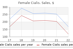
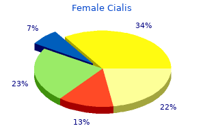
Female cialis 20mg low cost
This value (between 24 and 35 weeks of gestation) women's health center of grants pass cheap female cialis online american express, is an indication for cordocentesis and fetal transfusion (see p. As such, if Rh antibodies are found in the current pregnancy, it is an essential procedure to guide the management. The optical density difference at 450 nm wave length gives the prediction of the severity of fetal hemolysis. This can give indications when to terminate the pregnancy and when to give intrauterine fetal transfusion. Other methods of antenatal assessment of fetal well-being include: (1) Serial ultrasonography may detect fetal hydrops and anemia. The important features are: Polyhydramnios, placental thickness >4 cm, pericardial or pleural effusion, echogenic bowel, dilatation of cardiac chambers and enlargement of spleen and liver; (2) Doppler flow velocity wave forms in the umbilical artery, ductus venosus, middle cerebral artery have been used to detect fetal anemia and acidosis (see p. An intensive neonatal care unit, arrangements for exchange transfusion and an expert neonatologist are the basic requirements to tackle the affected babies. Delivery is to be done in all cases of immunized mothers with evidences of fetal hemolysis in utero. In mild affection, the pregnancy may be continued upto 38 weeks and then termination is to be done. In severe affection: It is reasonable to terminate the pregnancy around 34 weeks after maternal steroid administration (p. In every case of premature termination before 34 weeks, it is desirable to confirm the fetal lung maturation by measuring the L: S ratio in the amniotic fluid. In a specialized center where there is severe affection before 34 weeks, intrauterine fetal transfusion (intraperitoneal or intravascular) is done (p. Methods of delivery: (1) Amniotomy (low rupture of the membranes) is quite effective, if termination is done near term. Clamping the umbilical cord: In either methods, the cord is to be clamped as quickly as possible to minimise even minute amount of antibody to cross to the fetus from the mother. Collection of cord blood for investigation: Cord blood sample is to be taken from the placental end of the cut cord. It is indicated in selected cases where there is severe affection of the fetus in utero prior to 34 weeks. The advantages are: (i) Correction of fetal anemia and improvement of oxygenation; (ii) Improved fetal hepatic function. Intraperitoneal transfusion: Principle: Blood is transfused in the fetal peritoneal cavity under ultrasound guidance. Fetal anemia is corrected when the transfused erythrocytes are taken up by the sub-diaphragmatic lymphatics. The quantity of blood is to be calculated as number of weeks of gestation over 20 multiplied by 10 in milliliters. Severity of fetal affection is best assessed by fetal hemoglobin and hematocrit levels as determined by cordocentesis. Generally a fetus whose hemoglobin deficit is 2 g/dL or more from the mean of a normal fetus of corresponding gestational age (hematocrit < 30%) should be transfused. Procedure: Transfusion is generally made through umbilical cord vessel (vein) near its insertion into the placenta under real time ultrasound. Hematocrit level is checked at intervals during the procedure to determine the volume. Fetal injury, volume overload, preterm labor, fetomaternal hemorrhage are the common complications. Fetal surveillance with ultrasound and continuous electronic fetal monitoring is performed at the post transfusion phase. Betamethasone (24 mg in 3 divided doses) should be administered to the mother 24 hours before transfusion from 26 weeks onwards to enhance pulmonary maturity, in case delivery becomes necessary during transfusion. With the advent of wider use of prophylactic anti-D immunoglobulin, less and less problem babies are born and through exchange transfusion, the incidence of kernicterus has also been reduced. The replacement temporarily helps to tide over the crisis from anemia and hyperbilirubinemia for about two weeks. Thereafter, the baby is quite capable to get rid of the maternal antibodies by producing sufficiently his own Rh-positive blood. A plastic catheter of 1 mm diameter is passed about 7 cm beyond the umbilicus so as to place it in the inferior vena cava. Entire set should be air tight and to be periodically flushed with heparinized saline (1000 units in 100 mL) to prevent clotting. Occasionally, the level of conjugated bilirubin may remain higher and phototherapy should be continued; (4) Hypoglycemia (due to increased insulin secretion) is to be checked by blood glucose estimation post transfusion 4 hourly. Immediate complications: (1) Cardiac failure due to raised venous pressure and overloading of the heart; (2) Air embolism; (3) Clotting and massive embolism; (4) Hyperkalemia; (5) Tetany; (6) Acidosis; (7) Sepsis; (8) Hypocalcemia; (9) Hypoglycemia; (10) Coagulopathies due to thrombocytopenia. Delayed complications: (1) Necrotizing enterocolitis; (2) Extrahepatic portal hypertension due to thrombosis of portal vein; (3) Other complications are mostly attributed to prematurity, hyperbilirubinemia and hypoxia. These products are water soluble and therefore readily excreted in the bile and urine. Phenobarbitone increases the glucuronyl transferase enzyme activity in the fetal and neonatal liver to conjugate the bilirubin which hastens its clearance. With alloimmunization of the mother, the prognosis of the baby depends on: (1) Genotype of the father; (2) Genotype of the fetus; (3) Maternal antibody level. The age limit is arbitrary and is based on the fact that the outcome of the pregnancy is adversely affected beyond the specified age limit. There are two groups of patients: (1) One with high fecundity -a women married late but conceives soon after; (2) One with low fecundity-woman married early but conceives long after marriage. The latter one is prognostically more unfavorable so far as the obstetric outcome is concerned. After conception occurs following treatment of infertility (ovulation induction or assisted reproductive technology). Puerperium: (1) Increased morbidity due to operative interference; (2) Failing lactation. The perinatal mortality is increased due to prematurity, increased congenital malformation (trisomy 21) and operative interference. Considering the risks involved in pregnancy and labor, the patients are considered "high risk". They require meticulous antenatal supervision and should have a mandatory hospital delivery. The following principles are to be followed: (1) Result of induction is unsatisfactory and as such cesarean section is a preferred alternative; (2) Prenatal diagnosis and sonography (targeted) are done to exclude fetal genetic or structural anomaly; (3) Addition of another complication should be viewed with concern. The incidence has been gradually declining over the couple of decades due to acceptance of small family norm but it still constitutes to about one-tenth of the hospital population and accounts for one-third of the maternal deaths in the developing countries. Puerperium-(1) Increased morbidity due to intranatal hazards; (2) Subinvolution; (3) Failing lactation. As such they require adequate antenatal care and should have a mandatory hospital delivery. In the developing countries, too often the disaster is linked with inadequate or neglected antenatal, intranatal or neonatal care. Previous history of congenital deformity of the baby specially a neural tube defect should be excluded as there is likely chance of recurrence (see p. These antibodies are either IgG or IgM or both and bind to negatively charged phospholipids. There is inhibition in release of prostacyclin (vasodilator) from vascular endothelium with rise in the level of thromboxane (vasoconstrictor) from platelets.
Purchase female cialis mastercard
Chance of having a diabetic child is about 1-3% when the mother is only diabetic womens health issues buy female cialis 10mg, 6% when father is only diabetic, rising to 20% if both the parents are diabetic. Principles in the management are: (1) Careful antenatal supervision and glycemic control, so as to maintain the glucose level as near to physiological level as possible (2) To find out the optimum time and method of delivery (3) Arrangement for the care of the newborn. At times patient needs admission for stabilization of blood glucose and for monitoring the fetus. Diet-30 Kcal/kg for normal weight women, 24 Kcal/kg for overweight women and 12 Kcal/kg for morbidly obese women. Ideal plasma glucose (mg/dL) levels in women with pregestational diabetes should be: fasting < 95, premeal < 100; 2 hour P. Usually three meal regimen, with breakfast 25% of the total calorie intake, lunch 30%, dinner 30% and several snacks are quite suitable for most of the patients. Frequent blood sugar estimation is required as the urine examination for sugar is not informative. Monitoring of blood glucose by glucose meter can give an accurate idea about the control. In addition, glycosylated hemoglobin should be determined at the end of first trimester and trimonthly thereafter. Insulin therapy: When diabetes is first detected during pregnancy and cannot be controlled by diet alone, it should be treated with insulin. A post prandial (2 hours) plasma glucose level of more than 140 mg% even on diet control is an indication of insulin therapy. There is frequent change in insulin need during pregnancy and changes in the dosage are made in small increments at a time. Glycemic goals should be around 90 mg/dL before meals and not to exceed 120 mg/dL, 2 hours after meals. The patient should receive three to four daily injections of a regular (human act rapid) and an intermediate acting insulin (isophane), the latter is to be given before dinner. The aim is to maintain the blood sugar level as near to normal as possible without causing troublesome hypoglycemia. Use of subcutaneous insulin infusion by insulin pump is preferred as it is more physiological. Women are instructed on diet composition, insulin dose, recognition and treatment of hypoglycemia, hyperglycemia and ketosis, adjusting insulin dose in relation to exercise, food and sick days. Early hospitalization facilities: (1) Stabilization of diabetes (2) Minimizes the incidence of pre-eclampsia, polyhydramnios and preterm labor (3) To select out the appropriate time and method of delivery. Methods:Prior to the day of induction of labor, the usual bed time dose of insulin is administered. An intravenous drip of one litre of 5% dextrose is set up with 10 units of soluble insulin. Blood glucose levels are estimated hourly with a glucose meter and the soluble insulin dose is adjusted accordingly. If the labor fails to start within 6-8 hours or if the labor progresses unsatisfactorily, cesarean section should be performed. Cesarean section: the indications are-(1) Elderly primigravidae (2) Multigravidae with a bad obstetric history (3) Diabetes with complications or difficult to control (4) Obstetric complications like pre-eclampsia, polyhydramnios, malpresentation (5) Fetal macrosomia (> 4 kg). Continuous subcutaneous insulin infusion with insulin pump is preferred as it is more physiological. The insulin requirement suddenly falls following delivery and after the omission of the drip, pre-pregnant dose of insulin is to be administered or adjusted from the blood glucose level. Epidural or spinal anesthesia is better than general anesthesia as oral feeding could be started soon following the operation. To control blood glucose: (1) One liter of 5% dextrose drip is started with 10 units of soluble insulin (2) A general guideline for insulin infusion rate is, 1 unit per hour for blood glucose of 100-140 mg/dL, 2 units per hour for blood glucose of 141-180 mg/dL and 3 units per hour for blood glucose of 181-220 mg/dL is followed. Use of motorized syringe pump for insulin infusion is convenient (3) Hourly estimation of blood glucose levels is done with glucose meter and the insulin dose is adjusted accordingly. Fetal monitoring: Constant watch to note the fetal condition is mandatory, preferably with continuous electronic fetal monitoring. However, labor should not be allowed for more than an arbitrary 12 hours and should be augmented by low rupture of the membranes and oxytocin or delivered by cesarean section. Examination of the placenta and cord: Placenta is large, the cord is thick and there is increased incidence of a single umbilical artery. Microscopically, villi show edema and excessive syncitial knots, numerous cytotrophoblasts and thickened basement membrane. It may be precipitated with the use of mimetic agents (Isoxuprine) and corticosteroids. Management is done in an acute care unit where both neonatal care is also available. Parameters to assess are: Degree of acidosis, alterations in the level of arterial blood gas, blood glucose, ketones and electrolytes. A fresh blood glucose level after 24 hours will help to adjust the dose of insulin. The baby should preferably be kept in an intensive neonatal care unit and to remain vigilant for at least 48 hours, to detect and to treat effectively any complication likely to arise. All babies should have blood glucose to be checked within 2 hours of birth to avoid problems of hypoglycemia (blood glucose < 35 mg/dL). Improvement in the care of diabetes in pregnancy has reduced perinatal mortality significantly (< 5%). Low dose combined oral pills containing third generation progestins, are effective and have got minimal effect on carbohydrate metabolism. Main worry is their effect on vascular disease (thromboembolism and myocardial infarction). Other causes are:Nodular thyroid disease, sub-acute thyroiditis, hyperemesis gravidarum and trophoblastic disease. The maternal and fetal/neonatal complications in untreated hyperthyroidism: Maternal: Miscarriage, preterm delivery, pre-eclampsia, congestive cardiac failure, placental abruption, thyroid storm and infection. Thyroid stimulating antibodies can cross the placenta and produce neonatal thyrotoxicosis with increased neonatal death. The risk is increased if the antithyroid drug is stopped in late pregnancy or following surgery. Ultrasonography of the fetal thyroid gland is done when the mother is taking antithyroid drugs. Antithyroglobulin, antimicrosomal antibodies and thyroid stimulating immunoglobulin should be measured. Radioactive iodine uptake and scans should not be done during pregnancy as it will cross the placenta and damage the fetal thyroid gland permanently. Propylthiouracil is given at a daily dose of 300-450 mg and continued till the patient becomes euthyroid-the maintenance dose being 50 and 150 mg daily. Patients having marked tachycardia or arrhythmias should also have propranolol (blocking agent). Thyroidectomy, when required to relieve the pressure symptoms can be done safely in the second trimester with prior biochemical control. Preconceptional counseling: Considering the hazards during pregnancy, preconceptional counseling is important. Adequate treatment should be instituted to bring down the thyroid function profile to normal. Radioactive iodine therapy should not be given to patients wanting pregnancy within one year. Oral pill is to be withheld because of accelerated metabolism and disturbed liver function. The clinical association of hypothyroidism in pregnancy may be due to (i) first time diagnosis in pregnancy (ii) hypothyroid women who either discontinue thyroid therapy or who need larger doses in pregnancy (iii) hyperthyroid women on excessive amounts of antithyroid drugs (iv) women with lithium or amiodarone therapy. Primary hypothyroidism met in pregnancy is mostly related to thyroid autoimmunity.
Buy 20 mg female cialis
The olfactory system consists of several elements of the central nervous system: the olfactory nerves understanding women's health issues a reader buy female cialis paypal, bulbs, and tracts, the olfactory tubercle, the primary olfactory cortex, the entorhinal cortex of the parahippocampal gyrus, and the amygdala. The functions of this system include distinguishing isolated odors from other background odors, determining the concentration of the odor and being able to identify it at different concentrations, creating a representation of the odor, and pairing the odor with an associated memory. For many other species, olfaction is important for locating food and communication. Olfactory stimuli are sensed by specialized peripheral olfactory receptors in a part of the nasal mucosa called the olfactory epithelium. This specialized tissue consists of pseudostratified columnar olfactory epithelium located on the superior concha, the roof of the nasal chamber, and the upper portion of the nasal septum. The receptor cells are bipolar neurons with cilia at their dendritic endings in the olfactory epithelium. Interestingly, olfactory neurons are the only special sensory receptors that are the nerve itself. The molecules that are smelled or tasted alter the membrane potential of the receptors through ligand-receptor-mediated second messenger mechanisms that result in ion channel openings in the ciliary membrane. There are more than 1000 different receptor proteins in the olfactory cilia, with each neuron expressing a single receptor. Olfactory neurons are also unique in that they are short-lived neurons with an average life span of 30-60 days (figure 21-1). Aside from perceiving and distinguishing odors, they contribute to the sensation of taste. The ability to discriminate between different odorants is the result of differential expression of the receptor proteins in receptor cells across the surface of the mucosa, combined with selective convergence of axons from functionally related receptor cells to target cells in the olfactory bulb. These axons are grouped into fascicles before passing through the cribriform plate as the olfactory nerve. The olfactory axons terminate in the olfactory bulb in what are called the glomeruli. The olfactory bulbs, which rest on the cribriform plates, contain several types of neurons, including inhibitory interneurons and mitral cells. The mitral cells receive direct synaptic input from olfactory nerve fibers and project their axons in the lateral olfactory tract. Each mitral cell receives input from olfactory nerves expressing the same odorant receptor. Inhibitory interneurons also form synapses with mitral cell dendrites and inhibit mitral cells in surrounding glomeruli, producing lateral inhibition. The anterior olfactory nucleus is located in the olfactory stalk and contains groups of neurons. The olfactory portion of the anterior commissure originates in the olfactory stalk as one of these groups. Information from the ipsilateral olfactory bulb is received and transmitted to the contralateral olfactory bulb by way of the anterior commissure through this group of neurons. A portion of the fibers in the medial olfactory stria contain the axons of the anterior olfactory nucleus neurons which cross the anterior commissure to the contralateral olfactory bulb. The remaining fibers consist of the mitral cell axons and terminate in the ipsilateral olfactory tubercle within the anterior perforated substance. The lateral stria, or lateral olfactory tract, consists primarily of mitral cell axons and projects to the lateral margin of the anterior perforated substance, the piriform cortex, a small rostral portion of the entorhinal cortex, and the corticomedial amygdala. This pattern distinguishes the olfactory system as being the only sensory system in which second-order neurons (the mitral cells) project directly to the cerebral cortex and not to the thalamus. The primary olfactory cortex, or piriform cortex, is the key region involved in the conscious perception of smell. Lesions in this region have been shown to result in failure to discriminate between various odorants. The piriform cortex is also unique in that it consists of a three-layered cortex, making it phylogenetically older than the six-layered cortex of the visual, auditory, and somatosensory systems. Projections travel directly from the primary olfactory cortex to the lateral orbitofrontal cortex and indirectly through the magnocellular portion of the dorsomedial nucleus of the thalamus. Other communications important in olfactory discrimination include corticocortical connections between the temporal lobe and the orbitofrontal cortex. Olfactory impulses that reach the corticomedial amygdala are important in many species for the control of social behaviors. In humans, however, the behavioral significance of olfactory projections to the amygdalae has not been clarified. The function of the olfactory input to the hippocampus through the entorhinal cortex is also unclear. These pathways, however, are thought to integrate olfactory information with visual, auditory, and somatosensory impulses arriving from other association cortices. Through connections with the amygdala, the hippocampus is thought to participate in the integration of multisensory inputs into appropriate emotional and physiological responses to external stimuli. Fractures of the cribriform plate can result in injury to the olfactory bulbs or tracts. Direct insult to the olfactory neuroepithelium is the primary cause of the problem. Viruses can cause edema and hyperemia of nasal membranes, necrosis of cilia, and cellular damage. Anosmia results when there is a lack of regeneration following severe destruction of the epithelium. If the regeneration of receptor neurons and their central attachments are "misguided" to reach abnormal locations in the brain, patients may experience dysosmia, or distorted smell. Even though spontaneous recovery in some patients is theoretically possible, meaningful recovery is rare when marked loss has been present for some time. Less common causes of anosmia include olfactory groove meningiomas, frontal lobe gliomas, metabolic diseases, amphetamine or cocaine use, and Parkinson or Alzheimer disease. A complete anosmia will result in loss of the ability to recognize flavors as the olfactory and taste systems function together in the perception of flavors. He states that several months ago he was involved in a bar fight in which his nose and several other facial bones were fractured. After being discharged from hospital following this incident, he has not been able to smell. The maximal number of olfactory neurons is present at birth, and the number slowly decreases throughout life as neurons are damaged. Olfactory neurons sensing different but complimentary odorants synapse with the same mitral cell. This occurs at unpredictable times, typically lasts for several minutes, and then goes away. As the olfactory neurons pass through the tiny holes in the cribriform plate, they are susceptible to injury, as occurred in this case. Olfactory neurons are small, bipolar neurons that run a very short distance from the olfactory epithelium of the superior nasal cavity through the cribriform plate to the olfactory bulb in the olfactory groove of the frontal lobe. Depending on the degree of damage to the olfactory epithelium and the cribriform plate, the neurons may be able to regenerate, resulting in a return of the sense of smell. Although there are thousands of odorant receptors expressed in humans, each olfactory neuron expresses only one odorant receptor. The neurons expressing the same receptor project through the cribriform plate and synapse with mitral cells in the olfactory bulb. Mitral cells then project their axons down the olfactory tract directly to the olfactory cortex. The neurons in the olfactory epithelium have a lifespan of just a few months, after which time they die and are replaced by new olfactory neurons derived form the basal cell layer of the olfactory epithelium. The olfactory cortex, made up of the piriform cortex and the periamygdaloid cortex, is located on the medial aspect of the temporal lobe. The occipital lobe houses the primary visual cortex; discharge here would result in abnormal vision. The superior parietal lobe contains the primary sensory cortex, and a discharge here would result in abnormal sensation. The anterolateral frontal lobe is not a sensory area, but is involved in executive function.
Purchase female cialis 20mg online
Attempt to determine the exact amount of drug ingested women's health clinic ventura generic 10 mg female cialis amex, as the cyclic antidepressants have a rather narrow therapeutic window, and small excursions beyond the usual therapeutic range (2-4 mg/kg) may result in signifi cant toxicity. Anti muscarinic findings are commonly appreciated in poi soned patients, including dry skin and mucous membranes, diminished or absent bowel sounds, urinary retention, and sinus tachycardia. Sinus tachycardia is a very common early finding, but typically does not result in hemodynamic compromise. That said, severe poisonings frequently progress to induce wide complex tachycardias and refractory hypotension. Early subtle alterations in levels of consciousness can quickly progress to obtundation and coma. It not only provides rapid, distinc tive, and diagnostic findings suggestive of toxicity, but also facilitates the provision of targeted therapy. Keep in mind that patients with intentional overdoses may neither be reliable nor forthcoming regarding their ingestions. Gastrointestinal Decontami nation the induction of emesis with syrup of ipecac is no longer recommended given the potential for sudden decompensa tion and secondary aspiration. Administer activated char coal (1 g/kg) to all patients with intact airway reflexes as it will readily bind to and decrease the absorption of cyclic antidepressants. Orogastric lavage can be considered in symptomatic patients who present within an hour of ingestion after carefully weighing the benefits of removing a highly toxic drug against the inherent risks of the procedure. Norepinephrine (1 meg/min titrated to a max of 30 meg/min) is the agent of choice as it directly antagonizes the effects of cyclic antidepressants on the a-adrenergic receptors. Discharge Patients who remain symptom-free throughout an observa tion period of no less than 6 hours may be safely discharged home provided they are cleared from a psychiatric perspective. Benzodiazepines such as diazepam or lorazepam are the initial treatment of choice. Hypothermia occurs as the body loses heat from 1 of 4 major mechanisms: conduction, convection, evaporation, and radi ation. Convective (windy environments) and conductive (cold and wet exposures) mechanisms are responsible for most cases of accidental hypothermia. Primary hypothermia occurs when an otherwise healthy person is unable to compensate for an excessive exposure to cold tem peratures. Although most common in colder climates, hypother mia can occur in any environment. In the United States, hypothermia is respon sible for approximately 700 deaths annually, with more than half occurring in patients older than 65 years. History the potential for hypothermia is usually obvious in patients with significant exposures. Patients may present in wet clothing, be found outdoors in the cold weather, or be inappropriately dressed for the environment in which they live. In the United States, most hypothermic patients are either intoxicated or suffer from an underlying psychiatric illn ess or dementia. The history or presentation may be less obvious for patients with mild hypothermia or unknown exposures. Said patients typically present with nonspecific neurologic findings, including dizziness, confusion, slurred speech, or ataxia. It is imperative to immediately and completely undress the patient to remove any wet clothing and identify any signs of coexisting frostbite, trauma, sepsis, hypothyroidism, adrenal crisis, toxidromes, or cardiac dys function. Refrain from any unnecessary movement of the patient to avoid precipitating life-threatening dysrhyth mias, as hypothermic myocardium is exceptionally irritable. Finally, perform a comprehensive neurologic exam includ ing an evaluation for level of consciousness, pupill ary reactivity, and focal deficits. Mild Hypothermia Patients tend to present with shivering, tachycardia, tachy pnea, and hyperventilation. Moderate Hypothermia Patients with moderate hypothermia develop hypoventila tion, hyporeflexia, and an altered sensorium or stupor. Severe Hypothermia Patients with severe hypothermia may present with pul monary edema, areflexia, hypotension, and apnea and are extremely susceptible to ventricular fibrillation and cardiac arrest. As the hypothermia worsens, atrial fibrillation and eventually ventricular fibrillation often develop. Obtain a head computed tomography in patients who exhibit persistent alterations in mental status despite adequate rewarming and in those with any signs of cranial trauma. The absence of a known environmental exposure or any concern for secondary hypothermia should prompt an active search for potential etiologies. To be successful, the patient needs an intact shivering response and sufficient energy stores. Those who do demonstrate signs of myo cardial instability and/or cardiac arrest require active core rewarming. Serum hyperglycemia is actually more common secondary to a cold-induced inhibition of insulin secretion. Avoid treat ment with supplemental insulin in these patients, as this may precipitate iatrogenic hypoglycemia on rewarming. Hypothermia can impair the concentrating ability of the renal tubules, leading to a "cold-diuresis" with secondary dehydration and hypovolemia. Hypothermia impairs both platelet aggregation and the coagulation cascade, and patients may become profoundly coagulopathic. In spite of this, the laboratory measurement of the prothrombin time and partial thromboplastin time will be normal as blood samples are warmed to physiologic temperatures before running these tests. Peritoneal and pleural irrigation can also be performed after the insertion of percutaneous catheters. Emergent thoracotomy with internal cardiac massage and mediastinal irrigation with warmed saline is a very invasive technique, but has been used successfully in severely hypothermic patients with prolonged cardiac arrest. Standard Advance Cardiac Life Support medications (eg, atropine, lidocaine, arniodarone) are typically ineffective for the management of hypothermia induced dysrhythrnias. Discharge Patients without serious comorbidities who present with mild to moderate hypothermia and successfully undergo passive rewarming can be safely discharged, provided there is a warm environment for them to go. To prevent recur rent cold exposure, obtain social work consultation to arrange placement for undomiciled patients and admit to the hospital if unsuccessful. Part 12: Cardiac Arrest in Special Situations: 20 1 0 American Heart Association Guidelines for Cardiopulmonary Resuscitation and Emergency Cardiovascular Care. Anti-arrhythmic and vasopressor medications for the treatment of ventricular fibrillation in severe hypothermia: A systematic review of the literature. Admit all patients with evidence of cardiac instability and those undergoing active core rewarming to an intensive care unit setting. Do not discharge patients with cold-induced tissue injuries without first ensuring they have a warm, d ry place to go. Of the 2 types of injury, frostbite is the more devastating and requires more aggres sive treatment. That said, chilblains and immersion foot can also progress to significant disability and require prompt recognition and intervention. Although individuals at the extremes of age are at a higher risk for cold-induced tissue injuries, frostbite is fairly uncommon in these cohorts. Other areas of the body at risk for cold-induced tissue injury include the face (eg, nose, ears), buttocks and perineum, and penis.
Order female cialis 20mg without prescription
Pelvic assessment specially in primigravidae should be done during the initial examination women's health university discount female cialis 10 mg otc. Enquiry is to be made about the onset of labor pains or leakage of liquor, if any. Thorough general and obstetrical examinations including vaginal examination are to be carried out and recorded. Records of antenatal visits, investigation reports and any specific treatment given, if available, are to be reviewed. Ambulation can reduce the duration of labor, need of analgesia and improves maternal comfort. If, however, labor is monitored electronically or analgesic drug (epidural analgesia) is given, she should be in bed. Low pH of the gastric contents is a real danger if aspirated following general anesthesia when needed unexpectedly (see p. Fluids in the form of plain water, ice chips or fruit juice may be given in early labor. Intravenous fluid with ringer solution is started where any intervention is anticipated or the patient is under regional anesthesia. If the patient fails to pass urine specially in late first stage, catheterization is to be done with strict aseptic precautions. For practical purposes, the common analgesic drug used is pethidine 50-100 mg intramuscularly when the pains are well established in the active phase of labor. The drug should not be given if delivery is anticipated within two hours (For antidote see p. Abdominal palpation-(a) Uterine contractions: as regard the frequency, intensity and duration are assessed. The number of contractions in 10 minutes and duration of each contraction in seconds are recorded in the partograph (see p. To be of value, the observation should be made immediately following uterine contraction. To avoid confusion of maternal and fetal heart rates, maternal pulse should be counted. Vaginalexamination - (a) Dilatation of the cervix in centimeters in relation to hours of labor is a reliable index to note the progress of labor. The position and the station of the head are once more to be reviewed and the progressive descent of the head is ensured. One sterile sheet is placed beneath the buttocks of the patient and one over the abdomen. This is achieved by pushing the occiput downwards and backwards by using thumb and index fingers of the left hand while pressing the perineum by the right palm with a sterile vulval pad. If the patient passes stool, it should be cleaned and the region is washed with antiseptic lotion. The purpose of increasing the flexion of the head is to ensure that the small suboccipito-frontal diameter 10 cm (4") distends the vulval outlet instead of larger occipitofrontal diameter 11. The forehead, nose, mouth and the chin are thus born successively over the stretched perineum by extension. This simple procedure prevents the serious consequence of mucus blocking the air passage during vigorous inspiratory efforts. If it is found and if loose enough, it should be slipped over the head or over the shoulders as the baby is being born. Flexion of the the sub-occiput comes under the symphysis pubis so that lesser sub-occipitofrontal 10 cm (4") diameter emerges out of the introitus. This indirectly signifies that the bisacromial diameter is placed in the anteroposterior diameter of the pelvis. If there is delay, the head is grasped by both hands and is gently drawn posteriorly until the anterior shoulder is released from under the pubis. Traction on the head should be gentle to avoid excessive stretching of the neck causing injury to the brachial plexus, hematoma of the neck or fracture of the clavicle. Deliveryofthetrunk: After the delivery of the shoulders, the fore finger of each hand are inserted under the axillae and the trunk is delivered gently by lateral flexion. It facilitates drainage of the mucus accumulated in the tracheobronchial tree by gravity. The tray is placed between the legs of the mother and should be at a lower level than the uterus to facilitate gravitation of blood from the placenta to the infant. Two separate cord ligatures are applied with sterile cotton threads 1 cm apart using reef-knot, the proximal one being placed 2. Leaving behind a length of the cord attached to the navel not only prevents inclusion of the embryonic structure, if present, but also facilitates control of primary haemorrhage due to a slipped ligature. The cord is divided with scissors about 1 cm beyond the ligatures taking aseptic precautions so as to prevent cord sepsis. Presence of any abnormality in cord vessels (single umbilical artery) is to be noted. The cut end is then covered with sterile gauze piece after making sure that there is no bleeding. The purpose of clamping the cord on the maternal end is to prevent soiling of the bed with blood and to prevent fetal blood loss of the second baby in undiagnosed monozygotic twin. This is beneficial to a mature baby but may be deleterious to a pre-term or a low birth weight baby due to hypervolemia. But early clamping should be done in cases of Rh-incompatibility (to prevent antibody transfer from the mother to the baby) or babies born asphyxiated or one of a diabetic mother. Previously uneventful first and second stage can become abnormal within a minute with disastrous consequences. The principles underlying the management of third stage are to ensure strict vigilance and to follow the management guidelines strictly in practice so as to prevent the complications, the important one being postpartum hemorrhage. Desire to fiddle with the fundus or massage the uterus is strongly to be condemned. The patient is expected to expel the placenta within 20 minutes with the aid of gravity. If the patient fails to expel, one can wait safely upto 10 minutes if there is no bleeding. As soon as the placenta passes through the introitus, it is grasped by the hands and twisted round and round with gentle traction so that the membranes are stripped intact. Gentleness, patience and care are prerequisites for complete delivery of the membranes. If the spontaneous expulsion fails or is not practicable, because of delivery under anesthesia, any one of the following methods can be used to expedite expulsion. The body of the uterus is pushed upwards and backwards, toward the umbilicus while by the right hand steady tension (but not too strong traction) is given in downward and backward direction holding the clamp until the placenta comes outside the introitus. The pressure is to be withdrawn as soon as the placenta passes through the introitus. If the baby is macerated or premature, this method is preferable to cord traction as the tensile strength of the cord is much reduced in both the instances. The sterile gloved hand should be introduced and the placenta is to be grasped and extracted. The maternal surface is covered with greyish decidua (spongy layer of the decidua basalis). Normally the cotyledons are placed in close approximation and any gap indicates a missing cotyledon. The membranes-chorion and amnion are to be examined carefully for completeness and presence of abnormal vessels indicative of succenturiate lobe. An oval gap in the chorion with torn ends of blood vessels running upto the margin of the gap indicates a missing succenturiate lobe. The absence of a cotyledon or evidence of a missing succenturiate lobe or evidence of significant missing membranes demands exploration of the uterus urgently. The vulva and adjoining part are cleaned with cotton swabs soaked in antiseptic solution.
Purchase discount female cialis online
Contraindications to gastric lavage include the ingestion of hydrocarbons or other caus tic agents menstrual blood smell female cialis 10mg without a prescription, and potential complications include increased intracranial pressure, aspiration, and esophageal rupture. There are several different modalities available to enhance the elimination of poisons. Hemodialysis is ideal for smaller-sized poisons with small volumes of distribution (< 1 L/kg) and low degrees of protein-binding. Hemodialysis should also be performed in all patients with profound acidemia regardless of the etiology. Alkalinization of the urine is commonly initiated for ingestions of weak acids such as aspirin and phenobarbital. Circulating toxins will be preferentially converted to their conjugate bases in the alkaline environment and consequently trapped in the renal tubules, where they will be excreted in the urine. Antidotal therapy is important and necessary when managing the poisoned patient, but should never take priority over the s upportive measures already mentioned. Examples of selected focused therapy along with general indications are listed in Table 54-3. This is a reminder for clinicians to never hesitate in calling their regional poison center (1 -800-222 - 1 222) for assistance during any point in the care of the poisoned patient. Getting help early may facilitate a more focused work-up, prevent unnecessary laboratory and/or diagnostic studies, provide insight into potentially life-saving antidotal therapy, and assist with appropriate disposition making. Admission All patients with hemodynamic abnormalities, persistent mental status changes, and metabolic or acid-base irregularities should be admitted to an intensive care setting. Additionally, those who ingested medications that either require antidotal therapy or have prolonged or delayed toxic effects (eg, sulfonylureas, extended-release calcium channel blockers, or beta-blockers) also require admission to a critical care setting. Consult a nephrolog ist early to prepare for hemodia lysis in cases involving large ingestions or severe metabolic acidosis. Ethylene glycol, methanol, and isopropanol are the most common toxic alcohols associated with human poisoning. Toxic alcohols are often ingested in 1 of 2 ways, either unintentionally if placed in an inappropri ately labeled container, or intentionally by patients either attempting suicide or trying to become intoxicated when regular ethanol is not readily available. Of note, each of the 3 is capable of causing inebriation, with isopropanol being twice as intoxicating as ethanol. According to the National Poison Data System, more than 35,000 toxic alcohol exposures are reported to the American Association of Poison Control Centers yearly. Isopropanol is the most frequently ingested but causes the fewest number of deaths, whereas methanol is the least commonly ingested toxic alcohol but associated with the highest number of fatalities. Ethylene glycol is metabolized to glycolic acid, glyoxylic acid, and oxalic acid, all of which can produce systemic acidosis and acute kidney injury. Isopropanol is not converted to an organic acid but is rather metabolized into acetone, which can produce hemorrhagic gastritis and systemic hypotension in the absence of a concurrent acidosis. If either are unrecognized or untreated, all toxic alcohol ingestions can result in patient fatality. If a bottle or label is not available, ask the patient what kind of product was ingested. For example, antifreeze usually contains ethylene glycol, windshield-washing fluid usually contains metha nol, and rubbing alcohol usually contains isopropanol. Beyond attempting to iden tify exactly what was ingested, it is critically important to determine the time of ingestion, as this will affect the interpretation of laboratory results and impact patient management priorities. Traditionally a normal osmol gap is < 1 0 mEq/L, but in actuality will normally range between -7 to + 1 4. It is the toxic alcohol parent com pound that contributes to the osmol gap, and elevated gaps occur early after ingestion before the metabolism of the toxic alcohols into their poisonous byproducts. Ethylene glycol and methanol can result in a simultane ous anion gap metabolic acidosis and elevated osmol gap. Because isopropanol is not metabolized into an organic acid, ingestion typically results in an elevated osmol gap in the absence of an elevated anion gap. Because isopro panol is rapidly converted to acetone, a serum acetone level can help identify isopropanol poisoning in this setting. Calcium oxalate crystals on urine microscopy or urine fluorescence under ultraviolet light (due to the addition of fluorescein to most commercial antifreeze) may be clues for ethylene glycol poisoning, but the absence of either should not be used to rule out exposure. Patients arriving soon after an ingestion may appear well with unremarka ble physical exams, whereas those who arrive many hours after ingestion may be obtunded with unstable vital signs. Of note, the peak serum level of a toxic alcohol correlates poorly with the physical exam findings. As with all overdoses, perform a careful neurologic examination (mental status, cranial nerves, cerebellar findings, and motor strength). Ocular examination may reveal blurred vision, decreased visual acuity, retinal edema, optic atrophy, or hyperemia of the optic disc in methanol poisoning. Abdominal tenderness or blood on rectal examination may indicate isopropanol ingestion as it is classically associated with hemorrhagic gastritis. La boratory A basic metabolic panel is indicated to determine renal function, acid-base status, and calculate an anion gap (the normal range is 8-1 2 mEq/L). An osmol gap can be a useful screening test for toxic alcohol poisoning if calculated early after the ingestion. Computed tomography imaging of the head should also be considered when the mental status does not cor relate with the presumed exposure or associated t rauma is a concern. In the absence of a reported inges tion, an unexplained anion gap acidosis or elevated osmol gap should prompt further evaluation for a toxic alcohol. Obtain early nephrology consultation to facilitate emergent hemodialysis if the ethylene glycol or methanol level is >50 mg/dL to filter out the parent compound and any accumulated toxic metabolites. Emergent dialysis is also indicated in cases of renal failure or severe acidosis irrespective of the ethylene glycol or methanol level to remove any remaining toxic metabolites and improve the systemic acid-base status. All patients with abnor- Suspected or confirmed toxic alcohol ingestion Lab studies: Serum pH, electrolytes, and osmolal ity. Patients receiving fomepizole can typically be admitted to a regular hospital bed. A visual schematic for clarifying the temporal relationship between the anion gap and the osmol gap in cases of toxic alcohol poisoning. Discharge Unintentional ingestions with no evidence of acidosis or indications for antidotal treatment or hemodialysis may be safely discharged after appropriate poison prevention counseling. It is found in more than 1 00 combination pharmaceuticals (eg, cold and cough agents, sleep agents) and is present in multiple prescription opioid analgesics (eg, Vicodin, Darvocet). Toxic exposures to analgesics as a class have increased rapidly over the last decade. The maximum recommended safe dose is 4 g per day for adults and 60-90 mg/kg/day for children. Sulfonation and glucuronidation are the two primary mechanisms and produce nontoxic metabolites that are cleared in the urine. Patients with lower glutathione stores (chronically ill, malnourished, and alcoholics) and those with upregu lated cytochrome P450 activity (patients on certain medications including anticonvulsants and antituberculosis agents, chronic alcoholics) are more likely to suffer significant toxicity. Although all patients may not progress beyond the first stage after a toxic exposure, those that do tend to follow the following timeline. Stage 2 occurs between 24-48 hours post exposure and is known as the latent stage. Abdominal symp toms return, including pain and vomiting along with jaun dice and potential encephalopathy. Laboratory studies may reveal acidemia, hypoglycemia, renal failure, and coagu lopathy. Stage 4 lasts between 4 days and 2 weeks of the exposure and involves a progression to outright liver failure and death or complete patient recovery. Check a full panel of liver studies (aspartate aminotransferase, alanine aminotransferase, albumin, bilirubin) in all patients and follow serially, looking for evidence of worsening hepatotoxicity. Check a baseline complete blood count and follow serial hemoglobin levels in patients who develop coagulopathies. Order a metabolic panel to assess for electrolyte abnormalities and to calculate the anion gap, as a significantly elevated anion gap metabolic acidosis may be present.
Generic female cialis 20mg free shipping
Any factors that impede these responses can lead to heat exhaustion or heat stroke women's health ultimate bootcamp workout purchase 10 mg female cialis. Evaporation of sweat is the main mechanism through which the body dissipates heat. Elderly, infants, and those with chronic illness have decreased ability to adapt to hot conditions. Certain medi cations including antipsychotics, anticholinergics, beta blockers, and diuretics also interfere with sweat e vaporation and cooling. Alcoholics, those with decreased mobility, and some patients with chronic medical conditions including obesity, poor cardiac function, and scleroderma have impaired abilities to evaporate heat as well. Radiation, conduction, and convection of heat also allow the body to lose heat, but only when the ambient temperature is lower than body temperature. Patients complain of nonspecific symptoms and signs including weakness, dizziness, fatigue, nausea, vomiting, headache, myalgias, tachycardia, tachypnea, hypotension, and diaphoresis. Patients can exhibit a wide range of neurologic symptoms and signs, including ataxia, seizures, and hemiple gia. Multiorgan system failure consisting of hepatic, renal, and cardiac impairment may also be present in severe cases. Neuroleptic malignant syndrome and sero tonin syndrome should both be considered in any patient taking psychiatric medications. Malignant hyperthermia, although usually occurring in the context of inhalational anesthetic or succinylcholine use, should be considered if symptoms develop in a patient with a previous or family history of this condition. History Important factors to address in the history include a full description of the circumstances surrounding the heat exposure. Has the patient been in a non-air-conditioned apartment in the summer for several days, or has the patient been working outside while there is an elevated heat index Consider oral volume replacement with an electrolyte-containing solution if the symptoms are mild. If there is any concern for potential complications from comorbid conditions, N therapy and laboratory studies should be instituted. In both cases, treat the patient with ambient cooling, removal of heavy clothing, and rest. Start with 250-500 mL of normal saline and replace other electrolytes based on laboratory values. Completely expose the patient and mist with tepid water while a fan is blowing on him or her. Specially made cool mist fans are highly effective, but not available in most facilities. A Foley catheter device that provides continuous temperature evaluation or rectal temperatures recorded every 10 minutes is ideal. Patients may shiver during c ooling, which is counterproductive by producing heat. The number one factor that contributes to the morbidity and mortality of heat illness is the severity of underlying comorbid illnesses, not the absolute height of the core body temperature. Physical Examination Physical examination should involve complete exposure of the patient to remove heat-trapping clothing and to assess for any physical injuries. Electrocardiogram should be performed on all patients with heat exhaustion and heat stroke to evaluate for signs of ischemia or electro lyte abnormalities. Admission If the patient has any serious comorbid conditions or ill nesses, admission may be necessary. Consider that drown ing may have resulted from a primary medica l or traumatic insult. Patients who present and remain asymptomatic for 6 hours may be d ischarged from the emergency department. A 2005 report from the World Health Organization recommends that the term "near-drowning" be abandoned and instead to use the term "drowning inci dent" with a description of the outcome (death, morbidity, no morbidity). Drowning itself should be described as "the process of experiencing respiratory impairment from submersion/immersion in liquid. One estimate states that there is 1 death per 13 drowning inci dents, suggesting that underreporting likely occurs. Children make up the majority of fatal incidents, with peak ages of 1 -4 years and seasonal variability. Freshwater drowning is more common than saltwater, with bathtubs and pool as the most common locations. Patients may fall or j urnp into a body of water and their distress is immediately noted or alternatively may be found floating or at the bottom of a lake or pool after a period of t ime without being seen. Clinical effects of the s ub mersion/immersion event itself most often manifest as respiratory abnormalities including hypoxia, tachypnea, or abnormal lung sounds. At severe levels of illness, cardiac dysrhythmias may occur, and mental status can change. It will also be important to consider the potential of suicide; the medical status of the patient will dictate how urgently this assessment is needed. Physical Examination the order of the physical exam will depend on patient stability. Assess the unstable patient like a trauma patient, with a primary survey and management as necessary, fol lowed by a thorough secondary survey. Areas of focus include signs of external trauma, especially the head and neck, lung sounds to evaluate for water or emesis aspiration, skin color (cyanosis), core body temperature (rectal), and a neurologic e xam. Laboratory No specific laboratory testing is universally recommended for drowned patients. If the provider feels that significant aspiration occurred, or if the patient is unstable, useful labo ratory tests to determine the severity of injury include a blood count, serum electrolytes, and blood gases. Animal studies have shown that 1 1 mL/kg of aspirated hypotonic fluid are necessary before any effect is seen on hemoglobin, volume status, or electrolytes. In the majority of cases, hypoxia and meta bolic acidosis cause the resultant morbidity and mortality. To determine whether this is a primary or secondary drowning incident Secondary drowning refers to a drowning incident that occurred as a result of a medical event, drug or alcohol ingestion, or preceding trauma. For instance, a boater who drowns in a lake may have ingested a large amount of alco hol, causing him to fall into the water. A diver may strike her head on the bottom of the pool, causing prolonged submersion. Airway protection, management of hypoxia and hypothermia, and urgent/emergent treatment of traumatic injuries or medical emergencies take first priority. This may include placement of an endotracheal t ube, high-flow oxygen, active rewarming, and volume resuscitation. Patients who have aspirated large volumes may require positive pressure ventilation to recruit collapsed alveoli. Manage any cardiac dysrhythmias as recom mended by Advanced Cardiac Life Support protocols. Treatments once suggested but not currently recom mended are prophylactic antibiotics or steroids. In addition, hyperbaric chamber use has not been shown to be beneficial unless decompression illness complicates the drowning. Head and cervi cal spine imaging should be considered if the patient dived into the water. Any other traumatic injuries noted on exam or suspected by history should be imaged and e valuated as appropriate. Those who are intubated, have persistently altered mental status, are hypothermic, or require high-flow oxygen should be admitted to an intensive care unit. Cardiac monitoring is indicated for any patient with oxygen requirements or changes on chest radiograph. Discharged patients should be instructed to return for development of difficulty breathing, fever, or mental status changes. A new definition of drowning: Towards documentation and prevention of a global public health problem.
Generic 10 mg female cialis with mastercard
If in the early stages of the disease the diagnosis is in question womens health vanderbilt cheap female cialis 10mg amex, repeated evaluations at a later stage are warranted. The most difficult aspect of the diagnosis is to distinguish the idiopathic form from the secondary form. A positive response to the administration of levodopa helps to confirm the diagnosis. Some are produced in the soma of the neuron by the free ribosomes and the rough endoplasmic reticulum, packaged in vesicles, modified by the Golgi apparatus, and transported down the axon to the presynaptic terminal. Other neurotransmitters are produced by enzymes in the cytoplasm and concentrated in synaptic vesicles. The vesicles are stored in the terminal and await the signal for release into the synaptic cleft where the neurotransmitter can diffuse across to the postsynaptic membrane, bind to receptors, and effect a change in the cell. There are specific mechanisms in place to remove neurotransmitters from the synaptic cleft. Small-molecule transmitters are charged molecules that are derived from the metabolism of carbohydrates. The precursors to the neurotransmitters are enzymatically altered in the cytosol and concentrated into synaptic vesicles for storage. As in all biosynthetic pathways, there is generally one enzyme that regulates the production of the neurotransmitter and functions as the rate-limiting step for its production. It is synthesized from dietary choline and endogenous acetyl CoA by the enzyme choline acetyltransferase. Glutamate is the main excitatory neurotransmitter in the central nervous system and is synthesized from -ketoglutarate, an intermediary of the tricarboxylic acid cycle. It binds to several different receptor types and acts on both inotropic and metabotropic receptors. Glutamate is cleared from the synaptic cleft by glial cells, which then convert it to glutamine by glutamine synthase. Glutamine diffuses across the plasma membrane, is synthesized back into glutamate in the presynaptic terminal, and is then repackaged into vesicles. Glycine is likely synthesized from serine and is the major inhibitory neurotransmitter in the spinal cord. Both neurotransmitters bind to receptors that lead to the opening Cl- channels in the postsynaptic neuron. Glycine activity in the synaptic cleft is terminated by reabsorption into the presynaptic cleft via active transport. Tyrosine is first converted into l-dihydroxyphenylalanine (l-dopa) by tyrosine hydroxylase. The neuroactive peptides are produced by ribosomes on the endoplasmic reticulum of the cell body and, following modifications, are transported down the axon to the terminal. They are produced as a large precursor protein called a polyprotein in the membrane-limited organelles of the neuron. These larger proteins are cleaved to form the neuroactive peptides, which are removed by both diffusion and breakdown by extracellular proteases. Comparison of the biochemical events at cholinergic endings with those at noradrenergic endings. Neuroactive peptides differ from the small-molecule neurotransmitters in several ways. First, because they rely on protein synthesis and modification, neuroactive peptides can only be produced in the cell body. They are also taken up and concentrated within the synaptic vesicles, unlike the neuroactive peptides, which are packaged into vesicles by the Golgi apparatus. Because of the different processing steps, the type of synaptic vesicles also differs between the two classes. The vesicles for small-molecule neurotransmitters can be recycled quickly at the nerve terminal following exocytosis to produce more synaptic vesicles. The membrane that constitutes the vesicles for neuroactive peptides come from the Golgi apparatus and is transported from the cell body in a more time-consuming fashion. Despite these differences, neuroactive peptides and small-molecule neurotransmitters often coexist within the same neuron. They can be released together to function synergistically on the postsynaptic cell. Additionally, several different neuroactive peptides processed from a single polyprotein can be released into the synaptic cleft. Following release, the neurotransmitters must be removed from the synaptic cleft to prevent desensitization of the postsynaptic receptors and to allow future transmissions to occur. As learned previously, enzymes in the synaptic cleft degrade and inactivate certain neurotransmitters, such as acetylcholine. Neuroactive peptides are cleared more slowly from the synapse by simple diffusion. Most neurotransmitters, however, are taken up by the neuron to terminate their action. Transporter proteins in the neuron often rely on the electrochemical gradient for the active reuptake of the neurotransmitter. Based on clinical presentation and additional studies, you diagnose him with Parkinson disease. Which molecule is the immediate precursor in the synthetic pathway leading to the neurotransmitter involved in this disease She is ultimately diagnosed with fibromyalgia, a disorder that is associated with elevated levels of the neurotransmitter "Substance P. One of the proposed pathologic mechanisms of this disease is a lack of cholinergic neurotransmission in certain areas of the brain. By which of the following mechanisms is a drug that increases acetylcholine in the synaptic cleft most likely to act Substance P is a neurotransmitter that belongs to the neuroactive peptide class and, like all peptides, is synthesized in the cell body on the rough endoplasmic reticulum. After synthesis, these peptides are further processed by the Golgi apparatus, which also packages them into vesicles. These vesicles are transported down the axon via fast anterograde axonal transport to the presynaptic terminal, where they are secreted into the synaptic cleft when properly stimulated. While all of the above are potentially mechanisms by which the acetylcholine levels in the synaptic cleft could be increased, the most likely candidate is inhibition of acetylcholinesterase-mediated degradation. The primary method of removal of acetylcholine from the cleft is enzymatic degradation by acetylcholinesterase. Parkinson disease is associated with dopamine depletion of the nigrostriatal tracts. Acetylcholine is synthesized from choline and acetyl coenzyme A (acetyl CoA) by the enzyme choline acetyltransferase. In the synaptic cleft, acetylcholine is hydrolyzed by acetylcholinesterase into acetate and choline. In the synaptic cleft, glial cells facilitate the clearance of glutamate by converting it to glutamine, which then diffuses across the plasma membrane to be converted back to glutamate and then repackaged into vesicles for future release. She states that he started to have difficulty eating this afternoon and that he has been "drooling" more. He had his normal diet since last night but was given a special treat for his good behavior. Based on the clinical presentation, the baby is diagnosed with botulism, a disease that interferes with neurotransmission and the neuromuscular junction. Symptoms, including constipation, cranial nerve abnormalities, hypotonia, and respiratory difficulties, classically appear 12-36 hours following ingestion of the contaminated food. Parents will complain that breast-feeding babies will have poor suction and weak cries. Once the bacteria are established, they begin to produce the botulinum exotoxin, which is absorbed throughout the intestinal tract. The exotoxin makes its way to the presynaptic neurons of the neuromuscular junction, where they bind irreversibly to the presynaptic cholinergic receptors and enter the cell by endocytosis. Once inside the cell, the toxin functions as a protease and cleaves integral membrane proteins of the acetylcholine-containing synaptic vesicles.
