

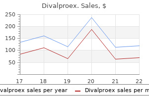
Purchase divalproex 500 mg amex
Motor neurone disease (amyotrophic lateral sclerosis) is a progressive disease of the corticobulbar and corticospinal tracts symptoms 6dpo order divalproex with visa. Progressive swallowing difficulties affecting mainly the oral and oropharyngeal stage of swallowing together with dysarthria and anarthria, account for much of the misery of the disease. Drug-induced Drugs can cause dysphagia directly by causing oesophagitis, or as part of their pharmacological action or normal side effects. The oesophagus at the level of the aortic arch is most commonly injured by both lack of neutralization of the saliva and contact by acidproducing drugs with a pH of less than 3, such as tetracyclines, doxycycline, vitamine C and ferrous sulphate. Broad-spectrum antibiotics and chemotherapeutic drugs may cause secondary viral ulceration or fungal infections. The size and shape of the foreign body will dictate where it lodges, but common sites are areas of constriction at the cricopharyngeus, at the level of the aortic arch and at the cardia. Bolus obstruction from large pieces of meat with or without a piece of bone, is often seen in elderly patients who cannot chew due to lack of teeth and patients with a benign or malignant oesophageal stricture. Patients present with painful dysphagia following recent ingestion of food or a foreign body. Adults tend to localize the level of the obstruction better than children or psychiatric patients. If obstruction is complete as in meat bolus obstruction, there will be drooling of saliva. Neck examination may reveal subcutaneous emphysema if pharyngeal or oesophageal perforation has occurred. A lateral soft tissue radiograph of the neck and a chest radiograph may show a radioopaque foreign body, widening or the presence of an air bubble at the postcricoid space or subcutaneous emphysema. It may be necessary to perform a contrast swallow with a nonionic contrast, which has the added advantage of not interfering with subsequent oesophagoscopy as it is clear. Ageing Presbydysphagia refers to swallowing difficulties due to ageing which affects all stages of swallowing. The oral phase is affected by loss of teeth and tongue connective tissue, reduced strength of mastication and weakness of the velopharyngeal reflexes. The pharyngeal phase is affected by decreased elevation of the larynx and prolongation of the pharyngeal transit time. In the oesophageal stage, there is prolongation of the upper oesophageal sphincter relaxation time and the oesophageal transit time. Despite these age-related changes and barium swallow abnormalities evident in one-third of otherwise healthy elderly individuals, few complain of dysphagia. However, as the swallowing mechanism is already compromised, severe swallowing difficulties may result from even minor insults, such as swallowing medications. The burns may be superficial and heal completely or be full thickness and repair by fibrosis with stricture formation and dysphagia. In the acute phase, flexible oesophagoscopy is mandatory to assess the extent of the oesophageal damage prior to placing a feeding gastrostomy. Small sharp foreign bodies, such as fish bones or spicules of meat bones, may lodge in the tonsil, base of tongue, vallecula or piriform fossa, the tonsil being the most common site. The patient will complain of a pricking sensation on swallowing and should be able to localize the site and side. Examination of the oral cavity, pharynx and larynx by inspection, palpation where appropriate and indirect laryngosopy and or nasolaryngoscopy will reveal most bones lodged in these areas. A lateral soft tissue radiograph of the neck may help if the bone is ossified and radio-opaque, but not if it is purely cartilaginous. Pharyngeal pouch is an example of a pulsion diverticulum, but its aetiology remains uncertain. Manometric studies suggest that it is associated with high intrapharyngeal pressures with a high resting tone in cricopharyngeus that is slow to relax. Overflow of food into the pharynx will cause regurgitation of undigested food and overflow into the larynx will Chapter 153 Causes of dysphagia] 2035 cause aspiration with bouts of coughing and eventually pneumonia. Middle-aged patients usually have reflux oesophagitis or a hiatus hernia and although cancer should be excluded this is more common in elderly patients. Globus pharyngeus is common in middle aged women and is rarely associated with serious disease. Dysphagia of short duration in an elderly male who smokes and drinks and which progresses from solids to liquids is classic of malignancy of the swallowing pathway. Referred otalgia in a patient with dysphagia is usually a sinister symptom and a poor prognostic sign. Neurological causes of dysphagia mostly affect the oropharyngeal phase of swallowing. Ingested foreign bodies tend to lodge at sites of constriction at the cricopharyngeus, at the level of the aortic arch and at the cardia. A contrast swallow for suspected perforation and/or aspiration should be with a low molecular weight, nonionic, water-soluble contrast medium. Oesophageal manometry can be helpful in patients with atypical chest pain and unexplained dysphagia. Twenty-four hour ambulatory oesophageal pH monitoring is the most accurate method of diagnosing gastroeosophageal reflux. A barium videofluoroscopy swallowing study is the gold standard for evaluating the swallowing mechanism and is particularly useful for the oral and pharyngeal phase. Although it affects the swallowing mechanism, this symptom being more common in women is not true dysphagia and rarely associated with serious disease. Although there may be a functional element to globus, it is associated with gastrooesophageal reflux with or without oesophagitis in a significant number of patients. If the symptom persists despite strong reassurance, some patients may be submitted to a barium swallow or rigid endoscopy to exclude other disease, notably tumours (see also Chapter 154, Globus pharyngeus). The suspected thyroid disease is confirmed with the help of thyroid function and thyroid antibody blood tests, fine needle aspiration cytology, ultrasound and thyroid scanning of the thyroid. A multi-observer study examining the radiographic visibility of fishbone foreign bodies. Videofluoroscopic evaluation in the assessment of swallowing disorders in paediatric and adult populations. Indications and techniques of endoscopy in evaluation of cervical dysphagia: Comparison with radiographic techniques. Hippocrates first mentioned the term in his treatises and regarded it as a disease of women being inextricably involved in the uterine axis from which all hysteria was believed to be derived at that time. Today, patients are referred for investigations to otolaryngologists and gastroenterologists and rarely to psychiatrists or psychologists, even though globus has been identified as the fourth most discriminating symptom of somatization disorder, after vomiting, aphonia and painful extremities. It is often a nebulous clinical diagnosis to make because the symptoms are variable within and between subjects and objective clinical findings are by definition absent. In the past 50 years the most popular organic explanations for the symptom are that globus can be an atypical manifestation of silent gastrooesophageal reflux2, 3, 5 or caused by oesophageal dysmotility. Older people can complain of globus sensation, but it is often difficult to differentiate this from age-related mucosal inflammation. The everyday experience of globus is reported more or less equally by both sexes9 although from those seeking medical attention, three of four subjects are women. The same authors also showed that the most important predictor of hospital attendance was the severity of the globus sensation. Although earlier studies suggested abnormally elevated upper oesophageal sphincter pressures in some patients with globus, subsequent work with more appropriate manometric equipment did not confirm this result. Only 3 percent (only one man) of 650 subjects with normal tonic upper oesophageal sphincter pressure had globus, but 28 percent of the 101 with elevated tonic pressures had globus. No relationship with reflux could be demonstrated, although only a minority had pH studies. In 1983, Gray described inferior constrictor strain swallows caused by increased awareness of contact between the epiglottis and the lingual tonsils. This could provoke a vicious circle of dry saliva swallows with increased frequency of swallowing, reduction in interswallow interval, aerophagy and failure of upper oesophageal sphincter relaxation.
Bacterial Polysaccharide (Xanthan Gum). Divalproex.
- Use as a bulk-forming laxative to treat constipation.
- Are there safety concerns?
- Use as a saliva substitute for dry mouth.
- What other names is Xanthan Gum known by?
- What is Xanthan Gum?
- Dosing considerations for Xanthan Gum.
- Are there any interactions with medications?
Source: http://www.rxlist.com/script/main/art.asp?articlekey=96358
Purchase divalproex on line
The histological picture reveals atypical lymphocytes treatment 7th feb cardiff generic divalproex 250mg without prescription, but distinct evidence of neoplasia is often difficult to identify. The exact clinicopathological picture is still not clear but may encompass a spectrum of disease, which includes the development of lymphoma in its pathological course. For all these situations the clinical picture combined with thorough investigation and histological analysis is essential. Relapsing polychondritis this is a rare disease typified by inflammatory changes in cartilaginous structures with subsequent degeneration and fibrosis. Nasal involvement is seen in 60 percent of patients and is heralded by sudden onset of redness, warmth, swelling and tenderness that subsides after two to four weeks but recurrence invariably happens weeks to months later. Cartilage is replaced by fibrous tissue with loss of function and cosmetic deformity. Corticosteroids are the backbone of management, although cyclosporine and methotrexate are sometimes required. The disease is unpredictable and may follow a relatively benign course, although involvement of laryngeal cartilage can lead to airway collapse or stenosis. The disease presents either acutely or insidiously taking many years to evolve and nasal mucosal disease is almost always present at some stage. Periorbital oedema and violaceous facial erythema are diagnostic of dermatomyositis. Systemic sclerosis may involve the face and is characterized by tightening of the facial skin and appearance of a beaked nose. Relapsing polychondritis involves the nasal cartilage in 60 percent of patients and may result in nasal deformity. Glandular types show a massive increase in sebaceous glands resulting in a pitted, dented, distorted and asymmetrical surface. Ectatic surface veins characterize the fibroangiomatous group and the actinic form is distinguished by distorting nodular masses of elastic tissue. Reasonable results are obtained with long-term antibiotics, particularly topical metronidazole and oral tetracyclines, and some successes have been reported with synthetic retinoids. Topical steroids should be avoided as they may cause rebound erythema but skin care and sun protection should be discussed. Carbon dioxide, argon and pulsed dye lasers have also been employed to good effect. Eczema and dermatitis these are interchangeable terms covering a wide variety of conditions and may involve facial skin. Its precise origin is unknown but genetic factors appear to be relevant and affected patients always have evidence of sun-damaged skin. Patients report a history of excessive flushing or blushing in appropriate situations. This increases in frequency, culminating in a persistent dark red erythema involving the central face. Papules, pustules and telangectasia are evident in the affected area, however, comedones are absent, and therefore differentiate the disease from acne vulgaris. A late and rare development of rosacea, arising almost solely in men, is rhinophyma. Chapter 134 Conditions of the external nose] 1715 atopic or endogenous causes, which exhibits scaling, vesicular lesions and exudation. Pruritus and scratching are the main symptoms and the disease runs a chronic course with eventual cutaneous thickening or lichenification. Contact dermatitis results from contact between skin and a substance in touch with the skin that produces epidermal damage. Erythema, oedema and vesicles typify the acute form, but over time lichenification, scaling and extensive epidermal damage ensues. Topical application of medication to the eye or nose, especially with external use of aminoglycoside preparations can produce a contact dermatitis with eczematous change. Tetracycline, sulphonamides and thiazide diuretics may generate rashes in sun-exposed areas including the face. Seborrhoeic dermatitis this disorder is typified by the presence of discrete erythematous patches, often with a superficial greasy yellow scale. Pityrosporum ovale, a normal commensal yeast, can be identified from the lesion site, although its precise relationship to the condition is unclear. Adult males are mostly affected, particularly those prone to scaling and dandruff. The central face, particularly in the nasal furrows and nasolabial folds, scalp, ears, eyebrows, sternum, axilla and groin are the usual sites and there may be an associated blepharitis or otitis externa. Topical or systemic triazole antifungal preparations are beneficial and salicylic acid or ichthammol can be employed for long-term care. Pemphigoid In an elderly patient, the appearance of tense bullae on a preceding rash or normal skin suggests a diagnosis of pemphigoid. The limbs and trunk are predominately involved, although the face and nose may be included. Perniosis Chilblains are tender, erythematous, itchy lesions that blister and ulcerate. This inflammatory reaction is an abnormal response to cold temperatures and appears on fingers, toes, legs, ears and nose. A genetic influence is suggested by positive family histories and systemic conditions, especially arterial disease, are relevant. Preventative measures to provide warmth are valuable but traditional techniques employing ultraviolet radiation probably have no value. Other types arise from exposure to hydrocarbon derivatives, phenols, steroids and androgenic hormones. Findings that appear to be important include elevated levels of sebum and free fatty acids in sebaceous glands associated with hair follicles, the presence of proprionibacterium acnes, dermal inflammation and increased levels of androgenic hormones. These factors conspire to produce inflammatory papules and pustules on the face, back and chest, which progress in severe cases to the development of nodules and cysts. Diet, skin care, antibiotics, synthetic retinoids and ultraviolet light are used to moderate the condition. Following the active phase of disease, permanent scarring can be lessened with dermabrasion techniques or laser therapy. Dermatological disease involving the nose may present as erythema, pustules, scaling or thickening, but is always part of a more widespread cutaneous reaction. Best clinical practice Facial rashes Rashes rarely if ever involve the face alone. Pityriasis versicolour is a skin eruption following sun exposure with [A comprehensive drug history should be sought as topical use of facial, nasal or ophthalmic medications can produce a localized contact dermatitis. Trends in non-melanocytic skin cancer treated in Australia: the second national survey. Different sun exposure patterns in the aetiology of basal cell and squamous cell carcinomas of the skin. The role of the human homologue of Drosophila patched in sporadic basal cell carcinomas. The number and distribution of benign melanocytic naevi in a healthy British population. Thickness, cross-sectional areas and depth of invasion in the prognosis of cutaneous malignant melanoma. Malignant melanoma of the extremities: a clinicopathologic study using levels of invasion (microstage). Deficiencies in current knowledge and areas for future research $ $ $ $ $ $ With the increasing involvement of otorhinolaryngologists in facial diseases, the surgeon must develop the knowledge, experience and ability to manage such pathology. Surgical excision of nasal and facial lesions must be directed at resecting disease, maintaining function and preserving aesthetic integrity. The nature of the lesion dictates the extent of resection and the margin of healthy tissue that must be removed.
Order 250mg divalproex with amex
Inspiratory stridor occurs with narrowing of the Chapter 171 Acute infections of the larynx] 2249 supraglottis or glottis medications hair loss purchase divalproex online from canada. Indirect laryngoscopy with a mirror is possible in most adults but flexible fibreoptic nasendoscopy is far superior and is recommended wherever possible. The specific features of note are the presence of inflammation and swelling of the supraglottis, glottis and subglottis; the space between the vocal cords and cord mobility; pooling of saliva in the hypopharynx. The usual cause is a virus associated with an upper respiratory tract infection, but laryngitis may also be secondary to infection of the tonsils or the chest. Inhalation of dusts and fumes or underlying allergy can induce laryngeal inflammation and may facilitate the development of acute laryngeal infection. Pathophysiology Why acute laryngitis occurs only in some and not all patients with a common cold is not fully understood. Patients will have a rough, deep voice of variable pitch that often disappears in midsentence and they may, on occasions become aphonic. Voice changes are usually preceded by the symptoms of a common cold and a sore throat. Examination will show erythema and oedema of the vocal cords and there may be excess secretions. Changes can, however, be subtle and not nearly as bad as the symptoms may suggest. Antibiotics should be reserved for more severe forms of bacterial laryngitis, persistent laryngeal inflammation, or in specific patients who rely on their voice professionally. Penicillin has no effect, but erythromycin, which is active against Moraxella catarrhalis, is associated with less vocal disturbance and reduced coughing at one to two weeks but no other significant effects. The likely explanation is that the residual dysfunction is due to persistent inflammation within the internal laryngeal muscles. There are, however, no definitive studies on the efficacy of steroids in such patients. Antiviral agents are not considered to be as effective as antibiotics and have significant side effects. Drooling, respiratory distress and hoarseness and oedema of the palatine arches and uvula may also be seen. Stridor is uncommon but tachycardia that is disproportionate to the pyrexia is an important sign that precedes airway obstruction. The main difference in children compared to adults is that acute epiglottitis progresses very rapidly and compromises the airway. There has been a marked decrease in paediatric epiglottitis since the introduction of vaccination against Haemophilus influenzae type b (Hib vaccine) in 1985 to prevent childhood meningitis. Prior to the introduction of vaccination, the ratio of epiglottitis between children and adults was estimated to be 3:1. The white cell count is important and significant elevation is likely to occur in patients with impending airway obstruction. Although radiographs have been considered to be unreliable, methods of improving their diagnostic accuracy have been described. A wide range of pathogens has been described in adults and these include Group A Streptococci, Streptococccus pneumoniae, Staphylococcus aureus and Klebsiella pneumoniae. In many patients, pathogens are not isolated, either from throat or blood cultures. Treatment should be with intravenous antibiotics and 100 percent humidified oxygen. This is much more likely in patients with rapidly progressive disease and occurs within hours of the onset of the illness. The characteristic feature of this specific form of laryngitis is subglottic oedema. Why the swelling affects the subglottic mucosa rather than the adjacent glottis is unknown. This dilemma has been studied in an animal model using rats infected with Sendai virus that is structurally and serologically related to parainfluenzae type I. The inflammatory response and migration of dendritic cells, neutrophils and lymphocytes is much greater in the subglottis compared to the glottis. The illness usually presents with a cough, sore throat, malaise and mild fever for two to four days. Deterioration can be rapid and signs include shortness of breath, stridor and inspiratory retraction of the soft tissues of the neck. The endoscopic appearance of the larynx will show a normal epiglottis, inflamed vocal cords and subglottic oedema extending into the trachea. Investigation Direct viral antigen detection by sampling mucus from the nasopharynx may be helpful in identifying the viral pathogen. Croup affects mainly young children, aged six months to three years, in which subglottic oedema leads to early respiratory distress and biphasic stridor. In contrast, adult croup is an uncommon condition that was first reported in 1990. The difference in pathogens between adults and children is probably due to memory Management Because of the risk of rapid deterioration, adult croup should be suspected and recognized early. The airway and oxygen saturation should be monitored and intubation considered if the airway is impaired. Broad-spectrum antibiotics are advisable to cover secondary infection, but there is no evidence to support antiviral agents. At present, there is no means of preventing infection but recent research is focussed on the way the virus binds to cells resulting in a cascade of molecular events that leads to the virus entering the cell. Fluoroquinolones are also likely to be effective in adults but there is no supporting data of effectiveness. There is no systematic review of the use of steroids, adrenergic agonists or pertussis-specific immunoglobulin. Erythromycin is thought to prevent patients from being infectious and is recommended as prophylaxis in affected households, but the effect has been described as modest compared with good quality vaccination. The bacteria produce endotoxin and exotoxins that induce an inflammatory response, stop cilia from functioning and cause epithelial cell necrosis. It is a notifiable communicable disease that affects all age groups and is transmitted by coughing and sneezing. However, the disease is still prevalent in the developing world and there has been a resurgence in Eastern Europe. The ease of world travel facilitates the transmission to countries where doctors will probably not have seen a case and the diagnosis may therefore not initially be considered. The disease is caused by Corynebacterium diptheriae and spreads by droplets from the upper respiratory tract of an affected individual. It affects nonimmunized children and susceptible adults, particularly the elderly. The usual site of infection is the tonsil and fauces, but it can also occur in the nasal cavities or spread to the larynx. Clinical features Whooping cough presents with symptoms of a runny nose, dry cough and mild pyrexia, similar to a common cold. The cough occurs in prolonged paroxysms after one to two weeks and is followed by gasping and the characteristic whoop in children. Clinical features the disease causes a severe sore throat, malaise, pyrexia and nasal discharge if the nose is affected. Examination of the throat shows a characteristic grey membrane in the oropharynx and this may spread to affect the larynx. Whether an infected patient becomes a carrier or develops a severe illness depends on the host response and virulence and toxigenicity of the organism. Since Corynebacterium diphtheriae is not easily identified, throat swabs from all patients with sore throats should be specifically screened. There may also be palatal petechiae, oral ulceration, splenomegaly and hepatomegaly. The heterophile antibody test is highly specific and if negative should be repeated.
500 mg divalproex with visa
The diagnosis is made either at endoscopy medications 2 times a day purchase 250mg divalproex, under a general anaesthetic, or by a contrast swallow. A nasogastric tube is passed into the oesophagus of the child lying prone and a nonionic contrast medium is injected down the tube as it is withdrawn. Vascular rings formed by anomalies of the great vessels may cause significant dysphagia depending on the abnormality, though they more commonly present with respiratory symptoms first. An anomalous subclavian artery (dysphagia lusoria) may present with dysphagia in adulthood. Other anomalies seen include a double aortic arch and an anomalous left pulmonary artery. Infections Infections are one of the most common causes of dysphagia and are obvious when they affect the oral cavity and oropharynx, but are more difficult to diagnose further down the tract. Acute onset of dysphagia during a coryzal-like illness suggests an infective cause for the dysphagia. Acute pharyngitis and tonsillitis are the most common cause presenting with fever, malaise and painful dysphagia, the degree depending on the severity of the inflammation. The initial viral infection usually predisposes to a secondary bacterial infection, most commonly a group A, beta-haemolytic streptococcus. Acute supraglottitis is now a rare cause of painful dysphagia in children, due to immunization with the Haemophilus influenzae type B vaccine. However, epiglottitis should be suspected in a child who becomes rapidly unwell with fever, stridor, painful dysphagia and drooling. The diagnosis is confirmed only after the airway has been secured by endotracheal intubation or tracheostomy. Supraglottitis in adults has a more protracted course, Traumatic Trauma to the head and neck, chest or cervical spine may be accidental or iatrogenic and may involve blunt trauma, penetrating injuries or compression effects or a combination of these. Chapter 153 Causes of dysphagia] 2031 stridor may not be present, and diagnosis can be confirmed by fibreoptic nasolaryngoscopy. Herpetic, fungal or cytomegalovirus mucosal infections can cause dysphagia and is usually seen in severely immunocompromised patients. In addition, candidal infections are more common in patients on broad spectrum antibiotics or steroids. However, candida affecting the hypopharynx and oesophagus may not be obvious to the clinician. Oesophagoscopy is the investigation of choice for diagnosis when a swab can be taken. Tuberculosis is a chronic infection that can cause dysphagia by either a mucosal lesion or compression of the oesophagus by enlarged lymph nodes. Abscesses of the head and neck spaces can result in significant painful dysphagia with drooling in patients who are already unwell with a high fever and torticollis of the affected area. Clinical examination may show erythema and oedema of the posterior larynx and lower pharynx, but usually a flexible oesophagoscopy is needed for diagnosis. The inflammatory changes seen in the oesophagus range from mild erythema, which is equivocal evidence of reflux, to erosions, confluent ulceration or stricture formation in more severe cases. Twenty-fourhour ambulatory oesophageal pH monitoring is the most accurate way of confirming the diagnosis and is useful when standard investigations are normal. The dysphagia is due to hyperkeratinization with web formation in the postcricoid region and can be seen on barium swallow, but may not always be found at rigid endoscopy. The dysphagia and the hyperkeratinization are treated with iron replacement, but the web may need dilatation. The condition is associated with the development of postcricoid carcinoma in a small percentage of patients. Systemic autoimmune disorders8 are associated with dysphagia in a large proportion of cases, though the mechanism differs between disease groups. Diagnosis of this group of conditions is based on the clinical picture and auto-antibody profile. Control of the disease with steroids and immunosuppressive drugs does not usually improve the dysphagia. Dermatomyositis is a diffuse inflammatory condition affecting skin and skeletal muscle associated with malignancy in a high percentage of patients. Benign pemphigoid and epidermolysis bullosa are characterized by subepidermal and submucosal blisters respectively, leading to scarring and obstruction of the pharynx and oesophagus. Rheumatoid arthritis may involve the cricoarytenoid joint with resultant hoarseness and dysphagia. Sarcoid is a granulomatous disease that may cause dysphagia by compression of the oesophagus by involved lymph nodes. Nutcracker oesophagus also causes noncardiac chest pain associated with dysphagia. Manometry shows normal peristaltic waves of high amplitude in the distal oesophagus. Neoplastic Both benign and malignant tumours may cause dysphagia by mechanical obstruction and also by neuromuscular invasion. In the oral cavity, most tumours are malignant and of these 95 percent are squamous cell carcinomas. The patient may present with a lump in the mouth or with an ulcer that may result in odynophagia. In the oropharynx, most malignant tumours are squamous cell in origin and usually ulcerative and a small percentage are lymphomas which present as smooth enlargements. The patient usually presents with a sore throat, referred otalgia or dysphagia and the tumour should be visible clinically. In the hypopharynx, rare benign tumours can be found such as leiomyomas, lipomas and fibrolipomas which cause dysphagia. The majority of tumours are squamous cell carcinomas, 60 percent occurring in the pyriform fossa. Large tumours are obvious from the symptomatology and should be seen on clinical examination. Small tumours and tumours of the postcricoid region or the cervical oesophagus can be more difficult to diagnose. In the oesophagus, benign leimyomas can be found occasionally but the majority of tumours are squamous carcinomas. Dysphagia of short duration in an elderly male who smokes and drinks and which progresses from solids to liquids is classical. Examination may show weight loss and anaemia, cervical lymphadenopathy and hoarseness with a bovine cough in advanced disease, but there may be little to find in early disease. In all cases of pharyngeal and oesophageal malignancy, the diagnosis is confirmed by biopsy performed under general anaesthesia in order to assess and stage the tumour. Oesophageal motility disorders these disorders can produce severe dysphagia in the absence of visible abnormalities, the diagnosis being made by manometry. Achalasia (cardiospasm) is due to failure of relaxation of the lower oesophageal sphincter with progressive dilation and hypertrophy of the oesophageal wall above. The condition is indistinguishable from Chagas disease, seen in patients from South America and which is due to infection by Trypanosoma cruzi and which destroys the ganglion cells. Patients complain of progressive dysphagia to fluids and then solids and eventually regurgitation of undigested food. Manometry is used to establish the diagnosis in early disease when the classic barium appearance has not yet developed and shows failure of relaxation of the lower oesophageal sphincter, absence of oesophageal peristalsis and a raised resting pressure in the oesophagus. Diffuse oesophageal spasm causes angina-like chest pain in the presence of normal coronary arteries and dysphagia. Manometry shows repetitive nonperistaltic, multipeaked contractions of high amplitude of the body of the oesophagus with intermittent normal peristalsis and incomplete lower oesophageal sphincter relaxation. Chapter 153 Causes of dysphagia] 2033 Nasopharyngeal carcinomas are known for their varied presentations and may present with cranial nerve palsies from skull base invasion resulting in speech and swallowing problems and hoarseness. Skull base tumours, such as craniopharyngomas and chordomas, may cause dysphagia by compression of the parapharyngeal space or invasion of the cranial nerves. Enlarged mediastinal lymph nodes due to lymphoma, or lymph node metastases, bronchial carcinoma or thyroid malignancy especially if retrosternal, can all cause dysphagia by external compression of the oesophagus. Neurological Neurological causes of dysphagia10 mostly affect the oropharyngeal phase of swallowing.
500mg divalproex free shipping
Stereotaxic amygdalotomy in the treatment of olfactory seizures and psychiatric disorders with olfactory hallucination medicine descriptions buy cheap divalproex. Inflammation, infection and neoplasia can spread in both directions and may happen both accidentally and intentionally. This relationship has been heightened by the advent of endoscopic sinus surgery in both a positive and negative sense. The average volume of the adult Caucasian orbit is 30 mL, 70 percent of which is occupied by retrobulbar and peribulbar structures. As it constitutes a fixed bony cavity, a 4-mL increase in retrobulbar tissue volume produces about 6 mm of proptosis. The infraorbital foramen and the optic foramen are separated by an average distance of 46 mm and the average distance from the posterior wall of the maxilla to the infraorbital foramen is about 25 mm. A, anterior ethmoidal foramen; P, posterior ethmoidal foramen; O, optic canal; E, lamina papyracea of ethmoid; M, maxilla; L, lacrimal bone. Anteromedially lies the fossa for the lacrimal sac, demarcated by anterior and posterior lacrimal crests. The roof is triangular and composed of the orbital plate of the frontal and lesser wing of the sphenoid. The superior margin has a supraorbital notch or foramen, transmitting the respective vessels and nerves, and in 50 percent of the population a frontal notch, lying more medially. The trochlea is a connective tissue sling anchoring the tendinous part of the superior oblique muscle to the orbital wall and the trochlear fovea, a small depression lying close to the superomedial orbital margin. In about 10 percent of individuals the ligaments attaching the pulley are ossified and the tendon runs in a synovial sheath within the pulley. The infraorbital foramen, lying halfway along the inferior rim, is vertically in line with the superior orbital notch and is continuous with the infraorbital canal. The anterior (and occasionally middle superior) alveolar nerves join the infraorbital nerve within the canal which, if damaged, may lead to denervation of the upper dentition. Chapter 132 Orbital and optic nerve decompression] 1679 Lateral wall the lateral wall is composed of: the greater wing of the sphenoid; the orbital surface of the zygoma; the zygomatic process of the frontal bone. The superior orbital fissure lies between the greater and lesser wings of the sphenoid. The fissure is at least 28 mm from the frontozygomatic suture at the rim, and due to this depth and the curvature of the lateral wall it is rarely at risk in intraorbital procedures. Inferiorly it is thickened to form the suspensory ligament of Lockwood, the importance of which becomes evident after radical maxillectomy. Different protocols may be required, dependent upon whether the sinus or orbital anatomy is to be optimally imaged. The orbital fissures are relatively larger and while an infraorbital foramen is usually present at birth, the canal may not be fully formed, remaining open to the orbital surface for some years. Resorption of bone happens with advancing age, leading to defects and widening of the fissures. The female orbit is, in general, more elongated and relatively larger than that of the male. The commonest example of this is thyroid eye disease where hypertrophy of the extraocular muscles and fat produce at least cosmetic embarrassment and at worst corneal exposure, ulceration and even prolapse of the globe. Involvement of the muscles may lead to diplopia and compression of the optic nerve at the orbital apex leading to visual loss. The creation of greater orbital volume by removal of one or more walls dates back to 1911 when Dollinger described removal of the lateral wall. A more satisfactory surgical decompression results from removal of the medial and inferior walls, either individually or combined. These procedures aim Periorbita the importance of the orbital periosteum lies in its ability to protect the orbital contents and to resist spread of infection and malignancy. It is adherent to the orbital margins, sutures, foramina, fissures and lacrimal fossa and is continuous with dura through the superior orbital fissure, optic canal and ethmoidal canals. It encloses the lacrimal fossa and surrounds the duct as far as the inferior meatus. It must, therefore, be dissected from its attachments with care, at the least to avoid troublesome prolapse of fat into the operative field. The medial canthal ligament comprises the preseptal and pretarsal heads of orbicularis oculi muscle and each of these has a superficial and deep component. The superficial heads fuse medially to form that part of the medial canthal ligament that attaches to the anterior lacrimal crest and the deep heads attach to the posterior lacrimal crest. A transnasal endoscopic approach may be utilized to remove the entire medial wall and medial part of the orbital floor, but in more severe cases a three-wall decompression via a lower eyelid swinging flap is most effective. Therapeutic options In addition to correction of thyroid status and cessation of smoking, other medical therapies for active thyroid eye disease include high-dose corticosteroids, low-dose orbital radiotherapy or immunosuppression with agents such as azathioprine. Where these fail, or are deemed inappropriate, surgical decompression may be undertaken primarily or as a secondary procedure. For an endoscopic approach, coronal scans carried out on wider window widths are required to show the detailed anatomy of the lateral nasal wall. A detailed discussion of the potential benefits and complications of the procedure must take place with the patient, in particular, highlighting the possibility of temporary or permanent double vision. The patient must be aware of the possible need for strabismus surgery and subsequent eyelid surgery, particularly for upper lid retraction. Additional haemostasis is achieved by positioning the patient in a reverse Trendelenberg position, by use of mild to moderate hypotensive anaesthesia and the application of topical 1:1000 adrenaline on ribbon gauze to the surgical field. An anterior portion of the middle turbinate is often removed to facilitate the dissection. The anterior and posterior ethmoids are exenterated, defining and skeletonizing the lamina papyracea. The dissection is continued from front to back, defining the skull base and ethmoidal vessels if easily apparent. The posterior ethmoids are opened as far as the sphenoid, which is then incorporated into the cavity by enlarging the natural ostium laterally. Although decompression is usually not necessary beyond the ethmosphenoid junction, it is an advantage to define this area in case a second procedure should be required in the future. Where there is severe visual impairment at presentation, it may be necessary to remove the thick bone of the orbital apex using an endoscopic drill specifically designed for this purpose. The largest possible middle meatal antrostomy is fashioned, mainly into the posterior fontanelle to allow adequate access to the medial floor of the orbit. The bone at the junction of the medial floor and medial orbital walls is thick and strong down-biting forceps are usually required or the bone drilled in exceptional cases. Care should be taken when removing the bone superiorly to avoid damage to the skull base, which might produce a cerebrospinal fluid leak and/or haemorrhage from the ethmoidal vessels. Anteriorly the bone may be removed with backbiting forceps, curettes or ball-tipped seekers as far as the anterior attachment of the uncinate process with the maxillary hiatus. Care should be taken not to damage the nasolacrimal duct that runs just anterior to this point. Lastly, removal of the bone across the floor of the orbit is undertaken, although it is rarely possible to reach the infraorbital nerve nor indeed advisable as this may lead to paraesthesia. Bone can be removed anterosuperiorly in the region of the frontal recess, but should be retained in the immediate vicinity of the recess to avoid occlusion of this area when orbital fat prolapses into the surgical field. Decompression will not occur unless the orbital periosteum is incised and this may be carried out with a sickle knife or disposable myringotome. These incisions can be connected by vertical cuts, although great care must be exercised to ensure that structures deep to the periosteum. Gentle pressure on the globe encourages fat prolapse into the surgical cavity, which can be encouraged by gently teasing out the fat to break up some of the septations, but taking care not to create significant bleeding or damage to underlying structures. Ideally, the orbital contents should fill the majority of the ethmoidal cavity but should not block the drainage of the frontal or maxillary sinuses which could lead to secondary infection.
Buy cheap divalproex
Arytenoid adduction may be used treatment nurse cheap divalproex 250 mg visa, in addition, to complete the closure but this part of the operation is not reversible. In patients where permanent vocal cord paralysis is expected, Teflon, a nonabsorbable material can be injected with extreme care to avoid overcorrection and airway obstruction as Teflon cannot be easily removed. Medical treatment of laryngopharyngeal reflux with a proton pump inhibitor is helpful in some cases of spasm. Localized injection of botulinum toxin into the cricopharyngeus at endoscopy28 or percutaneously,29 has been used to relieve the spasm of the cricopharyngeus temporarily and assess the need for cricopharyngeal myotomy. In debilitated patients botulinum toxin injections can be performed every four to six months. A cricopharyngeal myotomy is approached through a skin crease incision at the level of the cricoid cartilage. The transverse fibres of cricopharyngeus are found by retracting sternomastoid and the carotid sheath laterally and the laryngopharynx medially. The muscle fibres are divided, taking care not to damage the recurrent laryngeal nerve or the mucosa of the cervical oesophagus. Neurological patients with conditions such as motor neurone disease, brainstem stroke or myopathies who have difficulty in all three stages of swallowing may derive only limited or no benefit from cricopharyngeal myotomy. Chapter 159 Management and treatment of intractable aspiration] 2099 Narrow field laryngectomy Total laryngectomy allows the patient to breath through a stoma and is the most definitive method of separating the upper air and food passages. Communication can be re-established by tracheosophageal puncture and insertion of a voice prosthesis such as a Provox or Blom-Singer valve if coordination will allow it; if not, an artificial larynx can be used. Epiglottic flap closure of the larynx (epiglottopexy) this technique uses the epiglottis as a flap to close the laryngeal inlet. Modifications of this procedure include: wedge excision or striations of the epiglottic cartilage to decrease its tensile strength and decrease the incidence of posterior dehiscence of the flap;18 leaving the posterior flap open to allow phonation;19 suspension of the larynx to the mandible to increase protection of the airway;20 closure of the larynx at the level of the false cords to improve airway protection. Epiglottopexy has the advantage that it allows both phonation and swallowing in patients who have an open posterior flap and that it is potentially reversible endoscopically. The success of the procedure is dependent on the stent being the correct size for the patient to avoid leakage around it or extrusion. A tracheostomy is required in both types of stent but the technique is reversible as the stent can be removed. Oral intake is not tolerated in all patients in which case other means of feeding are required. Weisberger,30 in 1991, reported a reduction in aspiration with resolution of pulmonary complications in 24 of 25 patients fitted with solid endolaryngeal stents. In three patients the stents had to be replaced with larger ones and this was successful in two patients. However, only eight patients achieved adequate swallowing without aspiration after stent removal and six patients required a laryngectomy. Vertical laryngoplasty this technique involves fashioning a tracheostomy and then incising the laryngeal mucosa around the edge of the epiglottis, along the aryepiglottic folds, the arytenoids and the interarytenoid area. The epiglottis and supraglottic larynx can then be tubed by vertical suturing in two layers. In one patient aspiration was reduced enough to change from a cuffed tracheostomy tube to a silver tube to allow phonation but gastrostomy feeding continued. Subperichondrial cricoidectomy Subperichondrial removal of the cricoid cartilage was first developed in 1987 by Cummings as an alternative to narrow field laryngectomy and the technique was reported by Eisele et al. It separates the upper air and food passages permanently and reliably when recovery is not expected and can be performed under local anaesthetic in severely ill patients. A tracheostomy is fashioned and the cricoid cartilage is exposed and incised vertically. The cricoid is removed after both the inner and outer perichondrium is elevated circumferentially to allow removal of the cartilage. The inner perichondrium and subglottic mucosa are transected horizontally, inverted and closed horizontally, followed by closure of the outer perichondrium. The incidence of fistula formation is decreased by rotating the sternohyoid muscle into the space produced after separating it from the hyoid bone. Partial submucosal cricoidectomy this technique controls chronic aspiration in patients who have undergone extensive pharyngeal or base of tongue resections as it enlarges the pharyngeal inlet and narrows the laryngeal inlet. After fashioning a tracheostomy, a midline thyrotomy exposes the true cords, ventricles, false cords and posterior commissure, all of which are denuded of mucosa. Sasaki modified the technique by swinging a sternohyoid muscle flap to give an additional layer of closure. Tracheo-oesophageal diversion and laryngotracheal separation these two procedures, which were devised by Lindeman in 1975 and 197626, 27 for the prevention of aspiration, are effective, dependable and technically relatively uncomplicated. Both have a low complication rate and can be performed under local anaesthetic in debilitated patients. These techniques are acceptable to patients as the larynx is preserved with an intact neural input and, therefore, the possibility of subsequent reversal exists. Postoperatively, most patients will tolerate a normal diet depending on their neurological function. The main drawback of the procedure is loss of speech, although most patients manage with an electronic larynx. This allows direct drainage of secretions penetrating the larynx into the oesophagus. The trachea is divided between the fourth and fifth tracheal rings and the proximal trachea anastomosed end to side to an opening in the oesophagus. The proximal trachea is formed into a blind-ending pouch and the closure reinforced with rotated sternothyroid muscle. Concerns about accumulation of saliva and food residue in the blind-ending pouch have not been warranted as barium studies have shown emptying of the pouch when the patient is supine. This involves creation of an anterior tracheal mucosal flap following partial resection of the cricoid cartilage with the first and second tracheal rings anteriorly. Following a full evaluation of the problem and treatment of any correctable causes, a number of treatment options is available depending on whether recovery of neurological function is anticipated. When there is no chance of recovery the preferred treatments are narrow field laryngectomy and subperichondrial cricoidectomy. When recovery is possible, laryngotracheal diversion and separation are preferred. Deficiencies in current knowledge and areas for future research $ the evaluation of the treatment modalities for chronic aspiration are level 3 evidence on small patient numbers. Neurological disorders are the most common cause of chronic aspiration with cerebrovascular accidents being top of the list. Contrast swallow for suspected aspiration should be with a low molecular weight, nonionic, water-soluble contrast medium to minimize pulmonary complications. If aspirated, a high osmolar, water-soluble contrast medium can cause chemical pneumonitis or pulmonary oedema. Tumor necrosis factor-alpha mediates acid aspiration-induced systemic organ injury. Indications and techniques of endoscopy in evaluation of cervical dysphagia: comparison with radiographic techniques. Gelfoam paste injection for vocal cord paralysis: temporary rehabilitation of glottic competence. Laryngeal framework surgery for the management of aspiration in high vagal lesions. Subperichondrial cricoidectomy: an alternative to laryngectomy for intractable aspiration. Diverting the paralysed larynx: a reversible procedure for intractable aspiration. Clinical experience with the tracheoesophageal anastomosis for intractable aspiration. Treatment of dysfunction of the cricopharyngeal muscle with botulinum toxin A: Introduction of a new, noninvasive method. The tracheoesophageal diversion and laryngotracheal separation procedures for treatment of intractable aspiration. Laryngotracheal separation for intractable aspiration: A retrospective review of 34 patients. The roof and posterior wall merge smoothly and are formed by the mucoperiosteum which lies on the body of the sphenoid, the basisphenoid and the basiocciput. Inferior to the pharyngeal tubercle, mucosa covers the pharyngobasilar fascia which lies anterior to the basiocciput, arch of the atlas and body of the axis.
Syndromes
- Glucose test
- Bulging tissue through the cut, called an incisional hernia
- Silicone gel or patches
- Corpus callosum that is partly or completely missing
- Decreased alertness
- Stress reduction exercises
- Electroencephalogram (EEG)
Purchase divalproex 500mg without a prescription
This paper is a meta-analysis of the published endoscopic series and compares techniques and success rates treatment quadriceps pain purchase divalproex 250mg visa. Transferrin allelic variants may cause false positives in the detection of cerebrospinal fluid fistulae. Use of protein electrophoresis in the diagnosis of cerebrospinal fluid rhinorrhoea. Erfahrungen mit der abklarung von frontobasisdefekten, ein diagnostisches konzept. The endoscopic treatment of cerebrospinal fluid rhinorrhoea: the Nottingham experience. Use of fluorescein for detecting cerebrospinal fluid rhinorrhoea: A safe technique for intrathecal injection. Should prophylactic antibiotics be used in the management of cerebrospinal fluid rhinorrhoea following endoscopic sinus surgery Traumatic cerebrospinal fluid leakage: Risk factors and the use of prophylactic antibiotics. Extracranial repair of cerebrospinal fluid fistulas: Technique and results in 37 patients. Cerebrospinal fluid fistula: Clinical aspects, techniques of localization, and methods of closure. Endoscopic closure of postsurgical anterior cranial fossa cerebrospinal fluid leaks. Spontaneous cerebrospinal fluid rhinorrhea: Evolving concepts in diagnosis and surgical management based on the Mayo Clinic experience from 1970 through 1981. Transnasal endoscopic repair of craniofacial fistulae: A refined technique with long-term follow-up. Endonasal endoscopic closure of cerebrospinal fluid fistulas at the anterior cranial base. Study on spontaneous cerebrospinal fluid rhinorrhoe: Its aetiology and management. Transnasal endoscopic repair of cerebrospinal fluid rhinorrhea: An interposition technique. This histologic configuration is encountered in a number of infective inflammatory and neoplastic conditions of the nose and sinuses (Table 130. Inflammation of the blood vessels may be primary or secondary and is further classified according to the size of vessel affected (Table 130. It is found throughout the world, although there is a higher incidence in the southeast United States and Scandinavia where 64 out of 100,000 people were found to have pulmonary sarcoidosis in Stockholm. Infectivea Bacteria Tuberculosis Leprosy Rhinoscleroma Syphilis Actinomycosis Fungal Aspergillus Zygomycosis Dermatacietes Inflammatory Neoplastic Myobacterium tuberculosis Myobacterium leprae Klebsiella rhinoscleromatis Treponema pallidum Actinomyces israeli Asp. Aetiology the aetiology of sarcoidosis is unknown, but suspicion has fallen on various infective agents, chemicals (including beryllium and zirconium), pine pollen and even peanut dust. However, total plasma protein levels are raised in particular gamma globulin in those of African descent. The persistence of the granuloma may be ascribed to continued antigenic stimulation, failure of suppressor-regulating mechanisms and/or abnormalities in the regulation of the cytokine network. The mucosa refers to the tiny pale granulomas against an otherwise erythematous mucosa. This is generally very friable and leads to complaints of nasal congestion, crusting and mucopurulent discharge which is often blood stained (Table 130. Thus the sarcoid granuloma is characterized by epithelioid cells surrounded by lymphocytes and fibroblasts but devoid of caseation. However, the histological picture is not diagnostic and similar granulomas can occur in a number of other conditions including berylliosis and fungal disease. Clinical features Sarcoidosis is a multisystem disease primarily affecting the lower respiratory tract but which may involve the upper respiratory tract, often more frequently than previously realized. Ninety percent of patients with sarcoidosis will have evidence of thoracic involvement either within the lung itself or affecting intrathoracic lymph nodes and classically the disease has been staged dependent on the extent of this involvement. Symptom Nasal stuffiness and obstruction Crusting Blood-stained discharge Purulent discharge Facial pain Mucoid discharge Anosmia Revised from Ref. As a consequence, facial pain is experienced by one in five patients with nasal sarcoid and the sense of smell may be affected due to mechanical obstruction of the olfactory cleft by crusting or fibrosis or as a result of direct neuropathy. In addition to nasal manifestations, lymphoid hyperplasia and adenoidal enlargement can lead to otitis media with effusion and sleep disturbance may result from involvement of the tonsils, soft palate, naso- and oropharynx. The soft tissue of the supraglottic larynx may become thickened and increasingly swollen producing dyspnoea and change in voice quality. Diagnosis usually relies on a combination of histology, imaging, haematology and clinical acumen. The erythrocyte sedimentation rate, serum globulin and serum and urinary calcium levels are sometimes elevated and a full blood count may show mild anaemia, leucopenia, thrombocytopenia and eosinophilia, but clearly none of these tests are specific. This will also demonstrate secondary involvement of the sinuses though will not distinguish between active disease and secondary infection. Treatment Some patients with limited disease undergo spontaneous remission without specific treatment, though the majority are offered some form of systemic therapy. This consists of a combination of oral steroids, methrotrexate and hydroxychloroquine, depending on the severity of the disease. Coronal section through nasal septal cartilage showing an erosion by granulomatous lesions. Chapter 130 Granulomatous conditions of the nose] 1649 though endoscopic sinus surgery can be undertaken for secondary bacterial infection with success in selected cases. However, it is increasingly recognized that limited forms of the condition can occur. Age and sex the age of presentation in our own cohort of over 90 patients has ranged from 15 to 73 years, although it has been reported in children. Its inflammatory nature and its resemblance to polyarteritis nodosa suggests that it represents some form of hypersensitivity reaction with an immune response to an unknown stimulus. It has been postulated that this may be related to inhaled bacteria, which would explain the frequency with which the respiratory tract is involved. McDonald and De Remee27 studied biopsies subjected to immunofluorescence microscopy, but rarely found deposits of immunoglublin or complement. The tests may be done by indirect immunofluorescence or radioimmunoassay and titres correlate well with disease activity. The pulmonary lesions often cavitate and can be seen radiologically in most patients. These may be the presenting symptoms and the diagnosis may be aided by flexible bronchoscopy and lung biopsy. The frequency with which different sites are involved, both at presentation and subsequently, varies with the interest and specialty of the reporting physicians. Rapid diagnosis remains of great importance since a fulminating course with a fatal outcome can occur in as little as 48 hours. Duration of symptoms before a diagnosis is made remains highly variable and can be more than a year in some cases or even longer. These often include a variable degree of epistaxis, nasal obstruction and bloody crusts in the nose. Destruction of the intranasal structures including the septum may follow, leading eventually to nasal collapse. Minor nasal surgery and/or repeated biopsies during this period may add to the problem. Between 30 and 90 percent of patients will develop renal symptoms, although the organs may be spared in more limited forms of the disease. Both casts and red cells appear in the urine and early treatment is vital since damage is irreversible. Delayed or inadequate treatment is responsible for failure to control many orbital systems and loss of vision has been frequently documented.
Order divalproex canada
Other factors such as pharyngeal muscle tone and the biophysical compliance relationship between airway patency and critical closing pressure remain unsolved medications with aspirin buy divalproex uk. There is growing evidence that sleep apnoea is also associated with cerebrovascular disease,13 with the recognition that some subjects with strokes also have sleep apnoea. It is possible that sleep apnoea may become an important modifiable risk factor for cerebrovascular disease. These include waist circumference, triglyceride and glucose levels, and hypertension. In a crosssectional and longitudinal analysis performed in 1387 participants of the Wisconsin Sleep Cohort, up to 15 percent of those with moderate-to-severe sleep apnoea had diabetes mellitus (defined as an abnormal fasting sugar or diabetes diagnosed previously by their physician). The partner may be able to describe the breath-holding, associated gasping, movement arousals (associated with the apnoeas), and may possibly display a raised level of anxiety. Many couples may express marital strife and hence it is prudent for the clinician to regard their issues with compassion. Neck circumference is a useful measure; above 43 cm (17 inches) may be predictive. Other important examination features include a detailed nasal and oropharyngeal assessment including the Mallampati score,22 the presence or absence of retrognathia, craniofacial abnormalities and tonsillar hypertrophy. Associated medical conditions such as hypertension, diabetes and hypertriglyceridaemia should always be looked for during the consultation. Activity Sitting and reading Watching television Sitting, inactive in a public place. It assumes that when an individual has an apnoea or hypopnoea, the oxygen saturation falls. However, pulse rate variability can only be analyzed in the presence of sinus rhythm rather than in subjects with atrial fibrillation. It is standard practice that a dip of 4 percent oxygen saturation, for example from 94 to 90 percent, is regarded as more meaningful than a 2 or 3 percent dip. In general, it is accepted that young, less obese subjects may not have oxygen desaturations in the presence of apnoeas and hypopnoeas and therefore will not be identified by oximetry. The reasons why some patients desaturate and some do not are unclear but may be related to lung volumes, functional residual capacity or central respiratory response to apnoeas. Disadvantages include sensor failure at home and loss of signal (which may lead to repeat studies). As the name describes, central sleep apnoea originates from the central respiratory drive centre of the brain and occurs occasionally in subjects with cerebrovascular disease. Patients are taught how to apply the gadgets, either at home or in the hospital physiology department. The patient either goes home already set up or they prepare themselves later in the evening. The kits allow automated analysis of the data, but the author recommends that a trained clinician or technician scores the data, thus avoiding the possibility of the software incorrectly scoring apnoeas and hypopnoeas. An apnoea is regarded as a completed cessation of airflow for at least ten seconds regardless of whether there is an associated oxygen desaturation or arousal. Repetitive but intermittent oxygen desaturations were seen, particularly 2 and 3 percent dips. It must be pointed out that the respiratory kit actually measures change in pressure rather than strictly changes in flow within the oronasal cannulae, and a pressure transducer detects this. The changes in pressure signal are proportional to airflow and any change in signal can be regarded as a change in airflow. Other patients, such as those with neuromuscular disorders, may need in-hospital assessment, especially if assisted ventilation is considered. The patient stays overnight at a sleep centre, most of which have video monitoring throughout the night. In some centres the technician observes the signals from a central computer room during the night. In some centres, whether the technician stays overnight is dependent on staffing and funding. The key to accurate diagnosis and management is adequate training in assessing sleepdisordered breathing with expertise in sleep physiology analysis so that the clinician is aware of the limitations of each diagnostic procedure. Some centres use the term respiratory disturbance index, which incorporates the number of apnoeas, hypopnoeas and respiratory effort-related arousals. If home respiratory monitoring testing is carried out, an estimate of the number of hours slept by the individual is necessary. The top trace is the oronasal cannulae flow, the second the thoracic band, the third the abdominal band, the fourth trace is oxygenation and the fifth is pulse rate. Note the marked desaturations of oxygen with each apnoea, which recovers once the apnoea has ended, and because of lag time effect, the nadir of oxygen saturation always follows the apnoea. It is important to note that these criteria do not take into account the desaturation index nor the length of apnoeas and hypopnoeas. Patient preference will determine which mask is chosen, but those patients choosing a nasal mask must ensure no mouth leaks occur. All patients are reminded to clean their masks and strappings regularly to ensure prolonged longevity. All new machines have an internal mechanism enabling voltage switching between 220 and 110 volts depending on where the user is in the world. Patients differ as to what level of pressure is necessary to eliminate the vast majority of apnoeas and hypopnoeas. The method of determining this opening of airway pressure differs from centre to centre. Some use a mathematical equation and one example that has shown reasonable correlation with titration studies is predicted pressure (cm H2O) = (0. The starting pressure is usually approximately 4 cm H2O and the pressure is increased quickly until all apnoeas and hypopnoeas are eliminated. The Chapter 177 Obstructive sleep apnoea: medical management] 2321 troubleshooting, for example mask leaks or the need for supplemental oxygen in some cases. Claustrophobia can be a problem for some patients, and increasing the choice of mask from full-face, to nasal, to an interface that sits on the nostril edge (nasal pillows) may be one solution. However, with patience, education and reassurance (an experienced technician is key here), we manage to alleviate most claustrophobia issues. Nasal stuffiness is a common troublesome side effect, and although interface technology has greatly improved recently, it can lead to poor compliance. Although nasal corticosteroids and/or corrective surgery for mucosal thickening and polyps might be considered, the author believes that a full-face mask may be a simpler solution. Other side effects include skin abrasions and leaks and both are usually related to poorly fitting masks. The patient has to be reminded that a good fit rather than a tight fit is more likely to be successful in eliminating leaks without skin and eye trauma. Other problems include air swallowing and pulmonary barotraumas, although the latter is very rare. Air swallowing may be more likely if the pressure delivery is greater than physiological oesophageal sphincter pressure. Treatment failure can therefore lead to continued symptoms plus a continued risk of comorbidities, particularly cardiovascular, cerebrovascular diseases and diabetes. Other alternatives, such as weight loss therapy in those who are overweight should be considered as well as mandibular devices or other forms of surgery, as discussed elsewhere. The author feels that tracheostomy should be regarded as a last resort in very exceptional circumstances. However, those patients deemed to be a genuine threat to the public by continuing to drive while sleepy may be considered for notification by the sleep physician, despite potentially breaking rules of patient confidentiality. Mask components wearing out may cause leaks around the mask, but if the mask is well looked after it should last for up to a year. The equipment technically belongs to the centre providing the machine and therefore it is the responsibility of that centre to ensure that it is electrically safe.
Discount divalproex online master card
The argon laser produces ablation by contact with tissue and has poor bone ablation medications not to crush divalproex 500mg. The diode laser has a single-use fibre increasing the expense per procedure of this laser. An operating microscope with a 300-mm lens may be used Chapter 133 Dacryocystorhinostomy] 1695 opposed to endonasal techniques where it is straightforward. The main advantage is that consistently high success rates have been achieved by this approach but whether this is reflected in normal practice is debatable as the results from an audit of a nonselected group suggest. It produces minimal bleeding and low primary and secondary haemorrhage rates, a short operating time and less disruption of medial canthal anatomy and lacrimal pump function. The disadvantages of this approach are that laser precautions are required, the expense of the laser and, although there is a wide variation in success rates, there appears to be, overall, a higher failure rate due to stenosis and scarring at the rhinostomy site. In addition, reported results of individual techniques use different selection criteria, different reporting of successful outcomes and different times as the end point. Uncontrolled observational series show widely varying results for any given technique with the general impression supporting the conclusions of the above citation, however, clear evidence is lacking. Antiproliferative agents applied at the osteotomy site may reduce the fibrosis and hence reduce the failure rate. This has been demonstrated in other areas of ophthalmology where the application of antimitotic agents in trabeculectomy improved success rates in glaucoma. Seven randomized controlled trials comparing the effect of antimitotic agents against controls have been identified. The most impressive results for supporting their use in external surgery were those obtained by Liao et al. The results obtained statistical significance, in part, due to the low success rate in the conventional group. Overall success rates of 76 percent in the treated group versus 63 percent in the placebo group were obtained. While showing benefit in the treated group this failed to reach statistical significance although a type B error cannot be excluded. No difference was found after a minimum follow-up of nine months for either primary or revision surgery. There is good theoretical evidence to support their use and in vitro experiments but no clear benefit has been demonstrated. Congenital nasolacrimal duct obstruction usually happens at the distal end of the nasolacrimal duct and is membranous. Fortunately, more than 95 percent will undergo spontaneous resolution in the first 12 months, and in the remaining cases a further 60 percent in the succeeding 12 months. Thereafter, the standard treatment is syringing and probing, usually under general anaesthetic in this age group. Such an approach has the advantages that the correct passage of the probe can be confirmed with a more accurate diagnosis of the site of obstruction. Therapeutic procedures, such as an infracture of the inferior turbinate, syringing and stenting, can be undertaken at the same time. An expectant policy should be adopted in children, at least until 12 months of age. The best approach for common canalicular obstruction is uncertain and relies on the result of further studies. Where there is a dense fibrous membrane or stenosis two approaches may be considered. Insertion of Lester-Jones tube First described in 1962 by Jones,43 this involves the insertion of a permanent indwelling ceramic tube between the nasal cavity and the conjunctival sac at the medial canthus to act as a channel to drain tears and completely bypass the lacrimal drainage system. Is size of the ostium important in terms of successful outcome and what is the optimum size Two new operations for obstruction of the nasal duct with preservation of the canaliculi, and an incidental description of a new lacrimal probe. Nuovo metodo conservatore di cura radicale delle suppurazioni croniche del sacco lacrimale (dacriocistorinostomia). Endonasal laser dacryocystorhinostomy; a new approach to nasolacrimal duct obstruction. Investigation and endoscopic treatment for functional and anatomic obstruction of the nasolacrimal duct system. Balloon dacryocystoplasty: Results in the management of complete and partial obstructions of the nasolacrimal system. Transluminal balloon dilatation of the lacrimal drainage system for the treatment of epiphora. Balloon dacryocystoplasty: results and factors influencing outcome in 350 patients. Prospective randomized comparison of external dacryocystorhinostomy and endonasal laser dacryocystorhinostomy. Prospective randomized comparison of endonasal endoscopic dacryocystorhinostomy and external dacryocystorhinostomy. Value of nasal endoscopy and probing in the diagnosis and management of children with congenital epiphora. The cure of epiphora due to canalicular obstruction or trauma and surgical failures of the lacrimal passage. Pathology of the external nose produces not only concern for the patient with regard to causation but also apprehension as to the aesthetic consequence of the condition. The exterior of the nose, including the nasal vestibule, is composed of skin and this chapter is primarily concerned with cutaneous disease. The modern clinician must, therefore, have an insight into such problems that potentially cross the boundaries of several branches of medicine. The aim of this chapter is to detail the various pathologies affecting the external nose, providing a reference base for the facial specialist and an overview of the clinical history of diseases that involve this area. Facial plastic techniques for reconstructing nasal tissue loss are an essential treatment modality for the management of many of these conditions but are beyond the scope of this section. Further information is provided in Chapter 205, Grafts and local flaps in head and neck surgery and from key references at the end of the chapter. Cellulitis is similar to erysipelas but the surface lesion is less well defined as inflammatory changes involve deeper connective tissue layers. Appropriate oral or intravenous antibiotics are required depending on the severity of disease. Commensal bacteria residing in the upper respiratory tract are the main pathogens with 30 percent of patients intermittently harbouring Staphylococcus aureus in their nose and 10 percent carrying Streptococcus pyogenes in their nasopharynx. Bacterial infection involving the nose rarely results in severe or fatal complications with retrograde spread of infection along the valveless facial veins to the cavernous sinus. Thrombosis of this region produces periorbital swelling, chemosis, ophthalmoplegia and pupillary changes. Group A streptococcus is the most common pathogen but not the only identified organism and two distinct forms exist. The nonbullous form is characterized by transient vesicles that enlarge, burst and leave yellow crusts. The face is commonly affected by this latter group and is the more usual presentation in children. Venepuncture and blood culture are required as nephrogenic forms of streptococcus remain an important, albeit rare cause of glomerulonephritis. This is an inflammation of the nasal vestibular skin either from infective agents, trauma, particularly of the finger variety, or excoriation from rhinitis. Topical agents such as steroids or antibiotics are prescribed according to aetiology. A specific presentation of vestibulitis is the furuncle or common boil, which arises in the hair-bearing region of the nasal vestibule and results from Staph. Initially a tender nodule with surrounding inflammation and induration, it subsequently swells and generally discharges into the anterior nares.
Discount 250mg divalproex with amex
It has been said that this presents as the mirror image of adductor spastic dysphonia medicine emoji discount divalproex 250mg without prescription. More recently, botulinum toxin (Botox) has been used to reduce or eliminate the spasm and tightness found in adductor spasmodic dysphonia. The effect of Botox injection followed by voice therapy or no therapy has been examined in a cohort study with 27 patients who had adductor spasmodic dysphonia. However, the study supported the value of voice therapy in maximizing the duration of vocal improvement following Botox injections. In a study of 200 patients, 87 percent had laryngeal dysfunction and 45 percent presented with laryngeal dysfunction as their only symptom. Further, the voice gains made during therapy seem to be extended post-intervention. Hyperfunction occurs when the glottis closes too firmly, or when there is excess ventricular fold or pharyngeal level constriction. Prolonged use of improper vocal habits may result in the vocal folds adapting to the strain by forming nodules, oedema and various forms of hyperplasia. Thus hyperfunction/nodules might be viewed as a continuum of voice misuse/abuse leading to pathological change. Voice therapy to treat and manage hyperfunction is similar to that for treating patients with vocal nodules. Therapy includes approaches to reduce vocal fold tension, to reduce muscle tension in the laryngeal and pharyngeal regions and to restore normal tone to speech muscles and obviate the need for surgery. The limiting factor in both these studies was the lack of either comparative (with other therapy) or control (no therapy) group. Visual and auditory feedback may be given together; usually a biofeedback needle providing visual information. The subject is instructed to reduce general laryngeal muscle tension while speaking by, for example, reducing vocal loudness. Skin resistance and site of electrode placement may be variables that are difficult to control. In a further study, with 20 university graduate-level voice majors, vocal function exercises improved acoustic and videostroboscopic measures. Such disequilibrium can result from unrealistic fear, anxiety, depression, anger, unresolved conflicts, personality abnormalities, psychosexual confusion, conversion reactions, interpersonal relationship disruptions, poor self-confidence and puberty adjustment difficulties, as well as neuroses and psychoses. The chances of recurrence are high if the causes of the stress (and resulting disarray) are left untreated. A therapy programme using an electrolaryngograph (which gives visual traces from external electrodes either side of the larynx, at the level of the vocal folds, recording pitch) for visual feedback has been successful, albeit in only two patients who had incomplete mutation. The use of coughing, clearing the throat and grunting to achieve a lower pitch and then extending this from a cough into production of vowel /a/ has been advocated. Normally, this ensues over a period of six months or so, and then settles in the lower register. It is only when the duration of these changes last for over a year and the voice problems show no sign of decreasing, that the termed puberphonia or prolonged pubertal mutation would be used. Although psychosocial factors are often cited, such as difficulty with male identification or acceptance of adulthood, in reality there is no evidence that this is the case. There are three mutational disorders: mutational falsetto voice (where the person speaks continually in a falsetto voice); prolonged mutation (where the heavy and light registers alternate, such that the voice constantly fluctuates in pitch and timbre); and incomplete mutation (where the mutation has proceeded imperceptibly; but the voice is pitched too high; timbre is dull but there is no split register). This is an irreversible effect, so the person must be certain they do, indeed, wish to change gender as, once applied, the vocal deepening does not revert if there is a change of mind and testosterone intake is ceased. The most common presentation of a transsexual person with a voice problem to a speech therapy service is that of a male to female transsexual who now finds their (male) voice is unacceptable in terms of pitch and quality to their new (female) body/persona. This is a highly specialized area of speech therapy and should not be undertaken by an uninformed practitioner without access to more experienced support. Ideally, this should be from staff in a gender dysphoria programme with whom the speech pathologist can obtain advice and assistance. As with many habitual changes in voice, success from speech therapy demands an expertise in the voice disorder (from the therapist); commitment to voice change (from patient and therapist); compliance (from the patient) and specialist training and experience (from the therapist). These may include, for example, dealing with stoma humidification, returning to swimming, restoring sense of smell and taste, as well as psychological adjustment. Clinicians need to be sensitive to the preoperative state of the person about to undergo a laryngectomy. If he wishes to communicate verbally, rather than mime, gesture and/or write for the rest of his life, then the (usually male) laryngectomized patient is left with essentially three options of rehabilitating voice after this surgery. This can restore near-normal speech to the patient in terms of loudness and fluency, but can be slightly restricted in vocal (pitch) range. It may eventually provide a fluent, hands-free mode of communication, but is limited in range and loudness. Artificial larynges: these may be electronic or pneumatic in type and these devices may be regarded as easy to use. They provide intelligible speech, although they are often perceived as monotonous and always demand the use of one hand as they need manual operation. At that time, the feeding tube is removed and a correctly sized and type of silicon voice prosthesis is inserted. Speech therapy involves teaching the patient (and possibly family) how to clean, change and manage the silicon voice prosthesis in an ongoing manner, as well as how to use the prosthesis to speak. Direct voice therapy involves teaching the following: valving (closure of the tracheostome with finger, thumb or outer valve to direct air for speech); articulation (shape and speed of lip, teeth and tongue movements); rate (of speech) and phrasing (coordination of the closure/ opening of the tracheostoma with onset to/stopping of voice) as well as improving pitch, intensity and suprasegmentals. Once sound is achieved, the next step is to achieve consistency of voice without effort, and then to shape this sound into single syllables, words, phrases and, eventually, conversational speech. The electronic larynges are further divided into neck type, or transcervical (such as the Servox), or mouth-type or intraoral (such as the Cooper-Rand). Most devices have controls to alter pitch and loudness but, in reality, electronic artificial larynges are functionally mono-pitch and mono-loud as few patients have the linguistic skills and knowledge to raise and lower pitch controls during connected speech in conversation. The pneumolarynx consists of a stoma cover (usually plastic), inside which sits a rubber membrane, or reed, that vibrates as respiratory air crosses it. The tone which results is then transmitted, via an external tube, to the inside of the mouth, and the patient articulates around this tone to produce speech. All artificial larynges demand good manual dexterity and all have the disadvantage of occupying one hand when speaking. A voice prosthesis insertion and instruction on its use should commence as soon as oral feeding is re-established. Deficiencies in current knowledge and areas for future research $ $ $ Universal consensus on a categorization of voice disorders is lacking. The effect of vocal exercises on preventing voice damage in professional voice users (teachers, singers, etc. Voice problems among teachers: differences by gender and teaching characteristics. Description of laryngeal pathologies by age, sex, and occupation in a treatment-seeking sample. The Sickness Impact Profile: Development and final revision of a health status measure. Prevalence of reflux in 113 consecutive patients with laryngeal and voice disorders. Personality traits and psychological factors in voice pathology: A foundation for future research. A study of the effectiveness of voice therapy in the treatment of 45 patients with non organic dysphonia.
