

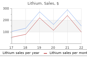
Generic lithium 300 mg
Passive compliance of the ventricle can be decreased by deposition of ibrin and collagen during scar formation or by hypertrophic thickening of the ventricular wall symptoms 10dpo purchase 300 mg lithium fast delivery. Both active and passive processes may be impaired together and are dificult to distinguish clinically. Compensatory Mechanisms and Remodeling When the heart fails to provide adequate cardiac output to meet tissue demands, a number of compensatory mechanisms are triggered. In the short term, these mechanisms are helpful in restoring cardiac output toward normal levels, but in the long term, they are detrimental to cardiac structure and function. Sympathetic Nervous System Activation Sympathetic activation of the heart is primarily a result of baroreceptor relex stimulation. However, because of impaired contractile ability, the failing heart may have reduced responsiveness to sympathetic activation. Sympathetic activation also causes venoconstriction, which redistributes blood and increases cardiac preload. Sympathetic constriction of arterioles helps to maintain blood pressure when cardiac output is reduced. The juxtaglomerular cells release renin and initiate the renin-angiotensin-aldosterone cascade, leading to salt and water retention by the kidney. Sympathetic activation is an early and immediate compensatory response to insuficient cardiac output. Sympathetic activation is a very effective means for increasing cardiac output in an acute process, such as volume depletion. However, in heart failure, sympathetic activation becomes a chronic process that is ultimately deleterious. The remodeled tissue is less functional and may predispose to worsening failure and cardiac dysrhythmias. The cardiac function curve lattens out at a certain point, and minimal beneit is obtained despite increasing preload. Patients with systolic failure have a cardiac function curve that is lat and shifted to the right of normal. These patients beneit from preload reduction, which will decrease systemic and pulmonary congestive symptoms and cardiac workload with little or no reduction in cardiac output. Myocardial Hypertrophy and Remodeling Hypertrophy of cardiac muscle cells is the third mechanism of compensation and generally takes much longer to occur than preload enhancement or sympathetic activation. Hypertrophy appears to result, in part, from a chronic elevation of myocardial wall tension. The development of high systolic pressures in the ventricle may be necessary to overcome a high afterload, such as occurs with arterial hypertension and aortic valve stenosis. Subsequently, decreased cardiac output to the kidney reduces glomerular iltration, resulting in luid conservation. Increased preload is a compensatory mechanism that enhances the ability of the myocardium to contract forcefully. An enlarged chamber volume causes the myocardial ibers to lengthen during diastole, which results in greater iber shortening during contraction (Frank-Starling mechanism). Under some conditions these triggers lead to effective hypertrophy and an increase in size and function, and in others they trigger apoptotic cell death. Systolic failure results in a shift of the curve to the right and a dampening of maximal stroke volume. A greater preload is required to achieve a given stroke volume compared with the normal ventricle. But over time, the signals that promote hypertrophy are thought to trigger a type of ventricular remodeling that contributes to progression of heart failure. A, Eccentric, in which muscle ibers grow in length and the chamber diameter increases. B, Concentric, in which muscle ibers grow in diameter and the ventricular wall becomes thicker. According to the law of Laplace, an increase in chamber radius or pressure will increase wall tension. The hypertrophic response increases wall thickness and helps to relieve wall tension. In summary, enhanced preload and cardiac hypertrophy may allow a heart to compensate for reduced ventricular function for an extended period. Unfortunately, these compensatory mechanisms, which serve to restore cardiac output to the tissues, also result in an increase in myocardial work and oxygen requirements and appear to cause pathologic remodeling. Unfortunately, these responses also increase myocardial workload and may perpetuate the heart failure. Sympathetic nervous system activation increases heart rate, contractility, arterial vasoconstriction, and renin release. Higher preload results in more forceful ejection of blood from the heart (Frank-Starling law) and improves cardiac output. Excessive neurohormones, volume overload, and high wall tension contribute to abnormal ventricular remodeling. Because of circulatory dynamics, left ventricular failure often leads to right ventricular failure-a condition termed biventricular failure. The etiologic process, clinical manifestations, and management of isolated right ventricular failure differ substantially from those for left ventricular and biventricular failure. Insuficient cardiac pumping is manifested by poor cardiac output, called forward failure, and by congestion of blood behind the pumping chamber, called backward failure. The clinical manifestations of left and right ventricular failure differ as a result of the anatomic location of the "backward" or congestive processes, but the forward effects of low cardiac output are the same. Inadequate perfusion of the brain may lead to restlessness, mental fatigue, confusion, anxiety, and impaired memory. Reduced perfusion of the kidney results in a decline in urine output (oliguria) with subsequent luid retention. Constriction of blood vessels serves to maintain blood pressure and redistribute reduced cardiac output to vital organs. However, this vasoconstriction also increases afterload, so the damaged left ventricle must generate more force to pump the same volume of blood. Depending on how much the afterload increases, the damaged left ventricle may not be able to pump suficient quantities of blood in to the circulation. If renal blood low becomes severely limited, the patient with left ventricular failure may develop kidney failure. Left-Sided Heart Failure Left-sided heart failure is most often associated with left ventricular infarction and systemic hypertension. Ineffective pumping of the left ventricle results in an accumulation of blood within the pulmonary circulation. As hydrostatic pressure builds within the pulmonary veins and capillaries, luid is forced from the capillaries in to interstitial and alveolar spaces, causing edema. Dyspnea, or breathlessness, occurs early in the progression of left-sided heart failure and may be considered the cardinal symptom. Dificulty breathing may be exacerbated by activity (dyspnea on exertion), lying down (orthopnea and paroxysmal nocturnal dyspnea), and blood volume expansion from excessive salt or luid intake. Orthopnea and paroxysmal nocturnal dyspnea are due in part to a redistribution of blood volume from the periphery to the heart when the individual lies down. The failing left ventricle is unable to effectively pump extra volume, and pulmonary congestion is worsened. Paroxysmal nocturnal dyspnea refers to intermittent attacks of severe dyspnea during the night and is a most distressing form of orthopnea. The individual experiences a feeling of suffocation and panic at not being able to overcome the dyspnea. Sitting or standing helps to relieve the dyspnea because blood pools in the extremities, reducing pulmonary hydrostatic pressure and congestion. In severe cases, sputum may be blood tinged, from breakage of fragile capillaries, and frothy, from luid buildup in the alveoli. The severity of pulmonary edema can be estimated from the location of crackles within the lung ields.
Discount lithium 300 mg amex
One heritable risk factor is a mutation of the retinoblastoma gene responsible for an infantile cancer of the eye that is associated with increased bladder cancer risk medicine keychain buy lithium 300mg on-line. Bladder cancer metastasis occurs directly through the bladder wall to adjacent organs. Once treated, tumors can recur at the original site, or an entirely new tumor may develop at another site. The sites of metastasis most commonly include the lymph nodes, liver, lungs, and bone. Other manifestations related to bladder cancer include urinary frequency and urgency; dysuria is an uncommon inding. But again, all these symptoms are also seen with other conditions involving the urinary tract. In most cases, diagnostic evaluation for bladder cancer is initiated because of the development of one or more of the previously listed clinical manifestations. A thorough history may reveal risk factors for bladder cancer, or physical examination and diagnostic testing may indicate another etiology. The recommended diagnostic test for suspected bladder cancer is cystoscopy, with biopsy of any questionable tissue and washings of free cells for cytologic examination. The sensitivity and speciicity of available tumor markers varies widely; at this time, no tumor marker is reliable enough to replace cystoscopy. The stage of the malignancy is an important contributor to treatment decisions, and also aids in the determination of prognosis. All of this information is used to guide the selection of the speciic treatment approach. The primary options are surgery, radiation therapy, chemotherapy, and immunotherapy. Chemotherapy or radiation therapy may be performed preoperatively to reduce tumor size and improve survival. Urine is drained from the reservoir often through a stoma created through the abdominal wall; however, substitute bladder reservoir (neobladder) procedures involve connecting the urethra to the reservoir, allowing the patient to void normally. In cases where the tumor is large or there are multiple bladder tumors, a radical cystectomy is performed, in which the bladder and surrounding nodes are removed; in men the prostate gland is also removed, and in women the uterus, ovaries, fallopian tubes, and part of the vagina are also often removed. External beam radiation therapy may be an acceptable alternative for patients who are not suficiently strong for radical surgical procedures. The normal defense mechanisms of the urinary tract are presented in detail in Chapter 27 with the discussion of pyelonephritis (infection of the kidney). By the time infection reaches the kidneys, the bladder and urethra are already infected. Inlammation of the urethra may lead to pain, burning, and urinary incontinence, and if it is attributable to an infectious organism, it may progress to infective cystitis. Urethritis can be caused by infection, external irritants, or, in women, insuficient estrogen levels. Infection of the urethra may be due to a wide variety of organisms, most often including Neisseria gonorrhoeae and Chlamydia trachomatis. The urethra is an estrogen-dependent structure, and postmenopausal women are at increased risk for irritation and inlammation of the urethra. Application of topical estrogen to the urethral opening helps maintain mucosal health. Otherwise, urethritis attributable to infection is often asymptomatic until the organism progresses to the bladder, causing cystitis. Cystitis Cystitis, or inlammation of the bladder lining, may result from bacterial, fungal, or parasitic infections, chemical irritants, foreign bodies. By far the most common cause of cystitis- and the focus of this discussion-is bacterial infection. Normally, bacteria are cleared from the bladder by the lushing and dilutional effects of voiding. The high urea concentration and osmolarity and the low pH in urine act as natural barriers, killing invading bacteria in a normal bladder environment. In the pediatric population, uncircumcised male infants less than 6 months of age and females less than 12 months of age have the highest prevalence. They are common occurrences during pregnancy, and have been associated with an increased risk of premature delivery and low birth weight. Escherichia coli is responsible for 80% of cases of bacterial cystitis, with Staphylococcus saprophyticus as the next most common causative pathogen. About 95% of bladder tumors originate from the transitional epithelium (urothelium) lining the urinary tract. Infections attributable to Neisseria gonorrhoeae or Chlamydia trachomatis are associated with sexually transmitted diseases but are typically limited to the urethra. The majority of patients with cystitis experience an acute onset of frequency, urgency, dysuria, and pain, often in the suprapubic area. Common symptoms of cystitis in children include fever, irritability, poor feeding, vomiting, diarrhea, and ill appearance. Uncomplicated infections, caused by common organisms, may be diagnosed on the basis of symptoms and a positive dipstick. Culture is recommended in situations in which the patient has manifestations suggesting pyelonephritis, fails to respond to empirical pharmacologic therapy, is pregnant, or has urinary calculi. A culture should be considered if the patient is immunosuppressed (because of the likelihood of atypical organisms) or has diabetes mellitus. Unresolved infections are those in which bacteriuria remains after the initial treatment. Unresolved infections are usually treated with targeted antimicrobials for 7 to 10 days. Urine culture and sensitivity are also necessary to ensure antibiotic effectiveness. In some women, recurrent infections are related to sexual intercourse, and a postcoital prophylactic antibiotic program is an effective treatment option. In children, administration of broad-spectrum antibiotics for 7 to 14 days is the treatment of choice. However, children younger than 5 years should keep taking prophylactic doses of antibiotics until radiographic evaluation is completed. Symptoms associated with bacteriuria in the elderly may include anxiety, confusion, lethargy, and anorexia as opposed to dysuria and fever. The diagnosis is often one of exclusion, and urinalysis and culture are included in the diagnostic evaluation. The speciic etiology is unknown and the pathophysiology is unclear, but a fundamental mechanism appears to be urothelial damage. Inlammation is activated and the damaged urothelium is less able to serve its protective functions, including production of mucus. Factors that predispose the elderly to cystitis include genitourinary abnormalities. The diagnostic workup should include a history, physical exam, urinalysis, and possibly a urine culture and cytology to rule out subclinical infection and bladder cancer. The foods and drinks most often associated with pain are alcohol, citrus fruits, coffee, carbonated beverages, tea, chocolate, and tomatoes. Hydrodistention of the bladder and instillations of potassium chloride will often reproduce the pain. Pelvic loor muscle training and bladder retraining may be helpful in moderating urgency. Amitriptyline and hydroxyzine are other drugs that have been prescribed for symptom management. It is most often due to infection, either from the bladder or associated with a sexually transmitted disease. Sexually transmitted diseases are conined to the urethra; infections of other etiologies may ascend to the bladder before symptoms present. Urethritis may also be due to external factors such as frequent catheterizations or poor personal hygiene.
Purchase genuine lithium on line
These products are often secreted by the cell as signaling molecules to other nearby cells symptoms 5 days before your missed period purchase lithium 150 mg on-line. In this case, the subunit of Gi is also activated and opens membrane potassium channels in the heart, which tend to slow the heart rate. A cascade of kinase activations is initiated resulting in a change in target gene transcription. The primary activator of guanylyl cyclase is a small lipid-soluble gas molecule called nitric oxide. It functions as a neurotransmitter in the brain and is an important smooth muscle relaxant in the vascular system. To be effective at communicating signals, all the receptor systems must be quickly turned off so that they can be responsive to the next incoming signal. Some drugs, such as caffeine and sildenail citrate (Viagra), are phosphodiesterase inhibitors that slow the normal breakdown of cyclic nucleotides and prolong their activity. Many of the intracellular signaling cascades rely on kinases that phosphorylate their target proteins so as to change their activity. The action of kinases is countered by numerous phosphatase enzymes that quickly cleave the phosphates off the target proteins and inhibit their activity. The cell also can regulate the activity and number of receptors on the cell surface. Receptors can be internalized in the cell where they are inactive but are available for later use, or they can be sent to lysosomes for degradation. Receptors that can bind ligand but do not produce a response are said to be uncoupled. The mechanisms that "turn off" signaling cascades are vitally important to maintaining a responsive communication system. Intracellular receptors are speciic for a particular ligand, just as surface receptors are. Because lipid-soluble ligands enter the cell directly, no second messengers are needed. Thyroid hormone enters the cell through carriers in the membrane and travels to the nucleus. When thyroid hormone inds its nuclear receptor, the complex dissociates and removes an inhibitory inluence on gene transcription. Cellular responses to these gene regulatory receptor complexes are slow in comparison to the cell surface receptor responses and generally last longer. Regulation of Cellular Growth and Proliferation In multicellular organisms such as humans, the growth and proliferation of cells and tissues must be strictly controlled to maintain a balance between cell birth rate and cell death rate. When the ligand binds to the receptor, an intracellular domain is changed in to an active coniguration that can interact with inactive trimeric G-proteins. The subunit is now in the correct conformation to reassociate with the subunits and await another signal from the receptor. In some cases the subunit also has functional activity and may regulate ion channels. A, It can be synthesized by enzyme-linked receptors that are activated by water-soluble ligands such as atrial natriuretic peptide. B, Nitric oxide is an important signaling molecule that is lipid soluble and can diffuse across the cell membrane. B, Receptor internalization temporarily reduces the number of receptors displayed at the cell surface. C, Receptor degradation results in a long-term reduction in receptors (down-regulation). D, the cyclic nucleotide second messengers can be degraded by phosphodiesterase enzymes to stop the intracellular cascade. E, Phosphatase enzymes counteract the phosphorylating activities of kinases and inhibit the intracellular cascade. Thyroid hormone is not lipid soluble and enters the cell through a carrier to interact with its intracellular receptor. When the ligand binds to its intracellular receptor, it forms a functional gene regulatory protein that affects the rate of transcription of its target genes. The response of the cell to intracellular ligands is generally slow and long lasting. Special intercellular communication systems function to regulate the replication of individual cells in the body. First, a variety of protein mitogens and growth factors are required in speciic combinations for growth and proliferation of particular cell types. Second, cells respond to spatial signals from the extracellular matrix (from integrin receptors) and neighboring cells (from cell adhesion proteins) that indicate how much room is available. Cycling cells proceed through G1, S phase (synthesis), G2, M phase (mitosis), and cell division. M phase, or mitosis, proceeds through six stages, beginning with prophase, in which the chromosomes condense and become visible, and ending with cytokinesis, when cell division is accomplished. Mitosis is responsible for the proliferation of body cells in which little genetic variation is needed or desired. A more elaborate cell division process, meiosis, occurs in the germ cells (egg and sperm), where signiicant chromosomal rearrangements occur (see Chapter 6). The cell cycle has been the subject of intense study in recent years because of its importance in cancer biology. Cancer cells continue to grow and divide unchecked, despite the lack of appropriate signals to stimulate them. Of particular interest are the events that prod the cell from its dormant state and cause it to begin the cycle. The Rb protein (or pRb) is of central importance in preventing a cell from proceeding through the cell cycle. The Rb protein can be induced to release the E2F transcription factors when appropriate mitogen signals arrive at the cell surface. The cell then proceeds systematically through the S phase (synthesis), G2, and M phase (mitosis). These proliferation-promoting signals at the cell surface are transmitted to the Rb protein by way of cyclin-dependent signaling pathways within the cell. Proteins called cyclins accumulate in the cell and then bind to and activate cyclin-dependent kinases (cdk). The cdk then phosphorylates the Rb protein, changing its afinity for E2F so that it is released. The mitotic spindle, a bipolar structure composed of microtubules and associated proteins, begins to form. Each is attached by its centromere to a microtubule that is also linked to the spindle pole. Activation of the receptor stimulates signaling pathways within the cell that increase cyclin proteins. The cyclins bind to cyclin-dependent kinases (Cdks) to form active enzyme complexes. The active cyclin-Cdk enzymes phosphorylate Rb protein (pRb), inducing it to release E2F transcription factors that initiate replication. In the absence of appropriate growth factor signals, the Rb protein functions to inhibit unwanted cell proliferation. Numerous mitogens have been identiied, and most cells require an appropriate combination of mitogen signals before they can enter the cell cycle. Somatic cells respond to growth factors by increasing cell size, whereas stem cell populations undergo cell division. Thus the same signaling ligands may have different effects depending on cell type and conditions. Similar signaling pathways may also trigger cell death (apoptosis) when cells have to be reduced or removed during tissue development and remodeling. The processes of abnormal cellular proliferation and cancer are further detailed in Chapter 7.
Lithium 300mg on line
It is through this junction that the young germinal cells medications 25 mg 50 mg buy generic lithium line, or primary spermatocytes, migrate and pass from the basal compartment to the basement membrane and then to the central or adluminal compartment of the seminiferous tubule. This barrier ensures that the more mature spermatocytes and spermatids located in the adluminal compartment are behind the barrier and theoretically maintained in a constant intratubular environment to support the development of maturing sperm cells. The proliferative phase involves division of the young germinal cells near the basement membrane (spermatogonia) either to replace their numbers or to produce daughter cells that will form spermatocytes. This division reduces the number of chromosomes to the monoploid number of 23 from the diploid number of 46. While sperm cells mature and move from the basement membrane to the adluminal compartment, the Sertoli cells have an important nutritional role in the spermatogenic process. However, the magnitude of functional decline of the male reproductive organs is variable. Active male germinal cells continue to produce spermatozoa (spermatogenesis), although the number of sperm produced declines proportionally over time. The testes become smaller as a result of the increased amount of connective tissue, ibrosis of the tubules, and decreased numbers of capillaries. The number of Leydig cells that produce testosterone decreases, leading to a decrease in testosterone level with aging. Sexually, the aging male has a longer refractory period after orgasm and decreased force of ejaculation. The axoneme, which runs the length of the tail, is composed of a central pair of tubules surrounded by a ring of nine pairs of tubules (the 9 + 2 pattern). This ring of tubules is surrounded by a supporting structure of nine noncontractile dense ibers. The mitochondria contain the enzymes required for the production of adenosine triphosphate, the energy source for the cell. These enzymes convert chemical energy from adenosine triphosphate to the mechanical energy of sperm cell movement to aid in fertilization of the egg. Mature spermatozoa are released in to the tubular lumen and rapidly low out to the rete testis and in to the epididymis. Although each spermatogonium, one of the primitive male germ cells, requires about 70 days to develop in to a mature sperm cell, or spermatozoon, within each tubule are spermatozoa in all stages of development. This characteristic allows new spermatozoa to be continuously produced across the male life span. The effects of aging on the male reproductive system are described in the Geriatric Considerations box. The oval head contains a nucleus that is highly condensed and stabilized by cross-links between its molecules, which makes it very resistant to physical injury during its passage and storage in the epididymis. An outer membrane, the acrosome, contains the enzymes required for penetration of the female egg before fertilization. The tail accounts for 90% of the length of the spermatozoon and is divided in to a middle piece, principal piece, and end piece. The spermatozoon derives its motile ability from the motor apparatus of the Once mature spermatozoa are released from the Sertoli cells in to the seminiferous tubules, they must pass through approximately 6 m of duct in the male reproductive tract before leaving the urethral meatus and being deposited in the vagina during sexual intercourse. From the seminiferous tubules, the spermatozoa are deposited in to the rete testis, a collecting chamber for all the seminiferous tubules. From the rete testis, the sperm travel through the efferent ductules, 12 to 20 channels that pass in to a single compact duct, the epididymis. The epididymis is a tightly convoluted duct that is divided in to three regions: the caput (globus major), the corpus (body), and the cauda epididymis (tail, or globus minor). It passes through the scrotum, traverses the inguinal canal in to the pelvis, and then passes behind the bladder to enter the prostatic urethra at the ejaculatory ducts of the verumontanum. It is joined by the ducts of the seminal vesicle before entering the ejaculatory ducts. As one passes in a proximal-to-distal direction from the efferent ducts to the vas deferens, the thickness of the muscle gradually increases. In the vas deferens, three interconnected smooth muscle layers form a thick muscular wall, with the ratio of wall thickness to lumen being the greatest in any human structure. Aside from serving as a conduit and storage depot for spermatozoa, the epididymis probably sustains maturational processes. Acrosome Principal piece of tail Head Nucleus covered by acrosome Neck Middle piece of tail are incapable of fertilizing eggs. It appears that the development of motility and increased fertility are acquired during transit through the epididymis. Initially, spermatozoa are carried in to the efferent ducts by luid from the rete testis. Within the efferent ducts, motile cilia within the lumen function to reabsorb testicular luid and help move spermatozoa in to the epididymis. Within the epididymis, the spermatozoa are probably transported by rhythmic contraction of the smooth muscle cells. In young men, approximately 200 million sperm can be found in the reservoir of the epididymis. With ejaculation, sperm from the distal part of the epididymis and vas deferens are deposited in to the prostatic urethra, where they account for less than 10% of the normal ejaculate. The physiology of erection is a complicated interaction of vascular, neurologic, and hormonal factors. Although erection has classically been thought of as a parasympathetic function, it is more complex. The presence of erections in patients with spinal cord injuries attests to the presence of relex erections. Such patients have an intact sacral spinal cord and its relex arc of afferent and efferent nerves below the site of spinal cord injury. The penis receives sensory innervation from the pudendal sensory nerves entering the sacral spinal cord. The pudendal nerve is a mixed nerve that provides motor innervation to the pelvic loor musculature and penile sensory ibers. The efferent nerve to the erectile tissue is provided by sacral parasympathetic ibers. Although erection is possible in patients with spinal cord injuries, in intact men it is a much more controlled process inluenced to a great extent by the cerebral cortex. Impulses may traverse the spinal cord from the cerebral cortex in the lateral columns and exit the spinal cord through sacral parasympathetic and possibly the thoracolumbar sympathetic nerves as well. Research indicates that erectile function cannot be fully explained by parasympathetic or sympathetic mechanisms; this observation has led to consideration that nonadrenergic and noncholinergic neuromodulators may be involved in such function. During emission, secretions from the periurethral glands, seminal vesicles, and prostate are deposited with sperm from the vasa deferentia and the cauda epididymis in to the prostatic urethra. Control of emission is mediated primarily through the sympathetic nerves, which stimulate contraction of smooth muscle in these genital structures. Next, the external sphincter relaxes and the perineal and bulbourethral muscles surrounding the bulb of the corpus spongiosum contract and expel the ejaculate from the posterior urethra and through the urethral meatus. The physiologic function of the secretory products of the accessory sex glands is uncertain. These secretions make up most of the seminal plasma, with the sperm and testicular luid probably composing less than 10% of the inal ejaculated semen volume. Although some investigators have demonstrated that sperm removed directly from the epididymis are capable of fertilization, these secretions most likely optimize conditions for sperm motility, survival, and transport in both the female and the male reproductive tracts. After ejaculation, the sperm that are deposited in the vagina swim away from the cholesterol vesicles upward in to the uterine luid, and they gradually lose much of their excess cholesterol during the next few hours. As the cholesterol is lost, the membrane at the head of the sperm becomes much weaker. Large amounts of calcium enter the sperm to increase the powerful whiplike motion of the lagellum beyond its previously weak, undulating motion. In addition, the calcium ions probably also alter the intracellular membrane covering the leading edge of the acrosome, thus making it possible for the acrosome to release its enzymes very rapidly and easily as the sperm penetrates the granulosa cell mass surrounding the ovum. These enzymes are released even more rapidly and easily as the sperm attempts to penetrate the zona pellucida of the ovum itself. Although sperm are anatomically complete and highly motile when ejaculated, the complex process of capacitation is necessary before the sperm are actually capable of fertilizing the egg.
Buy discount lithium line
The aims in stages 1 and 2 are to identify individuals at risk for progressive renal disease medications held for dialysis generic lithium 300 mg visa, and reduce those associated risks. Although they may be otherwise asymptomatic in stage 3, hypertension is nearly always noted. At stage 4, diagnosis is made because manifestations are usually very apparent with the signiicant decline in renal function. Decreased renal reserve is not associated with signs or symptoms of renal failure, largely because the remaining nephrons accommodate the additional workload. At this point, kidney function is already impaired and signiicant deterioration is possible if the kidney is stressed. At this point, the patient typically demonstrates the sequelae and complications of renal failure, seen as laboratory alterations and signs and symptoms associated with the inability of the kidneys to fulill their multiple roles within the body. Without interventions, when the remaining nephrons number less than 5% to 10% of normal, death is inevitable. Immune function, acid-base regulation, and the coagulation cascade are affected as well. In many cases, complications are interrelated, one contributing to or exacerbating the development of others. Additionally, a negativenitrogen balance exists because of escalated protein catabolism and decreased protein synthesis. Medications and concomitant diseases such as diabetes may cause nausea, vomiting, and slowed gastric emptying. Hypoalbuminemia in dialysis patients has a strong association with increased mortality and morbidity. However, caution must be used in interpreting these levels, because the serum level may be affected by the inlammatory response. The production of red blood cells by the bone marrow depends on numerous cofactors; perhaps the most signiicant of these is erythropoietin, which is produced by the kidneys. The problem is often further escalated by malnutrition, due to nutritional deicits of iron, folate, and vitamin B12. By stage 5 and the initiation of dialysis, approximately 66% of patients have hemoglobin levels <11 g/dl. It is also produced in association with hyperkalemia, when potassium ions in the blood are exchanged for intracellular hydrogen ions, lowering the pH of the blood (see Chapter 25). The kidneys lose their ability to secrete hydrogen ions or to produce bicarbonate, and with the limited capacity of the other buffers, pH can fall precipitously; coma develops and death will occur if the pH drops below 6. The respiratory system attempts to compensate for metabolic acidosis by increasing the rate and depth of respirations. Electrolyte Imbalances the loss of renal mechanisms involved in electrolyte balance result in the retention of potassium, phosphorus, and magnesium in the blood. Each of these imbalances is associated with speciic manifestations (see Chapter 24). The inability to eliminate phosphorus and the loss of the renal mechanisms involved in maintaining calcium balance result in mineral and bone disorders, which are discussed next. Dialysis itself can be a painful experience with frequent needle sticks and accumulation of uremic toxins. The primary cause of kidney disease, such as cystic kidney or diabetes (due to neuropathies or ulcerations), may result in pain. Many patients with renal disease experience a high rate of cardiovascular events, which are also associated with higher rates of depression. In addition, the disruption of social interactions and relationships, possibly attributable to dialysis and fatigue, contributes to depressive symptoms. The two most common causes are diabetes mellitus and hypertension, followed by recurrent pyelonephritis, glomerulonephritis, and polycystic kidney disease. In stage 4, planning for dialysis or transplant should begin, and in stage 5 renal replacement therapy is needed or death will ensue. Collaboration among the nurse, physician, clinical pharmacist, and dietitian is essential to attain optimal patient outcomes. Etiologies and risk factors for prerenal kidney injury should be identiied and, whenever possible, treated swiftly. Hypotension attributable to hypovolemia should be addressed; medications that might be contributing (antihypertensives, opioids) should be decreased or discontinued. Nutrition should be supported, and indwelling catheters and other invasive equipment should be removed as soon as possible to decrease the risk of infection. Two interventions that were previously staples of treatment of prerenal oliguria have been deemed to be harmful, or at the very least ineffective. Again, compared to placebo therapy, loop diuretics made no difference in the recovery of renal function. Whenever possible their use should be limited, and if these agents must be prescribed, their serum levels may need to be monitored. Postrenal etiologies should be avoided whenever possible, but if they occur they should be rapidly identiied and corrected. Because death is often due to cardiovascular pathologies, management of these risk factors assumes signiicant importance. Complications are typically evident by stage 3 or 4 and attention is directed to therapeutic interventions designed to minimize and treat these complications. Therapeutic and pharmacologic interventions are presented within the context of each complication listed in the following paragraphs. An in-depth presentation is beyond the scope of this text; discussion is simply an overview. Patients with edema, heart failure, or hypertension may need a 2 g/day sodium restriction. Avoiding malnutrition, preventing anemia, and countering disease- and drug-induced constipation are other aspects of nutritional management. Lists of foods high in sodium, potassium, and protein should be provided; patients can then be encouraged to identify their favorite foods and choose smaller portions or eat them less often. Additionally, diet-related risk factors for cardiovascular disease must be considered. This can all seem overwhelming to patients, so involvement of a dietitian and thorough education considering concomitant conditions, personal likes and dislikes, eating habits, and inancial resources is essential. Although a life-saving intervention, dialysis treatments have complications, some of which are life-threatening, and long-term morbidity remains quite high. During the treatment, the peritoneal cavity is slowly filled with dialysate through the catheter. Extra fluid and waste products are drawn out of the uremic blood and in to the dialysate. The patient instills about 2 quarts of dialysate in to the peritoneum through the catheter. The dialysate remains there for 4 to 5 hours or longer, before it is drained and discarded. Continuous hemoiltration and hemodialysis procedures ilter and dialyze the blood without interruption. This "gentler" continuous removal of wastes and blood helps avoid the hypotensive episodes caused by intermittent hemodialysis and its intermittent removal of large volumes of luid. Psychotherapy, exercise therapy, cognitive behavioral therapy, and music therapy all have demonstrated varying degrees of success. As with other conditions in which transplantation is indicated, the primary limiting factor is the availability of organs. For most of those who choose it, transplantation allows for increased independence, return to normal activities of daily living, and resumption of normal renal function. Thousands of patients receive kidney transplants each year in the United States, with a remarkable rise in transplant recipients over the last decade. This means that general health care providers will be caring for transplant recipients and the chronic medical conditions that accompany transplantation surgery. Common medical complications include cardiovascular disease, obesity, hypertension, dyslipidemia, diabetes, cerebrovascular disease, anemia, gout, depression, bone disease, malignancies, and infections.
Methi (Fenugreek). Lithium.
- How does Fenugreek work?
- Are there any interactions with medications?
- What is Fenugreek?
- Dosing considerations for Fenugreek.
- Are there safety concerns?
- Diabetes, high cholesterol, high triglycerides, stomach upset, decreased appetite, constipation, hardening of the arteries (atherosclerosis), gout, sexual problems (impotence), fever, baldness, and other conditions.
Source: http://www.rxlist.com/script/main/art.asp?articlekey=96717
Cheap 150mg lithium with visa
Oxygen therapy with or without mechanical ventilation may be necessary in severe cases medicine escitalopram buy cheap lithium 150 mg line. The more patients understand about their asthma, the better they are at self-managing their symptoms. Educational materials are available from the American Lung Association, the Asthma and Allergy Foundation of America, and the National Institute of Allergy and Infectious Diseases. Non-allergic (intrinsic) asthma is precipitated by exercise, stress, and exposure to pulmonary irritants, but no speciic allergen can be identiied. Drugs such as aspirin and exposure to occupational allergens have also been identiied as etiologic agents. The IgE binds to mast cells and causes them to release inlammatory chemicals in response to antigen. Avoidance of precipitating factors and use of prophylactic drug therapy are recommended. Bronchodilators, corticosteroids, and oxygen therapy are mainstays of treatment for an acute attack. Acute inlammation of the trachea and bronchi is produced most commonly (80% of the 12 million cases per year in the United States) by a variety of viruses such as inluenza virus A or B, parainluenza virus, respiratory syncytial virus, coronavirus, rhinovirus, Coxsackie virus, and adenovirus. Nonviral causes include Streptococcus pneumoniae, Haemophilus inluenzae, mycoplasma, moraxella, and Chlamydia pneumoniae. In chronic bronchitis, the thickness of the mucous glands increases and can be expressed as the Reid index, given by the following formula: (b - c)/ (a - d). Characteristic pathologic and clinical indings are described for each of these classiications. Clinically, pure forms of emphysema and chronic bronchitis are rare, and most patients present a combination of both of these obstructive processes. The major causes of chronic bronchitis are cigarette smoking (90% of cases),12 repeated airway infections, genetic predisposition, and inhalation of physical or chemical irritants. The National Center for Health Statistics reports a 3:1 ratio of annual cases of chronic bronchitis to emphysema. Pathologic changes in the airway include chronic inlammation and swelling of the bronchial mucosa resulting in scarring, increased ibrosis of the mucous membrane, hyperplasia of bronchial mucous glands and goblet cells, hypertrophy of bronchial glands and goblet cells, and increased bronchial wall thickness, which potentiates obstruction to airlow. During acute exacerbations, bronchial biopsy specimens have a 30-fold increase in the number of eosinophils. Hypertrophy of mucosal glands and goblet cells leads to increased mucus production; the mucus then combines with purulent exudate to form bronchial plugs. The narrowed airways and the mucous plugs prevent proper oxygenation and potentiate airway obstruction. High airlow resistance increases the work of breathing, leading to increased oxygen demands. The airways become inlamed and narrowed from capillary dilation, swelling from exudation of luid, iniltration with inlammatory cells, increased mucus production, loss of ciliary function, and loss of portions of the ciliated epithelium. Many viruses and mycoplasmal bacteria inhibit macrophages and lymphocytes, temporarily promoting secondary bacterial invasion. Microorganisms may also induce long-lasting hyperirritability of the respiratory tract with associated episodes of bronchospasm. The presentation of acute bronchitis is usually mild and self-limited, requiring only supportive treatment. Associated symptoms include low-grade fever, substernal chest discomfort, sore throat, postnasal drip, and fatigue. In children, the smaller airways are easily obstructed by inlammation, so that severe obstruction may occur. Associated inlammation of the larynx and trachea produces croup (see Croup Syndrome section in this chapter for further details). Diagnosis of acute bronchitis is usually based on the clinical presentation, with recent onset of cough being the distinctive hallmark. A chest radiograph is required to distinguish acute bronchitis (normal radiograph) from pneumonia (pulmonary iniltrates on radiograph). Acute bronchitis is predominantly caused by viruses (rhinovirus, coronavirus, adenovirus, inluenza virus). Viral infections do not respond to antimicrobial therapy, and symptoms resolve spontaneously in most normal, otherwise healthy individuals. Acute bronchitis caused by bacterial organisms responds well to antibiotic therapy. Codeine-containing medications are helpful in relieving the cough associated with bronchitis that interferes with sleep. Nonpharmacologic recommendations are to increase luid intake, avoid smoke, and use a vaporizer in the bedroom. The involvement of small pulmonary arteries related to inlammation in the bronchial walls and the compensatory vasoconstriction of pulmonary blood vessels from hypoxia produce pulmonary hypertension. In addition, widespread bronchial narrowing and mucous plugging produce ventilation-perfusion mismatch with hypoxemia and hypercarbia from impeded ventilation. The combination of hypoxia and hypercarbia increases pulmonary artery resistance and pulmonary hypertension. An enlarged right heart results in increased venous pressure, liver engorgement, and dependent edema. Manifestations of heart failure may occur during exacerbations of bronchitis and subside with appropriate treatment. Causes of bronchial wall destruction include infection from severe streptococcal or staphylococcal pneumonia, repeated bouts of acute bronchitis, infection with the mold Aspergillus fumigatus, presence of mucous plugs or foreign bodies, or deiciencies in immunologic response. If bronchiectatic lesions are localized, surgical resection of the affected portions of lung may be helpful. The typical patient is an overweight man or woman (1:2 male to female ratio) in his or her thirties or forties9,21,22,24 (or older) who presents with shortness of breath on exertion, excessive amounts of sputum, chronic cough, evidence of excess body luids (edema, hypervolemia), and a history of smoking. In addition, the patient often complains of chills, malaise, muscle aches, fatigue, loss of libido, and insomnia. Measures used to conirm the diagnosis include chest radiography, which may show increased bronchial vascular markings, congested lung ields, an enlarged horizontal cardiac silhouette, and evidence of previous pulmonary infection. Early pulmonary function testing before the onset of symptoms shows increased closing volume and a decrease in the maximal midexpiratory low rate. The electrocardiogram may reveal atrial dysrhythmias and evidence of right ventricular hypertrophy. Secondary polycythemia (increased numbers of red blood cells) related to continuous or nocturnal hypoxemia is common. Depending on the severity of the disease, the physical examination may reveal scattered crackles, rhonchi, and wheezes; use of accessory muscles to breathe; jugular vein distention; clubbing; and pedal and ankle edema. Table 22-1 lists the distinguishing features of both emphysema and chronic bronchitis. Because bronchitis and emphysema are most frequently seen in combination, the therapies are similar. The overall goals are to (1) block the progression of the disease, (2) return the patient to optimal respiratory function, and (3) return the patient to usual activities of daily living. Pharmacologic treatment involves the use of inhaled short-acting 2 agonists and inhaled anticholinergic bronchodilators, cough suppressants, and antimicrobial agents for infections. Inhaled or oral corticosteroids may also be used in the treatment of some patients for acute exacerbations. Theophylline products are used less frequently because of their narrow therapeutic range and toxicity. Home oxygen therapy has been demonstrated to retard the development of pulmonary hypertension and cor pulmonale in chronic bronchitis. Walking has proved to be the best form of exercise for increasing duration and intensity of activity. Symptoms are caused by narrowing of inlamed airways and increased mucus production. It is deined as a productive cough lasting more than 3 months per year for 2 or more consecutive years. Airway obstruction leads to poor ventilation of alveoli and impaired exchange of oxygen and carbon dioxide. Persistent hypoxemia causes a compensatory increase in red blood cell production (polycythemia). Emphysema tends to develop over a long period and thus is seen more frequently in persons older than 50.
Order generic lithium online
Hydropic swelling is characterized by a large acne natural treatment lithium 150 mg with amex, pale cytoplasm, dilated endoplasmic reticulum, and swollen mitochondria. With severe hydropic swelling, the endoplasmic reticulum may rupture and form large water-illed vacuoles. Generalized swelling in the cells of a particular organ will cause the organ to increase in size and weight. Intracellular Accumulations Excess accumulations of substances in cells may result in cellular injury because the substances are toxic or provoke an immune response, or merely because they occupy space needed for cellular functions. In some cases, accumulations do not in themselves appear to be injurious but rather are indicators of cell injury. A, Normal kidney tubule with cuboidal cells; B, early ischemic changes showing surface blebs and swelling of cells. However, with certain disorders, renal glomerular capillaries become leaky and allow proteins to pass through them. Renal tubule cells recapture some of the escaped proteins through endocytosis, resulting in abnormal accumulation. The abnormally folded intracellular proteins may cause 59 Abnormal metabolism Normal cell Fatty liver Protein mutation Protein folding, transport Lack of enzyme Complex Soluble substrate products Enzyme serious cell dysfunction and death if they are allowed to persist in the cell. If the chaperones are unsuccessful in correcting the defect, the abnormal proteins form complexes with another protein called ubiquitin. In some cases, the accumulated substances are not metabolized by normal intracellular enzymes. In diabetes, for instance, high serum glucose levels result in excessive glucose uptake by neuronal cells because they do not require insulin for glucose uptake. Some pigment accumulations are normal, such as the accumulation of melanin in tanned skin, whereas others signify pathophysiologic processes. Pigments may be produced by the body (endogenous) or may be introduced from outside sources (exogenous). In addition to melanin, the iron-containing substances hemosiderin and bilirubin are endogenous pigments that, when present in excessive amounts, indicate disease processes. Inorganic particles that may accumulate include calcium, tar, and mineral dusts such as coal, silica, iron, lead, and silver. Inhaled dusts cause chronic inlammatory reactions in the lung, which generally result in destruction of pulmonary alveoli and capillaries and the formation of scar tissue. Over many years, the lung may become stiff and dificult to expand because of extensive scarring (see Chapter 23). Deposits of calcium salts occur in conditions of altered calcium intake, excretion, or metabolism. Impaired renal excretion of phosphate may result in the formation of calcium phosphate salts that are deposited in the tissues of the eye, heart, and blood vessels. Calciication of the heart valves may cause obstruction to blood low through the heart or interfere with valve closing. Calciication of blood vessels may result in narrowing of vessels and insuficient blood low to distal tissues. Cellular stress may be due to an increased functional demand or a reversible cellular injury. Although the term adaptation implies a change for the better, in some instances an adaptive change may not be beneicial. Each of these changes is potentially reversible when the cellular stress is relieved. For example, lung damage resulting from tuberculosis often is apparent as calciied areas, called tubercles. With the exception of inorganic particles, the intracellular accumulations generally are reversible if the causative factors are removed. Atrophy Atrophy occurs when cells shrink and reduce their differentiated functions in response to a variety of normal and injurious factors. The general causes of atrophy may be summarized as (1) disuse, (2) denervation, (3) ischemia, (4) nutrient starvation, (5) interruption of endocrine signals, (6) and persistent cell injury. Apparently, atrophy represents an effort by the cell to minimize its energy and nutrient consumption by decreasing the number of intracellular organelles and other structures. For example, immobilization by bed rest or casting of an extremity results in shrinkage of skeletal muscle cells. Denervation of skeletal muscle results in a similar decrease in muscle size caused by loss of nervous stimulation. If the blood supply is totally interrupted, the cells will die, but chronic sublethal ischemia usually results in cell atrophy. Atrophic changes in the lower leg attributable to ischemia include thin skin, muscle wasting, and hair loss. Atrophy also is a consequence of chronic nutrient starvation, whether the result of poor intake, absorption, or distribution to the tissues. Many glandular tissues throughout the body depend on growth-stimulating (trophic) signals to maintain size and function. For example, the adrenal cortex, thyroid, and gonads are maintained by trophic hormones from the pituitary gland and will atrophy in their absence. Atrophy that results from persistent cell injury is most commonly related to chronic inlammation and infection. The biochemical pathways that result in cellular atrophy are imperfectly known; however, two pathways for protein degradation have been implicated. The second involves the lysosomes that may fuse with intracellular structures leading to hydrolytic degradation of the components. Certain substances apparently are resistant to degradation and remain in the lysosomal vesicles of atrophied cells. For example, lipofuscin is an age-related pigment that accumulates in residual vesicles in atrophied cells, giving them a yellow-brown appearance. Cells hypertrophy in response to increased physiologic or pathophysiologic demands. Cellular enlargement results primarily from a net increase in cellular protein content. Organ enlargement may be a result of both an increase in cell size (hypertrophy) and an increase in cell number (hyperplasia). For example, an increase in skeletal muscle mass and strength in response to repeated exercise is primarily the result of hypertrophy of individual muscle cells, although some increase in cell number is also possible because muscle stem cells (satellite cells) are able to divide. Physiologic hypertrophy occurs in response to a variety of trophic hormones in sex organs-the breast and uterus, for example. Certain pathophysiologic conditions may place undue stress on some tissues, causing them to hypertrophy. Hypertrophic adaptation is particularly important for cells, such as differentiated muscle cells, that are unable to undergo mitotic division. Estrogen, for example, leads to an increase in the number of endometrial and uterine stromal cells. Dysregulation of hormones or growth factors can result in pathologic hyperplasia, such as that which occurs in thyroid or prostate enlargement. Calluses and corns, for example, result from chronic frictional injury to the skin. The epithelium of the bladder commonly becomes hyperplastic in response to the chronic inlammation of cystitis. Metaplasia Metaplasia is the replacement of one differentiated cell type with another. This most often occurs as an adaptation to persistent injury, with the replacement cell type better able to tolerate the injurious stimulation. Metaplasia often involves the replacement of glandular epithelium with squamous epithelium. Chronic irritation of the bronchial mucosa by cigarette smoke, for example, leads to the conversion of ciliated columnar epithelium to stratiied squamous epithelium. Metaplastic cells generally remain well differentiated and of the same tissue type, although cancerous transformations can occur. Some cancers of the lung, cervix, stomach, and bladder appear to derive from areas of metaplastic epithelium. Hyperplasia Cells that are capable of mitotic division generally increase their functional capacity by increasing the number of cells (hyperplasia) as well as by hypertrophy. Hyperplasia usually results from increased physiologic demands or hormonal stimulation. Necrosis usually occurs as a consequence of ischemia or toxic injury and is characterized by cell rupture, spilling of contents in to the extracellular luid, and inlammation.
Order lithium 300mg line
Mitral regurgitation increases the work of the left atrium and ventricle and can lead to left-sided heart failure medications known to cause hair loss order lithium with amex. The portal of entry may be obvious, as with an overt infection, intravenous drug abuse, or invasive surgical or dental procedures. Sometimes the source may be less obvious, such as the gastrointestinal tract or the oral cavity. Once the organism enters the circulation, several factors inluence its ability to attack endocardial structures and cause disease. Acute infective endocarditis may theoretically develop in any individual if host resistance is low, if the organism is highly virulent, and if the bacterial invasion is suficiently large. Acute infective endocarditis usually affects individuals with previously normal valves and leads to death in a large percentage of patients. Intravenous drug abusers are particularly susceptible to acute infective endocarditis. Subacute infective endocarditis has a more insidious onset and generally affects individuals with some preexisting propensity for valvular colonization. Rheumatic heart disease, congenital heart abnormalities, mitral valve prolapse, calciied valves, and prosthetic valves are important predisposing factors. Immunosuppression and repeated exposure through intravenous drug abuse are other predisposing inluences. The valves on the right side of the heart commonly are infected in this population. Organisms associated with subacute infective endocarditis usually are not virulent enough to attack normal healthy endocardium but are able to gain a foothold in hearts with some underlying predisposition. Preexisting cardiac disease may allow the formation of platelet-ibrin deposits on the valves because of abnormal or stagnant blood low patterns. Antibodies against the invader may further assist attachment by causing clustering of organisms. The diagnostic indings in both acute and subacute infective endocarditis are much the same. In addition to the risk of embolization, vegetations may cause erosion or perforation of the underlying valve lealet. Myocarditis is an inlammatory disorder of the heart muscle characterized by necrosis and degeneration of heart muscle cells. Cardiomyopathy includes several disorders of the heart muscle that may be genetic or acquired but are noninlammatory. The division of these categories is somewhat arbitrary; however, the clinical course of myocarditis is generally acute and stormy, with recovery or death from cardiac failure occurring weeks to months after the onset of symptoms. In contrast, the cardiomyopathies generally evolve more insidiously over years, with few symptoms until the heart slips in to failure. Acute myocarditis is commonly characterized by left ventricular dysfunction or general dilation of all four heart chambers. The clinical course of acute myocarditis varies in severity from asymptomatic to rapidly evolving heart failure. Generalized symptoms related to the inlammatory process may be present, as well as electrocardiographic changes caused by myocardial cell death. Common presenting symptoms include fatigue, dyspnea on exertion, and dysrhythmia with associated palpitations. Many persons recover completely, whereas others have progressive disease that is manifested years later as dilated cardiomyopathy. Thus myocarditis and the cardiomyopathic forms of myocardial disease overlap and are dificult to separate. Therapy is supportive and usually includes therapy for heart failure (see Chapter 19). Immunosuppressive therapy may be considered for myocarditis associated with autoimmune disease or hypersensitivity reactions. Myocarditis is characterized by inlammation, leukocyte iniltration, and necrosis of cardiac muscle cells. Causes of myocarditis are many and include microbial agents, several forms of immune-mediated disease, and several physical agents. The true incidence of myocarditis is unknown because the diagnosis relies largely on circumstantial evidence. Most cases of myocarditis in the Northern hemisphere are associated with viral infections. Documenting a viral cause is often impossible, but a rising antibody titer supports the diagnosis. Direct viral cytotoxicity may occur to some extent, and the virus may evoke an immune response directed against the heart. In some countries, nonviral organisms are more commonly associated with myocarditis. For example, the protozoan Trypanosoma cruzi, which is endemic in areas of Central and South America, infects about 18 million persons worldwide. In some cases of myocarditis, the immune system reaction against the myocardium appears to be the primary cause. Several drugs, including penicillin, tend to evoke a hyperactive immune response in some individuals and may cause an allergic-type reaction that affects the myocardium. Toxins Cardiomyopathy Cardiomyopathies can be classiied by cause or by functional impairments. Those with known causes have been classiied as speciic cardiomyopathy by the World Health Organization (Table 18-8). The terms primary cardiomyopathy for dysfunction of unknown cause and secondary cardiomyopathy for myocardial dysfunction of known cause are also in clinical use. Deinitions of primary cardiomyopathy usually exclude hypertensive, ischemic, congenital, valvular, pericardial, and inlammatory myocardial disorders; however, classiication of cardiomyopathy continues to evolve as more is understood about the genetic contributions to the condition. Numerous factors are suspected in the initiation of dilated cardiomyopathy, including alcohol toxicity, genetic abnormality, pregnancy, and postviral myocarditis. Alcohol and its metabolites are toxic to heart muscle cells and are associated with thiamine and other nutritional deiciencies. Peripartum cardiomyopathy is the term applied to cases of dilated cardiomyopathy discovered just before or just after delivery. The etiology is unclear; however, inlammatory factors are implicated and a high incidence of lymphocytic activation has been reported. In some cases, dilated cardiomyopathy runs in families and has a presumed genetic basis. Postviral myocarditis is an attractive pathogenic mechanism for dilated cardiomyopathy, as previously discussed. Myocardial biopsy specimens often reveal signs of inlammatory injury; however, progression from acute myocarditis to dilated cardiomyopathy is dificult to document. In fact, dilated cardiomyopathy is a bit of a catch-all term invoked to cover cases of dilated congestive failure having no well-deined origin. Histologic examination of hearts with dilated cardiomyopathy reveals nonspeciic changes in the majority of cases. Caused by familial/genetic, viral and/or immune, alcoholic/toxic, or unknown factors, or is associated with recognized cardiovascular disease. Left and/or right ventricular hypertrophy, often asymmetric, which usually involves interventricular septum. Restricted illing and reduced diastolic size of either or both ventricles with normal or near-normal systolic function. Progressive ibrofatty replacement of right, and to some degree left, ventricular cardiomyopathy myocardium. Examples include systolic dysfunction with minimal dilatation, mitochondrial disease, and ibroelastosis. Speciic Cardiomyopathies Ischemic cardiomyopathy Valvular cardiomyopathy Hypertensive cardiomyopathy Inlammatory cardiomyopathy Metabolic cardiomyopathy General systemic disease Muscular dystrophies Neuromuscular disorders Sensitivity and toxic reactions Peripartal cardiomyopathy Presents as dilated cardiomyopathy with depressed ventricular function not explained by extent of coronary artery obstructions or ischemic damage.
