

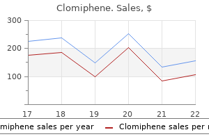
Purchase clomiphene 25mg on-line
Consequently pregnancy symptoms at 4 weeks purchase 25 mg clomiphene with amex, termination of pregnancy for maternal infection with Toxoplasma gondii has now become unusual, thanks to prenatal diagnosis along with the possibility of treatment of the infected fetus in utero via the mother, with the combination regimen of pyrimethamine and sulfonamides. These major advances in the field of diagnosis and therapy have facilitated a change in the indications for medical termination of pregnancy for toxoplasmosis almost exclusively for cases with severe lesions detected by ultrasonography. Prenatal diagnosis of fetal toxoplasmosis Since 1985, prenatal diagnosis of congenital toxoplasmosis has been reliably performed in women with suspected or confirmed Toxoplasma infection acquired during pregnancy. Availability of prenatal diagnosis has profoundly changed the management of fetal infection before birth through the use of specific algorithms for decisions Parasitology Toxoplasma gondii is a single-celled parasitic protozoan. Toxoplasmosis is spread by ingestion of oocysts or of cysts in their host tissue either by ingestion of undercooked meat or by congenital vertical transmission. After ingestion, the oocysts will spread in the organism and Genetic Disorders and the Fetus: Diagnosis, Prevention, and Treatment, Seventh Edition. Toxoplasma gondii can infect, replicate, and form cysts in all tissues that persist for the host lifetime. Epidemiology Worldwide seroprevalence of the parasite varies between 1 and 100 percent, depending on the environmental and socioeconomic conditions, including eating habits and health-related practices, general level of hygiene, host susceptibility, geographic location, and humidity of the soil. This decrease may be explained by reduced exposure to the parasite through changes in food habits and by improved hygiene practices in meat production. The incidence of toxoplasmosis among seronegative women depends primarily upon the prevalence in the general population and ranges from 0. Individual risk assessment may be favored and toxoplasma screening could be justified according to risk. The highest burden of congenital toxoplasmosis is in South America where the most pathogenic genotypes circulate, whereas the regions with the highest incidence are in the Middle East and some countries in Africa. The relevance of a universal screening program for pregnant women to detect seronegative women and perform subsequent serial screening for Toxoplasma through pregnancy depends upon such epidemiologic data. In France, in 2007, with a seroprevalence in French pregnant women of 40 percent, the overall prevalence of congenital toxoplasmosis was 3. In Europe, only four countries report the surveillance of congenital toxoplasmosis: Italy, Denmark, France, and Germany. In addition, multiple different cat exposures were assessed, but none were found to be risk factors for toxoplasmosis infection. Specifically, having a cat or kitten at home, cleaning the litter box, and owning a cat that hunts were not risk factors for T. Based on these established risk factors for primary toxoplasmosis, pregnant women (or women trying to become pregnant) should be appropriately advised by their obstetricians and primary care physicians on how to lower the risk of congenital toxoplasmosis. Prenatal diagnosis age at the time of maternal infection and should be considered before a decision is made about prenatal diagnosis. Because of a low rate and lack of specificity of clinical signs, a diagnosis of Toxoplasma infection is best established by systematic serologic screening of nonimmune pregnant women. This screening, which allows accurate and early diagnosis of maternal infection, is recommended on a systematic basis in France. Clinical signs are present in less than 40 percent of women and are often ignored and nonspecific: asthenia, low-grade fever, myalgia, and lymphadenopathy. The intrinsic sensitivity of the molecular prenatal diagnosis is crucial because parasitic loads are generally low, with a large proportion of infected amniotic fluids containing Toxoplasma loads of less than 10 tachyzoites per milliliter. Back in the late 1990s interlaboratory comparison studies reported some false-positive results. In some reports, a rather low sensitivity could be explained by an amniocentesis performed too soon after maternal infection (before detection of specific IgG) or too early during gestation. Sulfonamide, given with pyrimethamine and folinic acid, is the preferred treatment for an active infection. Spiramycin is the most commonly used macrolide to prevent placental passage of Toxoplasma in mothers who seroconvert. It is probably not effective in fetuses already infected and does not prevent, for example, neurotoxoplasmosis in immunosuppressed patients. The severity of congenital toxoplasmosis depends on gestational age at seroconversion: early fetal infections most likely result in fetal death or serious sequelae (ventriculomegaly associated with cerebral calcifications and chorioretinitis), whereas almost all fetuses infected during the third trimester are asymptomatic at birth. On the other hand, a specific antiparasitic treatment combining pyrimethamine and sulfonamide should be offered to otherwise infected fetuses without signs28 (see Table 26. The treatment regimen is administered orally via the mother and is designed to prevent and reduce infectious sequelae, due to its synergistic activity against T. Some can cross the placenta and reach the fetus; some only reach the maternal circulation. Pyrimethamine is a folic acid antagonist that can depress the bone marrow and induce macrocytic anemia, neutropenia, or thrombocytopenia. It Pyrimethamine 50 mg/day 946 Genetic Disorders and the Fetus transplacental transmission after amniocentesis or a low T. Therefore, in cases of negative prenatal diagnosis, careful ultrasonographic monitoring should be recommended to detect some rare cases of delayed symptomatic infections. After delivery, a placental examination should be performed together with a serologic follow up of the child, to rule out congenital infection. There was weak evidence for an association between early treatment and reduced risk of congenital toxoplasmosis. Further evidence from observational studies is unlikely to change these results and would not distinguish whether the association is due to treatment or to biases caused by confounding. The authors concluded that a large randomized controlled clinical trial would be required to validate the evidence for a potential benefit of prenatal treatment. Indeed, most infected newborns have no clinical signs but are at risk of developing visual impairment as a result of chorioretinitis in childhood or adolescence. Additional data are necessary to determine to what extent the available preventive options (prevention of maternal infection, early treatment of infection in pregnant women, preventive treatment of infected infants, or treatment of existing lesions) are effective in reducing the risk of severe visual impairment. Because new lesions or recurrence of existing lesions may appear late after birth, long-term follow-up studies are necessary to estimate the definite ocular prognosis. The longest follow up of children identified through detection of maternal infection in pregnancy is reported by Couvreur et al. However, no data were available on visual acuity, and no clear distinction was made between detection of new lesions and reactivation of existing lesions. Pyrimethamine and sulfadiazine were given in utero to 38 percent of fetuses and after birth to 72 percent of newborns. Seventy-nine (24 percent) children had at least one retinochoroidal lesion at a median follow up of 6 years. In 23 (29 percent) of them, at least one new event had been diagnosed up to 10 years after detection of the first lesions: reactivation of an existing lesion (one case), new lesion in a previously healthy location (19 cases), or both (three cases). Fifty-five children had lesions in one eye; of the 45 children for whom visual acuity data were available, 31 (69 percent) had normal vision. Twenty-four children had lesions in both eyes; of the 21 for whom final visual acuity data were available, 11 had normal vision in both eyes. Nevertheless, in this study the overall ocular prognosis of congenital toxoplasmosis is satisfactory when infection is identified early and treated accordingly. In one recent study from the same group, there was a significant reduction in risk since 1992 when monthly screening was introduced (59. Although these fundamental questions have not been subjected to any appropriately designed randomized controlled trial, a randomized Phase 3 trial is currently comparing the efficacy and tolerance of prenatal therapy with pyrimethamine + sulfadiazine versus spiramycin to reduce vertical transmission of T. Management at birth the approach depends on the infectious status of the fetus and the results of the neonatal examination. If the diagnosis of fetal infection was negative the following investigations should be performed: r parasitology of the placenta and the fetal blood; r neurologic and ophthalmologic examination; r ultrasound examination of the central nervous system; r neonatal immunologic status. New lesions of chorioretinitis seem to develop less often during the first year of life in children receiving several courses of treatment.
Purchase clomiphene 50 mg with amex
Molecular analysis or assay of glutaryl-CoA dehydrogenase activity in cultured fibroblasts confirms the diagnosis menopause 2 months no period discount clomiphene 50mg line. Specifically, large neutral amino acids should not be used during pregnancy, as they do not consistently alter maternal blood Phe levels. Sapropterin is a class C medication and may be used during pregnancy when the effects of not using it outweigh its potential adverse effects. There is no evidence of sapropterin-associated teratogenicity or adverse pregnancy effects, and anecdotal reports of successful pregnancy outcomes with its use are increasing. Maternal Phe requirements change significantly throughout gestation necessitating frequent testing and diet adjustments. Dietary overrestriction should be avoided as inadequate protein and calorie intake can contribute to increased maternal Phe levels. Use of medical food may provide the increased calories and protein required to support breastfeeding (640 kcal/day and 25 g protein per day). Preimplantation genetic diagnosis of phenylketonuria also has been reported (see Chapter 10). Intellectual disability, myoclonic seizures, hypertonicity of the extremities, drooling, and swallowing difficulties are frequently observed. Acute episodes of peripheral neuropathy are more common in Canadian patients than in Norwegian patients. It has been described in various ethnic groups, but is highly prevalent among FrenchCanadians from the Lac St John-Chicoutimi district of Quebec. A guanine-to-adenine change in the splice-donor sequence in intron 12 of the gene is extremely common in French-Canadian patients from Quebec. Therapy with a low-tyrosine, lowphenylalanine diet has resulted in rapid resolution of the oculocutaneous lesions, but no change in intellectual performance or behavior has been observed. A number of patients were detected by newborn screening, and those treated from infancy with a low-tyrosine and low-phenylalanine diet have had normal development. Those disorders involving defects of vitamin B12 and folate metabolism are discussed in Chapter 25. The majority of patients have been discovered as a result of routine newborn screening for the hypermethioninemia associated with cystathionine synthase deficiency. The enzyme defect is a partial deficiency of hepatic methionine adenosyltransferase activity. Enzyme activity is normal in erythrocytes, cultured skin fibroblasts, and lymphoblasts derived from these patients. Prenatal diagnosis by gene sequencing is available but of uncertain benefit given the usual benign nature of this disorder. In mildly affected patients, dislocation of the ocular lenses may be the only finding. Vascular complications resulting in heart attack or stroke are often the cause of death. Pyridoxine-responsive patients in general have milder clinical manifestations than pyridoxine-nonresponsive patients. Homocysteine does not appear teratogenic to the fetus, and treatments such as diet, B6, or betaine should be continued. Consideration of anticoagulation therapy is recommended during the final weeks of pregnancy and first postpartum weeks. More than 175 mutations are described, and over 95 percent of affected individuals have a detectable mutation. G307S mutation is prevalent in pyridoxine-nonresponsive patients of Celtic ethnic origin, whereas a p. Some patients with sulfite oxidase deficiency have a defect in the enzyme itself, but the majority have a defect in the molybdenum containing cofactor. The sulfite oxidase deficiencies are associated with severe neurologic impairment, including intractable neonatal seizures and developmental delay. Facial dysmorphism and eventually dislocated lenses have been observed in most patients. Additional metabolic defects are seen in molybdenum cofactor deficiency and are attributable to the added deficiency of xanthine oxidase. Patients excrete increased xanthine, and manifest hypouricemia and absence of urinary urothione. Additional testing to differentiate molybdenum cofactor deficiency includes the identification of xanthinuria and hypouricemia. Isolated sulfite oxidase deficiency is characterized by a variety of frameshift and early termination mutations. Biochemical findings include homocystinuria and hyperhomocysteinemia, with normal to low plasma methionine. For homozygotes or compound heterozygotes, the risk for coronary artery disease, stroke, and venous thrombosis is 20 percent increased over the general population. Symptoms usually appear within hours after birth and include marked hypotonia, apnea, seizures, and coma. The gene for the P-protein has been mapped to 9p13 and that of the T-protein to 3q21. A number of mutations have been identified in the glycine cleavage enzyme subunits. Its clinical features range from mild to severe failure to thrive, recurrent diarrhea, an- emia, hepatosplenomegaly, cataracts, dysmorphic features, and developmental delay. The enzyme defect is a deficiency of succinic semialdehyde dehydrogenase activity, which can be shown in lymphocytes and cultured lymphoblasts. The major clinical manifestations are facial dysmorphism, hepatomegaly, failure to thrive, skin lesions including multiple progressive ulcers of the lower extremities, telangiectases, erythematous rashes, and in some cases intellectual disability. The gene for human prolidase has been cloned and characterized and several molecular defects have been demonstrated in prolidase deficiency. Type I, characterized by a moderate degree of hyperprolinemia, is now considered a benign biochemical disorder. The rest had intellectual disability and seizures or abnormal electroencephalograms. A block in either of the first two steps of the proline metabolic pathways causes hyperprolinemia. The enzyme defect in type I is proline oxidase deficiency, which has been demonstrated only in liver. Disorders of renal amino acid transport There are four known renal amino acid transport disorders: cystinuria, affecting the transport of cystine and dibasic amino acids; Hartnup disorder, affecting the transport of neutral amino acids; familial iminoglycinuria, affecting the transport of glycine, proline, and hydroxyproline; and dicarboxylic aminoaciduria, affecting the transport of glutamic acid and aspartic acid. The renal and intestinal transport of cystine and the dibasic amino acids (lysine, ornithine, and arginine) are affected. Prenatal biochemical evidence of cystinuria in association with fetal hyperechogenic colon has been described. Seizures and cerebellar dysfunction including abnormal gait and dysarthria are common findings. Neuroimaging studies show loss of subcortical white matter and cerebellar atrophy. Two enzymes involved in the metabolism of d-2-hydroxyglutarate have been identified. Combined D-2- and L-2-hydroxyglutaric aciduria A report described three patients with combined d2- and l-2-hydroxyglutaric aciduria with neonatal onset of metabolic encephalopathy. In rare patients the defect is suspected to be in the metabolism of the riboflavin cofactor of these flavin-containing enzymes. The most severely affected patients present in the first days of life with severe hypoglycemia and metabolic acidosis, often with neonatal or infantile death. Other findings in these infants include: facial dysmorphism, macrocephaly, polycystic kidneys, and congenital heart disease. Patients can have milder forms of the disease with later onset in childhood or as adults. Organic acids in man: the analytical chemistry, biochemistry and diagnosis of the organic acidurias. Stable isotope dilution analysis of 3-hydroxyisovaleric acid in amniotic fluid: contribution to the prenatal diagnosis of inherited disorders of leucine catabolism. Application of fast atom bombardment with tandem mass spectrometry and liquid chromatography/mass spectrometry to the analysis of acylcarnitines in human urine, blood, and tissue.
Purchase generic clomiphene on-line
Relationship of sexual and physical abuse to pain and psychological assessment variables in chronic pelvic pain patients menstruation moon phases discount clomiphene 25mg otc. The psychological and physical benefits of pelvic ultrasonography in patients with chronic pelvic pain and negative laparoscopy. Diagnosis, treatment and follow up of women undergoing conscious pain mapping for chronic pelvic pain: a prospective cohort study. Characteristics of pathological findings in women with chronic pelvic pain using conscious minilaparoscopic pain mapping. A randomized controlled trial of medroxyprogesterone acetate and psychotherapy for the treatment of pelvic congestion. The use of medroxyprogesterone acetate 50 mg in the treatment of painful pelvic conditions: preliminary results from a multicentre trial. A randomized controlled trial of goserelin and medroxyprogesterone acetate in the treatment of pelvic congestion. A randomized, doubleblind crossover trial of sertraline in women with chronic pelvic pain. Laparoscopic uterosacral nerve ablation for alleviating chronic pelvic pain: a randomized controlled trial. Adhesion prevention and reduction: current status and future recommendations of a multinational interdisciplinary consensus conference. History, pelvic examination findings and mobility of ovaries as a 32 33 34 35 36 37 38 39 40 41 42 43 sonographic marker to detect pelvic adhesions with fixed ovaries. Laparoscopic adhesiolysis in patients with chronic abdominal pain: a blinded randomised controlled multicentre trial. Adhesion prevention agents for gynaecological surgery: an overview of Cochrane reviews. Is pelvic vein incompetence associated with symptoms of chronic pelvic pain in women The relationship between pelvic vein incompetence and chronic pelvic pain in women: systematic reviews of diagnosis and treatment effectiveness. Transvenous occlusion of incompetent pelvic veins for chronic pelvic pain in women: a 752 Pelvic Pain systematic review. Incidence and risk factors for chronic pelvic pain after hysteroscopic sterilization. Botulinum toxin type A for chronic pain and pelvic floor spasm in women: a randomized controlled trial. An enormous variation exists in the clinical presentation, from minimal descent to complete eversion of the vagina along with the uterus, bladder and rectum. Although it is a benign condition it can have a major impact on the quality of life. Skilful assessment and management is required to ensure appropriate treatment and improved outcome. The natural history of prolapse is not well known as longterm epidemiological studies are extremely rare. One study found that the incidence of prolapse to or beyond the hymen was 26% after 1 year of observation and 40% after 3 years. Over the same time period spontaneous remission rates at 1 and 3 years were 21% and 19% [7,8]. The most common form of prolapse is that of the anterior wall of the vagina (cystocele). Prolapse of the posterior wall (rectocele) is far less frequent and apical prolapse (descent of the uterus or vaginal vault if the patient has had a hysterectomy) the least common. Predisposing risk factors for the development of prolapse include vaginal childbirth, obesity, previous hysterectomy and age [9,10]. Vaginal birth is probably the principal risk factor, with avulsion injury to the levator ani during childbirth along with pudendal neuropathy and fascial damage the most common causes. Nulliparous women may also develop prolapse and a range of other conditions may contribute to the development of the disease [14]. Of these, age is the most significant contributor, with the incidence of prolapse doubling with every decade of life [5,8]. There is no clear consensus that a prior hysterectomy (unless it was performed for prolapse) is a risk factor for subsequent vault or vaginal prolapse [19]. Conditions associated with chronically increased intraabdominal pressure, such as chronic cough and heavy lifting, are also considered to be risk factors [15]. The levator plate provides indirect support for the vagina by acting as a platform against which the upper vagina and cervix are compressed during episodes of raised intraabdominal pressure. Narrowing of the urogenital hiatus also occurs with rises in intraabdominal pressure. The supports of the vagina are divided into three zones: the upper, middle and the lower. The fibres in the upper are largely vertical in orientation while the fibres supporting the middle section are attached to the side wall. The fibres surrounding the lower third are almost fused with the surrounding structures [20]. This reflects the different embryological origins of the vagina and determines the surgical approach to the repair of each level. It is not uncommon for women to be asymptomatic in the early morning and then for the symptoms to develop or worsen throughout the day with activity and be relieved by lying down. While some women may present with a single symptom of prolapse, they typically have a more complex presentation that can include urinary symptoms of incontinence, frequency, nocturia and voiding dysfunction; faecal symptoms of incontinence and obstructed defecation; and sexual dysfunction. In a large study of women with symptomatic vaginal prolapse, 87% reported urinary frequency and urgency, 73% reported urinary incontinence and 50% had symptoms of voiding dysfunction [21]. As stated above this may be because of shared aetiological factors and may not be a direct causal link. Bowel symptoms include the sensation of incomplete emptying and the need to manually assist defecation. The latter can include putting digital pressure on the perineum or splinting the posterior wall with the fingers during evacuation. Pressure on the perineum obviously has a different mechanism of action and there is no evidence that correction of a posterior wall prolapse in these cases will resolve the symptoms of obstructed defecation. Childbirth Age Obesity Genetics Occupation Pelvic anatomy the bony pelvis provides the architectural framework for the supports of the organs of the pelvis. The organs are supported by the fibres of the paracolpium (direct support) and by the levator plate (indirect support). The fibres of the paracolpium arise from a broad area on the pelvic side wall over the fascia of the piriformis muscle, sacroiliac joint and lateral sacrum. They insert into the lateral upper third of the vagina, with some fibres inserting anteriorly and posteriorly. These fibres are condensations of the endopelvic fascia and are composed of perivascular connective tissue and smooth muscle and contain blood vessels, lymphatics and nerves. They run in a predominantly vertical direction and their upper borders are continuous with the cardinal and uterosacral ligaments. The levator ani comprises the pelvic diaphragm muscles: the pubococcygeus, iliococcygeus, puborectalis and coccygeus muscles. Together they form a thin broad muscle arising anteriorly from the posterior aspect of the pubic bone just lateral to the symphysis pubis and laterally from the white line of the obturator internus muscle fascia and ischial spine. The right and left muscle bellies swing backwards and downwards to fuse together behind the anal canal and anterior to the coccyx to form the levator plate between these two structures. The anal Uterovaginal Prolapse 757 Sexual dysfunction is a common symptom in women attending a urogynaecology clinic [22]. A high percentage are not sexually active but cite the reason as lack of desire and arousal. Not only will this help with understanding what worries the patient most but can also be used for assessment of outcomes. It is also essential to establish which are the most worrisome symptoms and to clarify which symptoms the patient hopes will be corrected. Because of a lack of understanding of the longitudinal history of prolapse, counselling about the need for intervention can be extremely difficult.


Cheap generic clomiphene uk
Had the diameter been large contemporary women's health issues for today and the future pdf generic 100mg clomiphene with visa, excessive refraction would have caused spherical and chromatic aberrations, thus making the image indistinct. Raises body tube, puts the slide on the stage, uses mechanical stage to bring the object over the central aperture. Chooses the light source and correctly brings the objective lens into position; i. As the student gets used to handling the microscope in this and later experiments, she/he will realize that common objects of interest in microscopy such as, dust particles, cotton/wool/ silk/synthetic and other fibers, air bubbles, stain precipitate, etc. Then add a pinch of dust, starch powder, a few well-teased (dissected, separated) cotton, wool, and other fibers, drop of milk, and a few hairs to each drop of water separately. Then holding a cover-slip by its edges, place its edge in the edge of the water drop, and using a pencil point to support it, gently lower it on to the water drop. The usual house and garden dust contains inorganic, and organic matter-including silica, graphite, mica, carbon, calcium carbonate (from white washing, chalk, etc. These granules are oval or pear-shaped and usually have a hilum at their narrow ends. Concentric rings (lines) are seen, especially when stained blue with dilute iodine solution added to a watery suspension of starch powder. The human hairs (pili) are long, filamentous and cylindrical and cover most of the skin surfaces except palms and soles. Each hair has 3 layers: the inner medulla consists of cells containing pigment granules and air spaces. The granules usually lie singly and do not form aggregates or clusters (this is how you will see the granules on a blood smear when the stain dries up during staining, as mentioned in Expt 1-12. These granules have to be distinguished from platelets which form clusters of 2 to 12, and show a central darker and a peripheral lighter zone. A drop of diluted milk shows fat globules, most of which are round and of uniform size. They appear as darkish rings with a clear area in the centre, an appearance that changes when the focus is changed. The outermost layer, the cuticle, is a single layer of heavily keratinized thin, flat cells arranged like the tiles of a roof, with their free edges appearing as minute projections. If the focus is changed (as it always is during racking) the darker medulla appears lighter and the cortex appears darker. Undyed cotton fibers appear as long, ribbon-like, semi-transparent filaments which are spirally twisted at intervals. Two faint lines appear to enclose a light central zone throughout the twisted fiber. There are two common sources of blood for routine laboratory tests: blood from a superficial vein by puncturing it with a needle and syringe, or from skin capillaries by skin-prick. None of these samples can be called a representative sample because there are minor variations in their composition. But for routine hematological tests, however, these differences can safely be ignored. Explain what a blood sample is, what are its sources, and what are its main constituents. Collect capillary blood from a finger-prick, heel-prick, and earlobe prick, and precautions to be taken during a skin-prick. Name the various anticoagulants employed in hematological studies and their mode of action. Cotton swabs are likely to leave fibers sticking to the skin and provide an undesirable contact, or they may appear as artifacts in a blood film. Note After cleaning the skin, allow the alcohol to dry by evaporation (do not blow on it), because sterilization with alcohol is effective only after it has dried. Therefore, once the site has been cleaned and dried, it should not be touched again. Care must be taken to prevent contamination until the puncture wound has effectively closed/healed. The skin is a formidable barrier to the entry of foreign invaders and the first line of defence against bacteria and other disease-causing microorganisms which are present in abundance on the skin and in the air. Therefore, puncturing the skin always poses the danger of infection In order to achieve asepsis, the following aspects need to be kept in mind: 2. These tests are carried out for aiding in diagnosis and/ or prognosis of the disease or disorder. Sterilization of Equipment All the instruments to be used for collecting blood- syringes, needles, lancets, and cotton and gauze swabs-should preferably be sterilized in an autoclave. The old practice of boiling glass syringes and needles in tap water is now obsolete. Irradiated and sealed, single-use syringes, needles, lancets and blades are now freely available and are in common use. When larger amounts (say, a few ml that cannot be obtained from a skin puncture) are needed as for complete hematological and biochemical investigations, venous blood is obtained with a syringe and needle by puncturing a superficial vein. Cleaning/Sterilization of Skin Though it is impossible to completely sterilize the selected site for skin puncture, every aseptic precaution must be exercised. Note A Textbook of Practical Physiology have to be taken from the femoral vein, or the suitable anticoagulant. The blood is allowed to clot in the container and serum is collected as described later. When arterial blood is needed for special tests such as blood pH, gas levels, etc, an artery such as radial or femoral is punctured with a syringe and needle. Blood from a heart chamber, taken through a cardiac catheter, may be required for special tests. Differences Between Venous and Capillary Blood the differences between these two sources of blood are given in Table 1-1. Containers for Blood Sample A container is a receptacle into which blood is transferred from the syringe before sending it to the laboratory. Clean and dry 10 ml glass test tubes, collection bottles such as clean and dry 10 ml discarded medicine vials, glass bulbs, etc are the usual ones in use. A container may or may not contain an anticoagulant depending on whether a sample of blood/plasma, or serum is required. The blood is transferred to a container containing a Anticoagulants are substances employed to delay, suppress, or prevent clotting of blood. They are classified into 2 groups: the in vitro (outside the body) anticoagulants, and the in vivo (in the body) anticoagulants. The in vivo anticoagulants include: heparin and dicoumarol derivatives (warfarin, dicoumarin). Table 1-1: Sources and differences between Venous blood and Capillary blood Venous blood 1. It is obtained from a superficial vein by venepuncture A clean venepuncture provides blood without any contamination with tissue fluid There is less risk of contamination since sterile syringe and needle are used 1. Capillary blood It is obtained from a skin puncture, usually over a finger, ear lobe/or the heal of a foot Blood from a skin prick comes from punctured capillaries and from smallest arterioles and venules There is greater risk of contamination and transmission of disease as one may be careless about sterilization since skin prick is considered a harmless procedure these values are likely to be on the lower side since some tissue fluid is bound to dilute the blood even when it is free-flowing Capillary blood is not suitable for these purposes 4. Venous blood is preferable when normal blood standards are to be established, or when two samples from the same person are to be compared at different times 5. Trisodium citrate is the anticoagulant of choice in blood tests for disorders of coagulation. The negatively charged citrate ion is particularly useful for this purpose, usually in the form of sodium, ammonium, and potassium citrate. The citrate ion combines with calcium in the blood to form an unionized calcium compound. Along with other components, sodium citrate is used for storing donated blood in blood banks (see Expt 1-18), since it can be safely given intravenously. A mixture of ammonium oxalate and potassium oxalate in the ratio of 3:2 is an effective anticoagulant. Too much oxalate is hypertonic and damages all blood cells, while too little will not prevent clotting. Though each oxalate by itself (also sodium and lithium oxalate) can prevent clotting, a mixture is used since the ammonium salt increases cell volume while potassium salt shrinks them.
Cheap clomiphene 100mg
Blindness in the temporal fields of vision of both eyes is called bitemporal hemianopia (or hemianopsia) women's health center houston discount clomiphene 25 mg line. Blindness in the nasal halves of fields of vision of both eyes is called binasal hemianopia. It occurs when the uncrossed optic nerve fibers in the lateral parts of optic chiasma are damaged. In the case of retina, the natural stimulus of light requires minimum of energy to stimulate the rods and cones, while a mechanical stimulus requires many times the energy needed by the normal stimulus. The pressure produces an impression of a dark circular spot surrounded by a bright circle in the field of vision directly opposite to the point of pressure. These visual sensations are called pressure phosphenes and are caused by "inadequate" retinal stimulation. Draw a small cross on the board, then ask the subject to cover his left eye with a cupped hand, and to gaze fixedly on the cross with his right eye. Move a stick with a small white tip slowly on the board to the right of the cross until he can no longer see the white tip. Slowly bring the tip of the stick in vertical and oblique directions, from the periphery towards the roughly positioned blind spot, marking all the points when the white tip becomes visible. Join all these marks to obtain the outline of the projected image of the optic disk. The point at which the rays intersect in the eye is the nodal point, which can be assumed to lie 17 mm in front of retina. The distance of the nodal point from the board is 1 meter (the small distance from the nodal point to the cornea may be ignored). Cover your left eye with your left hand and hold the figure in front of your right eye. Fix your gaze on the cross (the more nasally located of the two marks), then move the figures towards and away from you until, at a certain distance, the spot disappears. The presence of the blind spot in the left eye can be confirmed by fixing the left eye on the spot and moving the figures towards or away from the eye till the cross disappears. Range of Accommodation the far point is the farthest point from the eye at which an object is seen clearly. Measure the far point in a manner similar to that used for the near point, remembring that if the subject is emmetropic (having normal vision), it will be infinitely far away. If the subject wears glasses, record the near and the far points with and without glasses. The nearest point at which an object can be seen clearly is called the near point. Seat the subject near a window, in good light, and ask him to cover one eye with a cupped hand. Hold a pencil in front of the other eye and slowly move it, preferably along a meter stick, towards the eye until it can no longer be seen in sharp focus. Near Response Seat the subject near a window and ask him to fix his eyes on a distant object. There is convergence of eyes, constriction of pupil, and increase in the curvature of the lens. Hold a burning candle to one side of his eye and observe the images of the candle flame from the other side. Ontheanteriorsurfaceofthelens(nearthe center of the pupil): the image is upright, somewhatlarger,andnotsobright. This shows that during accommodationfornearvision,theanterior surface ofthelensmovesforwards,i. The normal mechanism of vision fuses the two slightly different images into one togiveanimpressionofsolidity. They are the shadows cast on the retina by cellular and other debris in the aqueous and vitreous humors. Images of the retinal blood vessels reflected from the posterior surface of the lens also contribute to the floaters. A sudden appearance of new floaters, if accompanied by bright flashes in the peripheral field of vision, could indicate a retinal tear or detachment. Close one eye and look at the sky with the other and try to concentrate on what you see. You will observe small, circular, semitransparent, grey specks, or zigzag wispy filaments, or hair-like objects, or rows of cell-like structures that drift across the field of vision. If you try to focus on them, they drift away or sink down; and if you jerk your eye up they rise up, but sink down or float away once again. Factors Affecting Visual Acuity the visual acuity is a complex retinal and cortical mechanism that is affected by the following factors: A. This acuity of vision refers to the ability of the eye to recognize two point sources of light, or two parallel lines, as separate rather than one. It is expressed as minimum separable, ie, the minimum distance between two points or lines when they can be recognized as two. The nodal point lies at about the middle of the lens and is the optical center of the eye. In the Landolt ring chart, the gap in the ring is positioned at random in the 8 lines. In the E Test chart, the letter E is printed in 8 lines, the "legs" of the letters pointing in different directions. A person (or a child) who cannot read, has to indicate the direction in which the legs of each letter are pointing. It has a series of printed letters of varying sizes, black on a white background, arranged in eight lines. The subject is seated at a distance of 6 meters (20 feet) from a well-lighted chart and is asked to read the letters down the chart as far as she can read. A distance of 6 meters from the eye is considered as the practical far point because light rays from this distance are parallel. This chart is made up of reading material of various sizes with the smallest size at the bottom. The eye accommodates (adjusts) for near vision by increasing the refractive power of the lens, i. In this state, parallel rays of light coming from the distant object are brought to focus on the retina and the object is seen clearly. This blurring of the image on the retina acts as a stimulus for the reflex contraction of ciliary muscle, which pulls the ciliary body forwards and inwards. As a result, the lens ligament becomes lax, the tension on the lens capsule decreases, and the lens, due to its elasticity, bulges forwards. The increase in refractive power of the lens brings the image forwards onto the retina and the image becomes clearly visible. When the gaze is shifted to a distant object, the ciliary muscle relaxes and the lens becomes less convex. Amplitude of Accommodation the difference in the refractive power of the lens in the two states of complete relaxation and maximal accommodation is called the amplitude of accommodation. Of this 60 D, the cornea contributes 44 D, while the refractive power of the lens is 16 D in a young person. During maximal accommodation, the refractive power of the lens can add another 14 D to its refractive power. In this condition, parallel rays of light coming from a distant object are brought to a focus in front of the retina; the rays then diverge and form a blurred image on the retina. This is the condition in which parallel rays of light from a distance are focused behind the retina,(i. Reading and close work gradually becomes difficult and the person holds the reading material farther and farther away from the eye. Further processing occurs in lateral geniculate body and thalamus and then along specific pathways to the visual cortex. The human eye is sensitive to all wavelengths of light from 400 nm to 700 nm which constitute the visible part of the electromagnetic spectrum. Colors are perceived by cones that are concentrated in fovea and are sensitive to specific wavelengths of light.
25 mg clomiphene visa
Neonatal alloimmune thrombocytopenia is a potentially devastating disease that is a significant cause of morbidity and mortality in newborns pregnancy pillows safe 100 mg clomiphene, especially because of intracranial hemorrhage. Bleeding may also occur in the gastrointestinal tract, lungs, eyes, kidneys, and skin. Numerous platelet-specific antigens can induce maternal immunization during pregnancy, subsequently causing fetal platelet destruction. If homozygous, all his offspring will be obligate heterozygotes and incompatible with maternal antibodies. If heterozygous, his offspring would have a 50 percent risk of having inherited the antigen. Current management of pregnancies at risk suggest intravenous immunoglobulin and/or corticosteroids aiming to increase the fetal platelet count. However, the potential for intravenous immunoglobulin causing maternal hemolytic anemia has been recognized. The aim of fetal gene therapy is to introduce genetic material into the somatic cells of an affected fetus, but the window of opportunity is brief. The recognition of self and foreign antigens develops between 12 and 14 weeks of gestation. Although chorionic villus sampling might enable this effort, noninvasive prenatal diagnosis (see Chapter 11) will present another avenue. Certainly there has been success with a range of vector systems for postnatal gene therapy for hemophilia B, retinal blindness, and severe combined immunodeficiency. Insertional oncogenesis remains a worry while transplacental transfer may occur and spawn a maternal immune response. A maternal vector introduction could lead to gonadal transduction that would increase the risk of transmission to future offspring. Another novel therapeutic strategy, at least for dominant genetic disorders, is gene silencing. Normal external genitalia in a female with classical congenital adrenal hyperplasia who was not treated during embryogenesis. Prenatal dexamethasone treatment of children at risk for congenital adrenal hyperplasia: the Swedish experience and standpoint. Repeated antenatal corticosteroids: effects on cerebral palsy and childhood behavior. Long-term outcome of prenatal dexamethasone treatment of 21hydroxylase deficiency. Cognitive outcome of offspring from dexamethasonetreated pregnancies at risk for congenital adrenal hyperplasia due to 21-hydroxylase deficiency. Vindication of prenatal diagnosis and treatment of congenital adrenal hyperplasia with low-dose dexamethasone. Severe combined adrenal and gonadal deficiency caused by novel mutations in the cholesterol side chain cleavage enzyme, P450scc. Conception and pregnancy outcome in a patient with 11-bp deletion of the steroidogenic acute regulatory protein gene. The challenge of fetal dysrhythmias: echocardiographic diagnosis and clinical management. Blocked atrial bi/trigeminy in utero evolving in supraventricular tachycardia after birth. Anatomy of congenital complete heart block and relation to maternal anti-Ro antibodies. Autoimmuneassociated congenital heart block: demographics, mortality, morbidity and recurrence rates obtained from a national neonatal lupus registry. Outcome of children with fetal, neonatal or childhood diagnosis of isolated congenital atrioventricular block. Epidemiology, etiology, detection, and treatment of autoantibodyassociated congenital heart block in neonatal lupus. Clinical and genotype studies of cardiac tumors in 154 patients with tuberous sclerosis complex. Cardiac rhabdomyoma with long-term conduction abnormality: progression from pre-excitation to bundle branch block and finally complete heart block. Fetal paroxysmal supraventricular tachycardia without heart failure leading to ischemic damage. Successful in utero treatment of fetal goitrous hypothyroidism: case report and review of the literature. Experience with intraamniotic thyroxine treatment in nonimmune fetal goitrous hypothyroidism in 12 cases. Side effects of anti-thyroid drugs and their impact on the choice of treatment for thyrotoxicosis in pregnancy. Multivitamin/folic acid supplementation in the earliest weeks of pregnancy reduces the prevalence of neural tube defects. Incidence of fetal alcohol syndrome and prevalence of alcohol-related neurodevelopmental disorder. Fetal alcoa o hol spectrum disorders in Finland: clinical delineation of 77 older children and adolescents. Congenital structural anomalies in offspring of women with epilepsy a population-based cohort study in Finland. Pregnancy outcomes in women with epilepsy: a systematic review and meta-analysis of published pregnancy registries and cohorts. Factors influencing outcomes in the offspring of mothers with phenylketonuria during pregnancy: the importance of variation in maternal blood phenylalanine. Maternal hemolysis after intravenous immunoglobulin treatment in fetal 988 Genetic Disorders and the Fetus and neonatal alloimmune thrombocytopenia. Preimplantation genetic diagnosis for fetal neonatal alloimmune thrombocytopenia due to antihuman platelet antigen maternal antibodies. Delayed diagnosis of fetal and neonatal alloimmune thrombo- cytopenia: a cause of perinatal mortality and morbidity. As researchers continue to develop improvements to existing fetal interventions and as more health care centers worldwide begin to offer fetal interventions, medical and lay communities have become more familiar with and accepting of in utero fetal treatments. Fetal interventions have become so widely accepted that third-party payers routinely authorize reimbursement for the majority of these procedures. Perhaps most significantly, fetal surgery is now offered to improve the prognosis of fetal patients with nonlethal conditions, whereas in the past only fetal patients with lethal defects were considered suitable candidates for fetal intervention. However, safety and efficacy remain unproven for the major- ity of fetal procedures, and rigorous validation through clinical trials is necessary for the enterprise to make this transition in an ethically responsible fashion. Yet for families grappling with news of a devastating fetal diagnosis, the hope that fetal surgery can give their child a better outcome may overshadow the real risks of the procedure, as well as the fact that most fetal therapies remain experimental, with efficacy largely unproven. Accordingly, the guiding principle for offering fetal surgery has historically been that intervention should only be considered if maternal risks Genetic Disorders and the Fetus: Diagnosis, Prevention, and Treatment, Seventh Edition. In cases where hysterotomy, and its corresponding lifelong risk of uterine rupture, can be avoided, a more minor improvement in fetal outcome may be acceptable. Most centers performing fetal surgery have instituted oversight committees made up of members from various disciplines who are not involved in the fetal procedures to act in an advisory and quality assurance role. These committees are responsible for reviewing, usually at monthly intervals, all fetal evaluations and surgical procedures performed at their institution. Fetal surgical and anesthetic techniques Open and minimal-access techniques for fetal surgery continue to evolve as the goal of improving feasibility and safety spurs constant innovation. However, the techniques used today for administering anesthesia, monitoring both the mother and fetus intraoperatively, and maintaining fetal homeostasis are relatively standardized. Experimental work, in close collaboration with specialists in ultrasonography and perinatal obstetrics, begun in the late 1970s in fetal lambs and nonhuman primates.
Purchase clomiphene on line amex
The prefix deuter- refers to green color women's health clinic perth discount clomiphene uk, trit- refers to blue, and prot- refers to red color. The suffix -anomaly refers to color weakness while suffix -anopia refers to color blindness. Monochromats have only one cone system present, dichromats have two cone systems, and trichromats have all three cone systems but one may be weak. Physiological dichromatic vision is at the fovea centralis where only red and blue cones are present. The common defects of color vision, in order of occurrence are: Deuteranomaly, deuteranopia, protanopia, and protanomaly. If either red or green or both cones are missing, the person cannot distinguish red from green. However, though she can see the other colors, they are not seen in the way a normal person does. The average conversation voice frequency is 120 Hz in the males and 250 Hz in the females. Pitch discrimination is possible because different frequencies cause vibrations in different regions of the basilar membrane. Each segment of this membrane is thus "tuned" for a particular pitch- high-pitched sounds near the base of cochlea, and low-pitched sounds near the apex. Note While the human ear cannot perceive ("hear") ultrasounds, bats, dogs, and other animals can. The inaudible sounds are reflected from the organs and analyzed by a computer to provide a picture on the display screen. Nature and Characteristics of Sound Waves Sound waves are alternating regions of high- and low-pressure traveling through some medium. They are produced by some vibrating object and are perceived by us as the sound sensation. It is the psychological perception of the sound frequency; the higher the frequency, the higher the pitch. The entire audible range extends from 16 Hz to 20, 000 Hz (1 hertz = 1 cycle/sec). The intensity or loudness of a sound is the psychological term referring to the amplitude of the sound vibrations. This property refers to the sensation perceived when we hear 214 A Textbook of Practical Physiology a mixture of related frequencies, i. The ability to detect the position of the source of sound is called binaural effect (Consult Expt 2-24). Mechanism of Hearing the sound waves striking the tympanic membrane are magnified by the ossicles and set the basilar membrane to vibrate, which, in turn, causes movement of the hair cells of the organ of Corti. Since many fibers cross over from one auditory pathway to the opposite pathway in medulla, the primary auditory areas receive signals from both sides. The peripheral processes end on the hair cells of the organ of Corti, while the central processes, which form the auditory nerve, enter the upper medulla to synapse on the dorsal and ventral cochlear nuclei. The 2nd order neurons from these nuclei take different routes through the nearby olivary nuclei and the trapezoid bodies of both sides (some fibers end here), cross to the opposite side and turn upwards to form the lateral lemniscus. Audiometry [] Tuning-Fork Tests Before one can understand the principles on which the tuning-fork tests are based, one must understand Human Experiments what is meant by air conduction and bone conduction of sound. This mode of conduction of sound is called ossicular conduction, though it is commonly and misleadingly called air conduction. In this case, sound from a vibrating tuning fork, directly placed anywhere on the skull, can be heard in both ears by bone conduction. However, even loud sounds in the environment do not possess enough energy to cause vibrations of the skull bones and thus stimulate the organ of Corti; they usually take the air and ossicular route. The sense of hearing should first be tested with the whisper test, and then with a tuning-fork. Hold the stem of the tuning fork with the thumb and finger and set it into vibration by striking one of its prongs on the heel of your hand. When the sound stops, bring the prongs in front of the ear-the sound will become audible once again. After the subject stops hearing the sound, place the fork on your own mastoid process. Ask the subject if he hears the sound equally well in both the ears, or louder on one side. Sound is louder/better heard in deaf or deafer ear because of masking effect of environmental noise is absent on diseased side. Conduction deafness can be artificially created if you close one ear (say, left) of the subject with your finger. Set the tuning fork into vibration once again and place it on his head or forehead. Closing his ear produces a situation of conduction deafness, and demonstrates the masking effect of environmental noise. The auditory analyzer perceives the pitch, intensity or loudness, timbre or quality of sound, and the direction of the source of sound. The standard sound reference (faintest audible sound) corresponds to zero (0) db at a pressure level of 0. Loud stereo music with headphones on, or prolonged exposure to other loud noises causes selective loss of hearing. Infrasound refers to frequencies below 16 Hz and ultrasound refers to frequencies above 20,000 Hz (See above). The tuning-fork tests often provide valuable information, but cannot give quantitative estimates about the acuity of hearing. Furthermore, boneconducted vibrations reach all parts of the skull irrespective of where the fork is placed on the head. Thus, when testing bone conduction in one ear, the subject/patient will also be hearing sound in the other ear, which is likely to confuse him. The threshold is determined at each frequency and is then plotted as a percentage of normal hearing. Audiometry is thus the only reliable method to determine the nature and degree of deafness in a patient. The auditory system cannot separate the different components of total sound stimulation. Deafness: this refers to the inability of a person to hear either partially or totally. It is of two types: Conduction deafness, in which there may be wax 217 or a foreign body in the external auditory meatus, thickening or damage to tympanic membrane due to infection (otitis media), osteosclerosis (stapes gets fixed in oval window) interfere with hearing. Nerve deafness, in which there is damage to cochlear hair cells or auditory pathway, such as due to prolonged exposure to industrial sounds, or very loud music, or damage to 8th nerve by drugs. A cochlear implant is a device, which converts sounds into electrical signals that can be interpreted by the brain. The internal receiver relays signals to electrodes implanted in the cochlea where they generate impulses in the sensory fibers of the cochlear nerve. These signals are then propagated along the normal pathways to the brain where they are perceived as sounds. Selects either 216 Hz or 250 Hz tuning fork and strikes one of the prongs on the heel of her hand. Use a forceps to produce clicking noises behind, in front, and to each side of his head, one after the other, and ask him to locate the direction of sound in each case. Enter the results in your workbook, indicating the ability to localize the sound as excellent, good, fair and poor. Comments the ability to judge the position of the source of sound with both ears is called the binaural effect. When we want to localize a sound coming from a distance, we turn our head until the sound is equally loud in the two ears. After a few seconds, make a rattling noise near his ear by using a tin box containing some metal objects. Though the sensations of taste (gestation; gust = taste) and smell are closely related, they are anatomically distinct. They are commonly called chemical sensations because they are stimulated by chemicals (tastants, for taste, and odorants for smell) dissolved in oral and nasal mucus.

Purchase clomiphene with a mastercard
Human genetics policy guidelines for the management and prevention of genetic disorders women health tips buy clomiphene american express, birth defects and disabilities. Making mommies: law, preimplantation genetic diagnosis, and the complications of pre-motherhood. Infertility Treatment Act (1995) (as amended by the Health Legislation [Infertility Treatment and Medical Treatment] Act (2006)). Act containing rules regulating the use of gametes and embryos (Embryos Act) (2002). Media Release: Pre-implantation genetic diagnosis may be permitted in certain cases in the future. Preimplantation genetic diagnosis: a step by step guide to recent Italian ethical and legislative troubles. Japan Society of Human Genetics, Japan Society of Obstetrics and Gynecology, Japan Society for Pediatric Genetics, Japanese Society for Familial Tumor, Japanese Society for Gene Diagnosis and Therapy, Japanese Society for Inherited Metabolic Diseases, Japanese Society for Laboratory Medicine, Japanese Society for Massscreening and Japanese Teratology Society. Clinical Practice Guidelines, Pregnancy Outcomes After Assisted Human Reproduction No. Guidelines on Preimplantation Genetic Diagnosis with Human Leucocyte Antigen Tissue Typing. Directives for private healthcare institutions providing assisted reproduction services: Regulation 4 of the Private Hospitals and Medical Clinics Regulations. Ethical guidelines on the use of assisted reproductive technology in clinical practice and research. Professional selfregulation for preimplantation genetic diagnosis: experience of the American Society for Reproductive Medicine and other professional societies. Preimplantation genetic diagnosis: an overview of socio-ethical and legal considerations. Programme of Action adopted at the International Conference on Population and Development (Cairo, Egypt). Convention for the protection of human rights and dignity of the human being with regard to the application of biology and medicine: Convention on Human Rights and Biomedicine (Oviedo Convention) (1997). The changing landscape of genetic testing and its impact on clinical and laboratory services and research in Europe. Clinicians and patients confront a wide variety of ethical challenges in this context. The purpose of this chapter is to provide clinicians with conceptual and clinical tools to prevent and manage, in a professionally responsible fashion, ethical conflicts among members of the team or between team members, pregnant women, and their partners. To achieve this goal, the chapter begins with an account of the professional responsibility model of obstetric ethics,1 which appeals to two main principles of bioethics, beneficence and respect for autonomy. The professional responsibility model is then used as the basis for a detailed consideration of major ethical issues in the management of such pregnancies: the diagnosis of genetic disorders in the fetus; management of pregnancies complicated by genetic disorders; and research to improve the clinical management of fetal anomalies. On the basis of rigorous clinical judgment, informed by current science, especially evidencebased medicine, and a commitment to excellence in practice, the clinician should identify the strategies that are reliably expected to result in the greater balance of clinical benefits. The principle of beneficence has a long and illustrious history in the global pantheon of medical ethics. In Western medical ethics, for example, it dates back at least to the time of Hippocrates. Rather, the principle of beneficence was the primary consideration of the Hippocratic writers. This seemingly arcane point is not just historical but also conceptual and clinical: if Primum non nocere were to be made the primary principle of clinical ethics, then virtually all invasive aspects of healthcare, including many aspects of the diagnosis and management of genetic disorders, such as invasive prenatal diagnosis, would be unethical because of the clinical risks they involve for patients. If the primary goal of clinical management is to avoid harm, even drawing blood for noninvasive prenatal testing would become ethically suspect. In particular, all adult pregnant women should be assumed to possess the decision-making capacity to determine which clinical strategies for the clinical diagnosis and management of their pregnancies are consistent with their interests and which are not, unless there is reliable evidence of significant clinical deficits in their decision-making processes. In making decisions about their medical care, pregnant women may use values and beliefs that go far beyond health-related interests. Inasmuch as beneficence-based clinical judgment is limited by the scientific and clinical competencies of medicine, beneficencebased clinical judgment provides the physician no authority to assess the worth or meaning to the pregnant woman of her own non-health-related interests. This principle obligates the clinician to empower the pregnant woman to participate meaningfully in decisions about her healthcare by providing her with the clinical information that she needs (clinical information considered salient to diagnosis, treatment, and prognosis), supporting her to develop an adequate understanding of this information and relate it to her values and beliefs, and ensuring that her decisions are voluntary. In contrast, because of its insufficiently developed central nervous system, the fetus cannot meaningfully be said to possess values and beliefs. Thus, there is no valid basis for saying that a fetus has a perspective on its interests. Because of its centrality for the ethical management of pregnancies complicated by fetal anomalies, the topic of the fetus as patient requires careful consideration, a task to which we now turn. For Gregory and Percival a human being becomes a patient when he or she is presented to a physician or other healthcare professional and there exist clinical interventions that can benefit that individual clinically. An important advantage of the concept of the fetus as a patient is that the language of fetal rights or personhood has no meaning and, therefore, no application to the fetus in obstetric ethics, despite its popularity in public and political discourse in many countries. Thus, current controversies about "right to life," especially its possible limited application to patients from non-Western cultures, can be avoided in clinical judgment and decisionmaking about the management of pregnancies complicated by fetal anomalies. A major advantage of an ethical framework based on the concept of the fetus as a patient is that it keeps the ethics of the diagnosis and management of genetic disorders free from divisive disputes about fetal rights and about the fetus as an "unborn child. The ethical significance of the concept of the fetus as a patient therefore depends on links to its later becoming a child and, later still, achieving independent moral status. Viability should not be viewed as an exclusively biologic property of the fetus, but in terms of both biologic and technologic factors. Only by virtue of both factors can a viable fetus exist ex utero and subsequently become a child and later achieve independent moral status. When access to such technology is present, as is the case in the United States and other developed countries, viability occurs at approximately the end of 24 weeks of gestational age. For example, a patient with massive internal injuries with uncontrolled bleeding but without timely access to rapid transport to surgery is almost certainly nonviable, whereas the same patient with such access is viable. This is because technologic factors cannot result in the previable fetus becoming a child: this is simply what previable means. The pregnant woman is therefore free to withhold, confer, or, having once conferred, withdraw the status of being a patient on or from her previable fetus according to her own values and beliefs. Having made a decision to continue a previable pregnancy, the woman remains free to revoke that decision. The concept of the fetus as a patient has considerable clinical significance for genetic counseling because, when the fetus is a patient, directive counseling. Counseling regarding the viable fetus When the viable fetus is a patient, directive counseling for fetal benefit is ethically justified. However, the strength of directive counseling for fetal benefit varies according to the presence and severity of fetal anomalies. As a rule, the more severe the fetal anomaly, the less directive counseling should be for fetal benefit. Counseling therefore should be nondirective in offering a choice between nonaggressive management and termination of pregnancy, but directive in recommending against aggressive management for the sake of maternal benefit. On this account, no pregnant woman is obligated to her fetus to accept the risks to herself of experimental fetal intervention. Such conflict should be managed preventively through informed consent as an ongoing dialogue throughout the pregnancy, augmented as necessary by negotiation and respectful persuasion. If she does confer such status in a settled way, at that point beneficencebased obligations to her fetus come into existence, and directive counseling for fetal benefit becomes appropriate for these previable fetuses. Just as for viable fetuses, such counseling must take account of the presence and severity of fetal anomalies, extreme prematurity, and obligations owed to the pregnant woman. For previable pregnancies in which the woman is uncertain about whether to confer such status, we propose that the fetus be provisionally regarded as a patient. In particular, nondirective counseling is appropriate in cases of what we term near-viable fetuses3. Diagnosis of genetic disorders in the fetus Competence and referral in prenatal diagnosis the first component of the ethical concept of medicine as a profession generates the ethical obligation to provide competent genetic diagnosis and derives from both beneficence and respect for autonomy. Either principle alone, and certainly both in combination, requires clinicians to provide patients with accurate and reliable clinical information. To meet this ethical obligation, the clinician must address the following ethical considerations.
