

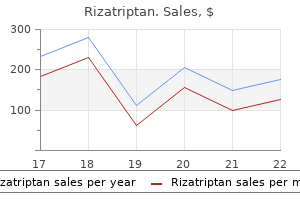
Buy discount rizatriptan line
Cross-section of the basal turn of the cochlear duct is shown in the box on the smaller orientation view ocean view pain treatment center discount 10 mg rizatriptan with amex. This view of a mid-modiolar section of the cochlea illustrates the position of the cochlear duct within the 2. Observe that at the top of the cochlea, scala vestibuli and scala tympani communicate with each other at the helicotrema. The scala media and the osseous spiral lamina divide the cochlea into the scala vestibuli and the scala tympani, which are filled with perilymph. The scala media (the space within the cochlear duct) is filled with endolymph and contains the organ of Corti. The axons of the spiral ganglion cells form the cochlear part of the vestibulocochlear nerve. Two cell types can be observed: a mesothelial cell, which faces the scala vestibuli and is bathed by perilymph, and an epithelial cell, which faces the scala media and is bathed by endolymph. The marginal cells, primarily involved in K transport, line the endolymphatic space of the scala media. The lower wall or floor of the scala media is formed by a relatively flaccid basilar membrane that increases in width and decreases in stiffness as it coils from the base to apex of the cochlea. The spiral organ of Corti rests on the basilar membrane and is overlain by the tectorial membrane. The spiral organ of Corti is composed of hair cells, phalangeal cells, and pillar cells. The apical surfaces of the marginal cells (M) of the stria are bathed by endolymph (E) of the scala media. Intermediate cells (I) are positioned between the marginal cells and the basal cells (B). The basal cells separate the other cells of the stria vascularis from the spiral ligament (SpL). Several other named cell types of unknown function are also present in the spiral organ. The number of cells forming the width of the continuous row of outer hair cells is variable. The width of the row gradually increases to five ranks of cells at the apex of the cochlea. Conductive hearing loss results when sound waves are mechanically impeded from reaching the auditory sensory receptors within the internal ear. This type of hearing loss principally involves the external ear or structures of the middle ear. Conductive hearing loss is the second most common type of loss after sensorineural hearing loss, and it usually involves a reduction in sound level or the inability to hear faint sounds. A conductive hearing loss may be caused by otitis media (ear infection); in fact, this is the most common cause of temporary hearing loss in children. Fluid that collects in the tympanic cavity can also cause significant hearing problems in children. Other common causes of conductive hearing loss include excess wax or foreign bodies in the external acoustic meatus or diseases that affect the ossicles in the middle ear (otosclerosis; see also Folder 25. In many cases, conductive hearing loss can be treated either medically or surgically and may not be permanent. Causes of acquired sensorineural hearing loss include infections of the membranous labyrinth. Sensorineural hearing loss not only involves a reduction in sound level but also affects the ability to hear clearly or to distinguish speech. A loss of the sensory hair cells or associated nerve fibers begins in the basal turn of the cochlea and progresses apically over time. The characteristic impairment is a high-frequency hearing loss termed presbycusis (see presbyopia, page 922). In selected patients, the use of a cochlear implant can partially restore some hearing function. The cochlear implant is an electronic device consisting of an external microphone, amplifier, and speech processor linked to a receiver implanted under the skin of the mastoid region. The receiver is connected to the multielectrode intracochlear implant inserted along the wall of the cochlear canal. This higher magnification photomicrograph of the cochlear duct shows the structure of the spiral organ of Corti. Relate this structure to the inset, which labels the structural features of the spiral organ. The sensory cells are divided into an inner row of sensory hair cells and three rows of outer sensory hair cells. The tectorial membrane extends from the spiral limbus over the cells of the spiral organ of Corti. The apical ends of the phalangeal cells are tightly bound to one another and to the hair cells by elaborate tight junctions. Its composition is similar to that of other extracellular fluids and to perilymph. Pillar cells have broad apical and basal surfaces that form plates and a narrowed cytoplasm. The inner pillar cells rest on the tympanic lip of the spiral lamina; the outer pillar cells rest on the basilar membrane. Its lateral free edge projects over and attaches to the organ of Corti by the stereocilia of the hair cells. Glycoproteins unique to the internal ear, called otogelin and tectorin, are associated with the collagen bundles. These proteins are also present in the otolithic membranes overlying the maculae of the utricle and saccule as well as in the cupulae of the cristae in the semicircular canals. Sound Perception As described on page 937, sound waves striking the tympanic membrane are translated into simple mechanical vibrations. In the internal ear, the vibrations of the ossicles are transformed into waves in the perilymph. Movement of the stapes in the oval window of the vestibule sets up vibrations or traveling waves in the perilymph of the scala vestibuli. Movement of the stereocilia of the hair cells in the cochlea initiates neuronal transduction. Hair cells are attached through the phalangeal cells to the basilar membrane, which vibrates during sound reception. The stereocilia of these hair cells are, in turn, attached to the tectorial membrane, which also vibrates. Thus, a shearing effect occurs between the basilar membrane (and the cells attached to it) and the tectorial membrane when sound vibrations impinge on the internal ear. Because they are inserted into the tectorial membrane, the stereocilia of the hair cells are the only structures that connect the basilar membrane and its complex epithelial layer to the tectorial membrane. The shearing effect between the basilar membrane and the tectorial membrane deflects the stereocilia and thus the apical portion of the hair cells. Ear Innervation of the Internal Ear the vestibular nerve originates from the sensory receptors associated with the vestibular labyrinth. This electron micrograph illustrates the configuration of stereocilia on the apical surfaces of the inner row and three outer rows of the cochlear sensory hair cells. A sound of specified frequency causes displacement of a relatively long segment of the basilar membrane, but the region of maximal displacement is narrow. The point of maximal displacement of the basilar membrane is specified for a given frequency of sound and is the morphologic basis of frequency discrimination. High-frequency sounds cause maximal vibration of the basilar membrane near the base of the cochlea; low-frequency sounds cause maximal displacement nearer the apex. The vestibular nerve is associated with equilibrium and carries impulses from the sensory receptors located within the vestibular labyrinth. The cell bodies of the bipolar neurons of the vestibular nerve are located in the vestibular ganglion (of Scarpa) in the internal acoustic meatus. Dendritic processes of the vestibular ganglion cells originate in the cristae ampullaris of the three semicircular ducts, the macula of the utricle, and the macula of the saccule.
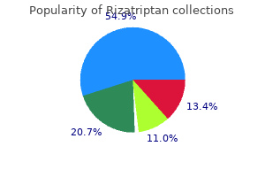
Purchase rizatriptan with amex
If body temperature continues to drop knee pain treatment yahoo purchase rizatriptan 10mg on line, control center signals muscles to contract involuntarily. In blood clotting, for example, certain chemicals stimulate more clotting, which minimizes bleeding (see chapter 14, p. Similarly, a positive feedback mechanism increases the strength of uterine contractions during childbirth (see chapter 23, p. Positive feedback mechanisms usually produce unstable conditions, which might not seem compatible with homeostasis. However, the few examples of positive feedback associated with health have very specific functions and are short-lived. Cardiovascular system Organic waste, excess salts, water Urinary system Blood Internal environment External environment Unabsorbed matter systems are delivered to all body cells by the cardiovascular system. The same blood that brings needed nutrients to cells carries away waste products, which are removed by the respiratory and urinary systems (fig. Homeostatic mechanisms maintain a relatively constant internal environment, yet physiological values may vary slightly in a person from time to time or from one person to another. Therefore, both normal values for an individual and the idea of a normal range for the general population are clinically important. Numerous examples of homeostasis are presented throughout this book, and normal ranges for a number of physiological variables are listed in Appendix C, Laboratory Tests of Clinical Importance. Within the axial portion are the cranial cavity, which houses the brain; the vertebral canal (spinal cavity), which contains the spinal cord and is surrounded by sections of the backbone (vertebrae); the thoracic (tho-rasik) cavity; and the abdominopelvic (ab-dom -no-pelvik) cavity. The thoracic cavity is separated from the abdominopelvic cavity by a broad, thin muscle called the diaphragm (diah-fram). Within the thoracic cavity are the lungs and a region between the lungs, called the mediastinum (mede-as-tinum). The remaining thoracic viscera-heart, esophagus, trachea, and thymus-are within the mediastinum. The abdominopelvic cavity, which includes an upper abdominal portion and a lower pelvic portion, extends from the diaphragm to the floor of the pelvis. The viscera within the abdominal cavity include the stomach, liver, spleen, gallbladder, kidneys, and the small and large intestines. The pelvic cavity is the portion of the abdominopelvic cavity enclosed by the pelvic bones. It contains the terminal end of the large intestine, the urinary bladder, and the internal reproductive organs. Orbital cavities, containing the eyes and associated skeletal muscles and nerves 4. Middle ear cavities, containing the middle ear bones Thoracic and abdominopelvic Membranes Thin serous membranes line the walls of the thoracic and abdominopelvic cavities and fold back to cover the organs within these cavities. These membranes secrete a slippery serous fluid that separates the layer lining the wall of the cavity (parietal layer) from the layer covering the organ (visceral layer). For example, the right and left thoracic compartments, which contain the lungs, are lined with a serous membrane called the parietal pleura. Normally, only a thin film of serous fluid separates the parietal and visceral pleural (plooral) membranes. However, the space between them may become significantly larger as a result of illness or injury. The heart, located in the broadest portion of the mediastinum, is surrounded by pericardial (per -kar de-al) membranes. The parietal pericardium is covered by a much thicker third layer, the fibrous pericardium. In the abdominopelvic cavity, the membranes are called peritoneal (per-i -to-neal) membranes. A parietal peritoneum lines the wall of the abdominopelvic cavity, and a visceral peritoneum covers most of the organs in the abdominopelvic cavity. Organ Systems A human body consists of several organ systems, each of which includes a set of interrelated organs that work together to provide specialized functions. A figure called "InnerConnections" at the end of some chapters ties together the ways in which organ systems interact. These parts protect underlying tissues, help regulate body temperature, house a variety of sensory receptors, and synthesize certain products. These parts provide frameworks and protective shields for softer tissues, serve as attachments for muscles, and act together with muscles when body parts move. By contracting and pulling their ends closer together, muscles provide the forces that move body parts. Integration and Coordination For the body to act as a unit, its parts must be integrated and coordinated. The nervous and endocrine systems control and adjust various organ functions from time to time, maintaining homeostasis. Nerve cells within these organs use a bioelectrical signal called an impulse (an action potential) in combination with a chemical signal (a neurotransmitter) to communicate with one another and with muscles and glands. Each neurotransmitter produces a rapid, relatively shortterm effect, making it well suited for situations that require immediate, but not necessarily long-lasting, responses. Other nerve cells receive the signals from these sensory units and interpret and act on the information. Still other nerve cells carry signals from the brain or spinal cord to muscles or glands, causing them to contract or to secrete products, respectively, in response. Chapters 10 and 11 discuss the nervous system, and chapter 12 discusses sense organs. Hormones, in turn, travel away from the glands in body fluids such as interstitial fluid and blood. A particular hormone affects only a particular group of cells, called its target cells. Hormonal effects last longer than those of neurotransmitters, making them well suited for responses that need to be maintained. Organs of the endocrine system include the pituitary, thyroid, parathyroid, and adrenal glands, as well as the pancreas, ovaries, testes, pineal gland, and thymus. It carries oxygen from the lungs and nutrients from the digestive organs to all body cells, where these substances are used in metabolic processes. Blood also carries hormones from endocrine glands to their target cells and carries wastes from body cells to the excretory organs, where the wastes are removed from the blood and released to the outside. It is composed of the lymphatic vessels, lymph fluid, lymph nodes, thymus, and spleen. This system transports some of the fluid from the spaces in tissues (tissue fluid) back to the bloodstream and carries certain fatty substances away from the digestive organs. Cells of the lymphatic system, called lymphocytes, defend the body against infections by removing pathogens (disease-causing microorganisms and viruses) from tissue fluid. The digestive system includes the mouth, tongue, teeth, salivary glands, pharynx, esophagus, stomach, liver, gallbladder, pancreas, small intestine, and large intestine. Chapter 18 discusses nutrition and metabolism, considering the fate of foods in the body. Specifically, oxygen passes from air in the lungs into the blood, and carbon dioxide leaves the blood and enters the air in the lungs and then moves out of the body. The nasal cavity, pharynx, larynx, trachea, bronchi, and lungs are parts of this system, discussed in chapter 19. Other parts of the urinary system store urine and transport it to outside the body. However, excretion (ek-skreshun), removal of waste from the body, is also a function of the respiratory system and, to a lesser extent, the digestive and integumentary systems. The male reproductive system includes the scrotum, testes, epididymides, ductus deferentia, seminal vesicles, prostate gland, bulbourethral glands, urethra, and penis. These structures produce and maintain the male sex cells, or sperm cells (spermatozoa). The male reproductive system also transfers these cells into the female reproductive tract and produces male sex hormones. The female reproductive system consists of the ovaries, uterine tubes, uterus, vagina, clitoris, and vulva. The female reproductive system also supports development of an embryo, carries a fetus to term, functions in the birth process, and produces female sex hormones. Because the passage of time is inevitable, so, too, is aging, despite common claims for the anti-aging properties of various diets, cosmetics, pills, and skin-care products.
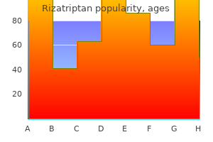
Discount rizatriptan 10 mg with mastercard
These spermatogonia are thought to be the stem cells of the seminiferous epithelium pain treatment in multiple sclerosis discount rizatriptan 10 mg mastercard. They divide at irregular intervals to give rise to either a pair of type Ad spermatogonia that remain as reserve stem cells or to a pair of type Ap spermatogonia. Type A pale (Ap) spermatogonia have ovoid nuclei with lightly staining, finely granular chromatin. ExpoDegenerative changes, such as apoptosis, premature sloughing of cells, or formation of multinucleated giant cells, are readily apparent after exposure to such agents. Vitamins, coenzymes, and microelements such as vitamin A, B12, C, E, -carotenes, zinc, and selenium have been shown to affect sperm formation. A recent study conducted in Denmark compared the sperm count in two groups of young men from rural and urban populations. A higher median sperm count (24%) was found in the men from the rural group compared with those from the urban group. Cryptorchidism, hypospadias, and factors such as low birth weight have been found to be important risk factors for testicular cancer associated with reduced semen quality and reduced fertility. A sedentary lifestyle may impair the ability to maintain the lower temperature of the testis in the scrotum. A higher than average scrotal temperature has been linked to failure of spermatogenesis. Prenatal exposure to estrogens can potentially inhibit fetal gonadotropin secretion and inhibit Sertoli cell proliferation. It has been shown to cause a major decrease in sperm count and infertility after human exposure. Most of these chemicals possess very weak estrogen properties and may affect fertility. Nitrogen mustard gas and procarbazine have been found to have toxic effects on spermatogonia. Proliferating cells are particularly sensitive to mutagenic agents and the absence of essential metabolites. Therefore, nondividing Sertoli cells, Leydig cells, and reserve stem cells, which demonstrate low mitotic activity, are much less vulnerable than actively dividing, differentiating spermatogenic cells. They undergo several successive mitotic divisions, thereby increasing their number. Thus, all of the progeny of an initial pair of Ap spermatogonia are connected, much like a strand of pearls. These cytoplasmic connections remain intact to the last stages of spermatid maturation and are essential for the synchronous development of each clone from an original pair of Ap cells. After several divisions, type A spermatogonia differentiate into type B spermatogonia. The appearance of type B spermatogonia represents the last event in the spermatogonial phase. Meiosis is described in detail in Chapter 3; a brief description of spermatocyte meiosis follows. Prophase of the first meiotic division, during which the chromatin condenses into visible chromosomes, lasts up to 22 days in human primary spermatocytes. This diagram shows the clonal nature of the successive generations of spermatogenic cells. The type A dark spermatogonia are recognized as the reserve stem cells in the testis, whereas the type A pale spermatogonia are the renewing stem cells. The type A pale spermatogonia undergo a series of synchronized cell divisions to either produce new type A pale cells or to form more differentiated type B spermatogonia that undergo further divisions into primary spermatocytes. All other spermatogenic cells remain connected by intercellular bridges as they undergo mitotic and meiotic division and differentiation of the spermatids. The cells separate into individual spermatozoa as they are released from the seminiferous epithelium. Further observations on the numbers of spermatogonia, spermatocytes, and spermatids connected by intercellular bridges in the mammalian testis. The paired homologous chromosomes, called tetrads because they consist of four chromatids, exchange genetic material in a process called crossing-over. During this exchange, the four chromatids rearrange into a tripartite structure called a synaptonemal complex. Through genetic exchange, the four spermatids produced from each spermatocyte differ from each other and from every other spermatid. After crossing-over is complete, the homologous chromosomes separate and move to the opposite poles of the meiotic spindle. Thus, the tetrads, which have been modified by crossing-over, separate and become dyads again. The two chromatids of each original chromosome (although modified by crossingover) remain together. The movement of a particular chromosome of a homologous pair to either pole of the spindle is random. This random sorting is another source of genetic diversity in the resulting sperm. The cells derived from the first meiotic division are called secondary spermatocytes. During metaphase of the second meiotic division, the chromosomes line up at the metaphase plate, and the sister chromatids separate and move to opposite poles of the spindle. These proacrosomal granules, rich in glycoproteins, coalesce into a membrane-bounded vesicle, the acrosomal vesicle, adjacent to the nuclear envelope. The position of the acrosomal vesicle determines the anterior pole of the developing sperm. Also during this phase, the centrioles migrate from the juxtanuclear region to the posterior pole of the spermatid, where the mature centriole aligns at right angles to the plasma membrane. The centriole initiates the assembly of the nine peripheral microtubule doublets and two central microtubules that constitute the axoneme of the sperm tail. In this phase, the acrosomal vesicle spreads over the anterior half of the nucleus. The portion of the nuclear envelope beneath the acrosomal cap loses its pores and becomes thicker. In this phase, the spermatid reorients itself so that the head becomes deeply embedded in the Sertoli cell and points toward the basal lamina. The condensed nucleus of the spermatid flattens and elongates, the nucleus and its overlying acrosome also move to a position immediately adjacent to the anterior plasma membrane, and the cytoplasm is displaced posteriorly. The cytoplasmic microtubules become organized into a cylindrical sheath, the manchette, which extends from the posterior rim of the acrosome toward the posterior pole of the spermatid. Male Reproductive System Spermatid Phase (Spermiogenesis) In the spermatid phase, spermatids undergo extensive cell remodeling as they differentiate into mature sperm. The haploid spermatids undergo a differentiation process that produces mature sperm, which are also haploid. The extensive cell remodeling that occurs during differentiation of the spermatid population into mature sperm (spermiogenesis) consists of four phases. These phases occur while the spermatids are physically attached to the Sertoli cell plasma membrane by specialized junctions. The centrioles, which had earlier initiated the development of the flagellum, now move back to the posterior surface of the nucleus where the immature centriole becomes attached to a shallow groove in the nucleus. They are then modified to form the connecting piece, or neck region, of the developing sperm. Nine coarse fibers develop from the centrioles attached to the nucleus and extend into the tail as the outer dense fibers peripheral to the microtubules of the axoneme. These fibers unite the nucleus with the flagellum; hence, the name connecting piece. Distal to the middle piece, a fibrous sheath consisting of two longitudinal columns and numerous connecting ribs surrounds the nine longitudinal fibers of the principal piece and extends nearly to the end of the flagellum. This short segment of the tail distal to the fibrous sheath is called the end piece.
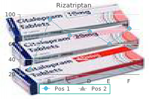
Order rizatriptan 10 mg on-line
Structure of the Wall the wall of the alimentary canal consists of four distinct layers that vary somewhat from region to region back pain treatment during pregnancy purchase generic rizatriptan canada. For example, when the stomach is full, waves of muscular contractions move along its wall from one end to the other (fig. These waves pass every twenty seconds or so and mix foods with the digestive juices secreted by the mucosa. In the small intestine, segmentation is a type of movement that aids mixing by alternately contracting and relaxing the smooth muscle in segments of the organ (fig. Because segmentation follows a back-and-forth pattern, materials are not moved along the tract in one direction. Propelling movements include a wavelike motion called peristalsis (peri-stalsis), in which a ring of contraction occurs in the wall of the tube and moves progressively along its length (fig. At the same time, the muscular wall just ahead of the ring relaxes-a phenomenon called receptive relaxation. As the peristaltic wave of contraction moves along the tube, it pushes the contents of the tube ahead of it. It causes the sounds that can be heard through a stethoscope applied to the abdominal wall. Millions of people have had the procedure, which has an advantage over endoscopy of not requiring sedation. Other parasympathetic impulses arise in the sacral region of the spinal cord and supply the distal half of the large intestine. The effects of sympathetic impulses on digestive actions usually oppose those of the parasympathetic division, meaning career cOrNer endoscopy Technician the young woman had undergone gastric bypass surgery five weeks earlier, and instead of being able to eat more foods, as she had anticipated, she was now unable to keep anything down. An endoscope is a flexible fiberoptic instrument that is passed through an opening in the body or inserted through an incision. Additional clinical training may take place on the job or in an internship program at a health-care facility. Many of the postganglionic fibers are organized into a network or nerve plexus within the wall of the canal (see fig. The submucosal plexus is important in controlling secretions by the gastrointestinal tract. The myenteric plexus of the muscular layer is more extensive and controls gastrointestinal motility. Parasympathetic impulses generally increase the activities of the digestive system such as motility and enzyme secretion. For example, sympathetic impulses inhibit mixing and propelling movements, but stimulate contraction of the sphincter muscles in the wall of the alimentary canal, blocking movement of materials through the tube. It is surrounded by the lips, cheeks, tongue, and palate and includes a chamber between the palate and tongue called the oral cavity, as well as a narrow space between the teeth, cheeks, and lips called the oral vestibule (fig. They consist of outer layers of skin, pads of subcutaneous fat, muscles associated with expression and chewing, and inner linings of moist, stratified squamous epithelium. A person rubs the inside of the cheek with a cotton swab or swishes with mouthwash and expectorates into a small tube. They contain skeletal muscles and sensory receptors useful in judging the temperature and texture of foods. The external borders of the lips mark the boundaries between the skin of the face and the mucous membrane that lines the alimentary canal. Appendix Cecum Tongue the tongue (tung) is a thick, muscular organ that occupies the floor of the mouth and nearly fills the oral cavity when the mouth is closed. Mucous membrane covers the tongue, and a membranous fold called the lingual frenulum (linggwahl frenu-lum) connects the midline of the tongue to the floor of the mouth. The body of the tongue is largely composed of skeletal muscle fibers that run in several directions. Muscular action mixes food particles with saliva during chewing and moves food toward the pharynx during swallowing. Other papillae contain taste buds, which are used in detecting sweet, salty, bitter, sour, and umami (see chapter 12, pp. Although most of the taste buds are located on papillae, some are scattered elsewhere in the mouth, particularly in children. It is covered with rounded masses of lymphatic tissue called lingual tonsils (tonsilz) (fig. Serosa Circular fold Mucosa Submucosa Microvilli Goblet cell Nucleus Longitudinal muscle Circular muscle Epithelium Lacteal Villi Capillaries Mucosa Lacteal Simple columnar epithelium Lymph nodule Intestinal gland Submucosa Mucous gland in submucosa Circular muscle Muscularis Nerve plexuses Serosa Longitudinal muscle Palate the palate (palat) forms the roof of the oral cavity and consists of a hard anterior part and a soft posterior part. The hard palate is formed by the palatine processes of the maxillae in front and the horizontal portions of the palatine bones in back. The soft palate forms a muscular arch, which extends posteriorly and downward as a cone-shaped projection called the uvula (uvu-lah). This action closes the opening between the nasal cavity and the pharynx, preventing food from entering the nasal cavity. In the back of the mouth, on either side of the tongue and closely associated with the palate, are masses of lymphatic tissue called palatine (palah-ti n) tonsils (fig. If the adenoids enlarge and block the passage between the nasal cavity and pharynx, surgical removal may be necessary. When tonsillitis recurs and does not respond to antibiotic treatment, the tonsils may be surgically removed. They are not considered part of the skeletal system because they have at least two types of proteins that are not found in bone, and their structure is different from that of bone. Teeth develop within sockets in the alveolar processes of the mandible and maxillae. The first set, the primary teeth (deciduous teeth), usually erupt through the gums (gingiva) at regular intervals between the ages of six months and two to four years. The ten primary teeth are anchored in each jaw from the midline toward the sides in the following sequence: central incisor, lateral incisor, canine (cuspid), first molar, and second molar. After their roots are resorbed, the secondary (permanent) teeth push the primary teeth out of their sockets. The secondary teeth usually begin to erupt at six years of age, but the set may not be completed until the third molars emerge between seventeen and twenty-five years of age. Sometimes these third molars, also called wisdom teeth, become wedged in abnormal positions in the jaws and fail to erupt. Chewing increases the surface area of the food particles, enabling digestive enzymes to interact more effectively with nutrient molecules. Each tooth consists of two main parts-the crown, which projects beyond the gum, and the root, which is anchored to the alveolar process of the jaw. The bulk of a tooth beneath the enamel is composed of a living cellular tissue called dentin, which is similar to bone but harder. Dentin, in turn, surrounds a central cavity (pulp cavity) that is filled with pulp. Blood vessels and nerves reach the central cavity through tubular root canals, which extend into the root. Tooth loss is most often associated with diseases of the gums (gingivitis) and the dental pulp (endodontitis). A thin layer of bonelike material called cementum, surrounded by a periodontal ligament (periodontal membrane), encloses the root. The ligament also contains blood vessels and nerves near the surface of the cementum-covered root (fig. Unless a dentist cleans and fills the cavity that forms where enamel is destroyed, the damage will spread to the underlying dentin. This fluid moist ens food particles, helps bind them, and begins the chemical digestion of carbohydrates. This is a favorable range for the action of the salivary enzyme and protects the teeth from exposure to acids in foods. Many minor salivary glands are scattered throughout the mucosa of the tongue, palate, and cheeks. Salivary Secretions the different salivary glands have varying proportions of two types of secretory cells, serous cells and mucous cells. Serous cells produce a watery fluid that contains a digestive enzyme, salivary amylase (ami -las).
Buy rizatriptan 10 mg free shipping
This device includes an electrical pulse generator and a lead wire that communicates with a portion of the myocardium pain treatment for small dogs buy 10mg rizatriptan with mastercard. The pulse generator contains a permanent battery that provides energy and a microprocessor that can sense the cardiac rhythm and signal the heart to alter its contraction rate. A device called a pacemaker-cardioverter-defibrillator attempts to correct ventricular fibrillation should it occur. They form a closed circuit of tubes that carries blood from the heart to the body cells and back again. The arteries and arterioles conduct blood away from the ventricles of the heart and lead to the capillaries, where substances are exchanged between blood and the body cells. Researchers create replacement blood vessels from cells and their products plus synthetic materials. One such blood vessel consists of smooth muscle cells seeded onto tubes of a biodegradable polymer. The cells secrete collagen and extracellular matrix, which replace the polymer, and then detergent is applied to remove the cells. Another approach uses rubber tubing and a nutrient solution that prompts the cells to produce extra elastin, which makes the replacement vessels more flexible. Because construction of tissues and organs in the laboratory (part of regenerative medicine) requires a blood supply, tissue engineers are creating cell-lined tubules using 3D printing to function as part of a semisynthetic, ex vivo ("outside the body") cardiovascular system. In normal development, angiogenesis is crucial to build a blood supply to serve a growing body. New blood vessels deliver nutrients, hormones, and growth factors to tissues and remove wastes. After a heart attack, for example, new vessels form in the remaining healthy cardiac muscle. As is the case for most biological processes, angiogenesis must be highly controlled. Two specific applications are healing hearts and removing extra capillaries in tumors and in eyes. At the same time, endothelial cells that are part of the tumor assemble into sheets, roll into tubules, and snake out of the tumor as new capillaries. Other cancer cells wrap around the capillaries, spreading out on this scaffolding into nearby tissues. For a time, maybe even years, these secondary tumors stay small, adhering to the outsides of the blood vessels that delivered them. From the observation by many surgeons that when a primary tumor is removed, secondary tumors grow, Harvard researcher Judah Folkman hypothesized that the primary tumor secretes antiangiogenesis factors that keep the secondary tumors small. Once factors that promote or block angiogenesis were discovered in the 1980s, researchers began to study the antiangiogenesis factors that keep secondary tumors small, to develop them as cancer treatments. The first antiangiogenesis drug to treat cancer became available in 2004, for colorectal cancer that has spread to other organs. Today antiangiogenesis drugs are also used to treat age-related macular degeneration, in which extra capillaries extend into the retina and block central vision. Coronary bypass surgery and angioplasty are treatments that restore blood flow by circumventing a blockage or opening up an artery, respectively. These vessels subdivide into progressively thinner tubes and eventually give rise to the finer, branched arterioles (ar-tere-olz). The wall of an artery consists of three distinct layers, or tunics, shown in figure 15. The innermost tunic, tunica interna (intima), is composed of a layer of simple squamous epithelium, called endothelium, that rests on a connective tissue membrane rich in elastic and collagen fibers. The endothelial lining of an artery provides a smooth surface that allows blood cells and platelets to flow through without being damaged. Additionally, endothelium helps prevent blood clotting by secreting biochemicals that inhibit platelet aggregation (see chapter 14, p. Endothelium also may help regulate local blood flow by secreting substances that dilate or constrict blood vessels. For example, endothelium releases the gas nitric oxide, which relaxes the smooth muscle of the vessel. It includes smooth muscle cells, which encircle the tube, and a thick layer of elastic connective tissue. The connective tissue gives the vessel a tough elasticity that enables it to withstand the force of blood pressure and, at the same time, to stretch and accommodate the sudden increase in blood volume that accompanies ventricular contraction. The outer layer, tunica externa (adventitia), is relatively thin and chiefly consists of connective tissue with irregular elastic and collagen fibers. It also contains tiny vessels (vasa vasorum) that give rise to capillaries and provide blood to the more external cells of the artery wall. The sympathetic branches of the autonomic nervous system innervate smooth muscle in artery and arteriole walls. Vasomotor fibers stimulate the smooth muscle cells to contract, reducing the diameter of the vessel. If vasomotor impulses are inhibited, the muscle cells relax, and the diameter of the vessel increases. Changes in the diameters of arteries and arterioles greatly influence blood flow and blood pressure. The walls of the larger arterioles have three layers similar to those of arteries (fig. The wall of a very small arteriole consists only of an endothelial lining and some smooth muscle cells, surrounded by a small amount of connective tissue (fig. Arterioles, which are microscopic continuations of arteries, give off branches called metarterioles that, in turn, join capillaries. The arteriole and metarteriole walls are adapted for vasoconstriction and vasodilation in that their smooth muscle cells respond to impulses from the autonomic nervous system by contracting or relaxing. Capillaries are extensions of the inner linings of arterioles in that their walls are endothelium-a single layer of squamous epithelial cells (fig. These thin walls form the semipermeable layer through which substances in the blood are exchanged for substances in the tissue fluid surrounding body cells. The openings or intercellular passageways in the capillary walls are thin slits where endothelial cells overlap (fig. The sizes of these openings, and consequently the permeability of the capillary wall, vary from tissue to tissue. For example, the openings are relatively smaller in the capillaries of smooth, skeletal, and cardiac muscle than they are in capillaries associated with endocrine glands, the kidneys, and the lining of the small intestine. Capillaries with the largest openings include those of the liver, spleen, and red bone marrow. Discontinuous capillaries allow large proteins and even intact cells to pass through as they enter or leave the circulation. The blood-brain barrier is not present in the pituitary and pineal glands and parts of the hypothalamus. Muscle and nerve tissues, which use abundant oxygen and nutrients, are richly supplied with capillaries. Tissues with slower metabolic rates have fewer capillaries, such as cartilage, or lack them entirely, such as in the cornea. If the capillaries of an adult were unwound and lined up end to end, they would be between 25,000 and 60,000 miles long. Slit Tissue fluid Capillary (a) Nucleus of endothelial cell Endothelial cell cytoplasm Lumen of capillary Cell junction the spatial patterns of capillaries also differ in various body parts. For example, some capillaries pass directly from arterioles to venules, but others lead to highly branched networks (fig. Because capillaries are physically organized in diverse ways, blood can follow different pathways through a tissue, attuned to cellular requirements. During exercise, for example, blood is directed into the capillary networks of the skeletal muscles, where the cells require more oxygen and nutrients.
Order rizatriptan 10 mg with visa
The release of acrosomal enzymes as the sperm touches the egg is the first step in the acrosome reaction treatment guidelines for back pain generic rizatriptan 10mg on-line. This complex process facilitates sperm penetration and subsequent fertilization and prevents the entry of additional sperm into the ovum. The basic changes in the structure of the key organelles of the spermatid are illustrated (see text for detailed explanation). Spermatids are no longer attached to each other and are released from the Sertoli cells. Spermatids are released into the lumen of the seminiferous tubules during the process called spermiation. Toward the end of maturation phase of spermatogenesis, elongated spermatids are released from Sertoli cells into the lumen of seminiferous tubule. This complex process called spermiation involves progressive removal of specialized Sertoli-to-spermatid junctional complexes and disengagement of spermatids from the Sertoli cell. The presence of the 1-integrins in the Sertoli-to-spermatid junctions, as well as increased activity of the integrin-linked kinase at the time of spermiation, suggests an enzymatic control of spermatid release. The rate of spermiation in the testis determines the number of sperm cells in the ejaculate of semen. Various pharmacologic treatments, toxic agents, and gonadotropin suppression result in spermiation failure, in which spermatids are not released but instead are retained and phagocytosed by the Sertoli cell. The acrosomal cap that covers the anterior two-thirds of the nucleus contains hyaluronidase, neuraminidase, acid phosphatase, and a trypsin-like protease called acrosin. Key structural features of the head (viewed in frontal and sagittal planes), the middle piece, and the principal piece of the spermatozoon are illustrated on the right. The middle piece is approximately 7 m long and contains the mitochondria, helically wrapped around the coarse fibers and the axonemal complex. These mitochondria provide the energy for movement of the tail and thus are responsible for the motility of the sperm. The principal piece is approximately 40 m long and contains the fibrous sheath external to the coarse fibers and the axonemal complex. The end piece, approximately the last 5 m of the flagellum in the mature sperm, contains only the axonemal complex. Newly released sperm cells are processed in the epididymis where they acquire motility and undergo further maturation. This process, which involves removal and replacement of glycocalyx components (glycoconjugates) on the sperm membrane, is called capacitation. These groupings or associations occur because intercellular bridges are present between the progeny of each pair of type Ap spermatogonia and because the synchronized cells spend specific times in each stage of maturation. Each recognizable grouping, or cell association, is considered a stage in a cyclic process. The series of stages that appears between two successive occurrences of the same cell association pattern at any given site in the seminiferous tubule constitutes a cycle of the seminiferous epithelium. The cycle of the seminiferous epithelium has been most thoroughly studied in rats, in which 14 successive stages occur in linear sequence along the tubule. These stages are not as clearly delineated as those in rodents because in man the cellular associations occur in irregular patches that form a mosaic pattern. Male Reproductive System Newly released sperm cells are nonmotile and are carried from the seminiferous tubules in a fluid secreted by the Sertoli cells. The fluid and sperm flow through the seminiferous tubules, facilitated by peristaltic contractions of the peritubular contractile cells of the lamina propria. They then enter the straight tubules, a short segment of the seminiferous tubule where the epithelium consists only of Sertoli cells. At the mediastinum testis, the fluid and sperm enter the rete testis, an anastomosing system of ducts lined by simple cuboidal epithelium (Plate 87, page 824). From the rete testis, they move into the extratesticular portion of the efferent ductules (ductuli efferentes), the first part of the excurrent duct system, and then into the proximal portion of the duct of the epididymis (ductus epididymis). As the sperm cells move through 4 to 5 m of the highly coiled duct of the epididymis, they acquire motility and undergo several maturational changes. The surface-associated decapacitation factor is added to inhibit the fertilizing ability of the sperm cells (page 811). These factors regulate flagellar activity through changes in protein phosphorylation, resulting from activities of protein kinases and protein phosphatases. For instance, pharmacologic stimulation of protein kinase A activity increases motility of sperm cells, whereas inhibition of protein phosphatase activity may initiate or stimulate such motility. This suggests that phosphatases have an important role in the regulation of sperm kinetic activity. Contractions of the smooth muscle that surrounds the progressively distal and larger ducts continue to move the sperm by peristaltic action until they reach the distal portion of the duct of the epididymis, where they are stored before ejaculation. Sperm can live for several weeks in the male excurrent duct system, but they will survive only 2 to 3 days in the female reproductive tract. They acquire the ability to fertilize the After injecting a pulse of tritiated thymidine, a specific generation of cells can be followed by sequential biopsies of the seminiferous tubules. In this way, the time required for the labeled cells to go through the various stages can be determined. Several generations of developing cells may be present in the thickness of the seminiferous epithelium at any given site and at any given time, which produces the characteristic cell associations. Autoradiographic studies have revealed that the duration of the cycle of the seminiferous epithelium is constant, lasting about 16 days in humans. It would then require approximately 12 days for the spermatozoon to pass through the epididymis. The length of the cycle and the time required for spermatogenesis are constant and specific in each species. This diagram shows each of the six recognizable cell associations (stages) that occur in the cycle of the human seminiferous epithelium. These stages of spermatogenesis are artificially defined according to changes observed in the spermatids during their various steps of differentiation. In 1952, six stages of human seminiferous epithelium were first described by Leblond and Clermont, and since then, they have been adopted by most researchers. In addition, the wave of the seminiferous epithelium describes the distribution of patterns of cellular association (spermatogenic stages) along the length of the tubule. In rodents and other mammals that have been studied, including subhuman primates, each stage occupies a significant length of the seminiferous tubule, and the stages appear to occur sequentially along the length of the tubule. A transverse section through the tubule usually reveals only one pattern of cell associations. There are no waves in the human seminiferous epithelium and the arrangement of spermatogenic stages along the seminiferous tubule is random. Patches do not extend around the circumference of the tubule, nor are they in sequence. Therefore, a transverse section through a human seminiferous tubule may reveal as many as six different stages of the cycle arranged in a pie-wedge fashion around the circumference of the tubule. Sertoli Cells Sertoli cells constitute the true epithelium of the seminiferous epithelium. They are the supporting cells for the developing spermatozoa that attach to their surface after meiosis. They have numerous spherical and elongated mitochondria, a well-developed Golgi apparatus, and varying numbers of lysosomes, lipid droplets, vesicles, and glycogen granules. This suggests that inclusion bodies might be involved in lipid transport and their utilization by the Sertoli cells. In mice and other rodent species, a particular cellular association occupies varying lengths along the tubule. Therefore, in a typical cross-section, only a single cellular association is observed. In humans, cellular associations occur in irregularly shaped areas along the tubule, and therefore, a cross-section typically shows two or more cellular associations. This complex is characterized, in part, by an exceedingly tight junction (zonula occludens) that includes more than 50 parallel fusion lines in the adjacent membranes. They are all oriented with their minus ends toward the apex and plus ends toward the base of the cell.
Rizatriptan 10mg cheap
The fifth pair of cranial nerves pain medication for the shingles rizatriptan 10 mg fast delivery, the trigeminal (tri-jemi-nal) nerves (V), are the largest and arise from the pons. Each sensory component includes three large branches, called the ophthalmic, maxillary, and mandibular divisions (fig. The ophthalmic division of each the trigeminal nerve consists of sensory fibers that conduct impulses to the brain from the surface of the eye; the tear gland; and the skin of the anterior scalp, forehead, and upper eyelid. The fibers of the maxillary division conduct sensory impulses from the upper teeth, upper gum, and upper lip, as well as from the mucous lining of the palate and facial skin. Fibers include axons of motor neurons as well as peripheral processes of sensory neurons. The motor branches supply the muscles of mastication and certain muscles in the floor of the mouth. Lacrimal nerve Ophthalmic division Lacrimal gland Eye A disorder of the trigeminal nerve called trigeminal neuralgia (tic douloureux) causes severe recurring pain in the face and forehead on one side. However, with surgery the patient loses sensations in other body regions that the sensory branch supplies. After surgery, care must be taken when eating or drinking hot foods or liquids, and the mouth must be inspected daily for food particles or damage to the cheeks from biting. They enter the orbits of the eyes and supply motor impulses to the remaining pair of external eye muscles, the lateral rectus muscles. Their sensory branches are associated with taste receptors on the anterior two-thirds of the tongue, and some of their motor fibers conduct impulses to muscles of facial expression (fig. Still other motor fibers of these nerves function in the autonomic nervous system by stimulating secretions from tear glands and certain salivary glands (submandibular and sublingual glands). It occurs from damage to the facial nerve from unknown causes, although it is thought to be linked to viral infections. Each of these nerves has two distinct parts-a vestibular branch and a cochlear branch. The neuron cell bodies of the vestibular branch fibers are located in ganglia near the vestibule and semicircular canals of the inner ear. These structures contain receptors that sense changes in the position of the head and, in response, initiate and send impulses to the cerebellum, where they are used in reflexes that maintain equilibrium. The neuron cell bodies of the cochlear branch fibers are located in a ganglion of the cochlea, a part of the inner ear that houses the hearing receptors. Impulses from this branch pass through the medulla oblongata and midbrain on their way to the temporal lobe, where they are interpreted. They are mixed nerves, with predominant sensory fibers that conduct impulses from the lining of the pharynx, tonsils, and posterior third of the tongue to the brain. Fibers in the motor component of the glossopharyngeal nerves innervate certain salivary glands and a constrictor muscle in the wall of the pharynx that functions in swallowing. The tenth pair of cranial nerves, the vagus (vagus) nerves (X), originate in the medulla oblongata and extend downward through the neck into the chest and abdomen. These nerves are mixed, including both somatic and autonomic branches, with the autonomic fibers predominant. Among the somatic components of the vagus nerves are motor fibers that conduct impulses to muscles of the larynx and pharynx. These fibers are associated with speech and swallowing reflexes that use muscles in the soft palate and pharynx. Vagal sensory fibers conduct impulses from the linings of the pharynx, larynx, and esophagus and from the viscera of the thorax and abdomen to the brain. Autonomic motor fibers of the vagus nerves supply the heart and many smooth muscles and glands in the viscera of the thorax and abdomen (fig. Each cranial branch of an accessory nerve joins a vagus nerve and conducts impulses to muscles of the soft palate, pharynx, and larynx. The spinal branch descends into the neck and supplies motor fibers to the trapezius and sternocleidomastoid muscles. They primarily consist of fibers that conduct impulses to muscles that move the tongue in speaking, chewing, and swallowing. All but the first pair are mixed nerves, and they provide two-way communication between the spinal cord and parts of the upper and lower limbs, neck, and trunk. Motor fibers conduct impulses to muscles that raise the eyelids, move the eyes, adjust the amount of light entering the eyes, and focus the lenses. V Trigeminal ophthalmic division Maxillary division Mandibular division Mixed Sensory fibers conduct impulses from the surface of the eyes, tear glands, scalp, forehead, and upper eyelids. Sensory fibers conduct impulses from the upper teeth, upper gum, upper lip, lining of the palate, and skin of the face. Sensory fibers conduct impulses from the scalp, skin of the jaw, lower teeth, lower gum, and lower lip. Motor fibers conduct impulses to muscles of mastication and to muscles in the floor of the mouth. Motor fibers conduct impulses to muscles of facial expression, tear glands, and salivary glands. Mixed Sensory fibers conduct impulses from the pharynx, tonsils, posterior tongue, and carotid arteries. Motor fibers conduct impulses to salivary glands and to muscles of the pharynx used in swallowing. Somatic motor fibers conduct impulses to muscles associated with speech and swallowing; autonomic motor fibers conduct impulses to the viscera of the thorax and abdomen. Motor fibers conduct impulses to muscles of the neck and back; some proprioceptor input. Primarily motor Motor fibers conduct impulses to muscles that move the tongue; some proprioceptor input. On each vertebra the vertebral notches, which are the major parts of the intervertebral foramina, are associated with the inferior portion of their respective vertebrae. For this reason, each spinal nerve, as it passes through the intervertebral foramen, is associated with the vertebra above it. The cervical spinal nerves are an exception because spinal nerve C1 passes supe- rior to the vertebra C1. Therefore, although there are seven cervical vertebrae, there are eight pairs of cervical nerves (numbered C1 to C8). There are twelve pairs of thoracic nerves (numbered T1 to T12), five pairs of lumbar nerves (numbered L1 to L5), five pairs of sacral nerves (numbered S1 to S5), and one pair of coccygeal nerves (Co). Each spinal nerve, except for the first pair, emerges from the cord by two short branches, or roots, which lie within the vertebral column. Portions of the peripheral nerves that result from branching are shown on the left. In early life, the spinal cord extends the entire length of the vertebral column, but with age, the column grows more rapidly than the cord. Thus, the adult spinal cord ends at the level between the first and second lumbar vertebrae, so the roots associated with the lumbar, sacral, and coccygeal nerves descend to their exits beyond the end of the cord, still within the vertebral canal. The ventral root (anterior, or motor, root) of each spinal nerve consists of axons from the motor neurons whose cell bodies lie within the gray matter of the cord. The dorsal root (posterior, or sensory, root) can be identified by an enlargement called the dorsal root ganglion. This ganglion contains the cell bodies of the sensory neurons whose axons (peripheral processes) conduct impulses inward from peripheral body parts. C2 C3 C4 C5 processes) of these neurons continue through the dorsal root and into the spinal cord, where they form synapses with other neurons or ascend to the brain (see fig. A ventral root and a dorsal root unite to form a spinal nerve, which extends outward from the vertebral canal through an intervertebral foramen (the dorsal root is usually absent from the first pair of spinal nerves). Each spinal nerve below C1 contains sensory fibers that reach the skin, and the region innervated is called a dermatome. A map of the dermatomes is useful in localizing the sites of injuries to dorsal roots or to the spinal cord. A dorsal branch (dorsal ramus) of each spinal nerve turns posteriorly and innervates the muscles and skin of the back, as figure 11. The main portion of the nerve, the ventral branch (ventral ramus), continues forward to supply muscles and skin on the front and sides of the trunk and limbs.
