

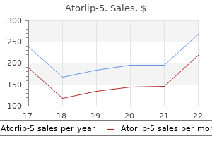
Purchase 5mg atorlip-5 otc
The nervous system is composed of such a heterogeneous population of cells that the specific trophic requirements for appropriate development and function are extremely complex cholesterol levels lab values effective 5 mg atorlip-5. Although the original studies on trophic factors defined their function in terms of their ability to support survival of different populations of neurons, recent studies have shown that these factors regulate many aspects of neuronal function. Current and future studies will provide additional insights into the mechanisms of trophic interactions, and especially whether they may be harnessed for therapeutic purposes. Evidence that embryonic neurons regulate the onset of cortical gliogenesis via cardiotrophin-1. Neurotrophins and their receptors: a convergence point for many signalling pathways. Studies on the physiological role of brain-derived neurotrophic factor and neurotrophin-3 in knockout mice. Amelioration of cholinergic neuron atrophy and spatial memory impairment in aged rats by nerve growth factor. Neurturin exerts potent actions on survival and function of midbrain dopaminergic neurons. Targeted disruption of the trkB neurotrophin receptor gene results in nervous system lesions and neonatal death. Essential role of the nerve growth factor in the survival and maintenance of dissociated sensory and sympathetic embryonic nerve cells in vitro. In the adult, the majority of cells are nonproliferative and the proportion of precursor and stem cells is dramatically reduced. Only a few tissues such as the skin and blood retain high levels of regular cell turnover in the adult, and they rely upon a continued source of stem cells for replenishment. By contrast, in the nervous system the majority of cells are generated during development, and cell division and morphogenesis are largely complete in the mammalian brain soon after birth. The role of these stem cells in building the brain in response to injury and their potential for repair is an area of intense study and progress. These characteristics do not, however, distinguish stem cells from precursors or progenitors. A stem cell differs from other cells in two essential ways: stem cells are multipotent, with the ability to give rise to multiple differentiated cell types, and stem cells are selfrenewing, with the virtually unlimited ability to make more of themselves. Studies by Till & McCulloch, 1961, established that cells in the bone marrow could reconstitute the immune lineage. Animals exposed to high levels of irradiation die, but can be rescued by infusion of bone marrow cells or purified hematopoietic stem cells. The entire blood-forming system could be restored by injecting bone marrow cells from a compatible donor. After about a week, the spleens of the injected mice contain colonies of cells, each from a single hematopoietic stem cell. Within this broad definition, there are various types of stem cells normally found at different stages of development and in different tissues. In this approach, animals that received lethal irradiation died, but those that received just a few purified hematopoietic stem cells had a completely restored immune system, which demonstrated the multipotency of the transplanted cells (Becker et al. Indeed, during the reconstitution of the immune system, clusters of hematopoietic cells infiltrated the spleen and formed colonies in numbers that reflected the original stem cells transplanted, and dissociation of those clusters could also reconstitute the immune system, demonstrating self-renewal. Thus far, no similarly powerful functional repopulation assay is possible with neural cells, but this may reflect the challenges of integrating neurons into existing circuits rather than the lack of reparative cells. The early studies led to successful allogeneic (from a genetically different person) hematopoietic stem cell transplants in the 1960s. Because they are isolated from very early embryonic tissues, they are the least restricted type and can give rise to the widest range of cell derivatives in the body, including germ cells. Because of their source and the potential that these cells can give rise to all cell types in the body and thus could be used for "cloning," the use of these cells for research or therapy has been associated with significant ethical concerns. One of the best-understood tissues that harbors stem cells in the adult is the blood or hematopoietic system. In healthy individuals, circulating red and white cells in the blood are replaced every few weeks from new cells generated in the bone marrow. Due to their self-renewing capability, these cells can be greatly expanded in number with specific growth factors. They do not divide again, which leads to the concept of a neuronal birthdate, and they are not turned over, so that any loss of neurons or glia in the adult is associated with loss of function. Classical studies by Altman and Das (1965; Altman, 1963) demonstrated the presence of new neurons in the adult mammalian hippocampus and olfactory bulb. The origin of many of these new neurons has been found to be among adult neural stem cells. In the mammal, neural development begins with induction and formation of the neural tube at about E 7. The walls of the neural tube contain neuroepithelial cells oriented like spokes on a wheel that will eventually divide dramatically to give rise to each of the major brain regions, and on a cellular level, to all the neurons and glial cells of the entire nervous system (see Development, Chapter 28). Neural stem cells expand early in development, and then give rise to neurogenic and then gliogenic lineages. Radial glia are stem cells Radial glial cells make up one of the earliest classes of cells to emerge from the neuroepithelium. Originally, radial glia were thought to be simply scaffolds that maintained the cytoarchitecture of the nervous system. However, several lines of evidence suggest that early radial glial cells have stem cell-like properties. In the developing cortex, initial retroviral lineage tracing identified "clones" of cells with a radial orientation and containing both neurons and glia. This observation suggested that these clones contribute to functional columns in the brain. Yet the key studies to demonstrate that particular stem cells give rise to particular progeny or derivates in situ remain incomplete. One example of a good critical approach has been to use Cre/loxP lineage tracing with different radial glial promoters, which reveals that many, if not most, neurons in the brain/ ventral telencephalon are derived from radial glia (Anthony & Heintz, 2008). More extensive genetic lineage analysis of not only radial glia but also their derivatives in vivo are needed to understand the quantitative contribution of stem cells to the adult brain and to understand key regulatory factors that direct the fate of their progeny. The neural tube rapidly acquires regional distinctions as a result of soluble inductive cues. Numerous studies have isolated stem cells from a variety of brain regions at various ages and have shown that these cells give rise to particular derivatives in vitro (see Temple, 2001). However, it is also clear that the progeny of neural stem cells can migrate long distances. Not only are the properties of neural stem cells regionally specified; in addition, the specification of their progeny may change during development. Stem cells isolated during the period of neurogenesis preferentially generate neurons in vitro while those isolated later in development during gliogenesis preferentially generate glia. Early in development, some neural tube cells in dorsal regions delaminate or migrate away from the neural tube as neural crest cells. Radial glial cells that initially span the wall of the neural tube give rise to clones of cells that include both neurons and glial cells (note green and yellow cells). Given their common embryological origins, it is not surprising that adult skin cells can be stimulated to give rise to neurons (Toma et al. Taken all together, this body of research results demonstrates the presence of neural stem cells that become increasing patterned as development proceeds. The molecular basis of this increasing restriction in the fate of cellular progeny during development and maturation is an area of current investigative interest. Even in embryonic tissues where they may be more plentiful, stem cells are intermingled with numerous progenitor and mature cells. To identify stem cells antigenically for isolation, investigators have worked to distinguish selective stem cell markers, as well as differentiated neural cell markers. Some stem cell markers reflect transcription factors present only at certain stages of differentiation, while other markers expressed on the cell surface are useful for prospective isolation. Commonly used stem cell markers in the nervous system as well as differentiated neural cell the strongest functional data for the existence of a selfrenewing, multipotent neural stem cell comes from in vitro studies. The neurosphere assay first developed by Reynolds and Weiss (1992) has emerged as a useful approach to expand and study neural stem cells and progenitors. In essence, dispersed neural cells are grown in suspension in Fibroblast Growth Factor 2 and/or Epidermal Growth Factor, during which stem cells proliferate and form floating cell aggregates called "neurospheres" while the vast majority of precursors and differentiated cells die.
5mg atorlip-5 sale
A de novo mutation affecting human TrkB associated with severe obesity and developmental delay cholesterol weight ratio atorlip-5 5 mg overnight delivery. During development, axons often need to navigate through long distances before finding their targets. Anterior and posterior retinal axons project to the posterior and anterior tectum, respectively. Similarly, the dorsal and ventral retinal axons innervate the ventral and dorsal tectum respectively. This reversal of projection is important for the rectification of the upside-down and back-to-front retinal image. This implies that Eph-mediated axon guidance is limited to short-range contact-dependent activity. EphR/ephrins are now understood to be important for axon growth and guidance in regeneration as well as during development and play a role in proliferation of astrocytes following injury (Goldshmit et al. Members of the EphR family play important roles not only in axon guidance, but in regulating the actin cytoskeleton, cell adhesion and cell migration in both health and disease states (Pasquale, 2008). Steering of an axon in different directions towards its destination is largely mediated by changes in the cytoskeleton in a structure at the leading edge of an axon known as the growth cone (Chs 6 and 32). The growth cone consists of finger-like protrusions known as filopodia interspaced by web-like structures called lamellipodia. Tyrosine phosphorylation appears to be an important signal-transduction mechanism in every step in the development of a neuron, starting from the survival and differentiation of stem cells, to axon guidance and synaptogenesis and to synaptic transmission at a mature synapse. Examples of each step are given on the right, indicating the receptors and their ligands. Mechanisms converting signals from extracellular cues into changes in intracellular cytoskeletal dynamics in the growth cone are gradually emerging. The extracellular cues for axon guidance are ephrins, semaphorins, Slits and netrins (Dickson, 2002). A close homolog of Nck, Grb4 binds to the phosphotyrosine residue on the cytoplasmic tail of ephrin and results in actin depolymerization. It is downregulated dramatically in mature muscle except at the neuromuscular junction. ErbBs at the neuromuscular junction are activated by another motor-neuron-derived factor known as neuregulin. Neurotrophins affect formation of both excitatory and inhibitory synapses through presynaptic as well as postsynaptic mechanisms. Tyrosine phosphorylation plays an important role in synaptic transmission and plasticity. As the role of neurotrophins and Trk receptors on synaptic function became recognized (see Ch. However, the importance of tyrosine phosphorylation on synaptic function is not restricted to Trks and neurotrophins. Fyn and Src activity may be downstream of the Trk signaling pathway (Minichiello, 2009) or may be regulated through some other aspect of synaptic activity. Tyrosine phosphorylation sites are present within the large intracellular loop between the third and fourth transmembrane domains. Mutagenesis studies have revealed the intracellular N-terminus as the tyrosine-phosphorylation site-bearing region, although other studies have suggested the presence of other tyrosine-phosphorylation sites. Neurotransmitter receptors and voltage-gated ion channels play a pivotal role in modulating synaptic efficacy and plasticity. Many of these receptors and ion channels in both the peripheral and central nervous systems are tyrosine phosphorylated (Davis et al. N-Methyl-d-Aspartate Receptors Glutamate is the major excitatory neurotransmitter in the central nervous system (Ch. They play an important role in the induction of synaptic plasticity and excitotoxicity. Each subunit has four transmembrane domains; both the N- and C-termini are in the extracellular space. Between the third and fourth transmembrane domains is a large cytoplasmic region containing a single conserved tyrosine residue in the cytoplasmic loop of each of the, g and subunits. Voltage-Gated Ion Channels Many voltage-gated ion channels also appear to be regulated by tyrosine phosphorylation (Reichardt, 2006). These include voltage-gated calcium, potassium and sodium ion channels whose function can either be enhanced or impeded by tyrosine phosphorylation. For example, voltage-gated L-type calcium ion channel activity is augmented by tyrosine phosphorylation. It consists of six transmembrane domains with both the N- and C-termini in the cytoplasmic side, carrying a number of potential phosphorylation sites. It is believed that both cytoplasmic ends of the channel impose an inhibitory tone on functional activity; upregulation of channel activity by protein phosphorylation may be attributed to the relief of this inhibition. The intracellular region between the third and fourth transmembrane domains contains a number of consensus protein phosphorylation sites. Ligandmediated negative regulation of a chimeric transmembrane receptor tyrosine phosphatase. Impaired long-term potentiation, spatial learning, and hippocampal development in fyn mutant mice. Neurotrophins and their receptors: Roles in plasticity, neurodegeneration and neuroprotection. Functional modulation of the nicotinic acetylcholine receptor by tyrosine phosphorylation. Src family kinases: Regulation of their activities, levels and identification of new pathways. The importance of being proline: the interaction of proline-rich motifs in signaling proteins with their cognate domains. Rescuing impairment of long-term potentiation in fyn-deficient mice by introducing Fyn transgene. TrkB and TrkC signaling are required for maturation and synaptogenesis of hippocampal connections. The biology of neurotrophins, signalling pathways, and functional peptide mimetics of neurotrophins and their receptors. Generally, this can be viewed as presynaptic neurons modulating the activity of postsynaptic neurons. Finally, given the complexities of transcriptional regulation, a brief discussion of possible sites for pharmacological intervention in the transcriptional process will be presented. Indeed, the recent sequencing of multiple genomes has revealed that the transcriptional machinery is largely conserved between different species ranging from yeast to human, reflecting the fundamental nature of the transcriptional process. These studies have revealed good sequence conservation of over 100 proteins known to be involved in the transcriptional process. This process of transcriptional regulation is multifaceted and involves the association of several additional proteins in particular arrangements with the transcriptional complex. The arrangement and identity of transcriptional accessory proteins, also called transcription factors, can be unique for individual genes.
Cheap atorlip-5 5mg without prescription
Plasmid-encoded toxin of enteroaggregative Escherichia coli is internalized by epithelial cells kresser cholesterol ratio buy atorlip-5 5mg lowest price. Pet toxin from enteroaggregative Escherichia coli produces cellular damage associated with fodrin disruption. Cytoskeletal effects induced by pet, the serine protease enterotoxin of enteroaggregative Escherichia coli. EspP, a novel extracellular serine protease of enterohaemorrhagic Escherichia coli O157:H7 cleaves human coagulation factor V. Identification and molecular characterization of EatA, an autotransporter protein of enterotoxigenic Escherichia coli. The serine protease motif of EspC from enteropathogenic Escherichia coli produces epithelial damage by a mechanism different from that of Pet toxin from enteroaggregative E. The secreted autotransporter toxin, Sat, functions as a virulence factor in Afa/Dr diffusely adhering Escherichia coli by promoting lesions in tight junction of polarized epithelial cells. Interferon-gamma selectively increases epithelial permeability to large molecules by activating different populations of paracellular pores. The sigA gene which is borne on the she pathogenicity island of Shigella flexneri 2a encodes an exported cytopathic protease involved in intestinal fluid accumulation. SepA, the 110 kDa protein secreted by Shigella flexneri: two-domain structure and proteolytic activity. Characterization of pic, a secreted protease of Shigella flexneri and enteroaggregative Escherichia coli. The Bacteroides fragilis toxin fragilysin disrupts the paracellular barrier of epithelial cells. Deamidation of Cdc42 and Rac by Escherichia coli cytotoxic necrotizing factor 1: activation of c-Jun N-terminal kinase in HeLa cells. Detection of cytotoxic necrotizing factor types 1 and 2 among fecal Escherichia coli isolates from Brazilian children with and without diarrhea. The enterotoxin from Clostridium difficile (ToxA) monoglucosylates the Rho proteins. Clostridium difficile toxins disrupt epithelial barrier function by altering membrane microdomain localization of tight junction proteins. Emergence of fluoroquinolones as the predominant risk factor for Clostridium difficile-associated diarrhea: a cohort study during an epidemic in Quebec. Claudin multigene family encoding fourtransmembrane domain protein components of tight junction strands. Clostridium perfringens enterotoxin utilizes two structurally related membrane proteins as functional receptors in vivo. Clostridium perfringens enterotoxin binds to the second extracellular loop of claudin-3, a tight junction integral membrane protein. Evidence that an approximately 50-kDa mammalian plasma membrane protein with receptor-like properties mediates the amphiphilicity of specifically bound Clostridium perfringens enterotoxin. Studies of Clostridium perfringens enterotoxin action at different temperatures demonstrate a correlation between complex formation and cytotoxicity. CaCo-2 cells treated with Clostridium perfringens enterotoxin form multiple large complex species, one of which contains the tight junction protein occludin. New insights into the cytotoxic mechanisms of Clostridium perfringens enterotoxin. Comparative biochemical and immunocytochemical studies reveal differences in the effects of Clostridium perfrin- 183. Clostridium perfringens enterotoxin fragment removes specific claudins from tight junction strands: Evidence for direct involvement of claudins in tight junction barrier. Role of C-terminal regions of the C-terminal fragment of Clostridium perfringens enterotoxin in its interaction with claudin-4. Binding of Clostridium botulinum C2 toxin to asparagine-linked complex and hybrid carbohydrates. Cellular uptake of Clostridium botulinum C2 toxin requires oligomerization and acidification. Binary toxin-producing, large clostridial toxin-negative Clostridium difficile strains are enterotoxic but do not cause disease in hamsters. Disruption of the epithelial apicaljunctional complex by Helicobacter pylori CagA. Interleukin-1 receptor phosphorylation activates Rho kinase to disrupt human gastric tight junctional claudin-4 during Helicobacter pylori infection. Production of ammonium by Helicobacter pylori mediates occludin processing and disruption of tight junctions in Caco-2 cells. Signal transduction pathways involved in enterohemorrhagic Escherichia coli-induced alterations in T84 epithelial permeability. Campylobacter jejuni activates mitogen-activated protein kinases in Caco-2 cell monolayers and in vitro infected primary human colonic tissue. Vibrio parahaemolyticus disruption of epithelial cell tight junctions occurs independently of toxin production. Infection of T84 cells with enteropathogenic Escherichia coli alters barrier and transport functions. Translocated EspF protein from enteropathogenic Escherichia coli disrupts host intestinal barrier function. Enteropathogenic Escherichia coli EspG disrupts microtubules and in conjunction with Orf3 enhances perturbation of the tight junction barrier. Intestinal barrier dysfunction by enteropathogenic Escherichia coli is mediated by two effector molecules and a bacterial surface protein. The bacterial virulence factor NleA is required for the disruption of intestinal tight junctions by enteropathogenic Escherichia coli. Comparative analysis of EspF from enteropathogenic and enterohemorrhagic Escherichia coli in alteration of epithelial barrier function. Shiga toxin-producing Escherichia coli can impair T84 cell structure and function without inducing attaching/effacing lesions. Escherichia albertii and Hafnia alvei are candidate enteric pathogens with divergent effects on intercellular tight junctions. Cadherin expression is required for the spread of Shigella flexneri between epithelial cells. Shigella flexneri regulates tight junction-associated proteins in human intestinal epithelial cells. Localization of dysfunctional tight junctions in Salmonella enterica serovar typhimurium-infected epithelial layers. Rapid disruption of epithelial barrier function by Salmonella typhimurium is associated with structural modification of intercellular junctions. A salmonella protein antagonizes Rac-1 and Cdc42 to mediate host-cell recovery after bacterial invasion. Disruption of epithelial barrier integrity by Salmonella enterica serovar typhimurium requires geranylgeranylated proteins. Apically exposed, tight junction-associated beta1integrins allow binding and YopE-mediated perturbation of epithelial barriers by wild-type Yersinia bacteria. Unmasking of intestinal epithelial lateral membrane beta1 integrin consequent to transepithelial neutrophil migration in vitro facilitates inv-mediated invasion by Yersinia pseudotuberculosis. Disruption of cell polarity by enteropathogenic Escherichia coli enables basolateral membrane proteins to migrate apically and to potentiate physiological consequences. The cell-binding domain of intimin from enteropathogenic Escherichia coli binds to beta1 integrins. A new biological agent for treatment of Shiga toxigenic Escherichia coli infections and dysentery in humans. The paradox therein is that the ingestion of food brings a myriad of foreign antigens and microbes that must be prevented from gaining access to the mucosa. Thus the single cell-thick layer of enterocytes responsible for nutrient absorption must also serve as a regulated barrier - the first line of defense in innate immunity. The trillions of bacteria that make the human colon their home are beneficial to their host when held at bay in the gut lumen; aiding digestion, generating butyrate and vitamin D, and limiting colonization of pathogens. However, commensal bacteria can be a serious health issue if they enter the circulation. In the case of intestinal inflammation, pathogens and commensal bacteria and antigens represent the triggers,1 whereas epithelial cells, dendritic cells, and macrophages are major sensors that respond to these triggers and direct any required immune responses.
Cheap 5 mg atorlip-5 with amex
N-methyl-Daspartate receptor antagonists abolish the maintenance phase of self-sustaining status epilepticus in rat cholesterol in boiled shrimp 5mg atorlip-5 visa. Barbiturates comprise another class of drugs commonly used therapeutically for anesthesia and control of epilepsy (Ch. Pentobarbital is also an anticonvulsant, but it is sedative at the effective concentration. Measurements of mean channel open times show that barbiturates act by increasing the proportion of channels opening to the longest open state (9 ms) while reducing the proportion opening to the shorter open states (1 and 3 ms), resulting in an overall increase in mean channel open time and Cl flux (Olsen & MacDonald, 2002). Picrotoxin works by preferentially shifting opening channels to the briefest open state (1 ms). It blocks the channel by interacting with the positively charged amino acid residues within the channel pore, consequently occluding Cl passage through the channel (Martin & Olsen, 2000; Olsen & MacDonald, 2002). A proper assessment of this phenomenon requires not only a behavioral assay of anesthesia but also in vitro models for the study of receptor function. In this regard, not only electrophysiological methods but also neurochemical measurements of Cl flux and ligand binding have been useful. This is seen with barbiturates and anesthetics in other chemical classes (Martin & Olsen, 2000; Olsen, 2001; Rudolph et al. Comparison of ligand-gated ion channels that vary in sensitivity to anesthetic modulation, using the chimera and sitedirected mutagenesis approach, has identified amino acids in the membrane-spanning domains that are critical for anesthetic sensitivity (Jurd et al. Residues binding the intravenous anesthetic etomidate were identified in the transmembrane domain by photoaffinity labeling and sequencing (Li et al. This suggests that the ethanol interaction may be an indirect action, but more likely is specific for certain receptor subtypes (Martin & Olsen, 2000; Olsen & Sieghart, 2008; Jurd et al. These observations led to the hypothesis that the neurosteroid binding site may be similar to the barbiturate site, but the sites of action for the two classes of drugs are not identical (Martin & Olsen, 2000; Li et al. The binding pockets for each class of ligand appear to be formed from several loops of amino acids. The 5 subunit has a unique specificity to bind most benzodiazepine ligands but not the sedative drug zolpidem (Olsen, 2001; Barnard et al. Subunit combinations containing the 4 or 6 subunit with a subunit bind benzodiazepine inverse agonists but not agonists and are moderately sensitive to Zn2 and neurosteroids. Combinations containing 4 or 6 with a subunit instead of a subunit do not bind traditional benzodiazepine-site ligands, but participate in tonic inhibitory currents that are sensitive to a subset of benzodiazepine ligands, and are relatively more sensitive to neurosteroids, other general anesthetics, ethanol, and Zn2 (Olsen & Sieghart, 2008; Rudolph et al. The data suggest a varied pharmacology and physiology associated with differing isoform combinations. Furthermore, a detailed understanding of the functional domains of the proteins may aid in rational drug design. Finally, plastic changes in subunit composition have been documented as a result of environmental experiences, giving new clues to understanding mechanisms of learning; disease states, such as epilepsy; and drug dependence, such as alcoholism (Martin & Olsen, 2000; Li et al. Gene targeting (knockouts) for several subunits reveal important phenotypic traits resulting from the loss of the receptor isoform. Mice lacking the become insensitive to neurosteroids, while mice lacking 5 show improved performance in hippocampus-dependent spatial memory acquisition (Martin & Olsen, 2000; Whiting, 2006; Rudolph et al. Further, the removal of benzodiazepine sensitivity in a selective subunit in a mouse using the gene knock-in technique has established that the 1 subunit plays a major role in the sedative and amnestic effects of benzodiazepines, part of the anticonvulsant effects, and little of the anxiolytic effects; the latter effects are more importantly mediated by the 2 subunit (Rudolph et al. The subunit selectivity for the drugs loreclezole (an anxiolytic) and etomidate (an anesthetic) allowed determination that a single residue in the M2 domain could account for this selectivity (2 3 1). When a mouse knock-in selectively removed the etomidate sensitivity of the 2 subunit, the animals showed reduced sensitivity to sedative effects of etomidate, but no reduction of the true anesthetic effects. In contrast, mutation of the 3 subunit to negate etomidate sensitivity of that subunit alone resulted in a mouse with no sensitivity to the anesthesia produced by etomidate. Clustering of inhibitory neurotransmitter receptors at developing postsynaptic sites: the membrane activation model. Philadelphia: American College of Neuropsychopharmacology, Lippincott, Williams & Wilkins. Subtypes of -aminobutyric acidA receptors: Classification on the basis of subunit composition, pharmacology, and function. This fact probably delayed recognition of other roles for purines as autocrine and paracrine substances and neurotransmitters. Today it is recognized that purines are released from neurons and glial cells and that they produce widespread effects on multiple organ systems by binding to purinergic receptors located on the cell surface. A nucleoside consists of a purine or pyrimidine base linked to a pentose, either d-ribose to form a ribonucleoside or 2-deoxy-d-ribose to form a deoxyribonucleoside. Three major purine bases and their corresponding ribonucleosides are adenine/adenosine, guanine/guanosine and hypoxanthine/ inosine. The three major pyrimidines and their corresponding ribonucleosides are cytosine/cytidine, uracil/uridine and thymine/thymidine. Chemical transmitters must be transported into synaptic vesicles against a concentration gradient (Ch. It has been postulated for some time that there must be a transporter for this purine, given that it is concentrated in vesicles at ~100 mM. Numerous studies have demonstrated that purines are critical signals utilized by glia in the central nervous system. Because astrocytes are slower signaling cells than neurons, it is not surprising that there is a diversity of pathways that can contribute to the release of purines from these glial cells. Equilibrative transport of adenosine allows bidirectional flow of adenosine and consequently the metabolic state of the cell to regulate extracellular adenosine. Increased expression of adenosine kinase in rodent models of epilepsy lead to enhanced clearance of adenosine and removal of this natural anticonvulsant. Unlike neurons, however, this release is likely to be a more tonic release pathway since astrocytes do not generate action potentials, which would be needed for fast synchronized release. Astrocytes are known to express connexin 43 (Cx43), which is able to form hemichannels as well as gap junctions. As will be discussed later, the release of these compounds contributes to the control of sleep homeostasis. Extracellular nucleotides are regulated by ectoenzymes Neurotransmitters are characteristically inactivated once they have been released in order to terminate their signaling activity. Pharmacological inhibition of these transporters leads to an extended lifetime of the neurotransmitter in the extracellular space, which is the mechanism of action of serotonin selective reuptake inhibitor antidepressants (Ch. Second, once released, extracellular enzymes can metabolize the transmitter into inactive products, which are then transported back into the neuron to provide a renewable source of transmitter. In histochemical studies, ecto-5-nucleotidase has been found to be associated with plasma membranes of glial cells and astrocytes. Soluble cytosolic 5-nucleotidases also exist and are involved in the formation of adenosine during increased metabolic activity. In rat brain, ischemia results in an upregulation of 5-nucleotidase on activated astrocytes. This is thought to increase the capacity of damaged tissue to form neuroprotective adenosine. There are several sources of extracellular adenosine Adenosine is not a classical neurotransmitter because it is not stored in neuronal synaptic granules nor is it released in quanta. It is generally thought of as a neuromodulator that gains access to the extracellular space in part from the breakdown of extracellular adenine nucleotides and in part by translocation from the cytoplasm of cells by nucleoside transport proteins, particularly in stressed or ischemic tissues. Attempts to measure intracellular adenosine are complicated by the fact that over 90% of intracellular adenosine may be weakly bound to this enzyme. The accumulation of high concentrations of adenosine under these conditions also leads to a large increase in inosine resulting from adenosine deamination. Intracellular adenosine can be reincorporated into the nucleotide pool upon phosphorylation by the cytosolic enzyme adenosine kinase. Deamination, which leads to a large accumulation of inosine, becomes the major pathway of adenosine metabolism when adenosine levels are elevated. Human genetic variations in adenosine deaminase do result in changes in the duration and intensity of sleep, a process that is known to be modulated by adenosine. The effect of adenosine is to increase oxygen delivery by dilating most vascular beds and generally to decrease oxygen demand by reducing cellular energy utilization. This resulting reduction in intracellular adenosine causes an inward concentration gradient promoting uptake of adenosine.
Generic 5mg atorlip-5
Neutralizing Nogo-A in cultured neurons with function-blocking antibodies or using neurons derived from neonatal Nogo-A knockout mice enhanced both the fasciculation and length of the neurites what type cholesterol in eggs cheap atorlip-5 line, suggesting a role for neuronal Nogo-A in adhesive and repulsive interaction (Petrinovic et al. Adult mutant mice with selective and conditionally targeted gene deletions have shown that reactive astrocytes also protected tissue and preserved function after spinal cord injury (Faulkner et al. In particular, a main problem is to overcome the inhibitory environment of the glial scar, without compromising the positive effects of glial cells. Despite the extensive use of neurotrophins to induce adult axonal regrowth and neuronal plasticity, results have been mixed, and many challenges remain. This lesion-induced plasticity following perinatal brain damage occurs in all systems studied, including the visual, auditory, sensorimotor and limbic systems. For example, following lesion to the motor cortex of two-day-old rat pups, the motor cortex from the opposite, unlesioned hemisphere reroutes cortico-efferent connections to bilaterally innervate subcortical areas including the striatum, thalamus, red nucleus, basilar pontine nuclei and spinal cord. In a series of experiments, adult rats were subjected to unilateral ischemic brain damage through middle cerebral artery occlusion and given antibodies to block Nogo-A. A dramatic recovery in skilled forelimb use was seen which correlated with new cortico-efferent plasticity from the unlesioned sensorimotor cortex to subcortical motor areas such as the red nucleus and the spinal cord. Furthermore, electrophysiological mapping studies of the opposite, unlesioned hemisphere showed a functional reorganization of the motor map, with new areas of the motor cortex now innervating the previously impaired forelimb (Emerick et al. The specificity of these new pathways indicates that developmental mechanisms of neuronal target recognition and synapse specification probably persist throughout life, thus assuring that under conditions of enhanced axonal plasticity and regeneration, functional networks can be formed (Bareyre et al. These aged animals showed dramatic improvement in skilled forelimb function (Markus et al. Another important aspect of these studies was that the time window for starting the treatment was extended to two months after stroke (Tsai et al. These findings open the possibility of treatment options for those suffering from chronic brain injuries. Even in the case of spinal cord injury where application of anti-Nogo antibodies results in regeneration of the cut axons, an additional important element for functional recovery is enhanced fiber growth from the unlesioned fibers, i. After high corticospinal tract injury in the rat at the level of the medullary pyramid and treatment with anti-Nogo antibodies, A. In the normal adult (A), cortical neurons project to the ipsilateral red nucleus and the contralateral spinal cord grey column. After a neonatal cortical lesion (B), cortico-efferent fibers spontaneously sprout to the contralateral denervated red nucleus and the ipsilateral, denervated spinal cord (gray). Schwab A few seconds of inattention on the road or in sports and you can end up wheelchair bound for the rest of your life. The initial anatomical experiments were complemented by the demonstration of functional recovery, as shown in a variety of locomotor and skilled movement tests. In contrast to these acute interventions, which all produced a similar enhancement of regeneration, compensatory fiber sprouting, and functional recovery, Nogo-A knockout mice had weaker and more variable outcomes, probable due to functional compensation by other inhibitory factors (Dimou et al. In collaboration with an industrial partner, we produced a human IgG-antibody with high binding affinity and function-blocking properties against human Nogo-A. Efficacy for regeneration was shown in macaques, and the absence of neurological as well as general side effects was tested extensively in the required two distant species, i. The trial is currently (2010) being conducted by Novartis in a network of leading spinal cord injury centers in Europe and North America. To show the efficacy of this growth- and regeneration-enhancing antibody, the availability of standardized, sensitive, functionally meaningful read-outs for the recovery of lost functions is crucial. Major efforts are devoted to the improvement of currently available assessment scales for locomotion, hand use, autonomic functions, spasticity, pain and daily life activities (Alexander et al. A clinical demonstration of enhanced functional recovery after spinal cord or brain trauma by a novel therapeutic approach based on an understanding of the underlying molecular and cell biological mechanisms would be extremely encouraging. Stimulation of the neuronal growth program, minimizing the barrier function of scars at lesions sites, combined suppression of several growth and inhibitory factors, and, ideally, bridging of large lesions by implants or cell grafts are approaches that are currently tested in animal models. Before they can be applied in combination to patients with spinal cord or brain injuries, however, safety and efficacy of each individual treatment need to be shown. Outcome measures in spinal cord injury: Recent assessments and recommendations for future directions. Cellular delivery of neurotrophin-3 promotes corticospinal axonal growth and partial functional recovery after spinal cord injury. Pinchergenerated Nogo-A endosomes mediate growth cone collapse and retrograde signaling. Survival and regeneration of rubrospinal neurons one year after spinal cord injury. Dendritic plasticity in the adult rat following middle cerebral artery occlusion and Nogo-a neutralization. Neuronal Nogo-A regulates neurite fasciculation, branching and extension in the developing nervous system. Neurotrophin-3 enhances sprouting of corticospinal tract during development and after adult spinal cord lesion. Axonal regeneration in the rat spinal cord produced by an antibody against myelin-associated neurite growth inhibitors. The cytokine network of Wallerian degeneration: Tumor necrosis factoralpha, interleukin-1-alpha, and interleukin-1-beta. Systemic deletion of the myelinassociated outgrowth inhibitor Nogo-A improves regenerative and plastic responses after spinal cord injury. Delayed anti-Nogo-A therapy improves function after chronic stroke in adult rates. Oligodendrocyte-myelin glycoprotein is a Nogo receptor ligand that inhibits neurite outgrowth. The Wlds protein protects against axonal degeneration: A model of gene therapy for peripheral neuropathy. Anti-Nogo-A antibody infusion 24 hours after experimental stroke improved behavioral outcome and corticospinal plasticity in normotensive and spontaneously hypertensive rats. Compensatory sprouting and impulse rerouting after unilateral pyramidal tract lesion in neonatal rats. Scope: Are neuroimmune interactions relevant only in the context of immune-mediated neurodegenerative disorders The immune system plays two essential roles necessary for the survival of complex organisms (Lo et al. Neuroimmunology is the specific study of the interactions between the nervous system and the immune system as well as the cross-regulatory impacts of these interactions on both immune and nervous system functions (Lo et al. For most of the 20th century, neuroimmune interactions were largely studied and characterized for their detrimental effects on nervous system function and for their contributions toward the onset and progression of neurodegenerative disease, autoimmunity and exacerbation of injury-induced loss of neuronal function (such as in spinal cord injury) (Carson et al. Immune functions were primarily aimed toward pathogen defense and were thus always cytotoxic. Neuronal function was incompatible with exposure to activated immune cells and their pro-inflammatory products. In this chapter, we will discuss the limitations inherent in the statements above. Specifically, we will detail multiple mechanisms by which the nervous system regulates and directs immune function toward what is needed and tolerated by the nervous system. Identification of multiple regulatory points also suggests multiple sites that can be disrupted by pathogens, toxins or genetic abnormalities to trigger, facilitate and/ or exacerbate the onset and progression of classic neurodegenerative disorders. Surprisingly, targeted disruptions in T-cell and macrophage functions have revealed previously unknown essential roles of the immune system in maintaining cognitive function even in the absence of overt infection and injury (Schwartz & Kipnis, 2011; Dilger & Johnson, 2008; Derecki et al. Supplementation of Hoxb8-deficient mice with wild-type bone marrow was sufficient to restore normal behavior (Chen et al.
Purchase 5 mg atorlip-5 mastercard
These synaptic rearrangements are not only dependent on activity cholesterol gene test 5 mg atorlip-5 with amex, but are also dependent on experience, reflecting the quality of the visual experiences of the open eye. However, this remarkable plasticity does not remain forever; when monocular deprivation is initiated later in life, the cortical domains do not change and more connections appear fixed. Experience and synaptic plasticity in learning and memory are discussed further in Chapter 56. In a way, however, explaining the structure of the brain is only part of the story, as the brain is notorious for allowing us to learn throughout our lifetimes. There is a close relationship between the experience-dependent brain development that we just discussed and the learning and memory that go on throughout adulthood. Processes of development and learning probably are based on similar neural mechanisms, but occur at different times in the life of the organism and involve different brain areas. The chick/ quail transplantation model: Discovery of the isthmic organizer center. Retinal axon growth cones respond to EphB extracellular domains as inhibitory axon guidance cues. Graded activity of transcription factor Runx3 specifies the laminar termination pattern of sensory axons in the developing spinal cord. Prevention of the first occurrence of neural-tube defects by periconceptional vitamin supplementation. Interaction between Reelin and Notch signaling regulates neuronal migration in the cerebral cortex. Genes and signaling events that establish regional patterning of the mammalian forebrain. Neuronal specification in the spinal cord: Inductive signals and transcriptional codes. Transgenic strategies for combinatorial expression of fluorescent proteins in the nervous system. In vivo imaging of presynaptic terminals and postsynaptic sites in the mouse submandibular ganglion. A role for Gbx2 in repression of Otx2 and positioning the mid/hindbrain organizer. Involvement of bone morphogenetic protein-4 and bone morphogenetic protein-7 in the differentiation of the adrenergic phenotype in developing sympathetic neurons. Floor plate and motor neuron induction by different concentrations of the amino-terminal cleavage product of sonic hedgehog autoproteolysis. Bone morphogenetic proteins are required in vivo for the generation of sympathetic neurons. Effect of a notochordal implant on the early morphogenesis of the neural tube and neuroblasts: Histometrical and histological results. Growth factors are proteins that regulate many aspects of cellular function, including survival, proliferation, migration and differentiation. In non-neuronal cells growth factors stimulate proliferation, but mature neurons are postmitotic and cannot re-enter the cell cycle. Consequently, when considered in the context of the nervous system, growth factors are frequently referred to as neurotrophic factors. These factors are critical for proper development of the nervous system from the earliest embryonic stages. Growth factors determine the fate of cells as they differentiate from being progenitors along either neuronal or glial lineages. In addition, during embryonic development, growth factors are crucial for regulating neuronal survival, determining cell fate and establishing proper connectivity. Many growth factors have now been identified that function in the brain, even factors that were originally identified in other systems, and there is an ever-expanding landscape of growth factor interactions with cellular populations in the nervous system, both during development and in the adult. In addition to the broad categories of neurons, astrocytes and oligodendrocytes, there are multiple types of neurons with a diversity of structure, function, localization, phenotype and projections, each with specific needs for trophic support. Understanding the complexity of these relationships is a major challenge (see Chaps. Finally, other factors that were initially discovered in non-neuronal systems and subsequently shown to have important roles in nervous system function will be touched upon. An important conclusion is that numerous growth factors, whether they were initially discovered in the nervous system or for effects on other cellular populations, have effects on neuronal and glial survival, development and function. More neurons are generated than are present in the adult, and there is a critical period of cell death during development when the afferent axons compete for access to the trophic factor (see Ch. Many studies have shown that providing an extra target (by grafting), or an excess of the neurotrophic factor itself, increases the number of surviving afferent neurons, while removing a peripheral target decreases neuron number. This process gave rise to the "neurotrophic factor hypothesis" in which the amount of target determines the number of surviving innervating neurons (Levi-Montalcini & Angeletti, 1968; Oppenheim, 1991). This mechanism suggests that the target secretes the neurotrophic factor; the ligand then binds to receptors at the axonal terminal and is internalized and retrogradely transported to the neuronal cell body. Glial cells are an important source of growth factors, particularly during development when the growing axons have not yet reached their targets. In peripheral neurons, Schwann cells also provide trophic support for regenerating neurons after injury to facilitate axonal growth and direct them to the denervated target. Genetic deletion of each of the neurotrophins in mice has provided information on the role of these factors in supporting survival of different populations of neurons. Neurons that compete successfully survive and make functional connections (white), while neurons that do not compete successfully, die (gray). For example, spinal cord motor neurons can be influenced by numerous growth factors, and the removal of one single factor does not result in the loss of the entire neuronal population. These observations also suggest that, although different factors may compensate for the absence of a single factor in supporting neuronal survival, they are not likely to be functionally redundant when all the factors are present. Thus, each neurotrophin may have specific and distinct functional effects on a target population. In order for any growth factor to have an effect, the responding cells must express the corresponding receptor for that factor. This growth factor is expressed at very low levels, primarily in the cortex and hippocampus consistent with the target-derived mechanism of action defined for peripheral neurons. This factor plays an important role in the initiation of myelination by Schwann cells, and in differentiation of oligodendrocytes during development and after injury, influencing the myelinating cells of both the peripheral and central nervous systems in distinct ways. Thus, it is important to realize that these neurotrophic factors affect not only neuronal survival, differentiation and function, but also glial function and interactions between neurons and glia. Infusion of this TrkA-Fc prevented the pain response in animal models of inflammation. The humanized version of some of these antibodies are in clinical trials for treatment of various types of inflammatory and chronic pain (Cattaneo, 2010). Several models of inflammation have demonstrated the efficacy of this therapeutic agent in preventing hyperalgesia. Additional approaches focus on blocking the receptor, either with antibodies or antagonists. All the antibody approaches have drawbacks associated with the size of the molecule potentially limiting its availability, as well as the possibility of generating an immune response. All of the strategies discussed have been tested in animal models and show efficacy in reducing hyperalgesia in multiple paradigms of inflammatory and neuropathic pain. Several of these approaches are currently in clinical development for therapeutic use (see also Pain in Chap. Tanezumab, a recombinant humanized mAb against nerve growth factor for the treatment of acute and chronic pain. However, all these neuronal populations can also be supported by other factors as well, and understanding the complexity of these growth factor interactions and whether they can have therapeutic application remains a challenge. Among the behavioral consequences of this genetic polymorphism is a decrease in cognitive abilities that are dependent on hippocampal function.
Purchase 5 mg atorlip-5
Glial cells can be activated in models of inflammation and may contribute to the inflammatory response by presenting antigens and producing cytokines cholesterol steroid generic 5mg atorlip-5 overnight delivery. Breaking this cycle of permeability and immune activation will likely be central to the development of therapies for inflammatory diseases of the gut. Therefore, with an improved understanding of the genetic basis for disease, understanding of the physiologic ramifications of the genetic predisposition, and how our gut microflora takes advantage of immunologically altered function, we will be able to identify, target, and more successfully treat specific patient populations. In addition, we are beginning to understand the importance of the gut microbiota in intestinal physiology and pathophysiology. The dendritic cell: its role in intestinal inflammation and relationship with gut bacteria. Toll-like receptor-4 is required for intestinal response to epithelial injury and limiting bacterial translocation in a murine model of acute colitis. No longer an innocent bystander: epithelial toll-like receptor signaling in the development of mucosal inflammation. Impaired barrier function as a result of inflammation can lead to increased transepithelial Chapter 78 Mechanisms and Consequences of Intestinal Inflammation 2091 6. Interaction between resident luminal bacteria and the host: can a healthy relationship turn sour Gut flora, Toll-like receptors and nuclear receptors: a tripartite communication that tunes innate immunity in large intestine. Innate immune activation through Nalp3 inflammasome sensing of asbestos and silica. Crucial role for the Nalp3 inflammasome in the immunostimulatory properties of aluminium adjuvants. Different bacterial pathogens, different strategies, yet the aim is the same: evasion of intestinal dendritic cell recognition. Intestinal dendritic cells: Their role in bacterial recognition, lymphocyte homing, and intestinal inflammation. Intestinal immune homeostasis is regulated by the crosstalk between epithelial cells and dendritic cells. In vitro-derived alternatively activated macrophages reduce colonic inflammation in mice. Nuclear factor kappaB is activated in macrophages and epithelial cells of inflamed intestinal mucosa. Nuclear factor-kappa B activation promotes restitution of wounded intestinal epithelial monolayers. The role of signal transducers and activators of transcription in T inflammatory bowel diseases. Post-transcriptional regulation of Smad7 in the gut of patients with inflammatory bowel disease. Chemokines and chemokine receptors in mucosal homeostasis at the intestinal epithelial barrier in inflammatory bowel disease. Intestinal epithelial responses to enteric pathogens: effects on the tight junction barrier, ion transport, and inflammation. Treatment of ulcerative colitis with adalimumab or infliximab: long-term follow-up of a single-centre cohort. Human leukocyte elastase induces keratinocyte proliferation by epidermal growth factor receptor activation. Oxidative study of patients with total body irradiation: effects of amifostine treatment. Cyclooxygenase 1 contributes to inflammatory responses in rats and mice: Implications for gastrointestinal toxicity. Exacerbation of inflammation-associated colonic injury in rat through inhibition of cyclooxygenase-2. Prostaglandin E2 receptor distribution and function in the gastrointestinal tract. Participation of thromboxane and other eicosanoid synthesis in the course of experimental inflammatory colitis. Effects of a thromboxane A2 receptor antagonist in an animal model of inflammatory bowel disease. Evaluation of the role of leukotrienes as mediators of colonic inflammation and ulceration in an animal model. Inhibition of leukotriene synthesis markedly accelerates healing in a rat model of inflammatory bowel disease. Reactivation of hapten-induced colitis and its prevention by anti-inflammatory drugs. Nitric oxide in inflammatory bowel disease: a universal messenger in an unsolved puzzle. Expression of nitric oxide synthase in inflammatory bowel disease is not affected by corticosteroid treatment. Role of inducible nitric oxide synthase expression and peroxynitrite formation in guinea pig ileitis. Induced nitric oxide promotes intestinal inflammation following hemorrhagic shock. Agonists of proteinase-activated receptor 1 induce plasma extravasation by a neurogenic mechanism. Deficiency of microvascular thrombomodulin and up-regulation of proteaseactivated receptor-1 in irradiated rat intestine: possible link between endothelial dysfunction and chronic radiation fibrosis. Induction of intestinal inflammation in mouse by activation of proteinase-activated receptor-2. Agonists of proteinase-activated receptor 2 induce inflammation by a neurogenic mechanism. Proteinase-activated receptor 2 is an anti-inflammatory signal for colonic lamina propria lymphocytes in a mouse model of colitis. Expression of protease-activated receptor-1, -2, -3, and -4 in control and experimentally inflamed mouse bladder. The peroxisome proliferator-activated receptor-gamma is a negative regulator of macrophage activation. Mechanisms of active intestinal inflammation and potential down-regulation via lipoxins. Lipoxin A4 stable analogs inhibit leukocyte rolling and adherence in the rat mesenteric microvasculature: role of P-selectin. Lipoxin A4 modulates transmigration of human neutrophils across intestinal epithelial monolayers. Pharmacological manipulation of granulocyte apoptosis: potential therapeutic targets. Apoptosis, oxidative metabolism and interleukin-8 production in human neutrophils exposed to azithromycin: effects of Streptococcus pneumoniae. Colonic bacterial superantigens evoke an inflammatory response and exaggerate disease in mice recovering from colitis. Interleukin 18 is a primary mediator of the inflammation associated with dextran sulphate sodium induced colitis: blocking interleukin 18 attenuates intestinal damage. Current concepts in radiation enteritis and implications for future clinical trials. Microscopic colitis: a large retrospective analysis from a health maintenance organization experience. Animal models of intestinal inflammation: new insights into the molecular pathogenesis and immunotherapy of inflammatory bowel disease. Bacterial-mucosal interactions in inflammatory bowel disease: an alliance gone bad. Evidence that stress contributes to inflammatory bowel disease: evaluation, synthesis, and future directions. Environmental factors affecting inflammatory bowel disease: have we made progress Stress-related modulation of inflammation in experimental models of bowel disease and post-infectious irritable bowel syndrome: role of corticotropin-releasing factor receptors. The role of psychological stress in inflammatory bowel disease: quality assessment of methods of 18 prospective studies and suggestions for future research. Clinical and experimental evidence of sympathetic neural dysfunction during inflammatory bowel disease. Neuronal activation in the nucleus of the solitary tract following jejunal lipopolysaccharide in the rat. Alterations in the brain-gut axis underlying visceral chemosensitivity in Nippostrongylus brasiliensis-infected mice.
Quality atorlip-5 5mg
Non-coated membrane invaginations are involved in binding and internalization of cholera and tetanus toxins cholesterol production cheap 5 mg atorlip-5 with visa. Heterogeneity of detergent-insoluble membranes from human intestine containing caveolin-1 and ganglioside G(M1). Hydrocortisone modulates cholera toxin endocytosis by regulating immature enterocyte plasma membrane phospholipids. Inhibition of heat-labile cholera and Escherichia coli enterotoxins by brefeldin A. Identification of an early brefeldin A sensitive event required for A1-peptide generation. Protein disulfide isomerase acts as a redox-dependent chaperone to unfold cholera toxin. Volunteer studies of deletion mutants of Vibrio cholerae O1 prepared by recombinant techniques. Accessory cholera enterotoxin (Ace), the third toxin of a Vibrio cholerae virulence cassette. Oligomerization of Vibrio cholerae cytolysin yields a pentameric pore and has a dual specificity for cholesterol and sphingolipids in the target membrane. The Vibrio cholerae haemolysin anion channel is required for cell vacuolation and death. The autophagic pathway: a cell survival strategy against the bacterial pore-forming toxin Vibrio cholerae cytolysin. Protective role of autophagy against Vibrio cholerae cytolysin, a pore-forming toxin from V. The Vibrio cholerae cytolysin promotes chloride secretion from intact human intestinal mucosa. Cloning and nucleotide sequence determination of a heat-stable enterotoxin gene from Vibrio cholerae non-O1. Purification and characterization of Vibrio cholerae non-O1 heat-stable enterotoxin. Amino acid sequence of heat-stable enterotoxin produced by Vibrio cholerae non-01. Rise of cytosolic Ca2 and activation of membrane-bound guanylyl cyclase activity in rat enterocytes by heatstable enterotoxin of Vibrio cholerae non-01. Interaction of heatstable enterotoxins with human colonic (T84) cells: modulation of the activation of guanylyl cyclase. Phosphorylation and activation of the intestinal guanylyl cyclase receptor for Escherichia coli heat-stable toxin by protein kinase C. In vitro hemolytic characteristic of Vibrio parahaemolyticus: its close correlation with human pathogenicity. Purification and characterization of a hemolysin produced by a clinical isolate of Kanagawa phenomenonnegative Vibrio parahaemolyticus and related to the thermostable direct hemolysin. Duplication and variation of the thermostable direct haemolysin (tdh) gene in Vibrio parahaemolyticus. Amino acid sequence of thermostable direct hemolysin produced by Vibrio parahaemolyticus. Mechanisms of chloride secretion induced by thermostable direct haemolysin of Vibrio parahaemolyticus in human colonic tissue and a human intestinal epithelial cell line. Contribution of Vibrio parahaemolyticus virulence factors to cytotoxicity, enterotoxicity, and lethality in mice. Molecular structure of the toxin domain of heat-stable enterotoxin produced by a pathogenic strain of Escherichia coli. A putative binding site for a binding protein on rat intestinal epithelial cell membranes. Structural features of Escherichia coli heat-stable enterotoxin that activates membrane-associated guanylyl cyclase. Mechanisms of net chloride secretion during rotavirus diarrhea in young rabbits: do intestinal villi secrete chloride Prevention of rotavirus gastroenteritis among infants and children: recommendations of the Advisory Chapter 77 Physiology of Host-pathogen Interactions 2069 89. The enteropathogenic Escherichia coli effector protein EspF decreases sodium hydrogen exchanger 3 activity. Aquaporins contribute to diarrhoea caused by attaching and effacing bacterial pathogens. Potent diarrheagenic mechanism mediated by the cooperative action of three enteropathogenic Escherichia coli-injected effector proteins. Downregulated in adenoma gene encodes a chloride transporter defective in congenital chloride diarrhea. Differential regulation of cholera toxininhibited Na-H exchange isoforms by butyrate in rat ileum. Positive and negative regulation of water channel aquaporins in human small intestine by cholera toxin. Astrovirus infection induces sodium malabsorption and redistributes sodium hydrogen exchanger expression. Clostridium difficile toxin A stimulates macrophage-inflammatory protein-2 production in rat intestinal epithelial cells. Substance P receptor expression in intestinal epithelium in clostridium difficile toxin A enteritis in rats. Neuromodulation of experimental Shigella infection reduces damage to the gut mucosa. Role of intestinal epithelial cells in the host secretory response to infection by invasive bacteria. Bacterial entry induces epithelial prostaglandin h synthase-2 expression and prostaglandin E2 and F2alpha production. Effects of indomethacin on Salmonella typhimurium- and cholera toxin-induced fluid accumulation in the porcine small intestine. Salmonella infection induces a hypersecretory phenotype in human intestinal xenografts by inducing cyclooxygenase 2. Secretion of microbicidal alpha-defensins by intestinal Paneth cells in response to bacteria. Pathogenic Escherichia coli increase Cl- secretion from intestinal epithelia by upregulating galanin-1 receptor expression. Galanin-1 receptor up-regulation mediates the excess colonic fluid production caused by infection with enteric pathogens. Rotavirus infection of murine small intestine causes colonic secretion via age restricted galanin-1 receptor expression. Cl- secretion in a model intestinal epithelium induced by a neutrophil-derived secretagogue. Vibrio cholerae hemagglutinin/lectin/protease hydrolyzes fibronectin and ovomucin: F. Association of protease activity in Vibrio cholerae vaccine strains with decreases in transcellular epithelial resistance of polarized T84 intestinal epithelial cells. In considering the role of these receptors/sensors in the initiation of modulation of enteric inflammation, it is critical that the target cell is clearly identified. They are located in the cell or endosomal membranes of a variety of cells including representatives of all of the major classes of immune cells, and respond to a wide range of microbial products. This distribution suggests that flagellin in the gut lumen can be ignored, whereas in the mucosa flagellin would indicate a breach of the epithelial layer by bacteria and the need to mobilize a response to this threat. A series of germline-encoded receptors (or sensors) has evolved for molecular signatures that are unique to microbes and are spread broadly over taxa. Thus, microbial-associated molecular patterns Physiology of the Gastrointestinal Tract, Two Volume Set. The former results in the formation of polyamines and hence promotes the deposition of collagen that would result in wound healing or could contribute to fibrosis. The dissociation of this dimer and the recruitment of the co-Smad, Smad4, produce a complex that can enter the nucleus and regulate transcription. The roles of chemokines (and cytokines) in innate and adaptive immunity are discussed in Chapters 41 and 79, so this section will serve as an overview of the involvement of these mediators in the inflammatory responses to be integrated with more detailed information in Chapter 79. The chemokines are classified into two major families based on a pair of cysteine residues that are juxtaposed. Evidence of a luminal factor triggering the development of enteric inflammatory diseases has focused attention on chemokine synthesis by the epithelium, the primary point of contact with the external environment. The majority of cell types will, under constitutive or stimulated conditions, produce one or more cytokines, with all classes of immune cells and epithelial cells as major sources. Often cytokine profiles are aligned with specific T cell phenotypes (Th1, Th2, Th17, Tr1, etc.
