

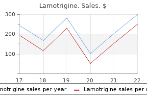
Order 50mg lamotrigine with mastercard
Sleep disturbance often precedes depression medications you cannot eat grapefruit with order lamotrigine online, being an independent risk factor for a first episode or recurrence of depression. The role of anxiety is less clear, but other studies emphasize the relationship between ruminations and an overarousal response. In the updated International Classification of Sleep Disorders there is also an emphasis on persistent sleep difficulties, adequate sleep opportunity, and daytime impairment. Comorbid medical and psychiatric disorders are frequently seen to accompany insomnia along with other sleep disorders. In a prospective 3year study of the natural course of insomnia, 46% of individuals with severe insomnia at baseline experienced persistent insomnia at all time points. Age alone does not cause insomnia if the individual is healthy, but comorbid medical conditions are often precipitating factors. Circadian rhythm disorders, shift work, parasomnias, and inadequate sleep hygiene can all be triggers for insomnia. Individuals are overwhelmingly concerned about sleep onset, return to sleep, and the unpredictability of sleep. Sleep diary monitoring is the most useful form of assessment, with additional questionnaires on beliefs and mood. Difficulty with sleep initiation and/or maintenance problems Despite adequate opportunity to sleep Daytime consequences as per other features Present for at least 3 months Occurring >3 times per week Other features Fatigue/malaise; attention, concentration, or memory impairment; impaired social, family, occupational, or academic performance; mood disturbance/ irritability; daytime sleepiness; behavioral problems. A recent metaanalysis explored 20 studies and found negative effects on verbal and working memory for zopiclone, and attention and speed of processing for zolpidem. Even a single dose of these zdrugs in healthy adults tested the following morning is associated with negative effects on cognitive function. Increasingly (offlabel) sedative antidepressants and more recently antipsychotics such as quetiapine are also being used for their sedating side effects. However, a 2014 review stated that there are no current studies to support the efficacy or safety of these in the treatment of insomnia. Circadin is 604 Part 18 Sleep Disorders a slowrelease melatonin derivative used to treat over 55yearolds with sleep maintenance insomnia, although there is currently only questionnaire data to support its efficacy. Overall management strategies in the treatment of insomnia Factual information sets boundaries and challenges inaccurate sleep attributions. Understanding what sleep is, how sleep changes with age, good sleep hygiene practices (reducing caffeine and alcohol etc. Sleepinitiation insomnia is improved with morning light and avoidance of evening light. Exercise can positively influence sleep quality, particularly in the late afternoon or early evening. Morning exercise with light exposure suppresses melatoninenhancing circadian rhythm and sets a constant waking time. Relearning sleep is about enabling the individual to perceive that the environment is now safe to let sleep happen and includes examination of external factors (heating, noise, violent others) and internal factors relating to previous experiences. Specific behavioral treatments such thoughts as "I shall be useless at work tomorrow," high levels of preoccupation and anxiety are reduced along with inaccurate thinking, thereby aiding sleep onset. Relaxation methods include progressive relaxation, imagery training, biofeedback, meditation, hypnosis, and autogenic training, with little evidence to indicate the superiority of any one approach. Relaxation techniques for reducing insomnia symptoms have an effective track record in lowering the overarousal and wired response. Finally, prevention needs to be extended to known extrinsic causes of certain sleep disorders and to alcohol, stimulants, or proprietary drugs that interfere with sleep. Encouraging individuals to seek advice early rather than selfadministering treatment is critical in reducing longterm complications. Conclusion Stimulus control Stimulus control is a reconditioning treatment forcing discrimination between daytime and sleeping environments. For the poor sleeper, the bedroom triggers associations with being awake and aroused. Treatment involves removing all stimuli that are potentially sleep incompatible (reading and watching television) and excluding sleep from living areas. Chronic insomnia disorder in most clinical situations is both multidimensional and complex. It is characterized predominantly by fatigue and poorquality sleep, where the individual often presents believing that their sleep has "gone away. Further reading Sleep restriction therapy Sleep restriction relates to the ratio of time asleep with time in bed, and involves recording average nightly sleep duration. The aim is slowly to reduce time in bed to match recorded sleep duration, which increases sleep efficiency and confidence. Thought stopping attempts to interrupt the flow of thoughts via "blocking" techniques, such as repeating the word "the" every 3 seconds. This procedure occupies the shortterm memory store used in the processing of information and, by not demanding attention, potentially allows sleep to happen. Cognitive restructuring challenges faulty beliefs that maintain both wakefulness and helplessness. Narcolepsy was first reported by Westphal in 1877 and the term was coined by Gelineau in 1880. Epidemiology the prevalence of narcolepsy with cataplexy in North America and Europe averages 0. Few studies have reported the prevalence of narcolepsy without cataplexy due to the requirement of a sleep study for diagnosis. Input from the limbic system and interaction with metabolic signals such as leptin and glucose allow hypocretin neurons to play a role in emotion, energy homeostasis, reward, addiction, and arousal. Interestingly, patients with hypocretin deficiency are less susceptible to stimulant abuse, suggesting a role for hypocretin in the regulation of drug addiction. This parallels the observation that indirectly stimulating monoaminergic transmission, using amphetaminelike compounds and antidepressants, improves narcolepsy symptoms. The occurrence of narcolepsy involves genetic predisposition and environmental triggers. Multiplex families are rare, but a 10 to 40 fold increase in relative risk is reported in firstdegree relatives. A 2009 pandemic H1N1 influenza virus in China was followed by a higher rate of narcolepsy, suggesting that in genetically susceptible individuals, narcolepsy might be triggered by a similarity between a region of hypocretin and a portion of a protein from the pandemic H1N1 virus or the H1N1 vaccine. This concept, known as "molecular mimicry," could explain that Tcells of the immune system primed to attack H1N1 could also crossreact with hypocretin and potentially cause the destruction of hypocretinproducing neurons. Narcolepsy typically begins in adolescence and early adulthood, although late adult onset or onset in prepubertal children is described in approximately 10% of cases. It frequently culminates with sudden sleep attacks (an overwhelming urge to sleep within minutes). The resulting sleep episode is usually brief, often associated with dreaming, and, in contrast to naps in other sleep disorders, frequently refreshing.
Purchase lamotrigine 200mg
The distribution of "neurogenic" pain (a typical consequence of root compression) is related to the segmental innervation of skin medicine x xtreme pastillas generic lamotrigine 100 mg, muscles, and bones (dermatome, myotome, and the sclerotome, respectively). Patients usually complain of neck pain radiating to the shoulder and upper arm (C5) and into the lower arm and hand into the thumb (C6), or the second, third, and fourth fingers (C7). Root involvement is accompanied by paresthesias and dysesthesias affecting the dermatome (in its distal part). Notably, the deltoid will be paretic in the C5 syndrome, the biceps and brachioradialis in the C6 syndrome, the pectoralis major, the triceps brachii muscle, and the finger extensors in the C7 syndrome, and the intrinsic hand muscles and index finger extensor in the C8 syndrome. C5 radiculopathy weakens the biceps reflex, which may be absent in C6 radiculopathy. Cervical radiculopathy is not accompanied by vegetative symptoms, as autonomic fibers do not leave the spinal cord via cervical roots. An acute posteromedial disc herniation in the cervical region may lead to spinal cord compression, causing myelopathy. This is clinically manifested by more or less pronounced longtract signs with paresthesias in lower limbs, accompanied by a spastic tetra or paraparesis (according to the level of the lesion) with lower urinary tract, anorectal, and sexual dysfunction. Cervical myelopathy due to cervical spondylosis occurs as a slowly progressive disorder, unless it is precipitately unmasked by (minor) neck injury. Lumbar disc disease the most commonly affected individuals are middleaged men, but a functionally significant episode of low back pain may be encountered by up to 80% of the adult population over a lifetime. The typical presentation is with low back pain, which may be accompanied by referred pain. The term "sciatica" is used for pain referred to the lower limb, but this is not necessarily indicative of nerve root involvement. In a minority of patients, there is spinal nerve/radicular involvement with "root pain" and neurological symptoms. As the most often involved nerve roots are L5 or S1, pain in these instances typically radiates down the whole lower extremity to its distal parts. The pain may be excruciating and make the patient more or less immobile, with accompanying paraspinal muscle spasm. Radicular neurological deficit (sensory, motor, and reflex) may be discrete or prominent, and may occur (or be revealed) after the acute pain episode. Local tenderness to palpation or percussion over the spinous processes may be present, and the patient often adopts a fixed posture, somewhat tilted away from the affected side. Pain may be exacerbated by increases in intraabdominal pressure (coughing, sneezing, defecating, etc. A posteromedial protrusion may affect several roots and lead to a cauda equina syndrome. The radiculopathies produce typical sensorymotor syndromes, but individual variability in segmental innervation exists. L4 radiculopathy leads to pain radiating down the distal lateral thigh and the anteromedial leg. The pain in L4 root compression is exacerbated by the patient lying on his or her stomach with the knee flexed and the hip extended. L5 radiculopathy leads to pain radiating to the anterolateral leg and the dorsum of the foot and hallux. Pain in the S1 syndrome radiates down the posterior leg to the heel, and the lateral aspect of the foot to the third to fifth toes. The L4 syndrome may partially weaken the quadriceps muscle, with hyporeflexia of the knee jerk and sensory loss along the anteromedial leg. The L5 syndrome leads to weakness of the ankle and particularly big toe dorsiflexion; walking "on heels" may be affected and weakness of ankle inversion also helps localize an L5 syndrome. The absence of the tibialis posterior reflex is helpful only in individuals with brisk jerks, which will allow the unequivocal demonstration of the contralateral reflex. In S1 radiculopathy walking "on toes" may be affected (due to paretic plantar flexors) and the ankle jerk is absent or very reduced. The sensory loss involves the heel, lateral surface of the foot, and the fifth toe. A cauda equina lesion is recognized by bilateral symptoms (which, however, are as a rule asymmetric). Pain radiates from the lower back to both legs and paresthesias may be bilateral and typically involve the perineal region. Distal lower limb paresis may or may not be present, but ankle jerks are asymmetrically reduced or absent. Urinary retention usually precedes urinary incontinence, which is of the overflow type. Urinary symptoms are the leading autonomic dysfunction in the acute stage, with anorectal and sexual dysfunction revealed in due course. The sensory deficit involves the affected segments and typically the lower sacral segments. The anal reflex is absent unilaterally or bilaterally; in most healthy subjects, pricking the perianal skin with a pin causes visible anal sphincter contraction. In a patient presenting with bilateral neurological symptoms and signs attributable to sacral segments, the question of a cauda equina versus conus medullaris lesion arises. In theory, conus medullaris lesions should demonstrate a combination of upper and lower motor neuron signs, but usually the signs of a lower motor neuron lesion predominate. Cauda equina lesions tend to be more asymmetric, with radicular pain and more pronounced lower limb deficits. Dissociated sensation, when found, distinctly diagnoses a (vascular) conus medullaris lesion. Sacral function (bladder, bowel, and sexual) deficits, and saddle sensory loss, are usually found in both. Cauda equina lesions are more common than those of the conus medullaris, with estimated annual incidence rates of 3. Root lesions of the axonal type probably get little true regeneration of the destroyed axons, but collateral reinnervation of the partially denervated muscle from the remaining axons can result in good functional recovery. Diagnosis the clinical picture of acute neck pain radiating to one limb with neurological deficit should be readily recognizable. The clinical examination defines the neurological deficit and thus the particular root syndrome. Plain radiography only helps to exclude serious disease (malignant, infectious), the readily and expectedly demonstrable degenerative changes being rarely of diagnostic relevance. In patients with isolated pain (even if radiating) but without a neurological syndrome, inflammatory causes and bonedestructive lesions should be ruled out. After the period of acute muscle denervation, reinnervation processes manifest themselves by "remodeling" of motor units. In practice, electrodiagnostics is particularly useful the generally good prognosis of disc disease dictates conservative treatment as the primary approach. Emergency surgery is indicated in acute spinal cord compression and the cauda equina syndrome. Treatment approaches to radiculopathy vary considerably in different centers and countries, and there are no generally agreed guidelines. In many hospitals (delayed) surgery is commonly performed for a variety of indications. A common indication is the situation when the symptoms (with significant pain) do not recede in weeks or months. However, there are no largescale controlled clinical trials demonstrating surgery to be superior to other treatments in the long term.
Syndromes
- Fatigue
- Fluorescein angiography (eyes)
- Brittle nails
- Brain injury
- Not having enough water in your body (dehydration)
- Hole in the bowel (bowel perforation)
- Neurological symptoms such as loss of feeling in the feet or legs, and memory loss
Order discount lamotrigine line
Like other inborn errors of metabolism medicine xifaxan lamotrigine 25mg with visa, urea cycle disorders cannot be distinguished from a variety of other acute conditions of the neonatal period, such as sepsis or perinatal hypoxia. An elevated plasma ammonia level accompanied by relatively normal electrolyte balance is highly suggestive of such a disorder. Epidemiology Treatment Acute episodes occurring in the immediate postnatal period or periodically thereafter are medical emergencies. The first and usually quite reliable method in older patients or those less severely affected is to feed an amino acid mixture lacking the branched chain amino acid precursors of the deficient enzyme step. This should be undertaken in collaboration with a metabolic disease specialist to prevent catabolism and exacerbation of the episode. This regimen promotes protein synthesis and utilizes the accumulated branched chain amino acids to complete the amino acid pool. The second approach to lowering the amino acid levels is dialysis, which effectively removes the excess branched chain amino acids (as well as the others present in normal amounts) from the body fluid space. This must be used in conjunction with proper nutritional support or body protein catabolism will occur and the salutary effect will be lost. Treatment of intercurrent catabolic episodes or of those patients presenting later in life is simpler. These events can often be managed with fluid and electrolyte support and administration of non branched chain amino acids. As with the treatment of all inborn errors, attention to adequate caloric intake, prevention of constipation, and special intervention during intercurrent illness are required. With transplantation, patients are able to eat a normal diet and do not have acute episodes of metabolic decompensation. Urea cycle disorders the urea cycle is an eightstep cycle consisting of six enzymes operating sequentially and two transporters, all of which are essential for the conversion of ammonia generated from either endogenous or exogenous amino acids to urea. The two transporters are both in the mitochondrial membrane, one transporting ornithine and the second transporting aspartate into the mitochondrion. With the possible exception of deficiency of the aspartate transporter (citrin deficiency), which may be more prominent in people of Japanese ethnicity, none of the disorders has any particular ethnic or geographic predilection. Clinical features Newborn patients present as a catastrophic encephalopathy, with lethargy, poor feeding, a weak cry and suck, hypotonia, coma, and death, if successful treatment is not instituted. Those with partial deficiencies may exhibit learning disabilities, hyperactivity, and psychiatric disorders. Pathophysiology and treatment Severe neonatal hyperammonemia constitutes a medical emergency and must be addressed immediately. The levels of ammonia and glutamine (with which it is in approximate equilibrium) must be reduced as rapidly as possible and prior to any determination of the precise site of the block. The most effective means of carrying this out is hemodialysis, which is more effective than peritoneal dialysis and certainly more effective than exchange transfusion, which has little utility or efficacy. Classic proximal urea cycle defects Anorexia Vomiting Cognitive and motor deficits Lethargy Ataxia Asterixis Brain edema Cytotoxic and vasogenic edema Hypothermia Seizures Coma Partial enzyme deficiencies Protein aversion Hyperactive behavior Selfinjurious behavior Strokelike episodes Psychiatric symptoms 664 Part 20 Pediatric Neurology hyperammonemia should be dialysis. In some larger centers, intravenous sodium benzoate combined with sodium phenylacetate is available and may be used to divert the ammonia from the urea cycle, to be excreted as benzoylglycine and/or phenylacetylglutamine. The resynthesis of glycine and glutamine in the body will then utilize one or two molecules of ammonia destined for the urea cycle to this synthetic reaction. Nutritional support with the guidance of a metabolic specialist can prevent the development of a catabolic state during treatment. Dialysis is a very effective method for reducing plasma ammonia and glutamine and the failure to do so, if it is being carried out effectively, is usually due to the ongoing catabolism in the body and the production of ammonia. When ammonia comes down to a level of 200 M, it then becomes imperative to add an essential amino acid mixture to the high calories being provided intravenously by glucose and lipids in order to inhibit endogenous catabolism and allow the body to begin to recover in a relatively normal manner, and to reestablish a balance in protein synthesis and breakdown. Hyperammonemia is the cause of the brain damage, possibly acting through glutamine, with which it is in equilibrium. The most obvious and visible effect of ammonia intoxication is cerebral edema, although a number of other pathological mechanisms may be operating simultaneously. The degree of neurological damage correlates best with the length of time the patient spends in a coma, and to a much lesser degree with the level of ammonia. Few patients escape severe neonatal hyperammonemia without permanent neurological injury. At this stage, specific therapy for each disorder generally does not occur, although the severity of argininosuccinic aciduria may be mitigated by high arginine infusion (600 mg/kg/ day in an infant). Deficiency in arginine biosynthesis is characteristic of seven of the eight urea cycle disorders and a lesser amount of arginine is used empirically in acute hyperammonemia of unknown etiology. Chronic treatment consists of adequate calories and fluids, a diet low in natural protein and supplemented with an essential amino acid formula and sodium phenylbutyrate. In developing countries phenylbutyrate may prove to be too expensive or otherwise unavailable, but sodium benzoate, which is readily available and inexpensive, may be a suitable, albeit less effective substitute. Liver transplantation is a definitive treatment for the hyperammonemia associated with the more acuteonset hyperammonemias and the outcome in the large majority of patients is favorable; the prior brain damage will, of course, be irreversible. Newborn screening is particularly useful in detecting milder cases of argininosuccinate synthetase and lyase and arginase deficiencies, but so far has proven less reliable in detecting disorders whose enzyme deficiencies occur earlier in the cycle. Propionic and methylmalonic acidemia these two disorders involve sequential steps in the disposition of the carbon skeletons of four amino acids, threonine, methionine, isoleucine, and valine. Defects in either propionylCoA carboxylase or methylmalonylCoA mutase cause the propionic acid or methylmalonic acid to accumulate behind the site of the block. Because propionic acid is a precursor of methylmalonic acid, it accumulates as well in methylmalonic acidemia. These metabolites are toxic to the brain, heart, and bone marrow and can cause severe illness and death in their most severe form. This B12 gets to the site of action only after having undergone a series of meta- bolic steps. The most common of these defects is referred to as cobalamin C deficiency and is characterized by elevated body fluid levels of both methylmalonic acid and homocysteine. Remethylation of homocysteine to methionine also requires an activated form of vitamin B12 and shares many metabolic steps with the activated form, which serves as a cofactor for the mutase reaction. Epidemiology Like any autosomal recessive disorder, forms of propionic and methylmalonic acidemia may be found in increased frequency in population isolates and/or in populations that practice cousin marriage as a social custom. Clinical features the largest number of patients with both disorders have severe enzyme deficiencies present in the newborn period. Initial symptoms include decreased suck, poor feeding, irritability, lethargy, hypotonia, and seizures, which progress to stupor and coma. In the past when these conditions were poorly recognized, death ensued in most instances. Other manifestations include bone marrow depression, acidosis, hyperammonemia, hypotonia, and cardiac failure. The signs and symptoms may mimic neonatal sepsis and the neonatologist should be alert to the possibility of conditions like this, particularly in suspected cases of sepsis that are atypical in some manifestation(s). In these jurisdictions, the level of suspicion in patients who have had a successful newborn screen will be altered and the position of these disorders on the list of diagnostic possibilities will be lowered. Laboratory abnormalities found in these disorders include metabolic acidosis with an increase anion gap, elevated blood lactate, ketonuria, hyperammonemia, and bone marrow depression. Ketonuria in a newborn is exceedingly rare and is a telltale sign of a disorder of organic acid metabolism. Pathophysiology the pathophysiology of these conditions is not known with certainty. The resemblance between propionylCoA and acetylCoA is great, and propionylCoA is thought to compete with acetylCoA for the active site of enzymes using the latter substrate, particularly the biosynthesis of acetylglutamate and activation of the urea cycle. Treatment In the initial phases of diagnosis, the two disorders may be indistinguishable until the results of newborn screening or diagnostic urinary organic acid analysis becomes available. The treatment will, therefore, be generic, seeking to reduce the level of accumulated toxic metabolite and simultaneously diminishing the production of the offending compound. The most effective way of lowering the levels of either of these readily excreted organic acids, which could accumulate rapidly in the period of catabolism following birth, is by dialysis. The most effective means of dialysis is hemodialysis, but when this is not available peritoneal dialysis is an acceptable alternative. Treatment should include vitamin B12, 1000 mg intramuscularly daily, until Chapter 166 Disorders of amino acid, organic acid, and ammonia metabolism 665 it is demonstrated that the patient does not have a vitamin B12responsive form of methylmalonic acidemia. The usual supportive measures of maintenance of electrolyte balance and hydration are important in these patients and in general follow nursery protocol. Infants rescued from the most acute manifestations of these disorders often will have suffered irreversible neurological damage and may over a period of years show mental retardation, growth delay, and a variety of neurological disabilities.
Buy generic lamotrigine
If the patient is intolerant of one statin xerostomia medications side effects purchase lamotrigine 100 mg fast delivery, it is appropriate to consider trying others, using lower doses, and occasionally alternateday therapy. It is almost always associated with a recent increase in dose or introduction of other drugs that impair statin metabolism. The most widely accepted theory is that inhibition of cholesterol biosynthesis causes a depletion of downstream intermediaries, leading to disruption of lipidrich cellular membranes and reduction of mitochondrial respiratory chain function. In addition, the loss of isoprenaline intermediaries impairs multiple cellular functions such as protein synthesis. The statininduced problems noted here have a presumed metabolic origin, and resolve on drug withdrawal. In 2007, a necrotizing myopathy was described associated with statin use, which persisted despite statin withdrawal but responded to immunosuppression. However, despite successful treatment, antibody levels remain elevated, suggesting that they are not directly pathogenic. Corticosteroid myopathy Acute and chronic myopathies are associated with short and longterm corticosteroid use, respectively. It is typically associated with highdose intravenous steroids, often combined with a neuromuscular blocking drug, but has been reported with lowerdose oral steroids or after an increase in the dose of maintenance steroids. Chronic steroid myopathy typically presents with painless proximal weakness affecting the pelvic girdle more than the shoulders. Patients have a limbgirdle pattern of weakness accompanied by respiratory muscle weakness and sometimes facial weakness. Characteristic clinical features are hypoxemia, hypercapnia, muscle rigidity, hyperpyrexia, metabolic acidosis, and rhabdomyolysis. Management, including the prompt administration of dantrolene, a ryanodine receptor antagonist, and supportive care, has significantly reduced mortality. Rarity of anti3hydroxy3methylglutaryl coenzyme A reductase antibodies in statin users, including those with selflimited musculoskeletal side effects. Malignant hyperthermia in Canada: Characteristics of index anesthetics in 129 malignant hyperthermia susceptible probands. Further reading 125 Critical illness neuromuscular disorders Nicola Latronico and Frank A. Limb and respiratory muscle involvement may cause limb weakness and paralysis, neuromuscular respiratory failure, prolonged dependency on mechanical ventilation, and longterm physical impairment. Difficulty in swallowing and ineffective cough may combine to cause pneumonia and lung atelectasis, which in turn may precipitate lifethreatening acute respiratory deterioration. Immobility, an expected complication of generalized paralysis, contributes to muscle wasting and weakness. Weakness may result from pathological processes involving the motor neuron with its axon and myelin sheath, the neuromuscular transmission, and the skeletal muscle itself. With persisting deficit, the energy supply and/or use is not restored and histological alterations ensue. Disturbed microcirculation and acquired sodium channelopathy with hypoexcitability or inexcitability of nerves and muscles are other potentially relevant mechanisms. In patients with septic shock or severe sepsis and coma, the incidence approaches 100%. Limb involvement is diffuse and symmetric, muscle tone is reduced, and sensation is impaired. Failed weaning from mechanical ventilation is a common feature, indicating weakness of the respiratory muscles. Electrophysiological changes of peripheral nerves and muscles may have a rapid onset within hours of normal action potential generation. Nerve histology is normal and muscle histology shows minimal changes, with loss of thick filaments in biopsies taken at an early stage of disease. Late biopsies demonstrate muscle fiber necrosis and atrophy, and nerve axonal degeneration. If a trial of spontaneous breathing is attempted, the patient becomes rapidly distressed and dyspneic, with rapid shallow breathing. Weakness of the pharyngeal and laryngeal muscles and of the expiratory muscles of the chest wall and abdomen leads to altered swallowing, impaired cough, inadequate clearance of secretions, and increased risk of pulmonary aspiration and pneumonia. Limb weakness can be severe enough to cause flaccid tetraparesis or tetraplegia, which in turn causes immobility and disuse muscle atrophy. Electrophysiological investigations are essential not only to define the cause, but also to help in outcome prediction. In the acute setting, simplified tests with peroneal nerve stimulation can be used to screen atrisk patients. Infection has subsided by the time neurological signs such as pain, paresthesia, numbness, and weakness in the limbs become evident. Serial electrophysiological investigations are needed to achieve the diagnosis and in gauging response to treatment. Propofol at high doses and for prolonged periods may rarely cause a fatal syndrome of cardiac failure, metabolic acidosis, hyperkalemia, hypertriglyceridemia, renal failure, and rhabdomyolysis. With the fully developed syndrome the diagnosis is easy, but incomplete forms can develop with mild early increase in serum creatine kinase; in these cases, prompt recognition is important to stop the propofol infusion and to avoid conversion into the overt syndrome with acute necrotizing myopathy. Electrical muscle stimulation and corticosteroids have no beneficial effect, with the latter possibly increasing the risk of longterm functional disability. Future studies of appropriate sample size should evaluate the impact of early rehabilitation on longterm measures of physical and mental performance in survivors of critical illness. An official American Thoracic Society Clinical Practice guideline: the diagnosis of intensive care unitacquired weakness in adults. Critical illness polyneuropathy and myopathy: A major cause of muscle weakness and paralysis. Exercise intolerance is caused by numerous conditions, many of which are nonneuromuscular, such as arthritis, heart or respiratory failure, recovery from longstanding disease, postinfectious conditions, bone deformities, and many others. Muscle pain, which emerges as exercise is undertaken, can be a reason for exercise intolerance, but chronic syndromes, with pain at rest that limits the initiation of exercise, are typically not categorized as exercise intolerance syndromes. Since exercise intolerance has many causes, this complaint is very common in the general population. It highlights the importance of being able to distinguish exercise intolerance caused by neuromuscular diseases from the very common symptoms of myalgia and fatigue in people without muscle disease. In many neuromuscular diseases in which there is muscle wasting, exercise intolerance is obvious from the disabling loss of muscle mass, and in such cases the term exercise intolerance is rarely used. The term is generally reserved for conditions in which low work capacity is disproportionate to the muscle mass of the person. Exercise intolerance is considered a hallmark of inborn errors of muscle metabolism, such as metabolic and mitochondrial myopathies, where it is caused by a mismatch between energy demand and supply. However, a number of nonmetabolic myopathies, preferentially muscular dystrophies, may also present with a picture characteristic of metabolic myopathies. Thus, exerciserelated muscle pain and even myoglobinuria are not rare in certain types of limbgirdle muscular dystrophies (types 2I and 2L) and dystrophinopathies. Exerciserelated pain is also commonly found in other muscle diseases such as facioscapulohumeral muscular dystrophy, channelopathies, and myopathy due to thyroid gland dysfunction. In channelopathies, such as myotonia congenita and paramyotonia congenita, the cause of the pain likely relates to the myotonia. In facioscapulohumeral muscular dystrophy and myopathy due to thyroid gland dysfunction, muscle wasting or pain usually explains the exercise intolerance. Symptoms evoked by such exercise therefore can suggest an underlying glycogenosis. Painful contractures are typically coupled with the exercise intolerance in these conditions. During prolonged, lowintensity exercise, muscle depends primarily on energy generated from oxidation of fatty acids. Symptoms provoked by such exercise therefore point toward a disorder of muscle lipid metabolism, and these patients do not develop contractures as in glycogenoses, but may experience tightness/stiffness of muscles that have been exercised. In contrast, patients with mitochondrial disease rarely experience contractures, stiffness, or muscle pain, but typically complain about being out of breath, which explains why these patients often have been seen by cardiologists and pulmonologists before referral to neuromuscular centers.
Order lamotrigine 50 mg line
There may be dissolution of the tympanic membrane with invasion of the middle ear treatment kidney cancer order lamotrigine paypal. Occasionally, the well-differentiated lesions may not be detected clinically until well advanced. Histopathology Epidermoid carcinoma of the external ear usually shows significant degrees of keratinization. Those showing a spindle cell morphology must be differentiated from melanomas and soft tissue tumours. In the cases with a canal origin evidence of origin from canal epidermis is usually present. In cases arising deeply within the ear canal there is usually a concomitant origin from middle ear epithelium and dissolution of the tympanic membrane. The association of such a neoplasm with marked desmoplasia may further delay the correct diagnosis. Prognosis and predictive factors Squamous cell carcinoma of the pinna is an aggressive disease with a high propensity for local recurrence. Tumours confined to the external ear usually have a good outlook after surgical therapy. The outcome of the disease following surgical excision is related to the clinical stage at presentation, the higher the stage the worse the outcome 1915. Metastatic spread of squamous carcinoma of the pinna and external auditory meatus to lymph nodes is unusual. Lesions arising in the canal have a worse prognosis because of the late diagnosis and invasion of adjacent structures. This section focuses on its occurrence as a primary tumour in the external ear canal. Definition A primitive malignant tumour with phenotypic and biological features of embryonic skeletal muscle. Advanced cases may present with aural discharge, facial weakness and swelling in the region of the ear 1116. Extensive destruction of the bone at the base of the skull, especially the petrous bone has been described. Histopathology Only the embryonal subtype of rhabdomyosarcoma is recognized as occurring at this site. The characteristics of this polypoid tumour are those of rhabdomyoblasts and primitive mesenchymal cells showing a variable degree of skeletal muscle differentiation loosely arranged but with condensation beneath the epithelium (cambium layer). Yolk sac tumour has been described as a polypoid tumour presenting in the external ear canal. However, this is histologically distinct, being composed of small round blue cells arranged in a vacuolated pattern with formation of Schiller-Duval bodies and expressing alpha fetoprotein 833. Histogenesis Although it is suggested that this tumour arises from striated muscle fibres in the middle ear, it seems more likely that the origin is from undifferentiated mesenchymal cells. Genetics Mutations in a region mapped to the short arm of chromosome 11 (11p15) have been associated with most embryonal rhabomyosarcomas. Complex structural and numerical chromosomal rearrangements have been associated with embryonal rhabdomyosarcoma. Prognosis and predictive factors Modern chemotherapeutic schedules have dramatically improved the outcome for children with this tumour. There is a distinct group arising in the head and neck of children, often very young, with a predilection for the palate, middle ear and orbit. Localization Most of the tumours arise in the middle ear with extension into the external canal as an "aural polyp". A A central area of necrosis is surrounded by "primitive cells" with a very high nuclear to cytoplasmic ratio. B this polypoid tumour has a "Grenz-Zone" between the neoplastic cells and the mucosal surface. The monostotic form affects both sexes equally; the polyostotic form is more common in females by a 3:1 ratio. The most recent attempts to define the disorder have focused on genetics and molecular biology. In the head and neck the skull and facial bones are affected in 10-20% of cases of monostotic disease and 50% of polyostotic cases. Other unusual sites include the internal auditory canal, the lateral semi-circular canal and the ossicles. In a retrospective analysis of patients with fibrous dysplasia affecting the skull base, Lustig et al found the temporal bone to be affected in 24%. Clinical features the main clinical features of disease affecting the temporal bone are: (i) progressive loss of hearing, mostly conductive but which can be sensorineural and profound in some cases, (ii) temporal bone enlargement with progressive bony occlusion of the external auditory meatus, (iii) facial nerve palsy in some patients when the process affects the seventh cranial nerve, (iv) constriction of the ear canal may result in development of an epidermoid cyst lateral to the tympanic membrane likened to cholesteatoma by Megerian et al 1698. Macroscopy the affected bone is often expanded and the marrow is replaced by firm grey/tan tissue depending on the proportion of bony, fibrous and cartilaginous elements. Histopathology the lesion consists of irregular trabeculae of woven bone arising abruptly from a bland spindle cell stroma. The trabeculae may be curved and shaped like letters in the Chinese ideogram and are devoid of a rim of osteoblasts. Secondary changes include osteoclast giant cells, foamy histiocytes and aneurysmal bone cyst formation. Prognosis and predictive factors Fibrous dysplasia has rarely been associated with malignant transformation including osteogenic sarcoma, fibrosarcoma and chondrosarcoma, but the temporal bone is not one of the sites where this change has been described. Beale Definition Benign bony enlargement of the deeper portion of the external auditory meatus. Therefore the distance between the epidermal surface and underlying bone is small, which may explain the propensity for exostoses of the tympanic bone to develop in those who swim frequently in cold water 2121. Osteoma and exostosis are often associated with infection of the external canal on the tympanic membrane side. Surgical removal may be required to enhance drainage as well as to relieve the conducting hearing loss. Histopathology the osteoma is a spherical, pedunculated lesion composed of cortical lamellar bone on the outside overlying trabecular bone with intervening marrow spaces. Both these lesions are distinct from the recently described benign fibro-osseous lesion of the superficial external canal 2121. Prognosis and predictive factors these are benign lesions with no potential for malignant transformation. Exostoses have also been observed in individuals who routinely use stethoscopes, eg cardiologists 550. Localization Osteoma is a very rare lesion, which is a single, unilateral, spherical mass on a distinct pedicle arising in the region of the tympanosquamous or tympanomastoid suture line. It has only occasionally been described outside the external auditory canal and the middle ear, developing in the mastoids, temporal bone internal auditory canal, glenoid fossa eustachian tube, petrous apex and styloid process. Note thin epidermal layer on the exostosis above and on the canal skin below and their proximity to the bone. In deeper sections, the exostosis merges gradually with the deep canal bone without pedunculation. Exostoses are common, broad-based lesions, often bilateral and symmetrical which are usually situated deeper in the ear canal than osteomas. In the bony portion of the normal external auditory meatus there are no adnexal structures and subcutaneous tissue and perios- A B. Henry Definition A benign vascular tumour with well formed, but immature, blood vessels, the majority of which are lined by plump, epithelioid (histiocytoid) endothelial cells. Most cases have a prominent inflammatory component in which eosinophils are a conspicuous feature. Epidemiology There is a wide age range with a peak in the third to fifth decades and women are affected more often than men 759, 1945. Etiology Whether angiolymphoid hyperplasia with eosinophilia is a reactive lesion rather than a neoplasm is still debated. Features cited as supporting a reactive process include a history of trauma (10 % of cases), its relationship around a larger vessel showing evidence of damage and the prominent inflammatory component 759,1945. Localization the lesion occurs most frequently on the head, particularly the forehead and scalp (often in the distribution of the superficial temporal artery) and in the skin of the ear and the peri-auricular area.
Order lamotrigine master card
Molecular genetics Molecular studies of these tumours are few and limited in number of cases medications drugs prescription drugs discount 50mg lamotrigine with amex. They show infrequent genetic loss at chromosomes 9p21, 8q, 5p, 16q and 12p 351, 1228,2408. Studies of the H-ras gene in these tumours have reported 18% mutations at codon 12 and/or 13 (one case) and no mutations at codon 61 2858. In one study, 8% of patients died of disease: 11% and 5% for major and minor gland tumours, respectively. Death correlated with high-grade histopatho- logic features in minor gland and parotid gland tumours, but not in patients with submandibular gland tumours 921. Death resulted from unresectable locoregional tumour, distant metastases or complications of adjunctive therapy 2609. Huvos Definition Adenoid cystic carcinoma is a basaloid tumour consisting of epithelial and myoepithelial cells in variable morphologic configurations, including tubular, cribriform and solid patterns. Clinical features the most common symptom is a slow growing mass followed by pain due to the propensity of these tumours for perineural invasion. Histopathology Tumours consist of two main cell types: ductal and modified myoepithelial cells that typically have hyperchromatic, angular nuclei and frequently clear cytoplasm. In the tubular form, well-formed ducts and tubules with central lumina are lined by inner epithelial and outer myoepithelial cells. They comprise 30% of epithelial minor salivary gland tumours with the highest frequency in the palate, followed by the tongue, buccal mucosa, lip and floor of mouth. The tumour occurs in all age groups with a high frequency in middle-aged and older patients. There is no apparent sex predilection except for a high incidence in women with submandibular tumours the cribriform pattern, the most frequent, is characterized by nests of cells with cylindromatous microcystic spaces. The solid or basaloid type is formed of sheets of uniform basaloid cells lacking tubular or microcystic formation. In the cribriform and solid variants small true ducts are invariably present but may not be immediately apparent. Each of these forms can be observed as the dominant component or more commonly as a part of a composite tumour 161,1663,1849,2016,2444, 2519. The stroma within the tumour is generally hyalinized and may manifest mucinous or myxoid features. In some tumours there is extensive stromal hyalinization with attenuation of the epithelial component. Tumours can extend along nerves for a considerable distance beyond the clinically apparent boundaries of the tumour. A Tubular form, composed of inner epithelial ductal and outer myoepithelial cells. Adenoid cystic carcinoma occasionally occurs with other different neoplasms (hybrid tumours) 505,1823,2297,2416. Pleomorphic carcinomas and sarcomatoid transformation of adenoid cystic carcinoma have been reported, mostly in recurrent and metastatic disease 397, 418. The t(6;9) (q21-24;p13-23) has been reported in several tumours and is considered to be a primary event in at least a subset of these tumours 657, 1220,1906,2238. Immunohistochemistry In differentiating between polymorphous low-grade adenocarcinoma and adenoid cystic carcinoma, Ki-67 immunostaining may be helpful 2680. Ki-67 and p53 have also been studied in these tumours 2844, but no clear association with outcome have been reported. Estrogen and progesterone receptor positivity has been reported in adenoid cystic carcinoma but the biological significance is currently unknown. Differential diagnosis Pleomorphic adenoma, polymorphous low-grade adenocarcinoma, epithelialmyoepithelial carcinoma, basal cell adenoma or adenocarcinoma and basaloid squamous carcinomas are the major entities to be differentiated from adenoid cystic carcinoma. Genetics Cytogenetics the most consistent, although not exclu- Molecular genetics Frequent losses at 12q (33%) 6q23-qter, 13q21-q22 and 19q regions (40%) have been reported 657. A recent genomic study identified new markers that may be helpful in future investigation of these tumours. Alterations of the p53 and Rb genes have been reported but no alterations in the K-ras have been found 2843. Prognosis and predictive factors Factors that influence survival include histologic patterns, tumour site, clinical stage, bone involvement and status of surgical margins 1849,2016,2439,2444, 2519. Generally, tumours composed of tubular and cribriform patterns pursue a less aggressive course than those with greater than 30% of solid component 2519. Other studies have failed to confirm the value of grading 2439,2444 and underscored the significance of tumour size and clinical stage as the most consistent predictors of clinical outcome in patients with these tumours 2442,2449. The 5year survival rate is approximately 35% but the long-term survival is poorer. Lymph node involvement is uncommon but has been reported to range from 5-25% and typically from tumours of the submandibular gland and is often due to contiguous spread rather than metastasis. Wide local and radical surgical excisions with and without post-operative radiation is the treatment of choice 54,339, 1849,2439,2444,2519. Radiation alone or with chemotherapy in the treatment of recurrent or metastatic disease has shown limited success. Radiotherapy, however, has been shown to improve local control in cases with microscopic residual disease 2670. Wenig Definition A malignant epithelial tumour characterized by cytologic uniformity, morphologic diversity, an infiltrative growth pattern, and low metastatic potential. Other intraoral locations are the buccal mucosa, retromolar region, upper lip, and the base of the tongue 342,697. Uncommon locations include major salivary and lacrimal glands, nasopharynx and nasal cavity 1299,2763. Bleeding, telangiectasia, or ulceration of the overlying mucosa occurs occasionally. The tumour cells are small to medium size and uniform in shape with bland, minimally hyperchromatic, oval nuclei and only occasional nucleoli. The striking feature of these carcinomas is the variety of morphologic configurations between tumours and within an individual tumour. The main microscopic patterns are: 1) lobular, 2) papillary or papillary-cystic (typically focal), 3) cribriform areas sometimes resembling those in adenoid cystic carcinoma, and 4) trabecular or small, duct-like structures lined by a single layer of cuboidal cells. The cells form concentric whorls or targetoid arrangements around blood vessels or nerves. Despite the innocuous cytologic appearance, the neoplasm always invades adjacent soft tissues and is uncapsulated. However, a higher proliferative rate (average 7%) has been reported by others investigators 2011. A total of 7 cases of which two were carcinoma ex-pleomorphic adenoma, have been reported. Alterations at 8q12 were found in two, 12q rearrangements in five, two showed a clonal t(6:9) (p21;p22) and one a monosomy 22 1651. A review of series with large numbers of cases and with long-term follow-up revealed a local recurrence rate between 9% and 17% and a regional metastases rate from 9-15% 342,697. Deaths attributed to tumour are unusual, and they occurred after prolonged periods 342,697. In studies which accepted tumours with a predominant papillary configuration a higher incidence of cervical lymph node metastasis was reported 697. So far described only in one series, all cases presented with a mass in the tongue, usually the posterior part, and synchronous metastases in lateral neck lymph nodes, but no distant spread. A characteristic feature is that some nearly solid islands have a glomeruloid arrangement of broad microfollicular papillae separated from a layer of peripheral columnar cells by a narrow cleft. Small numbers of tubules are seen, and occasional spindling of tumour cells may occur. The nuclei are uniform, pale and often overlap, closely mimicking those of papillary carcinoma of the thyroid. Myoepithelial markers, such as actin are either negative or only focally positive. Soares Definition A malignant tumour composed of variable proportions of two cell types, which typically form duct-like structures. The biphasic morphology is represented by an inner layer of duct lining, epithelialtype cells and an outer layer of clear, myoepithelial-type cells. Tumours arising in minor glands frequently present as ulcerated, submucosal nodules and have less well-defined margins. Rapid growth, facial nerve palsy and/or associated pain are suggestive of concomitant high-grade areas.
Whorlywort (Black Root). Lamotrigine.
- Are there any interactions with medications?
- Constipation, liver and gallbladder problems, causing vomiting, and other conditions.
- How does Black Root work?
- What is Black Root?
- Are there safety concerns?
- Dosing considerations for Black Root.
Source: http://www.rxlist.com/script/main/art.asp?articlekey=96774
Buy lamotrigine on line
Islands or strands of inactive-looking odontogenic epithelium are an integral component; they may be sparse but are often conspicuous medications used for bipolar disorder order 25 mg lamotrigine free shipping. This type shows foci of calcified material considered to be metaplastically produced dysplastic cementum/osteoid/ dentin. Synonyms and historical annotation Controversy exists as to concept and definition 628. Epidemiology Due to lack of uniform definition, data on relative frequency are wide-ranged and inconsistent. When considering the epithelium-rich type, the age range of 15 reported cases was 11-66 years with a mean of 40 years, with a female predominance of 2. From the above-mentioned source of epithelium-rich cases, more were located in the mandible giving a maxilla: mandible ratio of 1:6. A Periapical radiograph with a well outlined osteolysis with internal trabeculation. B Collagenous cell rich stroma with numerous strands of odontogenic epithelium without palisading of peripheral cells. When a relatively greater amount of collagen is evident, the term myxofibroma may be used. The borders of the tumour are usually well-defined and corticated but can be poorly defined or diffuse. Macroscopy Gross examination reveals a grey-white mass with a typical translucent mucinous appearance. The consistency varies from gelatinous to firm, depending on the amount of collagen present and fine white bands of collagen may be visible on the cut surface. Maxillary lesions tend to obliterate the maxillary sinuses as an early feature 1245. The high proton density resulting from water bound to the myxoid stroma gives the lesion a hyperintense signal in T2 weighted magnetic resonance imaging (right). The smooth periphery (left) and a few epithelial cells of the reduced enamel epithelium (right) exclude myxoma histologically. Rests of odontogenic epithelium are not obvious in most lesions and are not required for establishing final diagnosis. Histochemical studies show that the ground substance is rich in acid mucopolysaccharides, primarily hyaluronic acid and, to a lesser degree, chondroitin sulphate. Misdiagnosis of these entities should be avoided by correlation with the clinical and radiographic features 121,1313,2489. The microscopic differential diagnosis should also include myxoid nerve sheath tumours, chondromyxoid fibroma, low-grade myxoid fibrosarcoma and other myxoid sarcomas. Somatic genetics A study of 23 cases has shown that odontogenic myxomas are not associated with activating mutations of the Gs alpha gene 239. Odontogenic myxoma has been reported in a single case of tuberous sclerosis 1019 but is not otherwise associated with Carney complex or any known genetic lesion. Small lesions have been successfully treated in this way but larger lesions may require complete excision with free margins. Recurrence rates from various studies average about 25% but in spite of this, the prognosis is good. Recurrence usually follows incomplete removal within two years but may occur much later. The age range is from 8 up to 44 years, the mean being approximately 20 years 262. Localization the majority of cementoblastomas are located in the mandible, particularly related to the permanent first molar; association with a primary tooth is exceptional. Clinical features / Imaging the most common finding is a painful swelling at the buccal and lingual/palatal aspect of the alveolar ridges. Lower-lip paresthesia or a pathologic fracture of the mandible are rarely reported 262. Radiographically, the tumour is welldefined and is mainly of a radiopaque or mixed-density, surrounded by a thin radiolucent zone. Root resorption, loss of root outline and obliteration of the periodontal ligament space are common findings. Note the rim of connective tissue at the bottom corresponds to the peripheral radiolucent zone in. Macroscopy the tumour consists of a rounded or nodular mass attached to one or more tooth roots and is surrounded by a greyto-tan layer of irregular soft tissue 262. Histopathology A cementoblastoma consists of dense masses of acellular cementum-like material in a fibrous, sometimes rather vascular stroma that may contain multinucleated cells. The tumour mass blends with the root of a tooth with simultaneous root resorption. In the more mature parts of the tumour, basophilic reversal lines may produce a Paget disease-like pattern. At the periphery sheets of unmineralized tis- sue may be seen, often being arranged in radiating columns. The differential diagnosis includes osteoblastoma, the only distinctive criterion being the true connection with the surface of the root of a tooth in case of a cementoblastoma 2400. Prognosis and predictive factors In case of incomplete removal, together with the associated tooth, recurrence is common 262. The root surface is largely destroyed by the cementum-like tissue, while the pulpal tissue of the tooth still remained vital. A the bony trabeculae may exhibit irregular mineralization and may form an anastomosing lattice. B At higher magnification, the gradual transition between bone and fibroblastic background tissue is clearly displayed. Radiographs show a demarcated lesion that may have radiodense as well as radiolucent areas depending on the various contributions of soft and hard tissue components 261. The mineralized component may consist of woven bone, lamellar bone and acellular to poorly cellular basophilic and smoothly contoured deposits thought to be cementum. Due to the presence of this cementum-like material, ossifying fibromas have also been called cemento-ossifying fibroma. However, cementum is defined as a mineralized material covering the surface of the roots of the teeth and outside this location, its distinction from bone is equivocal and without clinical relevance 261,2401. Clinical presentation and radiographic appearance may be decisive (see osseous dysplasia). Additional but less typical features are multinucleated giant cells, pseudocystic stromal degeneration, and haemorrhages 644,2051, 2406. The stroma varies from being loose and fibroblastic to intensely cellular with minimal intervening collagen. The mineralized material consists of spherical or curved ossicles that are acellular or show sparsely distributed cells. Other features such as trabeculae of woven bone as well as lamellar bone, pseudocystic stromal degeneration and haemorrhages result in areas similar to an aneurysmal bone cyst. Multinucleate giant cells, and mitotic figures may be present, but are not specific for this variant of ossifying fibroma. These may contain irregular deposits of collagenous material in which threadlike calcifications are present. The long tubular bones, especially the femur, followed by the flat bones of the jaws, the skull (base of skull prior to neurocranium), and the ribs, are the most frequently affected sites in the skeleton 2067. Maxillary and mandibular involvement may lead to displacement of teeth, malocclusion and, rarely, root resorption 1759. Lesions extending to the orbit may cause visual impairment, while temporal bone lesions may produce hearing loss. Polyostotic fibrous dysplasia (mandible, base of skull) with extreme deformity of the mandible. Radiograph showing expansile osteolysis with irregular opacities of the mandible, extending into the ascending ramus up to the mandibular condyle.
Purchase 25 mg lamotrigine
They have been reported in adenoid cystic carcinomas in some studies 1214 treatment definition statistics cheap lamotrigine 25 mg without a prescription,1965 but in others they were absent, or present in only a few tumours 616,1214. Androgen receptors are present in over 90% of salivary duct carcinomas 711, 712,1265. A recent study showed immunoreactivity for androgen receptors in all their cases of salivary duct carcinoma, carcinoma ex pleomorphic adenoma and basal cell adenocarcinoma 1851. There was also staining for the receptors patients in poor health who are not good operative candidates, (4) patients with metastasis to a salivary gland or adjacent lymph node, (5) some examples of lymphoproliferative disease 763 or (6) in a primary soft tissue or skin appendage lesion arising in the area of a major salivary gland. The rate of correctly establishing a diagnosis as benign or malignant ranges from 81-98% in most recent reports. However, a specific diagnosis can only be made in approximately 60-75% of cases 668. False negative diagnoses due to inadequate sampling appear to be the most frequent error. Frozen section examination When considering all head and neck sites, the accuracy of frozen section diagnoses of the salivary gland is the most controversial. A review of 2460 frozen sections from 24 series revealed an overall accuracy rate for a benign or malignant diagnosis, excluding deferred diagnoses, of 96% 379,900,1697,2170, 2900. If one subdivides the salivary gland lesions into benign and malignant groups, the accuracy rate (98. This was frequently called mucoepidermoid carcinoma or adenoid cystic carcinoma 904. Mucoepidermoid carcinoma is the malignancy most frequently associated with a false negative benign frozen section diagnosis, while acinic cell carcinoma, adenoid cystic carcinoma, carcinoma ex pleomorphic adenoma and an occasional lymphoma have also caused difficulty. Recent changes in the staging system include a revision in the definition of T3 and the division of T4 into tumours that are resectable (T4a) and unresectable (T4b) 947,2418. Spiro and coworkers have successfully applied the criteria used for squamous cell carcinoma of the oral cavity, pharynx, larynx, and sinus to mucoepidermoid carcinoma 2305,2863. Genetics the goal of the molecular biological studies of salivary gland tumours is to define objective markers that may supplant the subjective phenotypic evaluation in the diagnosis, biological assessment and therapeutic stratification of patients with these tumours. Upon binding to its ligand, a signalling cascade is initiated to stimulate growth and differentiation of haematopoietic cells 835. Studies of C-kit in salivary gland tumours have largely focused on adenoid cystic carcinoma and findings vary considerably. C-kit expression appears to be restricted to adenoid cystic carcinoma 1215,2006 and myoepithelial carcinomas 1215 but absent in polymorphous low-grade adenocarcinoma 2006 and other types of salivary gland tumours 1215. None of the highly expressed tumours manifested genetic mutations at exons 11 & 17. The results confirm a previous study and underscore that a mechanism for gene activation and other genetic alterations may play a role 1117. A more recent study of this gene indicates high expression in other types of salivary gland neoplasms as well (adenoid cystic carcinoma, polymorphous low-grade adenocarcinoma and monomorphic types of adenoma) 636. The protein product acts as a transcription factor for cell differentiation, proliferation and death 636,1117,1485. The incidence of p53 expression in other benign, malignant and hybrid tumours is low and does not correlate with recurrence 1823. At present there is insufficient information on the correlation between p53 and outcome. These unsettling results reflect the lack of technical and interpretative uniformity in assessing this marker 2421. A study of adenoid cystic carcinoma using oligonucleotide array platform for 8920 human genes was recently reported 818. A recent study of the gene expression in a cohort of pleomorphic adenomas and in spectrum of malignant tumours has also delineated a potential genetic profile that may be used in the biological investigation of these tumours 1652. An association has been reported with dermal cylindromatosis in the setting of Brooke-Spiegler syndrome 1248. Prognosis and predictive factors Prognosis correlates most strongly with clinical stage, emphasizing the importance of early diagnosis. The microscopic grade and tumour type have been shown to be independent predictors of behaviour and often play an important role in optimizing treatment 1264,1918, 2440,2447,2449,2519. Locoregional failure of some types of salivary carcinomas results in a greater likelihood of distant metastasis indicating a need for aggressive initial surgery 2441. As might be expected, there is often a positive correlation between grade and clinical stage. The data suggest a biological role for members of this pathway in these tumours and their potential use as a target for therapy 887. Studies in salivary gland adenocarcinoma, including sali- Introduction 215 Acinic cell carcinoma G. Simpson Definition Acinic cell carcinoma is a malignant epithelial neoplasm of salivary glands in which at least some of the neoplastic cells demonstrate serous acinar cell differentiation, which is characterized by cytoplasmic zymogen secretory granules. They are usually circumscribed, solitary nodules, but some are ill-defined with irregular peripheries and/or multinodularity. Tumour spread Usually, acinic cell carcinomas initially metastasize to cervical lymph nodes and subsequently to more distant sites, most commonly the lung 670,960. Histopathology While serous acinar cell differentiation defines acinic cell carcinoma, several cell types and histomorphologic growth patterns are recognized. These are acinar, intercalated ductal, vacuolated, clear, and non-specific glandular and solid/lobular, microcystic, papillary-cystic, and follicular growth patterns 161, 478,668,1492,2290,2304. Acinar cells are large, polygonal cells with lightly basophilic, granular cytoplasm and round, eccentric nuclei. Acinic cell tumour is an inappropriate synonym since the malignant biologic behaviour of this neoplasm is well-established 2304. Affected patients range from young children to elderly adults with a fairly even distribution of patients from the second to the seventh decades of life. Localization the overwhelming majority, almost 80%, of acinic cell carcinomas occur in the parotid gland, and about 17% involve the intraoral minor salivary glands. Only about 4% develop in the submandibular gland, and less than 1% arise in the sublingual gland 668,2711,2886. Clinical features They typically manifest as slowly enlarging, solitary, unfixed masses in the parotid region, but a few are multinodular and/or fixed to skin or muscle. A third of patients also experience pain, which is often vague and intermittent, and 5-10% develop some facial paralysis. While the duration of symptoms in most patients is less than a year, it can be up to several decades in some cases 478,668,670, 1435,2445. Intercalated duct type cells are smaller, eosinophilic to amphophilic, cuboidal with central nuclei, and surround variably sized luminal spaces. Vacuolated cells contain clear, cytoplasmic vacuoles that vary in number and size. Non-specific glandular cells are round to polygonal, amphophilic to eosinophilic cells with round nuclei and poorly demarcated cell borders. Tumour cells are closely apposed to one another in sheets, nodules, or aggregates in the solid/lobular growth pattern. Numerous small spaces that vary from several microns to a millimetre or more in size characterize the microcystic pattern. Vacuolated cells in acinic cell carcinoma are similar in size to intercalated duct-type cells but have clear, cytoplasmic vacuoles, which sometimes distend the cellular membranes. A Clear cells in acinic cell carcinoma are similar in size and shape to acinar-type cells but have non-staining cytoplasm. C Sheets of tumour cells, acinar-type cells in this case, with few or no cystic spaces characterizes a solid growth pattern in acinic cell carcinoma. This variant, in particular, may be very vascular and haemorrhagic and sometimes phagocytosis of haemosiderin by luminal tumour cells is a conspicuous feature. In the follicular pattern, multiple, epithelial-lined cystic spaces are filled with eosinophilic proteinaceous material, which produces a thyroid follicle-like appearance. Although a single cell type and growth pattern often dominate, many tumours have combinations of cell types and growth patterns. Acinar cells and intercalated duct-like cells often dominate while the other cell types seldom do. The solid/lobular and microcystic patterns are most frequent, followed by the papillary-cystic and follicular patterns. A prominent lymphoid infiltrate of the stroma is associated with many acinic cell carcinomas 83,1717.
Purchase lamotrigine with mastercard
Knowledge of basic clinical features such as age treatment 3rd stage breast cancer buy lamotrigine in united states online, gender, and location can be extremely valuable in developing differential diagnoses of odontogenic tumours. Prior consensus conferences on taxonomy of odontogenic tumours, cysts and allied lesions 2048,2050,2051,2148, 2595 confirmed that the characteristic morphological and inductive relationship between the various parts of the normal tooth germ are reproduced, to a greater or lesser extent, in many of the tumours and tumour-like lesions of the odontogenic tissues. The observation of these features is important both in the identification of the lesions and in their classification. For example, normal dentin is easily identified because of its tubular structure, but if for some reason this tubular structure is absent it is difficult to distinguish between atypical poorly mineralized dentin (dentinoid) and atypical osteoid. However, if an osteoid-like tissue develops in direct juxtaposition to odontogenic epithelium, this relationship provides presumptive evidence that the material is dysplastic dentin. Subdivisions of "benign" lesions are then based on the types of odontogenic tissues involved: odontogenic epithelium with mature, fibrous stroma without odontogenic ectomesenchyme; odontogenic epithelium with odontogenic ectomesenchyme, with or without hard tissue formation; mesenchyme and / or odontogenic ectomesenchyme with or without the presence of odontogenic epithelium. It seems that one reason for these discrepancies may be found in the source of the data. Thus, the frequency of odontomas reported from these countries is probably underestimated. Ameloblastomas (A-S/Ms) on the other hand need a biopsy for confirmation of the diagnosis, and the radical treatment is often performed in a Medical Hospital. As it has been suggested that ameloblastomas are more common in Blacks than in Caucasians, it remains to be proved that the geographical variation suggested by the above data may also be based on ethnic differences. Benign odontogenic neoplasms (including hamartomatous lesions) seem to outnumber their malignant counterparts by a factor as high as 100 531. The majority of odontogenic tumours seem to arise de novo, without an apparent causative factor. Clinical features the large majority of odontogenic tumours occurs intraosseously within the maxillofacial skeleton, while extraosseous odontogenic tumours occur nearly always in the tooth-bearing mucosa. The clinical features of most benign odontogenic tumours are nonspecific; benign odontogenic tumours show slow expansive growth with no or slight pain. In contrast, pain is the first and most common symptom followed by rapidly developing swelling in nearly all malignant odontogenic tumours. Imaging Because odontogenic tumours may be composed of soft and hard tissues, their radiographic appearance will vary from radiolucent to radiopaque. Intraoral dental (plain) radiographs are usually the first means to identify the presence of an intrabony lesion. Clinical features / Imaging Clinical symptoms are identical to those of other malignant tumours in the maxillofacial region. Involvement of local lymph nodes and distant metastases may occur early in the course of the disease. Extensive jaw bone destruction with ill defined borders are characteristic and in addition a mixed radiographic pattern may be evident. Precursor lesions Developmental odontogenic cysts may contribute to the formation of certain odontogenic tumours and intraosseous squamous cell carcinoma. Experimental and family cluster studies of the molecular events associated with tooth development have resulted in the identification of over 200 genes playing a role in this area 464. Expression of both these genes in turn induces upregulation of additional genes, both in epithelium and ectomesenchyme as odontogenesis proceeds. Malignant odontogenic tumours Definition Most malignant odontogenic tumours are generally considered the counterparts of benign odontogenic tumours, but others such as the primary intraosseous squamous cell carcinoma are not. Slootweg Metastasizing ameloblastoma Definition Metastasizing ameloblastoma is an ameloblastoma that metastasizes in spite of a benign histologic appearance. Therefore, this diagnosis can only be made in retrospect, after the occurrence of metastatic deposits. Thus, it is clinical behaviour and not histology that justifies a diagnosis of metastasizing ameloblastoma. Confusion may also arise through the use of the term atypical ameloblastoma to denote lesions with fatal outcome for various reasons, either metastasis, histological atypia or relentless local spread 50. Metastatic deposits of ameloblastomas are mostly seen in the lung 1386,1439, 2405 but have also been reported at other sites. A A focus of comedonecrosis lies within an island of ameloblastic cells demonstrating cellular atypia, mitotic activity and individual cell necrosis. B Early carcinomatous changes are present within the lining of this cystic ameloblastic carcinoma. C Cords and seams of malignant cells surround and infiltrate a medium sized peripheral nerve. Cortical expansion often with perforation, may be present as well as infiltration into adjacent structures Histopathology Ameloblastic carcinoma is characterized by malignant cytologic features in combination with the overall histological pattern of an ameloblastoma. A tall columnar cellular morphology with pleomorphism, mitotic activity, focal necrosis, perineural invasion and nuclear hyperchromatism may be present. Peripheral palisading and so-called reverse or inverted nuclear polarity will be present. Atypical cells form nests and broad ribbons which may branch and anastomose with focal areas of subtle necrosis to more obvious central, comedo necrosislike areas. Clinical features / Imaging Approximately 2/3 of ameloblastic carcinomas involve the mandible 492,1813. Large, osteodestructive tumour of the right maxillary sinus with extension into the orbit. Often, multiple local jaw recurrences will precede this transformation, as well as prior radiation therapy 2405. Heralding the transformation is a corresponding shift from the typical radiographic presentation of an ameloblastoma of slow growth to one of more rapid bony expansion with infiltration of tumour beyond the buccal and lingual cortices, with invasion into the adjacent soft tissues 548. Histopathology Clusters or nests and islands of epithelium within a collagenous stroma are composed of a peripheral layer of polarized cells enclosing stellate to basaloid cells in the early transition or de-differentiation stage. Individual cellular features include pleomorphism, frequent mitotic figures, indistinct cell membranes, focal necrosis, loss of cellular cohesion, and infiltration along nerve bundles 548. Prognosis and predictive factors Proximity of the lesion to vital structures including the orbit, cranial base and pterygomaxillary fossa are important in gaining surgical access on one hand and clear surgical margins on the other, thus impacting on survival. As with the de novo ameloblastic carcinoma, prognosis must remain guarded over an observation period of several years. Islands with atypical cells and increased mitotic activity (left) are juxtaposed to typical ameloblastoma (right) with palisading and reversed nuclear polarity of peripheral cells. Differential diagnosis the differential diagnosis includes ameloblastomas which may show an occasional mitotic figure. Other odontogenic carcinomas including primary intraosseous squamous cell carcinoma and clear cell odontogenic carcinoma are usually distinct; however, some ameloblastic carcinomas have been reported to show clear cell features and yet others may contain a spindle cell component. Metastatic carcinomas should also be considered; however, they do not display ameloblastic features. Somatic genetics Aneuploidy has been found to be more frequently present in the ameloblastic carcinoma and may be used as a strong predictor of malignant potential of questionable lesions 1793. Prognosis and predictive factors Maxillary ameloblastic carcinomas demonstrate tumour-related deaths or pulmonary metastases in over one-third of cases 593. Mandibular counterparts behave in a similar manner, where local recurrences are likely to precede metastases 2363. The term "dedifferentiated ameloblastoma" has been applied when morphologic features of typical ameloblastoma were noted 1029,2405,2651. This in turn separates metastazising ameloblastoma since cytologic atypia is not a feature of this entity. Prior cases of so-called intraoral basal cell carcinomas (gingiva) may, in retrospect, be considered within this category as well 102,635,2033,2780. Most cases arise in older individuals (7th decade) 1029,2651, usually with a clinically proven longstanding ameloblastoma. Synonym Carcinoma ex peripheral ameloblastoma Epidemiology To date six cases of an ameloblastic car- 288 Odontogenic tumours cinoma arising within a preexisting peripheral ameloblastoma with a 1:1 gender distribution have been reported 2033. Clinical features/Imaging A gingival mass with variable surface alterations including irregularity, concavity, sessile, and pedunculated features as well as alveolar bone resorption have characterized the previously reported cases of transformed peripheral ameloblastoma. They are generally nontender, and may be associated with a developing inter-radicular radiolucency with separation of the roots of adjacent teeth 102,1674. Histopathology Nests, strands and follicles of recognizable ameloblastoma-type histology within the gingival soft tissues may be noted in association with variable degrees of squamous differentiation.
Cheap lamotrigine 200mg without prescription
Other diagnostic studies that are appropriate include laboratory testing for thyroid disease and autoimmune disorders in treatment 2 trusted 25mg lamotrigine. Side effects are primarily related to cholinergic muscarinic effects on the gastrointestinal and cardiopulmonary systems. Excessive amounts of pyridostigmine can cause fasciculations and increased weakness, sometimes leading to cholinergic crisis. There is a slow release form of pyridostigmine that may be useful overnight, but is not recommended for use during waking hours. There have been very few large, double blind, placebocontrolled studies, and recommendations are based on the clinical experiences described in the literature. Corticosteroids have been used for over 40 years and are generally accepted as being effective. The usual side effects of corticosteroids are common, but can be mitigated by alternateday dosing, the use of biphosphonates, and careful followup. It takes a minimum of 6 months and as much as 18 months before the full immunosuppressive effect is evident. It is usually well tolerated with minimal longterm risk of malignancy, but the patient must be carefully monitored for hepatic and bone marrow toxicity. The effectiveness of azathioprine as a single agent is less clear and has not been adequately studied. Other immunosuppressives generally accepted as being effective in a significant number of patients include cyclosporine and cyclophosphamide. Two controlled trials with mycophenolate mofetil failed to find efficacy, but issues with trial length and design have been raised. Other immunosuppressives that have been considered, although with less documented experience, include methotrexate, tacrolimus, and rituximab. The major problems with pheresis are venous access and complications of indwelling venous catheters, as well as potential cardiopulmonary effects of fluid and metabolic shifts. It has the same temporary effectiveness but is more easily given intermittently over months to years. The major ones include aminoglycosides, beta blockers, quinidine, procainamide, and magnesium products, but other medications including antibiotics and antiarrhythmics have been implicated. Emergency care of myasthenic patients Patients with swallowing or breathing problems and those with marked neck weakness and speech problems should be considered to be at risk for respiratory failure or aspiration pneumonia. They should be hospitalized and monitored closely, preferably in an intensive 526 Part 13 Neuromuscular Disorders care setting. Respiratory status must be followed closely with parameters that assess respiratory muscle function, usually forced vital capacity, and negative inspiratory force. Oxygen saturation is insensitive to respiratory muscle weakness and is an inadequate guide to respiratory failure. Patients with good swallowing who do not need airway protection may benefit from noninvasive ventilatory support. This causes muscle weakness and areflexia that improve after a few seconds of sustained contraction of the weak muscle. The passive transfer of human antibodies from antibodynegative cases to mice also induces disease. Difficulty in walking and standing from a seated position because of proximal leg weakness is nearly always the first symptom, but weakness of the arms is also a common and an early sign. The typical clinical pattern of weakness generally spreads proximally to distally on arms and legs and caudally to cranially, and finally reaching the cranial muscles. Tendon reflexes are usually hypoactive or absent, but may become apparent after voluntary muscle contraction. Postexercise facilitation of the improvement of muscle strength and transient normalization of tendon reflexes after a brief maximal contraction are present in only half of patients. There have been some reports of patients with features International Neurology, Second edition. The latter usually starts asymmetrically with hyperreflexia and apparent muscle atrophy. Lymphoma, leukemia, carcinoma of breast, prostate, stomach, colon, bladder, endometrium, and nonsmall cell and mixed lung carcinoma cases have been reported. It is effective and well tolerated, apart from perioral and digital paresthesias, which are common complaints. The most serious (but rare) adverse effects are seizures that appear to have a dosedependent risk. The therapeutic effect of azathioprine, a steroidsparing agent, in combination with prednisolone is best documented. For other immunosuppressive drugs, such as mycophenolate mofetil and cyclosporine, mostly limited series have been reported. Acute treatment with intravenous immunoglobulin or plasmapheresis is rarely required. Electromyography and electric stimulation of nerves in diseases of motor unit: Observations on myasthenic syndrome associated with malignant tumors. The toxin is excreted by the grampositive, sporeforming, anaerobic bacillus Clostridium botulinum. The "classic" form is food botulism, typically caused by homecanned food like fish, vegetables, and potatoes. Infant botulism and occult (adult) botulism occur when spores are ingested and the toxin is slowly released in the gastrointestinal system. A rare form is the complication after medical usage of the toxin in low quantities to overcome muscle overactivity. The potential use of botulinum toxin as a biological weapon should not be overlooked. The clinical syndrome of botulism varies by the mode of toxin access into the body. In foodborne cases there is ingestion of the toxin from illprepared food and the symptoms appear within hours after the meal. In infant and adult occult botulism the spores residing in the intestinal system slowly release the toxin and symptoms reappear over the course of several days. Similarly, in woundrelated cases the spores inhabit the oxygenfree environment of the tissue and the bacilli produce the toxin more slowly; this is also the case for the inhalational form. Botulism after medical use of the toxin is slow to appear due to the low quantities of the toxin injected. Regardless of access route, once disseminated the toxin produces a rather similar clinical picture of motor paralysis with autonomic failure. Symptoms and signs appear first in the cranial musculature, leading to ptosis, ophthalmoparesis, and speech and swallowing difficulties (the "4Ds": diplopia, dysarthria, dysphagia, and dysphonia). Motor paralysis develops rapidly in a descending pattern, affecting the upper limbs first. Autonomic failure manifests commonly as dry mouth, constipation, pupillary abnormalities, and often also urinary retention and hypotension. The fatality rate has markedly decreased in places with modern respiratory support facilities, but is still as high as 10%. The diagnosis of botulism may be missed if the clinical pattern is not recognized, as the only confirmatory tests include isolation of the bacilli and identification of the toxin in body fluids and tissues.
