

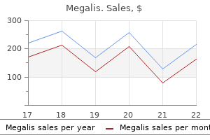
Cheap 20mg megalis
Vitamin K antagonists such as warfarin are used for primary and secondary stroke prevention in patients with high risk for cardioembolic stroke including individuals with atrial fibrillation or mechanical heart valves impotence exercises for men cheap 20 mg megalis fast delivery. The possible consequences of insufficient or excessive anticoagulation are extremely serious and often fatal, making it imperative to pursue good control but in some individuals, this can be difficult. Dabigatran inhibits the enzyme thrombin by directly binding to its active binding site. Both the factor Xa inhibitors and direct thrombin inhibitors indirectly block platelet aggregation and reduce thrombus formation. Laboratory Testing the functional integrity of the clotting system can be tested with a series of straightforward clinical tests that are sufficient for most individuals who present with ischemic stroke. The vast majority of individuals with ischemic stroke do not require esoteric testing to detect thrombophilia as such testing is shown to be low yield and wasteful of resources. The fibrinolytic system can be tested with the euglobluin lysis time and measuring D-dimer and fibrin split product levels. A variety of tests are available to test platelet function including the standard bleeding time assay, which is not performed routinely any longer and optical platelet aggregometry. Various bedside platelet function assay devices are available, but they have not gained widespread use. Thrombo-elastography can be used to measure whole blood coagulation, clot integrity, as well as platelet function. This technology is useful in surgical settings that employ cardiac assist devices or the artificial heart. Focal ischemic brain injury leads to local neurological deficits and a progression of histopathological changes depending on ischemic severity, location, and duration. In regions of developing infarction, acute neuronal alterations can progress through a series of phases including micro vacuolation, and ischemic and homogenizing cell change [1]. In other areas adjacent or remote to the evolving focal infarct, a slower progressive neuronal damage termed selective neuronal necrosis, which may have distinct histopathological mechanisms, is also commonly seen in human ischemic tissues. Because approximately a quarter of all strokes occur in white matter, injured axons and myelin sheaths are commonly observed in pathological specimens [2]. Rivaroxaban and apixaban can be monitored with chromogenic assays for anti-Xa levels. Individuals receiving unfractionated heparin who are suspected of experiencing heparin associated thrombocytopenia type 2 should be tested for antibodies against heparin/platelet factor 4 complexes. Spatial aspects of blood coagulation: two decades of the self-sustained traveling wave of thrombin. Metabolic syndrome, platelet activation and development of transient ischemic attack or thromboembolic stroke. Mechanisms underlying sporadic cerebral small vessel disease: insights from neuroimaging. In addition to neuronal changes, alterations in astrocytes, oligodendrocytes, vascular structures, and microglia/macrophage inflammatory responses are important components of the histopathological changes. This review will focus on the histopathological changes that characterize the cellular responses to cerebral infarction and selective neuronal necrosis. For this discussion, events at the tissue and cellular levels resulting from profound alterations in local cerebral blood flow and cerebral metabolism will be emphasized. Two common sources of embolization to the brain are the heart and internal carotid artery. A thrombotic arterial brain infarct may result from severe atherosclerosis of an artery leading to vascular occlusion and a severe drop in local cerebral blood flow. Common neuropathological characteristics of ischemic infarction, or pannecrosis, include a well-demarcated area of cellular necrosis including neurons, glia, and endothelial cells. This pathology is usually associated with a defined vascular territory of a major cerebral artery or a single vessel. In a fully developed infarct, the gray matter neuron cytoplasm stains intensely with acidophilic dyes (eosinophilic or red neurons) or fails to stain giving the appearance of ghost neurons. The surrounding neuropil adopts a spongy appearance, whereas other cells types such as astrocytes, oligodendrocytes, and endothelial cells may show evidence of early swelling and pale staining due to edema formation. Acute therapeutic interventions including thrombolytic agents or mechanical removal of thrombotic clots are used to restore blood flow and salvage ischemic tissues. In chronic stages, reactive astrocytes are observed around infarct borders that may impede axonal growth or cortical circuit plasticity by creating mechanical barriers and producing specific inhibitor factors. The cerebral vasculature plays an important role in the protection of neuronal cells through regulating levels of local blood flow to brain regions to maintain metabolic needs. In addition to serving as conduits of blood, cerebral microvessels control mechanisms important for cellular inflammation, secondary injury mechanisms, and tissue remodeling [4]. This barrier is formed by endothelial cell tight junctions and limited endothelial cell pinocytosis. Endothelial cells within the evolving infarct demonstrate light microscopic alterations including swelling and necrosis. During focal ischemia they can lose their cell permeability barrier properties, leading to edema formation and degradation of the vascular basal lamina. The transformation of pale to hemorrhagic infarction may result from reperfusion of an infarct or from the migration of an embolus. They represent the endogenous inflammatory cells within the central nervous system, exist in different morphological states, and become reactive following periods of cerebral ischemia and stroke [6]. Activation of microglia is an initial event of inflammation in ischemic stroke, which leads to the production of both pro- and antiinflammatory mediators [6]. In response to various signaling molecules, microglia proliferate and migrate to the site of injury. They can progress through various morphological stages characteristic of activation and phagocytosis including ramified, hyperramified, and amoeboid as seen using special stains. Because microglia are an important source of both injurious and reparative products, this "doubleedged sword" feature has encouraged research into the search for microglia-targeted therapeutic strategies for both reducing secondary injury processes and promoting reparative processes. Astrocytic changes include early signs of swelling and fragmentation within the evolving infarct. Over time, astrocytes become reactive and go through several patterns of morphological change. Perivascular swelling of astrocytic processes is a common occurrence that might lead in severe cases to cause luminal compression. Neutrophil infiltration is another early inflammatory event seen after severe cerebral ischemia and is frequently associated with microvessels coursing within the infarcted region. Over time neutrophils can migrate across the vascular endothelial barrier and infiltrate the ischemic tissue. Evidence for intravascular obstruction by polymorphonuclear leukocytes and platelets has been demonstrated [4]. Neutrophils and other white blood cells can also participate in secondary injury mechanisms through the synthesis and release of proinflammatory cytokines and oxygen free radicals. Currently, there is evidence that an ischemic penumbra exists in animals and humans after the occurrence of focal brain ischemia [8]. In contrast to the infarct central core, glia and endothelial cells are mainly preserved. This margin of the infarct is commonly called the ischemic penumbra and may also show evidence for delayed cell death. Vulnerable neurons within the infract core are generally felt to die mainly by necrosis and associated with robust inflammatory responses. Active areas of research continue to investigate the pathophysiology of the ischemic penumbra for therapeutic interventions. In this regard, repetitive episodes of cortical spreading depolarization have been implicated in the vulnerability of the penumbral tissue. Also, new imaging tools are being developed and tested to determine the occurrence of penumbral regions in individual patients with stroke to clarify salvable tissues for neuroprotective treatments. Axonal changes can occur relatively rapidly in white matter regions where severe ischemia is occurring.
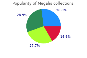
Order megalis mastercard
During the course of reperfusion erectile dysfunction drugs and glaucoma buy generic megalis 20 mg line, differences in p-Akt(Ser473) are observed in the penumbra versus the ischemic core. In the penumbral regions, transient increase of p-Akt (Ser473) occurs within the first few hours following reperfusion, but subsides by 9 h after reperfusion [1,3]. In the ischemic core, p-Akt (Ser473) decreases and remains at lower levels after reperfusion [1,3]. These differences are likely due to subtleties between global ischemia models and species. It is important to note that Akt phosphorylation at Ser473 is associated with, but not necessarily reflective of, full catalytic activity. Indeed, despite detecting increased p-Akt (Ser473) levels at early time points following focal ischemia, Zhao et al. Thus, although early phosphorylation of Akt at Ser473 occurs, this may represent an early response to injury that is muted before robust activation of the pro-survival signaling cascade in vulnerable ischemic regions. Significant data suggest an inverse correlation between phosphorylation or activity of Akt and cell injury in both focal and global ischemia models [3,7,9]. The presence of phosphorylated Akt or activated downstream components of the Akt signaling pathway does not overlap temporally with markers of apoptotic cell death. Thus, cells with activated Akt likely have not (yet) fully initiated cell death signaling, and may be still at a point of rescue or recovery. This is consistent with the lack of Akt activity despite increased phosphorylation at Ser473. Phosphorylated -catenin is highly unstable and quickly targeted toward degradation, and thus suppresses the expression of cell survival gene products. In cerebral ischemic settings, decrease of -catenin protein is associated with vulnerable ischemic regions. Proof of principle for the involvement of Wnt signaling in ischemic neuronal survival was provided by the observation that inhibition of Dickkopf-1 (Dkk-1, a negative regulator of Wnt signaling) increased neuronal survival following global ischemic injury. Akt contributes to neuroprotection by hypothermia against cerebral ischemia in rats. Evidence of phosphorylation of Akt and neuronal survival after transient focal cerebral ischemia in mice. Post-ischemic estradiol treatment reduced glial response and triggers distinct cortical and hippocampal signaling in a rat model of cerebral ischemia. Neuroprotective effect of sodium orthovanadate on delayed neuronal death after transient forebrain ischemia in gerbil hippocampus. Survival- and death-promoting events after transient cerebral ischemia: phosphorylation of Akt, release of cytochrome C and Activation of caspase-like proteases. However, Akt-mediated control of these targets has not been established in the context of cerebral ischemia. Hsps are a family of stress proteins thought to be involved in chaperone functions, such as protein folding, trafficking, and repair. Constitutively expressed members exist in all cell compartments, and appear essential for development and cellular function. Inducible forms can be induced following a variety of external stress including ischemia, but were originally described following heat stress [1]. Work over the past two decades has also established that some Hsps also function as cytoprotectants. Hsps have long been known to serve as protein chaperones in the sense that they assist in protein folding and the correct attainment of functional three-dimensional configuration, while preventing incorrect folding and protein aggregation [2]. They have also been shown to affect cellular signaling [2], and have been extensively studied in the setting of cerebral ischemia and demonstrated to provide protection against both global and focal cerebral ischemia. In studies of cerebral ischemia, Hsp70 was observed to be induced in brain regions that were relatively resistant to ischemic insults. Hence, the notion of a "molecular penumbra" was introduced, and raised questions as to whether this expression was an epiphenomenon of the injury, or an active participant in cell survival [3]. Subsequent studies using strategies to increase or inhibit Hsp70 expression have consistently shown that Hsp70 protects the brain and brain cells against experimental cerebral ischemia, neurodegenerative disease models, epilepsy, and trauma. Through its chaperone properties, it has been shown to reduce protein aggregates and intracellular inclusions [4]. In addition to their function in protein processing, Hsps appear to protect the brain by affecting several cell death and immune response pathways [1]. The best-studied class is Hsp70, the 70-kDa class that includes an inducible form also known as Hsp72, Hsp70i, or simply Hsp70. Newly generated Hsps can then bind denatured proteins and act as a molecular chaperone by contributing to repair, refolding, and trafficking of damaged proteins within the cell. During homeostatic conditions, inducible Hsp70 levels are low; however, its expression is significantly increased following injury. In experimental cerebral ischemia, Hsp70 has been shown to lead to neuroprotection [1]. Similarly, transgenic mice that overexpress Hsp70 are protected from these ischemic insults, whereas their deficiency exacerbates outcome [7,8]. Pharmacological induction of Hsp70 is also possible, and has been shown to protect the brain in experimental models. These compounds induce Hsp70 through their ability to inhibit Hsp90, and have been shown to protect the brain from experimental stroke and traumatic brain injury when given exogenously [6]. Hsp70 overexpression also appears to inhibit mitochondrial release of the proapoptotic Bcl-2 family member Bax [10], and directly inhibits the effector caspase, caspase-3 [9]. Hsp70 is also known to play a role in modulating inflammation caused by cerebral ischemia. As an antiinflammatory molecule, Hsp70 inhibited production of proinflammatory cytokines in cultured microglia and macrophages and in stroke models [6]. Although much of the work in brain ischemia surrounding the role of Hsp70 in modulating inflammation has focused on its role in intracellular signaling, work in related areas indicates that Hsp70 plays a different role extracellularly [12]. Hsp70 can be secreted by astrocytes and Schwann cells, or released by dying cells [2]. In these settings, Hsp70 may act as a danger signal, by acting upon receptors, such as Toll-like receptor-4 and -2, and leading to activation of immune cells and elaboration of proimmune molecules, thus potentially contributing to worsened damage. Mechanisms of Hsp70 Protection Hsp70 has been assumed to protect the cell via its chaperone functions. Indeed, overexpression of Hsp70 appears to prevent protein aggregation and redistribution of ubiquitin [2,4]. However, other studies have shown that Hsp70 can interfere with apoptosis at multiple levels. Mechanisms of this protective effect have largely been attributed to the ability of Hsp27 to interfere with apoptosis. Like Hsp70, Hsp27 has been shown to prevent mitochondrial release of cytochrome c and prevent formation of the apoptosome. Hsp27 may also directly interact with procaspase-3 and prevent Bax translocation to the mitochondria. Hsp70, which has shown consistent neuroprotective effects in different injury models. It appears to have multiple protective mechanisms, and can be pharmacologically induced with agents that have been tested in humans for other indications. Other stress genes and their respective proteins also hold promise as beneficial endogenous protectants, but may be less developed in terms of how this property may be applied clinically. Regulation of apoptotic and inflammatory cell signaling in cerebral ischemia: the complex roles of heat shock protein 70. Anti-inflammatory properties and pharmacological induction of Hsp70 after brain injury. The 70 kDa heat shock protein protects against experimental traumatic brain injury. Hsp70 inhibits heat-induced apoptosis upstream of mitochondria by preventing Bax translocation. Regulation of inflammatory transcription factors by heat shock protein 70 in primary cultured astrocytes exposed to oxygen-glucose deprivation.
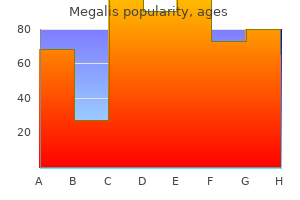
Cheap megalis 20 mg overnight delivery
Electron microscopic images of the arachnoid granulations show a series of channel-like structures [10] causes of erectile dysfunction in 60s generic 20 mg megalis otc. This occurs, for example, in bacterial meningitis or subarachnoid hemorrhage, where the white or red blood cells clog up the valve-like channels in the arachnoid granulations. In young obese women an idiopathic increase in intracranial pressure can occur [11]. In addition to unknown causes it can be due to medications, such as vitamin A and antibiotics, and to occlusion of the venous sinuses draining blood from the brain. Increased intracranial pressure can produce sixth nerve palsies as a remote effect because of the long course of the sixth nerve and the sharp bend over the clivus. Papilledema is the major finding; the vision is preserved until the swollen optic disc encroaches on the macula. It is possible to follow the severity of the papilledema by serial measurements of the size of the blind spot created by the optic disc. When vision is threatened, the optic nerve sheath fenestration can be done surgically. When the venous sinuses are blocked, there is some evidence that placement of a stent to open the sinus can be helpful [12]. However, there are no controlled trials to assess this approach, and some of the improvement may have occurred spontaneously. Enzymes are present in the vessel that degrade substances and prevent them from crossing the capillary. An extracellular matrix forms a basal lamina around the capillaries and arterioles. Brain tissue requires a constant supply of glucose and amino acids for normal metabolism. Cerebral capillaries have glucose transporter molecules that participate in the carrier-mediated transport across the capillary [13]. Abnormalities in the glucose transporter lead to reduced brain glucose and cellular damage. Monoamine oxidase, both A and B types, deaminate and inactivate biogenic amines, preventing them from entering the brain. Other enzymes identified in brain capillaries include choline acetyltransferase, which is important in choline metabolism, and -glutamyl transpeptidase, which is used as a marker for blood vessels. Insert: Basal lamina of cerebral capillary is shown in this electron micrograph tracing. The glucose transporter is found in higher concentration in the cerebral capillaries than in most systemic vessels. At low blood glucose concentrations the carrier is unsaturated and helps movement of glucose into the brain along with diffusion. At high serum glucose concentrations the carrier molecule becomes saturated, and any additional transport is by diffusion alone. Specific carrier molecules in the cerebral blood vessel also transport amino acids into the brain. Competitive inhibition of a carrier causes reduced transport of essential substances. For example, an amino acid carrier transports l-Dopa, which is used to treat Parkinson disease, into brain. When a large protein meal is digested, other amino acids compete for the carrier sites, and less of the drug is absorbed. Another example is found in patients with an enzyme deficiency that leads to accumulation of excessive amounts of phenylalanine in the blood. Phenylalanine competes with the essential amino acid, tryptophan, reducing its absorption into the brain. Reduction of levels of phenylalanine in the blood through dietary restrictions can prevent permanent brain damage. In other organs it plays an important physiological role in control of permeability. These enzymes are important in injury and repair, complicating attempts to block their actions with drugs. Several brain cells make matrix metalloproteinases and other proteases in response to injury [14]. Lipids dissolve in the membrane and equilibrate rapidly in brain with levels in the blood. The ability of a substance to cross the cerebral capillary determines its therapeutic potential. For example, heroin is a chemically modified morphine molecule that more rapidly enters the brain. Injection into the thecal sac can be used to increase the amount of the agent reaching brain tissue. Less dramatic forms of cell death occur with the orderly involution of a cell during apoptosis and with autophagy [15]. Another consequence of damage to the cell is brain edema: when the cells swell and the membrane is intact, there is cytotoxic edema; damage to the blood vessels results in vasogenic edema. Capillaries affected by the injury have increased permeability, which results in the movement of solutes and water into the brain. Either cytotoxic or vasogenic edema can contribute to the tissue damage and interfere with recovery. As the vessels enter the cortex they have glial limitans around them, but deeper in the cortex, they have the basal lamina. Antibiotics cause the bacterial cell walls to lyse with the release of toxic bacterial cell wall products, including lipopolysaccharide. Cytokines are formed, along with free radicals and proteases, which result in secondary damage to the blood vessel. Thus the antibiotic can control the infection, but the secondary damage can produce delayed vasogenic edema. In childhood meningitis treatment, steroids are given along with antibiotics to reduce the secondary damage from the inflammatory response, but use of steroids has not been adequately tested in adults with meningitis. Permanent occlusion of a blood vessel damages the cerebral capillaries in the ischemic tissue. Reintroduction of oxygenated blood into the damaged tissue allows free radicals to form and neutrophils to enter the tissue, worsening the ischemic damage. The critical factor is to reduce the time before reperfusion to reduce the subsequent reperfusion injury. At each interface between blood and brain, anatomic or enzymatic processes are present that block movement of substances that are not lipid soluble. Carrier-mediated processes transport nutrients essential to brain cell function, such as glucose and amino acids, into the central nervous system. Understanding the interaction of multiple factors in these processes will lead to more effective therapeutic agents. Five-year incidence of surgery for idiopathic normal pressure hydrocephalus in Norway. The morphology of cerebrospinal fluid drainage pathways in human arachnoid granulations. Diffuse brain oedema in idiopathic intracranial hypertension: a quantitative magnetic resonance imaging study. Diverse roles of matrix metalloproteinases and tissue inhibitors of metalloproteinases in neuroinflammation and cerebral ischemia. Many predisposing conditions such as dehydration, coagulopathies, pregnancy, trauma, surgical interventions, inherited collagen disorders, and autoimmune vascular diseases may result in cerebral vein thrombosis. However, in case of clinical manifestations developed, diagnosis must be done immediately in order to investigate and treat possible reasons. This chapter aims to present anatomical configuration of cerebral venous system regarding possible significant origin of "stroke. These arachnoid granulations (villi) are commonly found around the superior sagittal and transverse sinuses [1]. Superior sagittal, straight, and occipital sinuses join at the point of torcular herophili. Then they drain into transverse, sigmoid sinuses and internal jugular vein orderly.
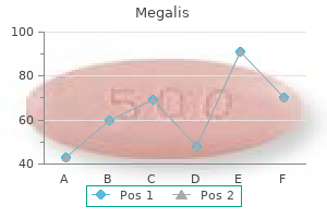
Megalis 20 mg on line
Accumulating experimental and clinical evidence suggests that stroke induces a rapid vasculogenic erectile dysfunction causes buy generic megalis 20mg on line, temporary immunodepression mainly mediated by the autonomic nervous system [3]. Stroke disturbs this normally well-balanced interplay leading to brain inflammation and immunodepression [2]. The hypothalamus is functionally linked with other autonomic centers allowing for a synchronization of glucocorticoid responses with the cholinergic pathway [1]. The sympathetic nervous system innervates primary and secondary lymphoid organs and thereby regulates immunity. Hence, overactivation of cholinergic as well as sympathetic pathways might cause immune dysfunction [4,5]. For example, the cholinergic pathway suppresses pulmonary macrophages and epithelial cells of the innate immunity thereby facilitating pneumonia after stroke [5]. Moreover, secondary lymphatic organs like spleen and thymus exhibit an atrophy after focal cerebral ischemia [4]. The extensive sympathetic innervation of immune organs and the presence of adrenergic receptors on almost all leukocytes indicate the strong influence of sympathetic activity on immune function [3]. Stroke induces an overactivation of the adrenergic system causing the release of catecholamines from sympathetic nerve terminals and the adrenal medulla [3]. This results in a pronounced antiinflammatory phenotype in lymphocytes, monocytes, and macrophages [1]. However, a more refined patient identification including biomarkers could be helpful. Overall, the mediated changes in neuroimmunomodulation result in a switch from a proinflammatory Th1 response to an antiinflammatory Th2 response, a rapid numerical decrease in peripheral blood lymphocyte subpopulations, changes in cytokine levels, and functional deactivation of monocytes. Impaired monocyte function results in insufficient antigen presentation and may, thus, contribute to reduced lymphocyte responses. The inflammatory cascade induced by acute stroke is usually linked to the progression of brain damage [2]. In fact, cellular immune responses to myelin-associated antigens and other brain antigens are seen in stroke survivors and seem to be more robust than in patients with multiple sclerosis [6]. One study demonstrated that autoimmune responses to myelin basic protein in patients with stroke are associated with a decreased likelihood of good outcome, even after adjusting for baseline stroke severity and patient age [6]. However, given that brain immune interactions after stroke might have protective as well as destructive effects in the brain itself and on the human organism as a whole, development of immunomodulatory strategies is not straightforward [2]. Further research on this topic is urgently needed since immunomodulation after stroke may not only allow for a prevention of poststroke infections but also might harbor further beneficial effects. One study demonstrated that B-lymphocyte responses to stroke occur in the brain of mice that subsequently developed cognitive deficits as well as in human subjects with stroke and dementia. In an animal model of stroke, preventive antibacterial treatment dramatically improved mortality and reduced infarct sizes [2]. A Cochrane systematic review on clinical trials assessing the efficacy and safety of antibiotics in the prevention of poststroke infections concluded that preventive antibiotic treatment reduces the risk of poststroke infections [1]. Two large randomized-controlled trials investigated whether or not prophylactic antibiotic therapy prevents poststroke infections and improves long-term outcome in acute stroke. These two trials are of considerable interest because they provide class 1 evidence recommending against preventive antibiotic treatment after stroke. Stroke-induced immunodeficiency promotes spontaneous bacterial infections and is mediated by sympathetic activation reversal by poststroke T helper cell type 1-like immunostimulation. Cholinergic pathway suppresses pulmonary innate immunity facilitating pneumonia after stroke. Blood-based immune and stress markers have been demonstrated to identify patients at high risk for poststroke infections as well as patients with unfavorable outcome. Many operations that were once considered experimental are now commonplace, with thousands of open heart procedures performed annually in the United States. At present, an estimated 1 million patients undergo cardiac surgery throughout the world every year. Neurological impairment is a well-known complication of cardiac surgery, resulting in longer hospitalizations, increased costs, and an escalation in morbidity and mortality. The types of neurological complications vary and can include peripheral neuropathy, encephalopathy, cognitive impairment, and stroke. Overall, in prospective studies, transient neurological complications have been noted in 61% of cardiac surgery patients [1,2]. Although refinements in surgical and anesthesia techniques have improved neurological outcomes, the number of elderly patients undergoing cardiac surgery has also increased, and thus cerebrovascular complications in particular continue to occur. One prospective study of 16,184 consecutive patients indicates that the specific stroke risk depends on the type of cardiac surgical procedure performed [3]. While the overall incidence of stroke in cardiac surgery is generally estimated to be 4. This was thought to be due to less aortic manipulation during off-pump surgery, but conflicts in the literature exist [1]. Stroke rates, as well as quality of life and cognitive function did not differ significantly between the two groups at 30 days and 1 year [8]. The potential mechanisms of cerebral infarction in the cardiac surgery population is explored in a later section. The risk of stroke was similar; however, patients with bioprostheses had a higher risk of aortic valve reoperation and a lower risk of major bleeding. Since then, there has been a growing comfort and a growing popularity for this intervention, especially to treat descending thoracic aorta pathology. This operation has generally fallen out of favor in lieu of off-pump techniques or a robotic approach. Placement of the Edwards Lifesciences Sapien valve requires a balloon inflation to expand the stent support structure of the valve. Irrespective of the specific valve used, both the valvuloplasty and valve deployment/ implantation are blamed for causing a shower of atheroemboli, which can flow directly up the carotid arteries to the brain, resulting in stroke [24]. In trials for Edwards Lifesciences Sapien valve, the stroke rate ranges from 5% to 7. Despite this concerning data, however, the embolic events do not clearly seem to correlate with clinical outcomes. Whether this reduction in lesion size correlates to improved neurological outcomes is uncertain. Prospective studies specifically evaluating neurological outcomes and interventions are necessary, as well as long-term clinical outcomes. The major concern regarding cardiac surgery in patients with carotid disease is whether the hemodynamic stress of heart surgery leads to underperfusion of areas supplied by already stenotic or occluded arteries, causing ischemic stroke. The chance of perioperative hypoperfusion stroke does escalate with increasing severity of carotid disease [1,4]. The best approach to the management of concomitant severe carotid and coronary artery disease remains unanswered. As of 2016, there is no consensus as to which surgical approach is superior [1,28]. The overall stroke risk was lower in patients who had intraoperative epiaortic ultrasound, compared with all patients undergoing cardiac surgical procedures [1,29,30]. However, these patients may still be candidates for mechanical endovascular acute stroke treatment [1]. Brain Embolism An argument against a hypoperfusion as a potential etiology for stroke in cardiac surgery patients is the timing of the infarct itself. If the mechanism of stroke were hemodynamic, the major circulatory stress would be intraoperative, and patients would awaken from anesthesia with the deficit. Postoperative emboli may arise from preexisting cardiac abnormalities (such as hypofunctioning ventricles, dilated atria, and aortic atheromas) or from postoperative arrhythmias such as atrial fibrillation. Additionally, postoperative activation of coagulation factors in cardiac surgery patients can promote hypercoagulability, precipitating occlusive thrombosis in atherostenotic arteries, and causing intraarterial embolism. Thromboembolic infarction often occurs in the days following surgery when cessation of anticoagulation is necessary. Evidence also links intraoperative and postoperative embolism to aortic ulcerative atherosclerotic lesions [1].
Cheap megalis 20 mg line
After each use erectile dysfunction doctors fort worth cheap megalis online visa, throw away the syringe and any drug left in it (ask pharmacist about disposal methods). When to take: Once or twice a week for etanercept, every other week for adalimumab and certolizumab, monthly for golimumab, infliximab is given several times a year. Consult your doctor or pharmacist if you should resume regular schedule or begin a new schedule. Time lapse before drug works: It will take several weeks before full benefits of the drug are noticeable. Over age 60: Use with caution in elderly patients, since infections are more common in this age group. Different drugs in this group are approved for use in children with certain medical disorders. It may also cause blood abnormalities or blood vessel inflammation with symptoms of vision changes, weakness, numbness or tingling, paleness, fever, or easy bruising or bleeding. What drug does: Decreases secretion of cholesterol into bile by suppressing production and secretion of cholesterol by the liver. Ursodiol will not help gallstone problems unless the gallstones are made of cholesterol. Rare: Mood or behavior changes, Continue, but call doctor right away continued nausea or vomiting or or seek emergency care for severe appetite loss, increase in seizures, symptoms. Clonazepam Diazepam Enzyme inducers* Felbamate Hepatotoxics* Lamotrigine Phenobarbital Phenytoin Primidone Rifampin May prolong seizure. Prolonged use: Talk to your doctor about the need for follow-up medical examinations or laboratory studies to check hearing acuity, kidney function, vancomycin serum concentration and urinalysis. It is recommended that patients combine use of this drug with a stop-smoking program (such as counseling, support groups and/or patient education). A lower dose is taken at the start of treatment and increased over the first few days. It may be continued for another 12 weeks to improve long-term success in quitting smoking. What drug does: It is a nicotine-free drug that acts on the brain to reduce cravings for cigarettes (and other tobacco products). This helps to decrease the desire to smoke and reduces the unpleasant smoking withdrawal symptoms. Infrequent: Strange dreams, gas, abdominal pain, upset stomach, taste changes, constipation, weak or tired feeling, vomiting. These include increased appetite, weight gain, tension, irritability, insomnia, headache and others. Symptoms may include depressed mood, agitation, being aggressive, other emotional changes and thoughts about suicide or possible suicide attempts. Common: Diarrhea, nausea, trouble sleeping, dizziness, dry mouth, change in sexual desire or function. Infrequent: Vomiting, abnormal dreams, blurred vision, dry eyes, migraine, sedation. Contact doctor right away if symptoms get worse or any there is any talk of suicide or suicide behaviors. Symptoms may include confusion, hair loss, yellow skin or eyes, dizziness, headache, skin irritation, pain in bones or joints, coma. Best sources are liver; yellow-orange fruits and vegetables; dark-green, leafy vegetables; milk; butter and margarine. Treatment and prevention of vitamin B-12 deficiencies in people who have had stomach or intestines surgically removed. Use nasal spray 1 hour before or 1 hour after eating hot foods or drinking hot liquids. What drug does: Acts as enzyme to promote normal fat and carbohydrate metabolism and protein synthesis. Colchicine Famotidine H 2 antagonists* Neomycin Para-aminosalicylic acid Potassium (extended-release forms) Ranitidine Vitamin C (ascorbic acid) Decreased absorption of vitamin B12. Estrogens* Increased likelihood of adverse effects from estrogen with 1 g or Iron supplements* Mexiletine Quinidine Salicylates* Tranquilizers* (phenothiazine) more of vitamin C per day. Also can raise blood calcium levels that can lead to damage to heart, blood vessels and kidneys. Anticonvulsants, hydantoin* Calcium (high doses) Calcium channel blockers* Calcium supplements* Cholestyramine Colestipol Cortisone Digitalis preparations* Diuretics, thiazide* Mineral oil Neomycin Nicardipine Phenobarbital Phosphorus preparations* Rifampin Vitamin D, other Decreased vitamin D effect. Prolonged use: Talk to your doctor about the need for follow-up laboratory studies to check calcium levels in your blood or urine. Skin & sunlight: Your skin may be more sensitive to sunlight when using this drug. Sunlight exposure (even brief periods) may cause a skin rash, itching, redness, other skin discoloration or a severe sunburn. When you begin using this drug, try to stay out of direct sunlight, especially between the hours of 10:00 a. Possible decreased effect of iron supplement in patients with irondeficiency anemia. Recommended dose of vitamin E- Increased benefit and decreased toxicity of vitamin A. Prolonged use: Talk to your doctor about the need for follow-up medical examinations or laboratory studies to check prothrombin time. What drug does: It prevents blood cells (platelets) from clumping together and forming blood clots in the arteries. Time lapse before drug works: Starts working in one to two hours, but may take a week or more for maximum benefit. Prolonged use: Consult with your doctor on a regular basis while taking this drug to monitor your progress and check for any unwanted effects. Serotonin is a brain chemical (neurotransmitter) that plays a role in emotional and psychological disturbances. Time lapse before drug works: Symptoms may improve after 2 weeks of use, but full benefit may take about a month. Prolonged use: Consult with your doctor on a regular basis while taking this drug to check for adverse effects and determine the need for continued treatment.
Buy megalis in india
Radiographic evaluation of the vessel wall has also revealed several distinguishing features of radiation vasculopathy impotence causes and cures buy 20mg megalis amex. Examples of the multivessel steno-occlusive disease and extensive collateralization of adjacent vessels is shown in. Thus, it may progress unnoticed for decades, until a critical threshold is reached and cerebrovascular ischemia occurs. Once identified however, radiation vasculopathy may progress faster than atherosclerotic arteriopathy. Therefore, it is important for physicians to recognize the potential symptoms of radiation vasculopathy in at-risk patients. Primary care or oncology specialists who regularly follows these patients should be aware of any complaints concerning for transient ischemic events. As of 2016, there are no detailed guidelines regarding screening with imaging studies for high-risk patients. As for patients who have received radiation to the head and neck, in 2007 the American Society of Neuroimaging recommended cervical carotid artery screening 10 years after treatment. Patients with history of radiation to the head and brain often have frequent brain parenchymal imaging performed to monitor for cancer recurrence, but these protocols may not include intracranial vessel imaging. Accordingly, evaluating physicians must still be vigilant of complaints that may represent transient neurological attacks and request vessel imaging for atrisk patients. With regard to procedural options for large vessel disease such as cervical carotid artery stenosis, both endarterectomy and stenting have been utilized to successfully treat radiationrelated lesions; however, one is not clearly superior over the other in all cases [14]. Radiation to the neck poses several obstacles to treatment that must be considered. Due to fibrosis of the vessel wall, scar tissue obscuring anatomical landmarks, fragile connective tissue, and poor wound healing, radiation therapy may result in a "hostile neck" with regard to open surgery. Tandem and atypical lesions with a propensity for restenosis pose barriers to endovascular options. Traditionally, patients undergoing corrective procedures for large vessel disease were felt to be at high risk for complication, particularly with open surgery. In this study, patients undergoing endarterectomy had higher rates of cranial nerve injury, whereas higher rates of late cerebrovascular events and restenosis were associated with stenting [14]. Given the heterogeneity of the lesions and potentially multiple comorbid variables, each patient should be evaluated on a case-by-case basis to determine optimal management. Additional consideration must be taken for patients undergoing subsequent surgical procedures to the head and neck-ligation of what might be thought of as a (A) (B) minor branch of the external carotid or thyrocervical trunk may prove catastrophic in the case of patients who are relying on collateral vessels in lieu of occluded major cervical vessels for brain perfusion. For intracranial pathology such as moyamoya syndrome, intracranial revascularization procedures such as encephalo-duro-arterio-synangiosis as per idiopathic moyamoya have been utilized with good success [9]. For both intracranial and extracranial disease, traditional stroke risk factors such as smoking, hypertension, hyperlipidemia, diabetes, and obesity should be addressed as these act additively on vessels damaged by radiation. Furthermore, radiation damage to the heart itself may cause myocyte loss and fibrosis of the myocardium and pericardium, leading to restrictive cardiomyopathy, diastolic heart failure, and conduction abnormalities [15]. Similar to cerebrovascular radiation vasculopathy, radiation-induced cardiac disease poses unique challenges. Pericardial fibrosis and scar tissue formation are barriers to surgical procedures. Heavy calcifications of the aortic arch and coronary arteries may impede endovascular approaches. On the day after a mandibular reconstruction, he was found to have watershed infarctions of the right frontal and parietal lobes. The angiogram showed bilateral common carotid artery occlusions and filling of the internal carotid arteries (arrowhead on right internal carotid artery) from the costocervical trunk via the inferior thyroid arteries through the superior thyroid arteries. On the right, the transverse cervical artery was anastamosed to the graft feeding the reconstructed mandible (arrow). Cardiovascular risk factors such as smoking, diabetes, and hypertension also have additive effects on the radiation damaged heart, and should be aggressively controlled. Akin to radiation vasculopathy, cardiac radiation damage may be more aggressive than traditional cardiac disease and frequent monitoring should be considered. Cerebral radiation necrosis: a review of the pathobiology, diagnosis and management considerations. Angiographic features, collaterals, and infarct topography of symptomatic occlusive radiation vasculopathy: a case-referent study. A historical prospective cohort study of carotid artery stenosis after radiotherapy for head and neck malignancies. Late cerebrovascular complications after radiotherapy for childhood primary central nervous system tumors. Characterization of radiation-induced cavernous malformations and comparison with a nonradiation cavernous malformation cohort. Ultrasonic analysis of plaque characteristics and intimal-medial thickness in radiation-induced atherosclerotic carotid arteries. Stenting versus surgery in patients with carotid stenosis after Previous cervical radiation therapy: systematic review and meta-analysis. Cardiovascular complications of radiation therapy for thoracic malignancies: the role for non-invasive imaging for detection of cardiovascular disease. Stereotactic radiosurgery guideline for the management of patients with intracranial arteriovenous malformations. Stereotactic radiosurgery for intracranial dural arteriovenous fistulas: a systematic review. In particular, radiation therapy is an attractive option in patients with vascular malformations not amenable to endovascular or surgical approaches. However, given the prolonged time course of radiation injury, eventual obliteration of the malformation may take several years. Furthermore, the radiation itself may precipitate hemorrhage, cavernous malformations, cyst formation, or injury to the brain tissue resulting in neurological dysfunction or seizures. Practitioners caring for these patients must be diligent in monitoring for the complications of radiation-induced vascular disease. In addition to ongoing research to further our understanding of both the angiodestructive and angiogenic effects of radiation, it may soon be possible to conduct large-scale randomized trials of therapies for patients with various forms of radiation vasculopathy and optimize treatment for patients with this increasingly common disease. David Liebeskind for assistance with the angiographic images of radiation vasculopathy. This misconception unfortunately has been propagated since the earliest descriptions of these diseases, describing a "Dissecting Aneurysm of the Aorta" [1]. While some aneurysms can be complicated by, and predispose to , dissection, and dissections can become aneurysmal over time, the disease processes are relatively distinct. Aortic aneurysm is an abnormal dilation of the diameter of the aorta usually caused by longstanding atherosclerotic disease, hypertension, and/or a history of smoking. This tear is often rapidly propagated within the three layers of the aortic wall to various extent due to the forces exerted by the systemic blood pressure, which quite commonly is elevated. Thus blood rushes directly into the separated wall of the aorta creating a false passageway for blood to travel. This "false lumen" of blood flow may thrombose, rupture, or obstruct blood flow to branch vessels, including coronary and carotid arteries, visceral organs, or the extremities. Dissection may also extend to the aortic valve sinuses and cause acute aortic insufficiency. Several classifications exist for aortic dissection, but the most common and functionally useful is the Stanford classification [2]. Aortic dissection occurring distal to the left subclavian artery are described as Type B. This distinction of location of the dissection dictates natural history, management, and surgical approach, if necessary. Left untreated, Type A dissections carry a mortality rate of 50% within 48 h, and 90% at 2 weeks. In contrast, Type B aortic dissections are usually managed medically, and have an overall lower short-term mortality, but with a progressive long-term mortality. Surgery for Type B dissection, when necessary, is usually to address complications of malperfusion or later development of aneurysm. Since brain perfusion arises normally from the aortic arch (carotid arteries) and its branches (vertebral arteries by way of the subclavian arteries), dissection involving this region can lead to acute neurological injury in the form of branch obstruction or embolism.
Order megalis 20mg without a prescription
However erectile dysfunction after vasectomy buy 20 mg megalis free shipping, a potential disadvantage is that they exhibit fewer similarities to the human brain as compared with gyrencephalic primates. Although unproven, this difference may limit the capacity of stroke experiments conducted in lissencephalic species to predict the effects of a promising therapy in humans. Old World primates are native to the African and Asian continents and include the family Cercopithecidae. They are gyrencephalic, possessing larger brains with a more complex cortical and subcortical organization and a vascular anatomy that resemble that of the human brain [3]. Due to their size, Old World species require specialized housing facilities with larger cages and more environmental enrichment than the smaller New World species. In general they also require greater resources and technical expertise for anesthesia, surgery, and recovery. Historically, the most common species used in stroke experiments has been the Papio anubis (baboon). Such research provided the foundation for stroke research to come by establishing the concept that therapeutic intervention could reduce infarction in brain-at-risk. However, baboons are large, requiring significant resources to house, and are highly aggressive, which limits the extent of clinical neurological evaluation that can be practically conducted. The vascular anatomy of the macaque closely resembles the human with less collateralization than the baboon [6]. Macaque monkeys are highly social and more easily trained than baboons, enabling researchers to conduct neurological evaluations before and after stroke more readily. However, these strokes have inherent variability in anatomical localization based on variations in collateral blood flow between subject animals and increased variability in neurobehavioral outcomes based on the mixed nature of deficits in a given stroke distribution. For example, we have described an experimental approach to preclinical testing of neuroprotectants [8] that predicted efficacy in a corresponding clinical trial [9]. This was achieved by matching the strokes produced in humans undergoing endovascular aneurysm repair [9] to a preclinical model in primates in which similar embolic strokes were produced by the intracarotid injection of emboli, resulting in identical strokes in the primates that could then be subjected to the therapeutic intervention before, or in parallel with, the clinical study [8]. Focal strokes of end-artery distributions like the lenticulostriate arteries have also been used [3]. Strokes in other anterior and posterior cerebrovascular distributions have not been widely pursued in preclinical studies. Although the transorbital method provides relatively consistent strokes as evaluated by imaging and histological analysis, the technique requires enucleation of the eye and prevents behavioral testing that requires binocular vision. The craniotomy approach results in a potentially painful incision site with decreased function of the temporalis muscle as it is dissected; however, in skilled hands the surgery is generally well tolerated and the animal is prepared for a full battery of behavioral tests when it recovers. This method also requires an occlusion of the posterior cerebral artery to decrease collateral flow. Other endovascular models employ autologous blood clots to achieve vessel occlusion. These models have the advantage of more closely modeling the pathophysiology underlying thromboembolic stroke in humans with the opportunity to achieve reperfusion with standard thrombolytic therapies; however, the variability in stroke distribution, volume, and neurological outcomes in endovascular models is high. This model can be undertaken with a minimal surgical opening and can be combined with thrombolytic therapies to achieve reperfusion. The strokes produced are generally smaller, are located in more distal vascular distributions and are variable in anatomic localization. Moreover, it can be defined using anatomical techniques that are commonly used in stroke research such as staining with the mitochondrial dye 2,3,5-triphenyl tetrazolium chloride. Therefore a transient ischemia model has been developed to facilitate recovery experiments; however, this produces stroke primarily in subcortical nuclei, sparing the cortex [3]. However, the baboon cerebral vasculature is critically different from that of humans due to a rich collateralization. Neurobehavioral assessments in this model are limited to observational scoring due to the aggressive nature of the baboon. Enucleation of the eye may obscure detailed cognitive, sensory, and motor neurobehavioral tasks requiring intact binocular vision. Thus it is possible to model both rapidly progressive strokes, as well as strokes that maintain a sizable ischemic penumbra for longer periods, such as those that are exhibited by patients who benefit from endovascular reperfusion or delayed thrombolytic therapy. To undertake these types of tasks, animals must be trained to achieve minimum baseline scores before stroke onset. For example, the two tube choice test is a test of hand preference and hemispatial neglect [3,8,10]. The hill and valley staircase task requires the monkey to reach through vertical slots in an acrylic panel attached to the front of the cage to retrieve food rewards from a five-step staircase. This task separates hemiparesis from hemisensory neglect by testing motor function in each arm in the isolated hemisensory field. Normal animals complete the task bimanually with pincer grip, by picking marshmallow pieces from two columns of three wells moving away from the cage on the left and right of center. Animals with hemisensory neglect will leave the marshmallows in the column on the affected side, whereas animals with hemiplegia will slowly gather rewards from both sides with the unaffected hand and mildly hemiparetic animals will lose pincer grasp in the affected hand but cup the hand and scoop marshmallow pieces out of the wells with the affected hand. With progressive motor impairment there is increased latency to complete the task. Cognitive tasks play an important role in assessing models of global cerebral ischemia and in follow-up of animals in chronic focal cerebral ischemia studies as memory and learning are impaired in both scenarios and can potentially affect human measures of stroke outcome. The simplest test of working memory in primates is the Delayed Response test in which the monkey is presented with an uncovered pair or matrix of wells with a food reward in a single well and is then required to recall the location of a food reward in a covered matrix of wells after a delay in time [3]. Visual discrimination can be added to this task by presenting monkeys with a shape or color on a card and then after a delay presenting the monkey with a matrix of covered wells with patterns or colors on the covers where a food reward is baited in the well matching the stimulus. The Object Retrieval Detour Task is designed to evaluate the cognitive elements of motor planning in reaching movements and has been used in cynomolgus macaques with chronic stroke. Higher cognitive function is assessed with a battery of three tests including the Delayed Nonmatching to Sample Task, the Delayed Recognition Span Task, and the Conceptual Set Shifting Task that have been used in studies of dementia in aged rhesus macaques. These tests have also been used to study hypertensive leukoencephalopathy in cynomolgus macaques. They evaluate recognition memory, pattern recognition, visuospatial memory, and executive functions. However, the validity of these surrogate measures in estimating functional outcome has been questioned. Surrogate measures have not been consistently linked to functional neurological outcome. Tissue salvage is commonly defined as a reduction in necrotic tissue on histological sections. Metabolic measures that correlate with tissue death have also been used in stroke studies. Of these the most common is the vital stain 2, 3, 5triphenyltetrazolium chloride that delineates regions with functional cellular respiration from those without, with close correlation to histological measures of tissue necrosis. Cellular homeostatic function is disturbed after stroke with both irreversible and reversible dysfunction; however, measures of some cellular functions that correlate strongly with tissue death have the advantage of defining tissue loss based on early loss of vital intracellular processes that may precede cell death. For instance, global protein expression and gene transcription have been used in limited studies to measure cellular function after stroke [10]. Chief among these may be flawed clinical trial design or execution, which may not have taken into account lessons already learned in lower-order species. Different strokes for different folks: the rich diversity of animal models of focal cerebral ischemia. The chief hypothesis in primate stroke studies is that using highorder species may allow researchers to simulate the human situation as closely as possible in the preclinical setting. Owing to their small size, rodents (the most commonly used animals in cerebrovascular research) present unique technical challenges when compared with humans. In addition, many biomedical research laboratories do not have access to technologies used routinely in a clinical setting. The proximal thermosensor is located several millimeters away from the distal sensor, outside of its thermal field, thereby allowing continuous monitoring of tissue temperature and compensation of baseline fluctuations. This thermal transfer includes not only convective effects induced by blood flow, but also the intrinsic conductive properties of the tissue. This was initially achieved by euthanizing the animal to determine the zero flow current. Modern systems enable quantification of tissue perfusion in absolute units by determining the conductive properties of the tissue from the initial rate of propagation of the thermal field and by subtracting this component from the total heat transfer as the determinant of the thermal convection component, making no-flow calibration unnecessary.
Generic 20mg megalis amex
This particular group of diuretics does not allow the unwanted side effect of low potassium in the blood to occur erectile dysfunction drugs history purchase megalis 20 mg overnight delivery. Diuretics, Thiazide-Drugs that act on the kidneys to prevent reabsorption of electrolytes, especially chlorides. These drugs include bendroflumethiazide, benzthiazide, chlorothiazide, chlorthalidone, cyclothiazide, hydrochlorothiazide, hydroflumethiazide, methyclothiazide, metolazone, polythiazide, quinethazone, trichlormethiazide. Dopamine Agonists-Drugs that stimulate activity of dopamine (a brain chemical that helps control movement). These include apomorphine, bromocriptine, cabergoline, pramipexole, quinagolide, ropinirole, rotigotine. Dopamine Antagonists-Drugs that interfere with dopamine production (brain chemical that helps control movement). These drugs include haloperidol, metoclopramide, phenothiazines, procainamide, thioxanthenes and others. Dosage Form-A dosage form is the physical form in which a drug is produced and dispensed, such as a tablet, a capsule, an injectable and others. It will detect enlargement of either heart chamber, but will not establish a diagnosis of heart failure or disease of the heart valves. Eczema-Disorder of the skin with redness, itching, blisters, weeping and abnormal pigmentation. This test is useful in the diagnosis of brain dysfunction, particularly in studying seizure disorders. Electrolytes-Substances that can transmit electrical impulses when dissolved in body fluids. These drugs include: alcohol (chronic use), barbiturates (especially phenobarbital), carbamazepine, darunavir, dexamethasone, efavirenz, glucocorticoids, glutethimide, griseofulvin, insulin, isoniazid, modafinil, nafcillin, nevirapine, norethindrone, omeprazole, oxcarbazepine, phenylbutazone, phenytoin, pioglitazone, prednisone, primidone, rifabutin, rifampin, rifapentine, saquinavir, secobarbital, St. Also included are charbroiled meats, cruciferous vegetables (such as broccoli and cabbage) and smoking. Epilepsy-Episodes of brain disturbance that cause convulsions and loss of consciousness. Erectile Dysfunction Agents-Medicines used to treat male impotence (the inability to develop and sustain an erection). These drugs include: Alprostadil, papaverine, sildenafil citrate, tadalafil, vardenafil, yohimbine. Ergot Preparations (Alkaloids)-Medicines used to treat migraine and other types of throbbing headaches. Also used after delivery of babies to make the uterus clamp down and reduce excessive bleeding. These drugs include erythromycin, erythromycin estolate, erythromycin ethylsuccinate, erythromycin gluceptate, erythromycin lactobionate, erythromycin stearate. Esophagitis-Inflammation of the lower part of the esophagus, the tube connecting the throat and the stomach. These drugs include: Systemic-chlorotrianisene, diethylstilbestrol, estradiol, estrogens (conjugated and esterified), estrone, estropipate, ethinyl estradiol, quinestrol. Estrogen agonists/antagonists-Drugs that 1) activate (agonism) estrogenic receptors in certain body tissues to produce an estrogen effect, or 2) block (antagonism) estrogenic effects in others. Eustachian Tube-Small passage from the middle ear to the sinuses and nasal passages. Extrapyramidal Reactions-Abnormal reactions in the power and coordination of posture and muscular movements. Some drugs associated with producing extrapyramidal reactions include amoxapine, antidepressants (tricyclic), droperidol, haloperidol, loxapine, metoclopramide, metyrosine, moclobemide, molindone, olanzapine, paliperidone, paroxetine, phenothiazines, pimozide, rauwolfia alkaloids, risperidone, tacrine, thioxanthenes. Fibrocystic Breast Disease-Overgrowth of fibrous tissue in the breast, producing non-malignant cysts. Flu (Influenza)-A virus infection of the respiratory tract that lasts three to ten days. Fluoroquinolones-A class of drugs used to treat bacterial infections, such as urinary tract infections and some types of bronchitis. These drugs include ciprofloxacin, enoxacin, gatifloxacin, gemifloxacin, levofloxacin, lomefloxacin, moxifloxacin, norfloxacin, ofloxacin, sparfloxacin. These drugs include dione anticonvulsants, hydantoin anticonvulsants, succinimide anticonvulsants, divalproex, methotrexate, oral contraceptives, phenobarbital (long-term use), pyrimethamine, sulfonamides, triamterene, trimethoprim, trimetrexate, valproic acid. Functional Dependence-The development of dependence on a drug for a normal body function. The primary example is the use of laxatives for a prolonged period so that there is a dependence on the laxative for normal bowel action. Ganglionic Blockers-Medicines that block the passage of nerve impulses through a part of the nerve cell called a ganglion. Ganglionic blockers are used to treat urinary retention and other medical problems. Generic Drug-A generic drug is the same as a brand name drug in dosage, safety, strength, how it is taken, quality, performance, and intended use. Gland-Organ or group of cells that manufactures and excretes materials not required for its own metabolic needs. Glaucoma-Eye disease in which increased pressure inside the eye damages the optic nerve, causes pain and changes vision. Glucagon-Injectable drug that immediately elevates blood sugar by mobilizing glycogen from the liver. Gold Compounds-Medicines which use gold as their base and are usually used to treat joint or arthritic disorders. H 2 histamine may be liberated at any point in the body, but most often in the gastrointestinal tract. Hangover Effect-The same feelings as a "hangover" after too much alcohol consumption. Hematocrit-A blood test that measure how much space in blood is occupied by red blood cells. Hemochromatosis-Disorder of iron metabolism in which excessive iron is deposited in and damages body tissues, particularly of the liver and pancreas. Hemolytics-Drugs that can destroy red blood cells and separate hemoglobin from the blood cells. These include acetohydroxamic acid, antidiabetic agents (sulfonylurea), doxapram, furazolidone, mefenamic acid, menadiol, methyldopa, nitrofurans, primaquine, quinidine, quinine, sulfonamides (systemic), sulfones, vitamin K. Hepatotoxics-Medications that can possibly cause toxicity or decreased normal function of the liver. Histamine-Chemical in body tissues that dilates the smallest blood vessels, constricts the smooth muscle surrounding the bronchial tubes and stimulates stomach secretions. Hives-Elevated patches on the skin that are redder or paler than surrounding skin and often itch severely. These include Atorvastatin, fluvastatin, lovastatin, pravastatin, pravastatin & aspirin, and simvastatin. Hormone Replacement Therapy-A medication (estrogen) or combination of medications (estrogen and progestin or estrogen and androgen) used for treatment of premenopausal and menopausal symptoms and for prevention of diseases that affect women in their later years. Hormones-Chemical substances produced in the body to regulate other body functions. Hyperglycemia-Causing Medications-A group of drugs that may contribute to hyperglycemia (high blood sugar). These include oral estrogen-containing contraceptives, corticosteroids, estrogens, isoniazid, nicotinic acid, phenothiazines, phenytoin, sympathomimetics, thyroid hormones, thiazide diuretics. Hyperkalemia-Causing Medications- Medicines that cause too much potassium in the bloodstream. The effects of hypersensitivity may be characterized by wheezing, shortness of breath, rapid heart rate, severe itching, faintness, unconsciousness and severe drop in blood pressure. A critically low blood sugar level will interfere with normal brain function and can damage the brain permanently. Hypoglycemia-Causing Medications-A group of drugs that may contribute to hypoglycemia (low blood sugar). These include acetohexamide, chlorpropamide, gliclazide, glipizide, glyburide, insulin, metformin, tolazamide, tolbutamide. Hypokalemia-Causing Medications-Medicines that cause a depletion of potassium in the bloodstream. These include adrenocorticoids (systemic), alcohol, amphotericin B (systemic), bronchodilators (adrenergic), capreomycin, carbonic anhydrase inhibitors, cisplatin, diuretics (loop and thiazide), edetate (longterm use), foscarnet, ifosfamide, indapamide, insulin, insulin lispro, laxatives (if dependent on), penicillins (some), salicylates, sirolimus, sodium bicarbonate, urea, vitamin D (overdose of). Hypomagnesemia-Causing Drugs-Drugs that may increase the loss of magnesium in urine.
