

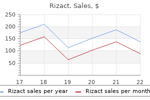
Discount 10mg rizact mastercard
Assuming a total number of 25 pain treatment agreement generic rizact 10 mg overnight delivery,000 to 40,000 genes in the mouse genome, a single treated male mouse should have between 25 and 40 different heterozygous mutagenized genes. In the case of multigenic phenotypes, segregation of the mutations in the next generation allows the researcher to focus on monogenic traits. In each generation, 50% of the mutations are lost, and only the mutation underlying the selected phenotype is maintained in the colony. Screening for dominant phenotypes is popular because breeding schemes are simple and a great number of mutants can be recovered through this approach. About 2% of all firstgeneration offspring mice display a heritable phenotypic abnormality. For example, in renal glomerular development, the phenotype of a genetic mouse strain with a tendency to development of congenital nephrosis. This approach has been successfully used to identify genes involved in neural development. The advent of genetically modified mice that express fluorescent proteins revolutionized cell lineage and mapping studies allowing high-resolution live visualization of morphogenetic events both in situ and in cultured organ explants. Targeted labeling of cells with fluorescent proteins can be achieved by driving expression of fluorescent proteins under direct control of a cell-specific promoter. This Cre-driven strategy is particularly valuable in cell lineage tracking and fate mapping analysis because both the progenitor and its subsequent derivatives become fluorescently labeled. This third method allows for the incomplete and pulse labeling of certain cell lineages, permitting the tracking of the fate and migratory behavior of individual cells in real time. This continuing feature stems from the facts that all of these organisms possess excretory organs designed to remove metabolic wastes from the body and that genetic pathways involved in other aspects of invertebrate development may serve as templates to dissect pathways in mammalian kidney development. Major contributions to our understanding of the function of polycystic and cilia-related genes have been made from studying C. Furthermore, studies on myoblast fusion and neural development in Drosophila-two processes that may not appear to be related to kidney development at first glance-have provided major clues to the development and function of slit diaphragms. The pronephros found in larval stage zebrafish (Dario rerio) consists of two tubules connected to a fused, single, midline glomerulus. The zebrafish pronephric glomerulus expresses many of the same genes found in mammalian glomeruli. The pronephros of the clawed frog Xenopus laevis has also been used as a simple model to study early events in nephrogenesis. As in the fish, the pronephros consists of a single glomus, paired tubules, and a duct. Together, the phenotypes of these knockout mice have provided an initial molecular hierarchy of early kidney development. Although Six1 and Eya1 may act in a complex together, the Six1 phenotype is somewhat different, in that a histologically distinct mesenchyme is present at 11. This understanding has been gained primarily through the phenotypic analysis of mice carrying targeted mutations that affect kidney development. Additional information has been gained by identification and study of genes expressed in the developing kidney, even though the targeted mutation, or knockout, either has not yet been performed or has not affected kidney development or function. This section categorizes the genetic defects on the basis of the major phenotype and stage of disrupted development. It must be emphasized that many genes are expressed at multiple points of renal development and may play pleiotropic roles that are not entirely clear. As previously mentioned, the organ culture system has been in use since the seminal experiments, beginning in the 1950s, of Grobstein, Saxen, and their colleagues. The embryonic neural tube was found to be able to substitute for the epithelial bud, and experiments involving the placement of the inducing agent on the opposite side of a porous filter from the mesenchyme provided information about the degree of contact required between them. A large series of experiments using organ cultures provided information about the timing of appearance of different proteins normally observed during the induction of nephrons and about the intervals that were crucial in maintaining contact between the inducing agent and the mesenchyme to obtain induction of tubules. Mice carrying mutations in any one of these genes do not have kidney abnormalities; however, triple-mutant mice for these genes demonstrate a complete absence of metanephric kidney induction. A novel approach to the organ culture system involving microinjection and electroporation has also yielded insights as to a possible function of the Wt1 gene in early kidney development. Blockade of Flk1 after the organ had been in culture for 48 hours had no effect, indicating that the angioblast-derived signal was required to initiate kidney development but not to maintain continued development. Flk1 signaling is also required to initiate hepatocyte differentiation during liver development. Nephric duct specification fails in Pax2/Pax8 mutants but not in the case of Lhx1 deficiency, in which only the caudal portion of the nephric duct degenerates. Nephronectin gene (Npnt) knockout mice exhibit renal agenesis or severe hypoplasia. Hence, dysregulation of mesenchymal cell adhesion causes the failure to attract and induce the ureteric epithelia. This fact is another example of how signaling through the extracellular matrix intersects with growth factor signaling to influence morphogenesis. The importance of basement membrane assembly in the development of other renal structures is emphasized by genetic studies on the genes Lama5 and Lamb2, which encode for laminins 5 and 2, respectively. Loss of Lama5 causes either renal agenesis or disruption of glomerulogenesis, whereas deficiency of Lamb2 leads to a defective glomerular filtration barrier. The formation of patent lumens within epithelial tubules of the kidney also depends on coordinated cell adhesion. Thus, cell adhesion molecules may suppress cell division to regulate distinctive aspects of renal branching and tubulogenesis. The radial arrangement of elongated collecting ducts together with the loops of Henle (derived from the nephrogenic mesenchyme) establishes the corticomedullary axis by which nephron distributions are patterned. Further elongation of the newly formed collecting duct network after birth is partly responsible for the postnatal growth of the kidney. Elongation of the collecting ducts is regulated by oriented cell division, a process dependent on Wnt7b and Wnt9b. Oriented cell division of the collecting duct epithelia therefore requires reciprocal signaling with the surrounding interstitial stromal mesenchyme. Conditional inactivation of Cttnb1 (-catenin) using a Tcf21-Cre transgene (which is expressed in the interstitial stroma) results in hypoplastic kidneys lacking medullary and papillary regions. Convergent extension involves the coordinated intercalation of elongated epithelial cells that thereby narrows and effectively lengthens the ducts. This mechanism was proposed on the basis of the reconfigured orientation of elongated cells in Wnt9b mutant collecting ducts. Interestingly, genetic removal of Gli3 on an Shhnull background restores the expression of Pax2, Sall1, cyclin D1, N-Myc, Gli1, and Gli2, providing physiologic proof for the role of Gli3 as a repressor of the Shh pathway in renal development. However, the ligand of Esrrg remains to be identified, and little is known about its downstream targets. Incorrect positioning or duplication of the bud leads to abnormally shaped kidneys and incorrect insertion of the ureter into the bladder, with resultant ureteral reflux that can predispose to infection and scarring of the kidneys and urologic tract. However, in contrast to the Foxc1 phenotype, ureters in the Slit2/Robo2 mutants undergo remodeling allowing their insertion into the bladder. The Wnt family was originally discovered as the wingless mutation in Drosophila and, in mammals, as genes found at retroviral integration sites in mammary tumors in mice. Thus, Wnt9b is the closest candidate identified to date, which is likely to be the crucial molecule produced by the bud that stimulates induction of the nephrons. These findings suggest that Wnt9b and Wnt4 likely bind distinctive receptor complexes, with Wnt4 acting downstream of Wnt9b. Trps1-null mutant kidneys are hypoplastic and distinctively lacking glomeruli and renal tubules. Renal vesicle formation is distinctively compromised in the absence of Trps1, with a concomitant depletion of the cap mesenchyme. The exact role of Cited1 in the condensing mesenchyme remains poorly understood because Cited1- and compound Cited1/Cited2-knockout kidneys have apparently intact mesenchyme-to-epithelial transitions. It is not clear, however, whether the closely related Cited4 is upregulated and functionally compensates in the absence of Cited1 and Cited2.
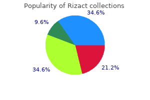
Buy cheapest rizact and rizact
In general kidney pain treatment natural purchase 5 mg rizact visa, the higher incidence of ovarian cysts (physiologic and pathologic) in reproductive-age women renders this group at higher risk of torsion compared to other age groups [9, 30, 31]. Isolated fallopian tube torsion is a rare cause of acute abdomen that is generally seen in women of reproductive age and less commonly found in prepubertal and perimenopausal women. Isolated torsion of the fallopian tube should be considered in patients with a medical history of hydrosalpinx and hematosalpinx (which complicate inflammatory conditions), pregnancy, previous tubal surgery, tubal reconstruction or ligation, and ovarian or paraovarian cysts. Nevertheless, isolated tubal torsion can occur even with an otherwise normal fallopian tube and without ipsilateral ovarian involvement. In general, isolated fallopian torsion is rare in a normal fallopian tube, and tubal abnormalities are risk factors for isolated fallopian tube torsions [32, 36]. Paraovarian and Paratubal Cysts Paratubal or paraovarian cysts account about 9% to 10% of all adnexal masses. Clinically, it is difficult to distinguish a paraovarian cyst from an ovarian cyst [37, 38]. Torsion of large paraovarian or paratubal cysts has been reported in several studies. Paratubal cysts should be taken into consideration as one of the differential diagnoses of abdominal pain in prepubertal girls [13]. A retrospective study that was done to compare adnexal torsion characteristics and torsion recurrence rates in a pre- and postmenarchal pediatric and adolescent females <18 years old with surgically diagnosed adnexal torsion found that there was a higher prevalence of paraovarian cysts on preoperative ultrasound in the postmenarchal group compared with the premenarchal group (20. Dysgerminoma is a malignant ovarian tumor that is most frequently seen during adolescence. The risk of torsion increases by at least eight-fold following tubal sterilization surgery. However, hysterectomy with ovarian conservation is not a risk factor for torsion [3, 23]. Infertility Treatments and Ovarian Torsion Ovulation induction to treat infertility may cause multiple large ovarian follicular cysts, and large cysts carry an increased risk of torsion [47, 59]. Operative laparoscopic conservative treatment (detorsion or unwinding the twisted adnexa) was performed in all of the patients. It is postulated that this is because the ovaries become displaced outside of the lesser pelvis [63]. Adnexal tumors at high risk for torsion and malignancy should be strongly considered for aggressive management during pregnancy [66]. Ovarian masses in pregnant women with adnexal masses 4 cm had a 1% to 6% lower incidence of torsion compared with ovarian masses in nonpregnant women [3, 70]. In a nutshell, pregnancy is a risk factor for torsion even in the absence of a predisposing factor. Ovarian Torsion in Neonates In neonates, complex masses may represent in utero or neonatal torsion; the risk of malignancy is extremely small in this age group. Most neonatal cysts are simple cysts and are mainly follicular cysts that originate as a result of maternal estrogens [71]. Prenatal torsion of the neonate is often diagnosed by routine ultrasonography during pregnancy in the second and third trimesters [73]. However, it is most commonly seen in adolescent (prepubertal) girls during the postovulatory period. In childhood, small cysts due to follicular development and atresia are common and not associated with a pathologic state [71]. However, ovarian tumors must be considered in children, especially in those with a large, persistent complex mass that has solid components. The most common ovarian tumor of childhood is mature cystic teratoma, followed by stromal tumors such as cystadenomas. Ovarian cysts are extremely common in adolescence because of persistent anovulation or ovulation dysfunction. Surgery was done for all patients, and the ovary was necrotic at the time of surgery in all cases. In addition, the associated medical and surgical treatment of the mass in this age group increases mortality. Recurrence of Ovarian Torsion the risk of recurrence is higher in premenarchal girls compared to postmenarchal girls. A common risk factor of recurrence of adnexal torsion in premenarchal girls is the occurrence of a first torsion episode in normal adnexa during the premenarchal period [27]. Another study examined the risk of recurrence of torsion of normal adnexa in postmenarchal women. This study found that torsion recurrence rates were higher in the twisted normal adnexa group compared to the abnormal adnexa group (P <. However, using laparoscopic management of adnexal torsion to spare adnexa by simply untwisting may predispose to recurrent torsion of normal adnexa [42]. Furthermore, torsion of normal adnexa recurs more often on the ipsilateral side [27, 42, 47]. The presentation is often nonspecific; patients my present with a variety of signs and symptoms that are often associated with other causes of acute abdomen, such as appendicitis, pyelonephritis, and nephrolithiasis [9]. Symptoms include acute-onset lower abdominal or pelvic pain; the pain fluctuates and radiates to the loin, back, or thigh. Before the sudden pain (acute), patients may experience an intermittent cramping pain for days or occasionally even for weeks. Premenarchal girls tend to mention a diffuse intermittent abdominal pain as the main presenting symptom because it is difficult for them to localize the pain. In addition, the clinical presentation of adnexal torsion in pregnant and nonpregnant women is the same [20, 23, 60, 80]. On abdominal examination, generalized abdominal tenderness, localized guarding, and rebound and palpable abdominal mass may be found. A clinical/pathologic study of torsion of ovarian tumor found palpable abdominal masses in the majority of cases (79. On vaginal examination, cervical excitation, adnexal tenderness, and adnexal mass may be seen [48]. Diagnosis Torsion of the ovary around its ligamentous supports may result in loss of its blood supply. Delay or misdiagnosis can result in the loss of the affected ovary and subsequent reduced reproductive capacity. Moreover, the diagnosis of this condition can be difficult due to a vague clinical presentation, particularly in intermittent torsion, and the differential diagnosis can include several other gynecologic and surgical emergencies. Adnexal/Ovarian Torsion 141 Clinical Presentation and Differential Diagnosis the ovaries are often difficult to palpate, so physical examination findings often do not suggest the diagnosis. Renal colic typically presents with sudden onset of severe unilateral colicky pain radiating from the loin to the groin, which comes in waves, very similar to torsion. In appendicitis, the pain is localized to the right iliac fossa, with localized guarding and tenderness. A history of sudden-onset, stabbing, sharp pain should raise the suspicion of hemorrhage from a functional cyst. Fibroid degeneration and torsion of pedunculated fibroids are not unusual and should be considered in women known to have fibroids. A rupture of a vessel over a fibroid is also a rare but reported cause of acute abdominal pain and intraperitoneal hemorrhage [83, 84]. Laboratory Investigations Basic laboratory investigations need to be performed to rule out other causes of acute abdominal or pelvic pain and include urine analysis, pregnancy tests, and full blood count. No single or combined markers have been identified that improve diagnostic accuracy in adnexal torsion. Torsion results in an ischemic insult to the ovary, and markers of ischemia or ischemia-reperfusion injury could theoretically be raised in the serum of women with torsion. The most common and easiest marker to examine is C-reactive protein, an acute phase protein that is increased in the presence of inflammation; the white blood cell count is also often measured and is increased in approximately 50% of women with adnexal torsion [88]. Unfortunately, neither of these markers has been found to be useful in the diagnosis of torsion because of low sensitivity and specificity.
| Comparative prices of Rizact | ||
| # | Retailer | Average price |
| 1 | Price Chopper Supermkts | 153 |
| 2 | Foot Locker | 688 |
| 3 | Office Depot | 520 |
| 4 | Delhaize America | 808 |
| 5 | Ross Stores | 549 |
| 6 | A&P | 385 |
| 7 | Trader Joe's | 460 |
| 8 | True Value | 290 |
Purchase cheap rizact on-line
Esser S knee pain treatment youtube purchase rizact cheap online, Wolburg K, Wolburg H, et al: Vascular endothelial growth factor induces endothelial fenestrations in vitro. Rasch R: Prevention of diabetic glomerulopathy in streptozotocin diabetic rats by insulin treatment. Osterby R: Morphometric studies of the peripheral glomerular basement membrane in early juvenile diabetes. Reiser J, Kriz W, Kretzler M, et al: the glomerular slit diaphragm is a modified adherens junction. Drenckhahn D, Schnittler H, Nobiling R, et al: Ultrastructural organization of contractile proteins in rat glomerular mesangial cells. Petermann A, Fees H, Grenz H, et al: Polymerase chain reaction and focal contact formation indicate integrin expression in mesangial cells. Schlondorff D: the glomerular mesangial cell: an expanding role for a specialized pericyte. Zenker M, Aigner T, Wendler O, et al: Human laminin beta2 deficiency causes congenital nephrosis with mesangial sclerosis and distinct eye abnormalities. Hakroush S, Cebulla A, Schaldecker T, et al: Extensive podocyte loss triggers a rapid parietal epithelial cell response. Barajas L: the ultrastructure of the juxtaglomerular apparatus as disclosed by three-dimensional reconstructions from serial sections. Hackenthal E, Paul M, Ganten D, et al: Morphology, physiology, and molecular biology of renin secretion. Sauter A, Machura K, Neubauer B, et al: Development of renin expression in the mouse kidney. Pricam C, Humbert F, Perrelet A, et al: Gap junctions in mesangial and lacis cells. Kaissling B, Kriz W: Variability of intercellular spaces between macula densa cells: a transmission electron microscopic study in rabbits and rats. Barajas L, Muller J: the innervation of the juxtaglomerular apparatus and surrounding tubules: a quantitative analysis by serial section electron microscopy. Bachmann S, Kriz W: Histotopography and ultrastructure of the thin limbs of the loop of Henle in the hamster. Dorup J: Structural adaptation of intercalated cells in rat renal cortex to acute metabolic acidosis and alkalosis. Bergeron M, Guerette D, Forget J, et al: Three-dimensional characteristics of the mitochondria of the rat nephron. Bergeron M, Thiery G: Three-dimensional characteristics of the endoplasmic reticulum of rat renal tubule cells: An electron microscopy study in thick sections. Coudrier E, Kerjaschki D, Louvard D: Cytoskeleton organization and submembranous interactions in intestinal and renal brush borders. Kerjaschki D, Noronha-Blob L, Sacktor B, et al: Microdomains of distinctive glycoprotein composition in the kidney proximal tubule brush border. Angermuller S, Leupold C, Zaar K, et al: Electron microscopic cytochemical localization of alpha-hydroxyacid oxidase in rat kidney cortex. Bachmann S, Velazquez H, Obermuller N, et al: Expression of the thiazide-sensitive Na-Cl cotransporter by rabbit distal convoluted tubule cells. Kaissling B, Bachmann S, Kriz W: Structural adaptation of the distal convoluted tubule to prolonged furosemide treatment. Loffing J, Loffing-Cueni D, Hegyi I, et al: Thiazide treatment of rats provokes apoptosis in distal tubule cells. Bachmann S, Bostanjoglo M, Schmitt R, et al: Sodium transportrelated proteins in the mammalian distal nephron-distribution, ontogeny and functional aspects. Ackermann D, Gresko N, Carrel M, et al: In vivo nuclear translocation of mineralocorticoid and glucocorticoid receptors in rat kidney: differential effect of corticosteroids along the distal tubule. Lonnerholm G, Ridderstrale Y: Intracellular distribution of carbonic anhydrase in the rat kidney. Kaissling B: Structural aspects of adaptive changes in renal electrolyte excretion. Stanton B, Janzen A, Klein-Robbenhaar G, et al: Ultrastructure of rat initial collecting tubule. Quantitative correlation of structure and function in the normal and injured rat kidney, Berlin, 1982, Springer-Verlag. Wolgast M, Larson M, Nygren K: Functional characteristics of the renal interstitium. Kaissling B, Le Hir M: Characterization and distribution of interstitial cell types in the renal cortex of rats. Schiller A, Taugner R: Junctions between interstitial cells of the renal medulla: a freeze-fracture study. A histological study on the number of droplets in salt depletion and acute salt repletion. Guan Y, Chang M, Cho W, et al: Cloning, expression, and regulation of rabbit cyclooxygenase-2 in renal medullary interstitial cells. Zhuo J, Dean R, Maric C, et al: Localization and interactions of vasoactive peptide receptors in renomedullary interstitial cells of the kidney. Barajas L, Powers K, Wang P: Innervation of the late distal nephron: an autoradiographic and ultrastructural study. Newstead J, Munkacsi I: Electron microscopic observations on the juxtamedullary efferent arterioles and Arteriolae rectae in kidneys of rats. This rate of blood flow, approximately 400 mL per 100 g of tissue per minute, is significantly greater than that observed in other vascular beds considered to be well perfused, such as heart, liver, and brain. Although the metabolic energy requirement of urine production is relatively high (approximately 10% of basal O2 consumption), the renal arteriovenous O2 difference reveals that blood flow far exceeds metabolic demands. In fact, the high rate of blood flow is essential to the process of urine formation as described later. The kidney contains several distinct microvascular networks, including the glomerular microcirculation, the cortical peritubular microcirculation, and the unique microcirculations that nourish and drain the inner and outer medulla. In Chapter 2 the gross anatomy of the kidney and arrangement of tubular segments were described. Therefore, complete obstruction of an arterial segmental vessel results in ischemia and infarction of the tissue in its area of distribution. In fact, ligation of individual segmental arteries has frequently been performed in the rat to reduce renal mass and produce the remnant kidney model of chronic renal failure. Morphologic studies in this model reveal the presence of ischemic zones adjacent to the totally infarcted areas. Thetissuehasbeenmade transparent by dehydration and clearing procedures after injection. A single nephron is also drawn to show the interlobular artery entering into the glomerular capillary network. These vessels, which most often supply the lower pole,5 may be the sole arterial supply of some part of the kidney. Within the renal sinus of the human kidney, division of the segmental arteries gives rise to the interlobar arteries. Glomeruli are classified according to their position within the cortex as superficial. The capillary network of each glomerulus is connected to the postglomerular (peritubular) capillary circulation by way of the efferent arterioles. Both the nomenclatures and the patterns of the renal arterial system are similar in most of the mammals commonly used experimentally. For example, the main arterial branches that lie beside the medullary pyramid are called interlobar, even in animals such as rodents that have but a single lobe. Efferent vessels emerging from glomeruli branch to form the cortical postglomerular capillary network. However, on the basis of studies of the vasculature of a unique set of juxtamedullary nephrons, most of the preglomerular pressure drop between the arcuate artery and the glomerulus occurs along the afferent arteriole. If the substance is neither metabolized nor synthesized in the kidney then its rate of appearance in the urine equals its rate of extraction from the blood. The blood extraction rate is equal to the renal plasma flow rate multiplied by the difference between the arterial and renal venous plasma concentrations. Nephrons originating from midcortical glomeruli have proximal and distal convoluted tubule segments lying close to the interlobular axis in the region above the glomerulus of origin.
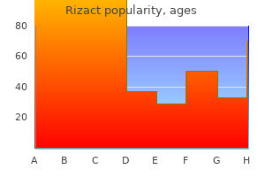
Trusted 5mg rizact
In livers from these mice pain treatment with opioids purchase 10mg rizact fast delivery, Angpt2 expression is elevated whereas Angpt1 expression is significantly attenuated, although Tie2 receptor expression in unchanged. Because Angpt1 and Angpt2 have distinctive roles in endothelial sprouting, vascular stabilization, and remodeling,315 it can be argued that Sox17 and Sox18 play redundant or synergistic roles in coordinating normal postnatal angiogenesis. There is evidence from other model systems that vascular development is required for patterning and terminal differentiation of adjacent tissues. For example, vascular signals and basement membrane produced by adjacent endothelial cells are required for differentiation of the islet cells of the pancreas. Understanding co-patterning between the vasculature and its immediate neighbors will be a challenging task, given the complex nature of various tissue-tissue communications involved, although progress in this effort will likely be facilitated by a growing arsenal of genetic tools. Although most of these cells cease to express renin in the adult, they appear to reexpress renin in stress conditions and are recruited to the afferent arteriole. Deletion of Dicer1 from renin-expressing cells results in severe reduction in the number of juxtaglomerular cells, reduced renin production, and lower blood pressure. Postmitotic mature podocytes, on the other hand, normally lose E-cadherin expression and atypically express vimentin, an intermediate filament protein more common among mesenchymal cells but absent in most epithelial cells. Podocytes ensheathe the glomerular capillaries, with their foot processes effectively forming the final layer of the glomerular filtration barrier. Proteinuria develops from loss of these genes, thus underscoring the importance of normal podocyte maturation in the establishment of the glomerular filtration barrier. Nephrin, a huge transmembrane adhesion molecule with multiple immunoglobulin-like motifs, has been shown to be a structural component of the slit diaphragm. A, Scanning electron micrograph of podocytes and their interdigitating foot processes (falsely colored to highlight the spatial relationships between neighboring foot processes). The topologic organization of slit diaphragm components remains unknown but it is likely that the larger adhesion molecules nephrin and Fat1 bridge juxtaposed foot processes. In effaced foot processes, the actin cytoskeleton has been remodeled into a meshlike network of randomly oriented filaments. Genetic removal of Flt1 from podocytes leads to foot process effacement and proteinuria. The secreted sFlt1 has been shown to act as an autocrine factor in podocytes, associating with glycosphingolipids and mediating podocyte cell adhesion, nephrin phosphorylation, and actin polymerization. It is likely that endothelial cells also produce factors required for terminal differentiation of podocytes, although these factors are currently unknown. Lumens form during glomerulogenesis through apoptosis of a subset of endothelial cells. Surviving endothelial cells flatten considerably and develop fenestrations and complex glycocalyces. Mendelsohn C, Batourina E, Fung S, et al: Stromal cells mediate retinoid-dependent functions essential for renal development. Muller U, Wang D, Denda S, et al: Integrin alpha8beta1 is critically important for epithelial-mesenchymal interactions during kidney morphogenesis. Michos O, Cebrian C, Hyink D, et al: Kidney development in the absence of Gdnf and Spry1 requires Fgf10. Saxen L: Organogenesis of the kidney, Cambridge, 1987, Cambridge University Press. Cui S, Ross A, Stallings N, et al: Disrupted gonadogenesis and male-to-female sex reversal in Pod1 knockout mice. Fierlbeck W, Liu A, Coyle R, et al: Endothelial cell apoptosis during glomerular capillary lumen formation in vivo. Giardino L, Armelloni S, Corbelli A, et al: Podocyte glutamatergic signaling contributes to the function of the glomerular filtration barrier. Levinson R, Mendelsohn C: Stromal progenitors are important for patterning epithelial and mesenchymal cell types in the embryonic kidney. Grobstein C: Inductive epitheliomesenchymal interaction in cultured organ rudiments of the mouse. Miyamoto N, Yoshida M, Kuratani S, et al: Defects of urogenital development in mice lacking Emx2. Sariola H, Saarma M, Sainio K, et al: Dependence of kidney morphogenesis on the expression of nerve growth factor receptor. Durbeej M, Soderstrom S, Ebendal T, et al: Differential expression of neurotrophin receptors during renal development. Sainio K, Saarma M, Nonclercq D, et al: Antisense inhibition of low-affinity nerve growth factor receptor in kidney cultures: power and pitfalls. Tufro A, Teichman J, Woda C, et al: Semaphorin3a inhibits ureteric bud branching morphogenesis. Berry R, Harewood L, Pei L, et al: Esrrg functions in early branch generation of the ureteric bud and is essential for normal development of the renal papilla. Ji J, Li Q, Xie Y, et al: Overexpression of Robo2 causes defects in the recruitment of metanephric mesenchymal cells and ureteric bud branching morphogenesis. Gao X, Chen X, Taglienti M, et al: Angioblast-mesenchyme induction of early kidney development is mediated by Wt1 and Vegfa. Vainio S, Lin Y: Coordinating early kidney development: lessons from gene targeting. Chi X, Michos O, Shakya R, et al: Ret-dependent cell rearrangements in the Wolffian duct epithelium initiate ureteric bud morphogenesis. Packard A, Georgas K, Michos O, et al: Luminal mitosis drives epithelial cell dispersal within the branching ureteric bud. Schneider T, Reiter C, Eule E, et al: Restricted expression of the irreC-rst protein is required for normal axonal projections of columnar visual neurons. Venugopala Reddy G, Reiter C, Shanbhag S, et al: Irregular chiasm-C-roughest, a member of the immunoglobulin superfamily, affects sense organ spacing on the Drosophila antenna by influencing the positioning of founder cells on the disc ectoderm. Auerbach R, Grobstein C: Inductive interaction of embryonic tissues after dissociation and reaggregation. Grobstein C: Morphogenetic interaction between embryonic mouse tissues separated by a membrane filter. Laclef C, Souil E, Demignon J, et al: Thymus, kidney and craniofacial abnormalities in Six 1 deficient mice. Takemoto M, He L, Norlin J, et al: Large-scale identification of genes implicated in kidney glomerulus development and function. Yoshida Y, Miyamoto M, Bo X, et al: Overview of kidney and urine proteome databases. Miyamoto M, Yoshida Y, Taguchi I, et al: In-depth proteomic profiling of the normal human kidney glomerulus using twodimensional protein prefractionation in combination with liquid chromatography-tandem mass spectrometry. Batourina E, Tsai S, Lambert S, et al: Apoptosis induced by vitamin A signaling is crucial for connecting the ureters to the bladder. Zhao H, Kegg H, Grady S, et al: Role of fibroblast growth factor receptors 1 and 2 in the ureteric bud. Watanabe T, Costantini F: Real-time analysis of ureteric bud branching morphogenesis in vitro. Hoshi M, Batourina E, Mendelsohn C, et al: Novel mechanisms of early upper and lower urinary tract patterning regulated by RetY1015 docking tyrosine in mice. Fougerousse F, Durand M, Lopez S, et al: Six and Eya expression during human somitogenesis and MyoD gene family activation. Sajithlal G, Zou D, Silvius D, et al: Eya 1 acts as a critical regulator for specifying the metanephric mesenchyme. Vainio S, Muller U: Inductive tissue interactions, cell signaling, and the control of kidney organogenesis. Uchiyama Y, Sakaguchi M, Terabayashi T, et al: Kif26b, a kinesin family gene, regulates adhesion of the embryonic kidney mesenchyme. Kobayashi H, Kawakami K, Asashima M, et al: Six1 and Six4 are essential for Gdnf expression in the metanephric mesenchyme and ureteric bud formation, while Six1 deficiency alone causes mesonephric-tubule defects. Bouchard M, Souabni A, Mandler M, et al: Nephric lineage specification by Pax2 and Pax8. Grote D, Souabni A, Busslinger M, et al: Pax 2/8-regulated Gata 3 expression is necessary for morphogenesis and guidance of the nephric duct in the developing kidney.
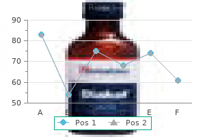
Generic rizact 5mg without a prescription
The trabeculae of the primary and secondary spongiosa may also be decreased in number and thickness joint and pain treatment center thousand oaks purchase rizact 5 mg with amex, and there is an inappropriate retention of hypertrophic chondrocytes within the primary spongiosa. While most commonly observed in growing rodents, the physeal effects of angiogenesis inhibitors have also been described in nonhuman primates (Ryan 1999). Although generally characterized by an increased thickness of the physis, other characteristics such as effects on the primary and secondary spongiosa and age dependency of the lesion appear to vary depending on the signaling pathway that has been interrupted. In addition to these pharmacologic agents that directly impact cell signaling, physeal and/or articular cartilage lesions have been induced by other agents as well. Semicarbazide administration results in a spectrum of osteochondral lesions of both physeal and articular cartilage (Takahashi et al. In addition to thickening of the growth plate, there is degeneration of hypertrophic chondrocytes and deformation and fissures of the articular cartilage. The metaphyseal bone immediately subjacent to the physis becomes thickened and disorganized. Thickening of the articular cartilage does not occur, but there are soft tissue changes, including formation of a vascular pannus and thickening of the synovial lining of the joint. In aging F344 rats, focal degenerative changes and fissures between the physis and epiphysis have been described (Yamasaki and Inui 1985). In dog toxicity studies, focal defects and/or overt necrosis of physeal cartilage can be observed (Yamasaki 1995). In toxicity studies, this effect will be more frequently observed in young rats due to their rapid bone growth. Similar lesions have been reported with a variety of xenobiotics, which affect signaling pathways important in endochondral ossification. Note the marked increased in physeal thickness in the young rat compared with the older rat. Distinct from decreases in physeal thickness is the relatively infrequent case where there has been premature closure of the physis such as has been observed following retinoid administration (De Luca 2000). Osteoblast hyperplasia was characterized by single or multiple foci of well-differentiated osteoblast-like cells. The focal lesions may fill enlarged trabecular spaces but lack significant disruption of the adjacent bone. The interior of the lesion has a paucity of cells, 726 Toxicologic Pathology and lacunae may lack cells all together. At the periphery of the lesion, there are often more delicate trabeculae covered by a layer of osteoblasts. The external surface of the neoplasm is smooth, and there is no evidence of invasion. Osteoma occurs at a low incidence in control rats and mice in 2-year carcinogenicity studies, and increased incidences have been observed following glucocorticoid (Zwicker and Eyster 1996) and parathyroid hormone fragment administration (Vahle et al. Microscopically, the lesion is characterized by an intramedullary pattern of disorganized trabeculae of immature bone, often accompanied by a fibrovascular stroma. There are typically moderate numbers of large, well-differentiated osteoblasts arranged along trabecular surfaces. There is typically minimal cytologic atypia, and low numbers of mitotic figures may be present. In studies where osteoblastoma has been observed, the lesion was often first recognized at a lower magnification as a focus of disorganized trabeculae. The presence of tumor osteoid is the essential diagnostic feature and may be limited to small amounts of wispy, eosinophilic matrix within a highly cellular lesion. Due to their often large and destructive nature, osteosarcomas are often visible at necropsy and often have gross or microscopic metastasis to the lung. While most frequently affecting the appendicular skeleton, they may arise at a variety of sites and, although rare, may occur as an extraskeletal osteosarcoma (Pace et al. Osteosarcomas have been induced in rodents by a variety of agents, including radiation, viruses, glucocorticoids (Zwicker and Eyster 1996), and parathyroid hormone (Vahle et al. The chondrocytes are well differentiated and are sparsely populated throughout an abundant matrix. These neoplasms are highly cellular and composed of generally well-differentiated, large basophilic cells within lacunae, surrounded by variable amounts of cartilaginous matrix. These tumors are described as being minimally invasive but readily metastasize to the lung (Gregson and Offer 1981). Osteochondromas are focal proliferations of lamellar bone covered by a cap of cartilage that consists of irregularly arrayed, hypertrophic chondrocytes. These lesions are to be differentiated from osteophytes, which form in the epiphysis in the setting of degenerative joint disease. Histologically, the neoplasm is characterized by pleomorphic-shaped cells, intermixed with variable amounts of collagenous matrix. Similar to fibrosarcoma in other locations, the mitotic index is increased, and cytologic features include significant nuclear and cellular pleomorphism. If possible, it is important to obtain adequate sections to clarify if the lesion truly represents a primary tumor of bone, rather than an extension of a soft tissue fibrosarcoma. A key differential is the occurrence of osteosarcomas, which have a primarily fibroblastic component; however, these lesions would contain at least a limited amount of tumor osteoid. In the early stages, a reduction or lack of uniformity of staining of the articular cartilage can be detected. Although this can be detected in routinely stained H&E sections as a focal loss of basophilia, early changes are best characterized via special stains such as toluidine blue. The lesion progresses to include fibrillation, irregularities, erosions, and ulcerations of the articular surface, and clusters or nests of chondrocytes (chondrones) in the adjacent cartilage. As the cartilage is progressively damaged, biomechanical changes begin to directly impact the subchondral bone, resulting in an increased thickness and density of the subchondral bone plate. In some cases, a cystlike space (subchondral bone cyst) will develop subjacent to the region of articular cartilage damage. Additional sequelae can include the formation of osteophytes on the periarticular bone surface, synovial cell hyperplasia, and joint capsule fibrosis and ossification. Due to the wide spectrum of histologic features that may occur, the nomenclature used to tabulate the findings in toxicity studies could include either a general term of osteoarthritis or a listing of specific components of the lesion. The decision on which terms to use depends on the nature and severity of the particular change. In studies supporting osteoarthritis research, it is typical to score various components of the lesion (Gerwin et al. As a spontaneous change, the frequency of osteoarthritis varies with the age and species but can be observed in any of the commonly used laboratory species used in toxicology studies. Multiple strains of mice have age-related spontaneous cartilage degeneration that most often affects the medial compartment of the knee joint and can be quite severe. The lesion is most prominent in the sternum, and histologic features include focal necrosis with loss of chondrocytes, fragmentation of the matrix, nest of proliferating chondrocytes adjacent to the lesion, and, in some cases, distinct areas of cavitation. Caloric restriction is reported to decrease the severity of the change (Kawahara et al. Often referred to as drug-induced arthropathies, these entities are primarily characterized by degeneration of the articular cartilage and lack a significant inflammatory component. One of the most well-characterized articular cartilage toxicities is quinolone-induced arthropathy (Burkhardt et al. In severe cases, the changes are associated with lameness during the in-life evaluation, and at necropsy, affected animals may have gross lesions of vesiculation and detachment of the superficial layer of cartilage in multiple joints. It has been suggested that the pathogenesis is due to an inhibition of proteoglycan synthesis (Yabe et al. Semicarbazide administration results in a spectrum of osteochondral lesions including chondrocyte degeneration and deformation and fissures of the articular cartilage (Takahashi et al. Infections within the joint typically present as the presence of edema, congestion, and an infiltrate of inflammatory cells of varying composition within the synovium and may expand the periarticular soft tissues. With time, inflammation of the joint leads to synovial hyperplasia, pannus formation, fibrosis, and, ultimately, erosion or ulceration of the articular cartilage with loss of subchondral bone.
Syndromes
- Infertility
- Hemorrhoids (common cause of bright red blood)
- The amount swallowed
- Kidney, liver, nerve, and muscle conditions
- Diabetic acidosis (also called diabetic ketoacidosis and DKA) develops when substances called ketone bodies (which are acidic) build up during uncontrolled diabetes.
- Cramping
- Dark urine
Cheapest generic rizact uk
Nerve bundles follow the arterioles and innervate the smooth muscle of the blood vessels or terminate within the hematopoietic cell compartment (Travlos 2006a) advanced pain treatment center ky purchase rizact on line amex. Endosteal cells with a thin layer of connective tissue line the inner surface of the bone cavities and the trabecular meshwork or spicules in the cavities (Sharkey and Hill 2010). Spindloid to stellate stromal cells extend from the endosteal surface into the hematopoietic space and produce factors involved in hematopoiesis in addition to providing a supporting network for hematopoietic cells, adipocytes, and blood vessels via production of structural fibrils (collagen, reticulin, laminin, fibronectin; Sharkey and Hill 2010). Adipocyte tissue within the marrow consists of both brown and white types, likely providing both structural and hematopoietic support (Sharkey and Hill 2010). The hematopoietic compartment is extravascular and located in the spaces between the marrow venous sinuses. In adults, hematopoiesis is closely associated with bone tissue and cells (osteoblasts and osteoclasts) and occurs within 200 m of bone (Valli and Jacobs 2000). There is also a close relationship between the hematopoietic cells and the venous lining cells, including the flat endothelial cells and the reticular outer layer. The basement lamina between the sinusoids and hematopoietic cells is thin and interrupted. The endothelial cells do not have tight junctions, a specialization of marrow endothelial cells facilitating passage of blood cells into the vascular space (Lichtman 1981). Within the hematopoietic compartment, the erythroid and megakaryocytic cells are adjacent to the venous sinuses, while the myeloid cells (granulocytes, monocytes/macrophages) and lymphocytes are located near the endosteum and arterioles (Sharkey and Hill 2010). Hematopoiesis occurs in cords composed of differentiating hematopoietic cells, stromal cells, adventitial reticular cells, adipocytes, and endothelial cells. Megakaryocytes (Mega) are located near venous sinuses, shedding platelets directly into the sinus. In normal marrow, the proportion of myeloid (paler staining cells) to erythroid (darkly staining cells) is approximately 1:1, with predominantly mature megakaryocytes distributed throughout the compartment. The first identifiable erythroid precursor is the rubriblast, followed by the prorubricyte, basophilic rubricyte, polychromatophilic rubricyte, metarubricyte, reticulocyte, and erythrocyte. During the maturation process, erythroid cells decrease in size with condensation of the nucleus, resulting in a decreased nuclear-cytoplasm ratio. Additionally, erythroid cells lose cytoplasmic organelles, acquire hemoglobin resulting in decreased cytoplasmic basophilia, and undergo nuclear pyknosis, with eventual extrusion of the nucleus to form a reticulocyte. Erythropoiesis occurs within erythroblastic islands featuring maturing erythroid cells surrounding a central macrophage. Myeloid precursors display round to indented to segmented eccentrically located nuclei and moderately basophilic cytoplasm. The first identifiable myeloid precursor is the myeloblast, followed by the promyelocyte/progranulocyte, myelocyte, metamyelocyte, band forms, and mature segmented neutrophils, eosinophils, and basophils. Similar to the erythroid series, as myeloid cells mature, there is a decrease in size with a concomitant decrease in the nuclear-cytoplasmic ratio. As maturation progresses, secondary granules appear consistent with the granulocytic cell type, that is, neutrophilic, eosinophilic, or basophilic granules. In rats and mice, as myeloid cells mature from the promyelocyte stage, the cells may develop as "ring forms" characterized by a generally round nucleus with a central "hole. In marrow preparations, the myeloid ring forms are found together with the typical indented to bandshaped myeloid cells. Monocytes mature from monoblasts, which are similar in appearance to the early myeloid/granulocytic precursor cells. Immature lymphoid cells are larger and more basophilic than the mature lymphocytes, which are uniformly distributed within the hematopoietic compartment. Differentiation between erythroid and lymphoid cells is difficult upon examination of routine histologic sections. Platelet production begins with the megakaryoblast, which is a large, single nucleated cell with deeply basophilic cytoplasm. The megakaryoblast undergoes endomitosis, resulting in a larger multinucleated cell with increased amounts of moderately basophilic cytoplasm. As maturation progresses, the megakaryocyte cytoplasm develops numerous eosinophilic granules. Emperipolesis, the movement of blood cells (neutrophils, erythrocytes, lymphocytes) within megakaryocytes, is fairly common with up to 5 percent of megakaryocytes containing blood cells in normal bone marrow from humans (Centurione et al. Emperipolesis differs from phagocytosis in that the blood cells exist temporarily within the megakaryocyte (Harvey 2001). Marrow smears prepared from male cynomolgus monkey dosed with vehicle control for 4 weeks. Myeloid cytoplasmic granulation is more apparent in nonhuman primates compared to rodent or dog. Mixture of immature and mature erythroid and myeloid cells with ring forms and lower numbers of small lymphocytes (note two lymphocytes in upper right corner of field). Inset photos show progression of ringed metamyelocytes with widening of central hole. Ring forms in the myeloid series are a normal finding in rodent marrow smears, but are considered a dysplastic change in other laboratory animal species. Stem cells have two defining characteristics: capability for self-renewal and capacity to form differentiated or specialized cell types (Overmann et al. This asymmetric cell division allows for the maintenance of a pool of undifferentiated stem cells and development of lineage-specific hematopoietic precursors (Overmann et al. For example, the erythroblastic island consisting of a central macrophage surrounded by maturing erythroid cells was one of the first identified hematopoietic niches. In this specialized microenvironment, the central macrophage assists with erythropoiesis by providing iron for hemoglobin synthesis and is important in the removal of the erythroid nucleus during maturation and phagocytosis of defective cells (Abboud and Lichtman 2001; Chasis and Mohandas 2008; Sharkey and Hill 2010). Basophils derived from unipotential BaPs mature in the bone marrow, whereas mast cell progenitors leave the marrow, enter circulation, and mature in peripheral tissues (Radin and Wellman 2010). The bipotential and unipotential progenitor cells will continue to differentiate into the morphologically identifiable rubriblast, megakaryoblast, myeloblast, or monoblast precursor cells, followed by multiple cellular divisions to form mature erythrocytes, platelets, granulocytes, or monocytes. B-lymphopoiesis continues in the marrow, whereas T-lymphocyte stem cells migrate to the thymus (Kaushansky 2006; Overmann et al. In this alternative model, murine lymphoid development is dependent on epigenetic silencing of myeloid genes with a gradual stop of myeloid maturation (reviewed in Doulatov et al. Once maturation is complete, erythrocytes and leukocytes migrate through the bone marrow venous sinuses to enter the bloodstream, whereas platelets are released directly into the bloodstream through cytoplasmic projections of the megakaryocytes into the venous sinuses (Gasper 2000; Travlos 2006a). Following release from the bone marrow, reticulocytes mature to erythrocytes within 24 to 48 hours in the blood or spleen (Fernandez and Grindem 2000). Red blood cell production is complete within 4 days, granulocyte production is complete within 6 days, and thrombopoiesis is completed in 4 days (Valli et al. Mature red blood cell life span is approximately 30 days in mice, 50 days in rats, 120 days in dogs, and 100 days in nonhuman primates (Barnhart 2010; Valli et al. Mature granulocytes are replaced three to four times a day with a peripheral circulation time of 6 to 8 hours, and they do not reenter blood circulation following egress into the tissues. Platelet life span is approximately 3 to 5 days in rabbits and rats and 7 to 9 days in larger species (Valli et al. Nonclinical toxicologic assessment of the hematopoietic system begins with evaluation of hematology results and histopathologic examination of bone marrow. In most instances, information derived from in-life findings, toxicokinetic and hematologic data, and bone marrow histopathologic evaluations are sufficient for determination of compound-related effects on hematopoiesis. The decision to perform cytologic examination of bone marrow smears should be made on a case-by-case basis, following discussions between the anatomic and clinical pathologist and in consultation with the study director and project team safety expert. Additional considerations for performing marrow smear examinations include dose response, tolerability, severity, and reversibility of compound-related findings as well as the stage of compound development and impact or usefulness of cytologic evaluation on the clinical safety plan (Reagan et al. If smear assessments are performed in early toxicity studies, cytologic examinations may not be needed for later studies, although this should be decided on a case-by-case basis. Routine bone marrow smear evaluations are not recommended for carcinogenicity studies (Reagan et al. Additional evaluations of bone marrow tissue include flow cytometric analysis, which may be performed in conjunction with bone marrow smear evaluation or as a stand-alone assay and electron microscopy and clonogenic assays for mechanistic information on compound-related changes in hematopoiesis (Reagan et al. Histopathologic examination of bone marrow is used to assess marrow cellularity, megakaryocyte numbers and morphology, presence of focal lesions such as inflammation or necrosis, estimation of myeloid and erythroid proportions, and assessment of iron stores, especially in conjunction with iron staining. Cytokines and growth factors that support the survival, proliferation, or differentiation of each type of cell are shown in red. For simplicity, the three types of granulocyte progenitor cells are not shown; in reality, distinct progenitors of neutrophils, eosinophils, and basophils or mast cells exist and are supported by distinct transcription factors and cytokines.
Order 10mg rizact amex
While there are few direct-acting thyroid carcinogens in laboratory animals pain treatment center sawgrass purchase line rizact, several nongenotoxic compounds, including natural goitrogens, drugs, environmental chemicals, and other agents, significantly influence the development of proliferative lesions in rodent thyroid. Virtually all compounds that induce thyroid follicular tumors in rodents have been shown to interfere with this negative feedback system (Thomas and Williams 1999). Any compound that interferes with thyroid hormone synthesis, secretion, or metabolism will significantly impair the hypothalamic-pituitary-thyroid hormone axis and potentially result in thyroid follicular hyperplasia and tumorigenesis. Xenobiotic compounds that alter any step in the synthesis, secretion, or metabolism of thyroid hormones can lead to goiter or neoplasia in susceptible species. Interference with uptake of iodine by the thyroid gland occurs in rodents exposed to anions that act as competitive inhibitors of iodide transport, including perchlorate, thiocyanate, and pertechnetate (Capen 1994; Crofton 2008). Perchlorate competes with iodine for uptake by the thyroid gland, which results in the decreased availability of iodine for the thyroid gland, which in turn results in hypothyroidism (Yu et al. The kinetics of inhibition of iodine uptake by perchlorate in humans and rodents is very similar; however, species differences in thyroid hormone biology prevent extrapolation between rodents and humans in terms of downstream adverse effects in humans (Miller et al. Interference with thyroid peroxidase is caused by various thionamides (thiouracil, thiourea, propylthiouracil, methimazole, aminotriazole, carbimazole), aniline derivatives (sulfonamides, para-aminobenzoic acid, para-aminosalicylic acid, amphenone), and phenols (resorcinol, phloroglucinol, 2,4-dihydroxybenzoic acid) (Heath and Littlefield 1984; Takayama et al. However, thiocarbamide drugs (methimazole, propylthiouracil, carbimazole) are used to block thyroid hormone synthesis in hyperthyroid patients, and occupational exposure to workers exposed to chemicals with similar chemical structure (aminotriazole herbicide, thiourea) can cause hypothyroidism (Baccarelli et al. Excess dietary iodide is known to cause inhibition of thyroid hormone synthesis in animals and humans, leading to decreased thyroid hormone levels, goiter, and hypothyroidism (Capen 1994). Iodide excess interferes with uptake of iodine by the thyroid gland and with normal colloid proteolysis by decreasing lysosomal protease activity, thereby blocking the release of T3 and T4 from thyroglobulin. As previously discussed, various pigments resulting from metabolism of compounds such as minocycline, other tetracycline derivatives, or the antipsychotic clozapine accumulate in the thyroid gland and alter thyroid function. Inhibition of thyroid hormone secretion due to abnormal colloid proteolysis in Gunn rats leads to altered thyroid function (Gomba et al. Inhibition of 5-deiodinase, the major enzyme responsible for conversion of T4 to active T3, leads initially to increases in serum T4. This is followed by conversion of T4 by the same enzyme, 5-deiodinase, leading to marked increases in the inactive form of T3, reverse T3 (r T3). Induction of hepatic microsomal enzymes by xenobiotics is a well-known phenomenon in rodents that occurs due to species-specific differences in the metabolism of thyroid hormones. Chemical induction of microsomal hepatic enzymes causes increased metabolism and excretion of thyroid hormones. Phenobarbital induces follicular cell hypertrophy and hyperplasia and increases thyroid weight in rodents through increased hepatic clearance of T4 due to glucuronidation. Even in areas where there is severe iodine deficiency and endemic goiter reported in human subjects, there is no evidence of increased risk for thyroid neoplasia. Furthermore, such compounds would likely have a significant toxicologic effect in humans before exposure would increase risk for thyroid tumorigenesis (McClain 1995). However, there are a number of mutagens that have been used to induce thyroid tumors in laboratory animals. Aromatic amines, polycyclic hydrocarbons, azo dyes, dichlorobenzene, 2-acetylaminofluorene, nitrosamines, and nitrosoureas have all been used to induce thyroid tumors in rodents (McClain 1989; Thomas and Williams 1999). A significant mutagenic inducer of thyroid neoplasms in humans and laboratory animals is irradiation (x-ray or iodine radioisotopes). Studies have indicated an increased incidence of thyroid neoplasia in children in areas of nuclear accidents and who are exposed to a high degree of irradiation, such as at the Chernobyl nuclear power plant (Jargin 2011), the Marshall Islands (Land et al. Follicular carcinomas induced by irradiation occur at a higher rate in males than females, whereas castration reduces the incidence in irradiated males versus females (Capen 1994). Goitrogens, including dietary deficiency or excess of iodine, are powerful promoters of thyroid carcinogenesis and have been used to significantly increase the induction of thyroid neoplasia (Kanno et al. Working in concert with thyroid C-cells, the parathyroid is responsible for maintaining physiologic levels of calcium and phosphorous necessary for adequate bone integrity and basic metabolic processes. Diseases or compounds that cause alterations in calcium homeostasis can alter the response of the parathyroid, resulting in proliferative lesions including tumorigenesis. Rats have relatively few of these secretory granules, while they are numerous in the mouse (Capen and Rosol 1989). Active chief cells have increased numbers of organelles and secretory granules, resulting in increased cytoplasmic electron density. In some species such as dogs and rats, multinucleate cells have also been observed in active glands (Capen 1983; Meuten et al. Quiescent chief cells, also called "clear cells," occur in small clusters or are singly interspersed among chief cells and are larger with more abundant pale cytoplasm and large hyperchromatic nuclei (Hardisty and Boorman 1999). It is rapidly secreted from secretory granules in parathyroid chief cells in response to relatively small decreases in serum calcium (Capen and Rosol 1989). Calcitonin decreases serum calcium and promotes movement into cells, primarily through effects on bone; it decreases bone resorption by inhibition of osteoclast function and increases phosphate mobilization to bone. Additionally, it blocks renal reabsorption of calcium and phosphorous and increases their excretion, and it decreases intestinal absorption by inhibiting gastrin and gastric acid secretion (Greco and Stabenfeldt 2007). Physiologic release of calcitonin regularly occurs as a result of stimulation by gastrointestinal hormones (gastrin, pancreozymin, glucagon) following meals to prevent postprandial hypercalcemia and protects the maternal skeleton during pregnancy against excessive calcium and phosphorous loss (Capen and Martin 1989a). Although evidence is lacking that xenobiotics affect the functions of calcitonin, C-cell function may be altered by any compound interfering with calcium homeostasis. Vitamin D3 is required for calcium absorption from the proximal small intestine and phosphorous absorption from the distal small intestine. This hormone is produced entirely in the skin through conversion of a provitamin, 7-dehydrocholesterol, to inactive vitamin D3 (25-hydroxyvitamin D) by cleavage via exposure to ultraviolet light in the epidermis. Inactive vitamin D3 is hydrolyzed in the liver and then in the kidney to the active molecule, 1,25-dihydroxyvitamin D. Alterations in vitamin D3 levels can have a significant impact on calcium homeostasis. Conversely, ectopic thymus may also be present within or adjacent to the parathyroid glands. Parathyroid tumors that arise within the precardiac mediastinum are derived from ectopic parathyroid tissue (Rosol and Capen 1989). Parathyroid cysts have been reported as spontaneous lesions in dogs and rats and occasionally other species. These occur as a result of persistence of the duct connecting the parathyroid to the thymic primordium during development. Parathyroid cysts have been reported in carcinogenicity studies in rats exposed to dihydrotachysterol and calcium acetate secondary to necrosis of chief cells and subsequent retention of necrosis cellular debris and secretory material (Hardisty and Endocrine Glands 847 Boorman 1999). Congenital thyroid cysts and dilated cystic follicles were twice as common in males as in females. Similar to the thyroid, diffuse lymphocytic parathyroiditis is associated with infiltrates of lymphocytes, plasma cells, and macrophages, and in severe cases, parathyroid tissue is eventually replaced by lymphocytes and fibrosis. While the parathyroid gland is an uncommon target for direct injury by chemicals, any compound that alters calcium homeostasis may result in changes in this organ. Animals with diffuse lymphocytic parathyroiditis may have significant degeneration and subsequent atrophy of the parathyroids, leading to loss of chief cells and replacement by fibrosis (Lupulescu et al. The presence of such a tumor in one parathyroid gland can lead to diffuse atrophy of the adjacent and contralateral parathyroids as a result of negative feedback on the chief cells (Rosol and Capen 1989). Finally, cancer-associated hypercalcemia may be associated with hypercalcemic states and atrophy of the parathyroids. Primary hematologic malignancies cause local bone destruction and resorption leading to increased serum calcium levels. Similarly, solid tumors that metastasize to bone may contribute to significant hypercalcemia through bony remodeling (Komatsu et al. All of these conditions result in atrophic changes that include loss of cytoplasm, reduction in organelles and secretory granules, and increased lipid and lipofuscin deposition in the cytoplasm (La Perle and Capen 2007). With marked atrophy, parathyroid glands may be undetectable or reduced such that only a small number of inactive chief cells remain interspersed in the connective tissue stroma and abundant adipose tissue. Vacuolar changes in parathyroid chief cells have been observed in rats following acute, subchronic, and chronic exposures to 2,2-methylenebis (4-ethyl-6-tert-butylphenol), a synthetic antioxidant (Takagi et al. Focal hyperplasia, less common than diffuse, may be unilateral or bilateral and may arise within preexisting diffuse hyperplasia. Large focal hyperplastic lesions may be difficult to differentiate from small adenomas, and the only discriminating factor to separate the lesions may be the lack of compression by hyperplasias (Botts et al. Focal hyperplasia is uncommon in laboratory species and usually nonfunctional (Rosol and Capen 1989). Conversely, bilateral, diffuse chief cell hyperplasia and hypertrophy, secondary to nephropathy, are the most common parathyroid lesions in the F344 rat and very common in hamsters and some strains of mice, where they 848 Toxicologic Pathology are most often associated with renal amyloidosis (Frith and Chandra 1991; Hardisty and Boorman 1999; Pour 1983). Parathyroid glands are rarely targets of toxicity by chemicals, but administration of compounds that interfere with calcium homeostasis or a chronic low-calcium diet can result in chronic stimulation of chief cells and diffuse hyperplasia.
Buy cheap rizact 5 mg line
Dilated ducts are typically lined by flattened to cuboidal epithelium with a thin connective tissue wall back pain treatment vibration order rizact line, and the lumen contains 986 Toxicologic Pathology proteinaceous residue, cell debris, and sometimes inflammatory cells. Marked dilation of ducts or alveoli may be visible grossly as white secretion-filled cysts and are referred to as galactoceles (van Zwieten et al. In rodents, duct dilation is a fairly common finding, particularly in older females, but has also been described in up to 5% of males in 2-year bioassays using Sprague-Dawley rats (van Zwieten et al. In macaques, cystic changes in lobular or ductal elements of the mammary gland are a common incidental finding usually observed in parous females (Cline 2007). In older animals, they may be associated with various hyperplastic lesions (Wood et al. While dilation of the mammary duct system has been reported in the general canine population (Hampe and Misdorp 1974; Miller et al. Dilation of mammary ducts has occasionally been reported as an effect of treatment in nonclinical safety studies, as with the administration of thalidomide to dogs (Teo et al. Spontaneous fibrosis is often observed in older rats, mainly around ducts (periductal), though it may also occur in intralobular or interlobular areas (Boorman et al. In mice, replacement of adipose tissue with fibrous connective tissue is reported in older animals in association with decreased size of the mammary gland (Kenney et al. Fibrosis of the mammary gland may also be induced experimentally and has been reported in association with iodine deficiency (Eskin et al. Fibrosis and inflammation may also be seen in association with the testing of implants of foreign materials (Greaves 2007). In many cases, it may be used to convey a physiological response to an abnormal hormonal stimulus. The lack of clarity around the term in veterinary pathology has arisen for a variety of reasons, including differences in morphology due to species and strain, the use of inconsistent or imprecise terminology often borrowed from human medicine, and a lack of descriptive clarity in the literature (Cline 2007; Greaves 2007). Given potential differences in usage and application, it is always important to consider the biological context when interpreting this change. Much of our understanding of mammary gland hyperplasia comes from work done in rodents and humans. While mammary tumors often arise in the terminal ductal regions of humans and many rodent models (Allred et al. Additionally, there is considerable evidence that hyperplasia is a risk factor for cancer and in some cases part of a continuum leading to the mammary gland cancers (Allred et al. Therefore, recognition and appropriate characterization of mammary gland hyperplasia in nonclinical safety studies can have important implications for human risk assessment. Reproductive System and Mammary Gland 987 In laboratory animals, mammary epithelial hyperplasias are generally divided into lobuloalveolar (acinar) and ductal forms. This division is based on the different biological responses between these compartments and perhaps also on morphologic classification of human breast cancers, which are predominantly ductal in origin. Lobuloalveolar hyperplasias can indicate general enlargement of the lobular unit (relative to surrounding tissue) or increased number of cells or cell layers within a subset of acini in a lobule. When features such as anisocytosis, anisokaryosis, piling or crowding of epithelium, or loss of cellular polarity are present, the hyperplasia may be classified as atypical. Typically, there is an increase in the number of ducts and/or alveoli per unit area, with or without duct dilation and secretory material in ducts and alveoli (Creasy 2008b). In these atypical foci, the epithelial cells are often enlarged with hyperchromatic nuclei and basophilic cytoplasm and the proliferating cells may assume a variety of arrangements extending into the lumen. Hyperplasia of the ductal epithelium is not a common change in younger rats, but may be observed following chronic exposure to some xenobiotics or as an age-related change (Creasy 2008b). With the exception of keratinized nodules, murine mammary hyperplasias are generally considered to be precursors of mammary carcinomas (Medina 2002). The keratinized nodule is a nonpalpable lesion that appears grossly as gray-white keratin-filled focus. Histologically, it consists of a few alveoli completely filled with desquamated cells and/or keratinized material accompanied by stromal mononuclear cell infiltration and fibrosis (Tsubura et al. A unique characteristic of the plaque is its appearance and regression in response to hormonal stimulation (Tsubura et al. Recently, efforts have been made to apply classification schemes from human breast pathology (based upon histology and immunohistochemistry) to canine lesions for comparative analysis and clinical prognosis (Ferreira et al. The utility of using such an approach in nonclinical safety testing is unknown at the present time. Spontaneous hyperplasias are infrequently observed in dogs in nonclinical safety studies, as the animals in these studies are usually less than 4 years of age (Warner 1976). Experimentally, female dogs administered exogenous estrogens exhibit duct proliferation and ectasia without increased alveolar development, while exposure to progestins induces mammary lobuloalveolar growth and neoplasia (Bhatti et al. Lesions are typically subdivided by location as either lobular (acinar) or ductal, which may include terminal ducts within lobular units. The frequency of intraductal hyperplasia in macaques varies depending on animal age and sectioning pattern, but has been reported in up to 42% of control animals in chronic studies (Cline 2007; Tavassoli et al. In intact female rhesus macaques, chronic administration of high doses of estrogen resulted in diffuse hyperplasia of the mammary gland with multilayered ductal epithelium after 2 to 4 weeks, full lobular development with ducts lined by one to two luminal layers of epithelium by 10 weeks, and morphology similar to pseudopregnancy with large lobules containing secretory alveoli after 5 months of treatment (Geschickter and Hartman 1959). An intact male macaque treated in this same study responded similarly to the females, although the response was somewhat delayed (Geschickter and Hartman 1959). Progestins administered alone to ovariectomized macaques do not induce clear changes in mammary gland morphology (Cline et al. Moreover, a number of genetically modified mouse strains have been developed for the study of mammary biology and neoplasia and many of these have been reviewed (Cardiff et al. Investigative studies in these and other targeted breast cancer models often use specialized nomenclature and 990 Toxicologic Pathology diagnostic criteria, which are considered to be outside the scope of this chapter. Here, we will focus specifically on mammary tumors commonly encountered in the setting of chronic toxicity or carcinogenicity testing studies. In all species, a number of factors may play a role in susceptibility to treatment-related development of mammary tumors. In rodents in particular, other factors such as age at the time of exposure, sex, hormone status, and nutrition have been shown to influence whether or not exposure to a xenobiotic or physical agent such as radiation induces the development of mammary tumors. The impacts of these factors in the development of mammary tumors in various species of laboratory animals have been recently reviewed (Cline 2007; Greaves 2007; Russo 2015; Davis and Fenton 2013). As would be expected, there is variability among laboratory animal species with respect to the types, morphology, and biological behavior of mammary tumors. Despite this heterogeneity, mammary tumors in all species are generally amenable to classification using a relatively simple system, based first upon whether they are of epithelial or stromal (mesenchymal) origin and second, whether they exhibit benign or malignant features. In the dog in particular, epithelial mammary tumors are further subclassified as "simple," if the proliferative component is limited to tubular or alveolar structures or "complex," if there is proliferation of both glandular structures and myoepithelial elements in a myxoid stroma. In rodents, the neoplastic epithelial cells are relatively well-differentiated and arranged in tubular/alveolar, cystic, or papillary structures, sometimes containing secretory material (Rudmann et al. In a survey study of dogs with proliferative mammary lesions, 17% had either simple or complex adenomas (Mouser et al. In addition, "basaloid" adenomas, comprised of neoplastic cells resembling basal-cells, have been described in dogs as a result of treatment with contraceptive steroids and this morphology has been clarified (Goldschmidt et al. In some adenomas, the neoplastic cells may be arranged in papillary fronds on delicate cores of collagen extending into the lumen of multilocular or cystic structures separated by collagen trabeculae (papillary adenoma or cystadenoma). The neoplastic cells in fibroadenomas are well-differentiated and typically arranged in tubular or alveolar structures separated by abundant collagen containing well-differentiated fibrocytes (Russo 2015). In larger tumors, the fibrous component may predominate making differentiation from fibroma problematic. Some authors have taken the position that fibroadenomas are fibromas arising within the mammary gland that displace and eventually destroy resident glandular tissue (Greaves 2007). In some cases, fibroadenomas may contain unusual growth patterns with cellular atypia that occasionally given rise to adenocarcinomas (van Zwieten 1984), which should be distinguished as a separate entity from primary adenocarcinoma (Rudmann et al. Among adenocarcinomas, commonly recognized growth patterns include tubular, papillary, micropapillary, cribriform, and solid, several of which may be present within a single lesion. In macaques, mammary gland carcinomas often include comedo-type morphology with extensive areas of necrosis and mineralization (Wood et al. Some carcinomas may contain areas of squamous differentiation and, if extensive, may be termed an adenosquamous carcinoma; in rodents, this term is used if squamous cells occupy more than 25% of the lesion (Rudmann et al. Carcinomas may also be subdivided on the basis of lobuloalveolar versus ductal location/cell of origin and then further subdivided as non-invasive (lacking penetration of the basement membrane; carcinoma in situ) or invasive (demonstrable penetration of the basement membrane).

Cheap rizact line
The cranial/superior mesenteric ganglion is located near the left adrenal gland where the cranial mesenteric artery leaves the aorta treatment pain right hand order 10mg rizact visa. The parasympathetic ganglia are within the target organs, including the heart and prostate (two organs where parasympathetic ganglia are often visualized). Ganglia of the enteric portion of the autonomic component of the nervous system lie in two layers in the wall of the gastrointestinal system (esophagus to anus). These ganglia are usually examined as part of the evaluation of the organs within which they reside. Exposure to neuronal toxicants may be greater in peripheral ganglia than in the brain due to differences in their respective barriers. Dead ganglion neurons may not appear as dead brain neurons, and careful study primarily with H&E-stained sections may be required to visualize dead ganglionic neurons since the various neuronal degeneration/necrosis stains may not function as they do to detect dead neurons in the brain and spinal cord. This is particularly true for neuronal and microglial reactions as these may be early or transient and may or may not leave remnants that can be observed at a later time. Conventional wisdom, especially among toxicologists and regulatory personnel but also many pathologists, suggests that if a particular test article is toxic to neurons, then daily administration of that test article for 90 days is going to produce a much more pronounced effect (lesion) as compared to giving a single dose of the test article. This conventional wisdom is usually wrong, although a cumulative exposure and effect is certainly possible after chronic administration. For many chemicals/test articles, the initial exposure defines the main toxicity event in terms of neuronal (and possibly microglial) effects. The neuronal effects, although somewhat widespread, primarily affect large neurons in the cingulate gyrus. So if the brains are examined 7 days or more after initial exposure, the lesion is likely to be missed because the remnants of earlier necrosis are no longer visible. You must look early to have the best chance of detecting neuronal necrosis, and the use of selective stains is an important diagnostic aid (see Section 22. What is noted at the end of a 90-day, 28-day, or even 14-day study may miss the change or not accurately reflect the magnitude of the initial morphologic change. This technique will be based upon current knowledge, training, and available support. There is no wrong way to evaluate the nervous system provided an acceptable subset of tissue is evaluated at the right time and with the proper techniques. From a pathology perspective, most morphologic evaluations are limited to light microscopic examination, often with all tissues embedded in paraffin and sections stained with H&E. Fortunately, this is often quite sufficient to provide a complete and thorough evaluation. As noted several (but never enough) times already, the key to nervous system evaluation is to examine sufficient tissue, especially in the brain. The following guidelines provide some suggestions for the evaluation of certain anatomic structures and microscopic lesions. Myelin/peripheral nerve: True demyelinating lesions may sometimes be detected with traditional H&E staining, but actual visualization of myelin surrounding peripheral nerve fibers, evaluation of the G ratio (axon diameter/total myelinated nerve fiber diameter), Nervous System 1105 and determining the mechanism of subtle axon and/or myelin degenerations may (and usually does) require osmium postfixation, resin embedding, and toluidine blue (or other myelin) stain of nerve cross sections. Guidelines that recommend "plastic" embedding of peripheral nerves have been misinterpreted over the years. Osmication is the key to obtaining highly detailed preparations of nerve cross sections that allow an assessment of myelin. Unmyelinated nerve fibers: In general, examination of these very small structures requires the transmission electron microscope. Immunohistochemical stains may be sufficient to verify the presence of unmyelinated nerve fibers (for example, in lesions where there is proliferation of unmyelinated axons), but even these stains do not allow for an evaluation due to the small size of the axons. The unmyelinated, sensory axons in the epidermis may be visualized and counted using Protein Gene Product 9. Examining these epidermal nerve fibers may allow for an early and sensitive means of detecting a peripheral/distal neuropathy, especially those affecting the sensory portion of the peripheral component of the nervous system. These evaluations are usually performed on skin from the plantar aspect of the foot from rats, mice, and monkeys. The sections required for evaluation (typically thick frozen sections in the 50 micron range), are easier to consistently produce in non-haired skin and the thicker epidermis of the footpad allows for a wider "canvas" against which to view the nerves. Rigid attention to tissue harvest (without damaging the epidermis), fixation, processing, staining, and evaluation are required to obtain meaningful results (Hermann 1999; Lauria 2005). The advantages of some immunohistochemical techniques are presented elsewhere in this chapter. Morphometrics/stereology may be the only means of detecting cell (or cell structure) losses (or gains) if the change is not sufficient to be obvious with visual inspection. For example, the loss of sensory neurons in the dorsal root ganglia may not be detectable if there is no residual damage that is still present at the time of morphologic investigation. However, using stereologic methods to do neuron counts may reveal a loss of neurons. More information on stereology may be found in a variety of references (Butt 2014; Mouton 2002; Gundersen 2011). Dark neurons are present in immersion-fixed brain tissue, but also occur in perfusion-fixed specimens (Garman 2011b). Dark neurons are characterized by a shrunken, dark (basophilic) nucleus and cytoplasm, often with prominent dendrites. Handling of the brain after necropsy and immersion fixation in formalin tends to exacerbate the appearance of this artifact. While large neurons seem to be affected most often, and it may be difficult to find a section of immersion-fixed brain tissue that is without dark neurons among the pyramidal cells of the cerebral cortex, any collection of neurons can be affected. This may be because these cells are organized linearly, which lends them to a thorough visual analysis. There may be some variation in appearance of necrotic neurons, as not all cells would be degenerating/dying in the exact same time frame, though the time frame may be quite narrow. Such stains can increase the sensitivity of detecting necrotic neurons down to a single neuron, provided the examiner is experienced regarding the interpretation of background staining. Typical dark neurons (artifactual change) near the surface of the cerebral cortex. The longer arrows in the center and center/bottom show normal (appearing) neurons. The disintegrating neurons are very readily apparent, even at medium power, as black structures against a pale yellow background. This stain provides for an exquisitely sensitive means of detecting disintegrating neurons and neurites. This stain, while not quite as easily examined as the Amino Cupric Silver stain, can be performed on paraffin-embedded tissue, thereby providing a means for assessing even subtle neuronal necrosis from an additional section from the same blocks used to produce the standard H&E section. Cupric silver is the best means of demonstrating necrotic neurons and is the method of choice if a directed study is designed to allow for the necessary preparatory techniques. The disadvantages of using the cupric silver technique are as follows: animals must be fixed via perfusion fixation (always a good idea for studies desiring optimal neuropathology endpoints), cacodylate buffers are required, the brains must be sectioned frozen, and the staining technique is somewhat difficult to perform. This approach may allow for a somewhat more efficient review of the H&E-stained slides, since neuronal necrosis will already have been ruled in or out. The central neuron contains numerous granules and lacks distinction between nuclear and cytoplasmic compartments. In both neurons, the nuclear envelope is disintegrated, the cytoplasm is filled with fine granules (autophagolysosomes; see [a]), and there is condensed chromatin (brightly eosinophilic granules). The dye may be staining polyamines that accumulate after cell death or degeneration of membrane-bound molecules, or the stain may be labeling cleaved microtubule proteins (Schmued et al. For the spinal cord, because the neurons are relatively compacted in a defined space, there is seldom the need for specialized neuronal necrosis stains, although even in the spinal cord, these stains enhance detection of dead neurons. In the dorsal root ganglia and in sympathetic/parasympathetic ganglia, the classic "red dead" necrotic neuron is seldom (if ever) encountered. Fluoro Jade does not appear to label necrotic neurons in dorsal root ganglia (experience of the primary author). In some of these autophagic neurons, there is the suggestion of a prior nuclear outline. Ultrastructurally, affected neurons have condensed chromatin, a disintegrated nuclear membrane, and autophagolysosomes.
Generic 10 mg rizact with mastercard
Therefore pain management for osteosarcoma in dogs rizact 10 mg for sale, in high-dose toxicology studies when receptor specificity may be lost, cardiotoxicity could be the result of competing and unbalanced exaggerated pharmacology resulting in a net vasoconstriction of the coronary arteries. The modes of action associated with cardiovascular effects of non-cardiovascular drugs are common, some are poorly understood, while others are equivocal. Additionally, cardiovascular toxicity may arise indirectly from drug interactions. While progress has been made in selecting compounds with a lowered potential risk of Torsades de Pointes and cardiac arrhythmias, there are still significant cardiac safety hurdles that must be overcome, and many of these are unpredictable based on the nonclinical toxicology data. This level of concern for thrombotic events was increased when it was shown that both celecoxib (McAdam et al. Results of several studies in humans suggest that this class of agents carry an increased risk of drug-related myocardial infarction and stroke due to thrombosis (Cairns 2007; Fitzgerald 2004; Horton 2004; Topol 2004); thus, regulatory authorities have recommended a "black box" warning. Prostacyclin is a vasodilator, inhibits platelet aggregation and adhesion, reduces cholesterol uptake and metabolism, and inhibits smooth muscle proliferation and remodeling (Mitchell and Warner 2006). In contrast, Thromboxane A2 is a potent vasoconstrictor, prothrombotic because it promotes platelet aggregation, facilitates loading of cholesterol, and stimulates smooth muscle proliferation and vascular remodeling (Fitzgerald and Patrono 2001; Vane et al. Oral doses of aspirin pass through the portal vein where platelets are exposed to peak levels prior to hepatic metabolism, (Cairns 2007; Pedersen and Fitzgerald 1984). Therefore, regular low doses of aspirin produce a cumulative inhibition of platelet thromboxane with little or no major effect on endothelial prostacyclin, thus creating an antithrombotic environment within the vascular wall and this is related to the vasculoprotective effects of this drug (Cairns 2007). Additionally, with the reduction in prostacyclin locally and systemically, there is an imbalance of thromboxane, leading to platelet aggregation and thrombosis. Clinically, this adverse effect in humans has led to withdrawal of anorexigens and pergolide from the United States market (Elangbam 2010). While the mechanism responsible for the pathogenesis of valvulopathy is not clear, the prevailing weight of evidence suggests that this response is an "off target" effect that was not initially predicted in routine, nonclinical toxicology studies. Furthermore, there were no well-established animal models or predictive screens to accurately identify this hazard in toxicology studies. Positive inotropic agents can cause valvular lesions primarily on the mitral cusps, characterized as foci of hemorrhage, hemosiderin-laden macrophages, and/or deposits with some ground substance (Greaves 2007). These so-called "jet lesions" are the result of local blood flow changes or 770 Toxicologic Pathology actual mechanical trauma to the cusps, because there may be a relationship with the morphological changes and the intensity of the inotropic effect (Greaves 2007). The increased thickness of the valves can lead to valvular regurgitation, and has been associated with these appetite suppressants (Andrejak and Tribouilloy 2013; Connolly et al. Carcinoid heart disease is a neuroendocrine malignancy in humans, related to oncogenic enterochromaffin cells which produce abundant serotonin. Inflammation is not prominent and there is no change in the structure of the valve. The pathophysiology of this condition is attributed to a sustained long-term increased secretion of vasoactive substances that includes serotonin. Furthermore, both phentermine and fenfluramine can cause an increase in circulating levels of serotonin (Zolkowska et al. Additional tools, such as "biomarkers," can improve safety hazard identification and aid in clinical risk assessment and enhance diagnosis and prognosis of adverse events, both nonclinically and clinically. In toxicology studies, structural damage to myocardium such as myocardial degeneration and necrosis, valvular pathology, cardiac hypertrophy and dilatation, and vascular injury are the most pressing issues of concerns to the practicing toxicologic pathologist. There is a need in safety testing for noninvasive, specific, and sensitive translational biomarkers for evaluating cardiovascular injury and remodeling. The challenge in toxicology is that such markers, as well as the bioanalytical methods, must be applicable across multiple species, including humans (Table 18. However, there is an emerging interest in evaluation of plasma biomarkers because, based on data from animal models, the value of such laboratory biomarkers to predict nonclinical and clinical cardiotoxicity has been recognized (Vasan 2006; Walker 2006). Scientific advancement within the field of biomarkers has enabled exploration of potentially novel biomarkers for nonclinical qualification to be used in conjunction with histopathology that will provide a first-tier evaluation for cardiac hazard identification (Walker 2006). Based on these data, a second tier of investigative studies can then be developed to better characterize the findings, and address issues such as time of onset and relationship to systemic exposure. The combination of the first- and second-tier data can then be used to develop an appropriate clinical risk assessment and risk management strategy. Furthermore, the troponin (cTn, T, and I) assays have comparable cardiac specificity in laboratory animals and humans (Fredericks et al. The application of cardiac troponin assays for cardiac toxicity hazard identification in nonclinical safety studies is well documented (Bertinchant et al. For comparable types of myocardial injury, cTn, T, and I assays perform similarly with good translational concordance to humans (Bertinchant et al. In nonclinical safety animal models of cardiotoxicity, the time to peak concentration, clearance rate, and correlation with histopathology has been studied (Bertinchant et al. Despite the specificity of cTn, the results of cross validation studies suggest that there is variability in cross reactivity in the dog versus cynomolgus and rhesus monkeys when several automated clinical assays were evaluated using cTn positive serum from Han Wistar or Sprague Dawley rats (Walker 2006). Therefore, caution must be exercised when using clinical commercial assays to analyze non-human specimens because of unpredictable cross reactivity and possible non-compatabilty (York et al. When mortality, cardiac histology, and cardiac biomarker response was compared using three outbred stocks of Sprague Dawley rat, differences in the response to a common cardiotoxic molecule were found (Schultze et al. These results suggest that in nonclinical safety studies, there is a need for recognition of the potential differences in cardiotoxic response between stocks of Sprague Dawley rats, and that this should be considered when selecting rats for cardiovascular safety studies, particularly if cardiovascular toxicity is suspected (Schultze et al. Although used, cardiac troponin has not been qualified for clinical drug development (Berridge et al. There are at least two concerns with cardiac troponin use in clinical trials; the difference between assays and the interpretation of values between lower limits of quantification and the 99th percentile for each assay. There is one clinically available cTnT assay and several cTnI assays that have different reference ranges and so care must be taken to ensure that the same assay is used across sites within a clinical trial. Values above the 99th percentile of the reference range is commonly used to detect cardiac structural damage, but the difficulty is in the interpretation of values below the 99th percentile but above the lower limits of the assay, which has become more of an issue with the higher sensitivity assays where most subjects have a value above the lower limits of quantification. This increased difficulty in data interpretation is because release of cardiac troponin into circulation is thought to be associated with damage to the cardiomyocyte. This low molecular weight protein is found abundantly in muscle (cardiac and skeletal), is released rapidly following the onset of injury, and when used in conjunction with cTn measurements, provides strong diagnostic evidence of cardiac injury (Kabekkodu et al. However, for non-clinical safety studies, this marker lacks cardiac specificity because in the rat there is significant skeletal muscle distribution, particularly in type 1 muscle fibers such as in the soleus muscle (Zhen et al. A major concern for pathologists and toxicologists is determining the time course between elevation of a cardiac specific marker and the morphologic changes of myofiber necrosis. Collagen homeostasis can be evaluated by measurements of cleaved products, rates of synthesis, processing, post-translational modification and degradation (Ahmed et al. In addition to studying the extracellular matrix remodeling pathways, additional markers such as the natriuretic peptides could be included in this multipanel (Dunn et al. It has been suggested that in using such an approach, a multi-biomarker panel that includes changes in extracellular matrix fibrillar collagen homeostasis can be of considerable value in assessing structural remodeling (Zile et al. A multi-biomarker panel would be needed due to the sensitivity and specificity of the individual biomarkers, for example, carboxy-terminal propetide from procollagen type 1 was determined to have 75% sensitivity and 78% specificity to detect severe myocardial fibrosis in patients with left ventricular hypertrophy (Gonzalez et al. Data from these studies can be used to evaluate their usefulness and applicability in nonclinical models of cardiac remodeling. Such a nonclinical panel could be the basis for developing a prediction algorithm of cardiac hypertrophy and remodeling that could be used in investigative, nonclinical, and clinical studies. Although not a biochemical marker, imaging is a translational marker of importance for detecting cardiac hypertrophy. Sheer stress and change from laminar to turbulence flow can induce expression of genes such as tissue factor, platelet-derived growth factor, and intracellular adhesion molecules (Gotlieb 1990). Expression of adhesion molecules stimulates pavementing and adherence of inflammatory cells, and this response contributes to cell migration and lipid accumulation within the vascular wall in diseases such as atherosclerosis (Ross 1999). They appear to have different embryonic origins including thoracic aorta-neuroectoderm; abdominal aorta-mesenchymal and; coronary arteries-intracardiac mesenchyme (Ross 1999). In mature animals, these terminally differentiated cells can exhibit contractile as well as synthetic phenotypes (Miano et al. In contrast, vasospasm and increased vascular reactivity are associated with anabolic steroids (Sullivan et al. In toxicology studies, determining a potential hazard of the vascular system is accomplished primarily by physiological hemodynamic measurements (vasoconstriction/vasodilation), and morphological evaluation in rats, dogs, and monkeys.
