

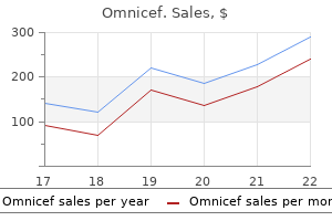
Order omnicef 300 mg with amex
Diagnosis can be easily made with inspection of the retina where angiomas can be identified antimicrobial agents and chemotherapy order omnicef without prescription. Prolonged antibiotic therapy is only 320 Answers advocated if urinary tract infection of prostatic origin is suspected. Within the forebrain, neuroanatomists recognize thalamic nuclei, hypothalamic nuclei, and several additional aggregates. Again, these groupings are based on the observation that the embryonic forebrain divides into thalamic, hypothalamic, and several other regions before it gives rise to the individual brain nuclei in those divisions. Having dealt with the fundamentals of brain nomenclature, let us quickly survey the large assemblages of nuclei you will encounter most often (see the online brain atlas for more detailed images). It is sometimes referred to as the rhombencephalon because it is rhomboid in shape, especially in embryos. Its surface is thrown into numerous tiny folds that make it look like a miniature version of the remaining brain. Its name in Latin means "marrow," which nicely describes its position deep underneath the other brain regions. The pons (Latin for "bridge") is named for the fact that it connects the cerebellum to various other brain regions. The midbrain (or mesencephalon) lies rostral to the hindbrain in most animals and superior to it in humans. Its two main divisions are called colliculus (meaning "little hill") and tegmentum, respectively. The colliculus has superior and inferior subdivisions that are best known for processing visual and auditory information, respectively. The tegmentum contains diverse cell groups, including several that modulate activity in other brain regions. For example, the superior colliculus (optic tectum) is large in most fishes, reptiles, and birds, and the inferior colliculus is large in dolphins and bats. Although the midbrain is functionally and embryologically distinct from the hindbrain, neurobiologists often lump these two brain regions together under the umbrella term brainstem. The corpus callosum is a large bundle of axons that interconnects the two cerebral hemispheres. The diencephalon abuts the midbrain and is divided into several parts, of which the thalamus and hypothalamus are the most prominent. These include the striatum, named for its streaked (striated) appearance in tissue sections, and the pallidum, which in stained tissue sections often appears pale (pallid). Similar laminae are also seen in the cerebellar cortex, but the number of layers is greater in the cerebral cortex. Divisions of the Cerebral Cortex How many laminae does the cerebral cortex contain The prefix neo- means "new" and indicates that thiscallosumis significantly Corpus a Corpu cortex Corpus callosum m more complex in mammals than in non-mammals. An important feature of the neocortex is that it is divisible into several cortical areas that differ from one another in their pattern of lamination (how thick and dense the various layers are), in their conStriat Striatum tr at Striatum Piriform m nections, and in their physiological functions. Small mammals have fewer than a cortex or ex cortex Pallidum Pal dum allid a Insula nsula sula a dozen cortical areas. Their number in humans is debatable, butm reasonable estimate is that humans possess ~100 cortical areas. Both sections were stained so that neuronal and glial cell bodies are purplish blue. The simplest answer is that folding allows the large, sheet-like neocortex to fit inside a reasonably sized skull, much as a sheet of paper will fit inside a cup only if it is folded or crumpled. Imagine trying to cram such a large sheet of tissue into your skull without folding it first! Thus, neocortical folding is an example of good biological design: it evolved because it elegantly solved the problem of how to cram a large cortical sheet into the skull. The same design principle explains the even more intricate folding of the cerebellar cortex. Just posterior lies the parietal lobe, and just inferior to this is the temporal lobe. The frontal and parietal lobes are separated by a major sulcus called the central sulcus, but the boundaries between the other lobes are not so easily defined. The medial aspect of the temporal lobe contains the piriform and hippocampal cortices, which were mentioned already. The anterior tip of the temporal lobe contains an almond-shaped structure called amygdala ("almond" in Latin and Greek), which comprises several brain nuclei and a few cortical areas. Shown here is what would happen to a human brain if you inflated it and, thus, smoothed the neocortical folds (gyri are green, sulci are red). As you can see, the inflated brain would take up more volume and, therefore, be more difficult to fit into a skull. The white labels indicate major sulci and the insula, a cortical region that is normally hidden from view by the temporal lobe. The olfactory bulb protrudes rostrally from the cerebral hemispheres and is quite large in many vertebrates. However, the retina begins its development as part of the forebrain, positioned between the diencephalon and telencephalon. The embryonic retina gradually moves away from the other forebrain structures and toward the skin. When the migrating retina reaches the skin, a lens begins to form and the eyeball gradually takes shape. This may explain why the retina, like many other forebrain components, is a complex structure with multiple laminae (see Box 1. The primate neocortex is commonly divided into four major lobes named after the overlying frontal, parietal, occipital, and temporal bones. Because neurons are discussed in detail in Chapter 2, we here limit ourselves to a brief survey. It conducts electrical impulses known as action potentials, which propagate along axons toward the dendrites or cell bodies of other neurons. The small gaps between the axon of one neuron and the dendrites or cell body of the next neuron are called synapses. Instead, when an action potential travels down an axon and approaches a synapse, the synapse releases molecules called neurotransmitters. These neurotransmitters diffuse across a tiny synaptic cleft to the other side of the synapse, where specialized receptor molecules await them. The binding of the neurotransmitters to their receptors elicits small electrical currents in the postsynaptic neuron. Depending on the neurotransmitters and receptors involved, these postsynaptic currents may be excitatory or inhibitory. The former increase the likelihood that the postsynaptic neuron will fire an action potential of its own; the latter decrease it. Excitatory synapses are more common than inhibitory synapses, at least in the cerebral cortex, but both are critical for normal brain function. People used to think that the human brain contains 10 times as many glia as neurons, but recent studies have shown the glia-to-neuron ratio for the entire human brain to be closer to 1. Protruding from the neuronal cell body are several branching dendrites (only the thickest branches are shown) and an axon, which is thinner than the dendrites. Shown on the right is a drawing of a real neuron that was visualized by filling it with molecules of biocytin. The investigators then took a small sample from this homogenate and stained it with antibodies that specifically bind to neuronal cell nuclei. By counting the number of stained nuclei in the sample, the investigators could estimate how many neurons were in the entire homogenate; all the unlabeled nuclei were assumed to come from glial cells.
Diseases
- Varadi Papp syndrome
- Spinal muscular atrophy type 2
- Ectrodactyly diaphragmatic hernia corpus callosum
- Glycogen storage disease type VI
- Livedoid dermatitis
- Hereditary hyperuricemia
- Weaver-like syndrome
Purchase 300mg omnicef with amex
For example infection vaginale discount 300 mg omnicef, the hand region of the cortical motor map is expanded in highly skilled pianists and violinists, relative to nonmusicians. Because violinists make faster, more differentiated movements with the fingers of their left hand than their right, you would expect violinists to show a disproportionate enlargement of the hand area in the right motor cortex, which controls movements of the left hand. Given that fine motor control requires detailed sensory feedback, you might also expect the sensory representation of the hand to be enlarged in skilled violinists. These observations raise an interesting question: did the sensory and motor maps change as a result of extensive practice, or did the musicians become good at their craft because their cortex was unusual to begin with A tentative answer to this question comes from the observation that the degree of cortical sensory and motor enlargement tends to correlate with the number of years of musical training. Thus, the data suggest that, the more you practice, the more the cortical areas related to the practiced skill will grow. An interesting demonstration of motor cortex plasticity involved monkeys whose motor cortex had been chronically implanted with electrodes at two different locations, sites A and B. At the beginning of the experiment, electrical stimulation at these two sites evoked two very different movements. The experimenters then stimulated site B every time the neurons at site A fired an action potential. After two days of such stimulation, while the monkey was moving freely around its cage, the experimenters again stimulated sites A and B separately. They found that the evoked movements were now quite similar at the two sites, with the movements elicited by stimulation at site B having changed more than those evoked at site A. More work is needed to determine whether this plasticity involves the sprouting of new connections or just Can experiences rewire the Brain Because neural activity during imagined movements correlates with activity during actual movements, researchers can use those correlations to construct the decoder. In fact, providing a subject with visual feedback on the success or failure of their attempts to use the robot arm greatly improves their rate of progress in controlling that arm. Over several days, such feedback leads to changes in the "movement tuning" of the recorded neurons. It took several months of regular practice, but now the subject can feed herself a chocolate bar. Either way, the finding implies that the internal wiring of the motor cortex can be modified by electrical stimulation. Researchers are now exploring how this knowledge might be used to develop better neural prostheses, which allow patients with paralyzed limbs to control robot arms or other machines (see Box 3. One form of neuronal plasticity that we have not yet discussed is experience-dependent growth of the entire brain or major brain regions. To demonstrate this kind of large-scale plasticity, Mark Rosenzweig and his colleagues in the 1960s studied the brains of laboratory rats that were housed in two very different environments. Some rats were housed by themselves in small cages without toys or opportunities for exercise. A second group of rats was given daily maze training and housed in roomy cages with social companions, toys, ladders, and running wheels. As Rosenzweig discovered, the rats living in the impoverished environment ended up having a significantly smaller neocortex than the rats that had been living in the enriched conditions. Most dramatically, the brains of orphans who were institutionalized under atrociously deprived conditions in Romania (under the Ceauescu regime) were 16% smaller, on average, than control brains. Whether these neural and behavioral effects of developmental deprivation can be reversed, at least to some extent, remains unclear. Similarly, it remains unclear why the effects of deprivation are more severe in some children than others, even when the deprivation was equally severe. You have now learned that even adult brains exhibit diverse forms of neuronal plasticity. They may change the strength of some existing synapses, form novel connections or eliminate established ones, change the number of neurons representing Does Neural plasticity Cause Learning and Memory Based on a mountain of evidence, neuroscientists now know that experience-dependent neuronal plasticity may be found in many different brain regions, including the hippocampus, sensory and motor cortices, and the cerebellum. Although plasticity in these brain areas tends to accompany learning and memory, providing convincing evidence that the neural changes cause the learning or the memories is difficult. To establish a causal link between learning and neural plasticity, researchers can ask whether preventing the plasticity impairs the learning. It is important to note, however, that negative results in such experiments do not disprove a causal link because other mechanisms may compensate. For example, the auditory cortex is probably involved in tone conditioning, even though animals with lesions of the auditory cortex can still associate a tone with noxious stimuli, most likely using plasticity in some subcortical circuits (see Chapter 14). Gain-of-function experiments that determine whether boosting plasticity is sufficient to enhance learning and memory can also provide evidence for causal links; but negative results are, once again, inconclusive. However, negative results in such gain-of-function experiments would be difficult to interpret. The manipulation might have targeted an insufficient number of neurons, or additional mechanisms may have to be altered before a gain-of-function becomes evident. One other important way to test for causal links between neuronal plasticity and memory is to ask whether the degree of plasticity correlates with the amount of learning animals exhibit. For instance, the hypothesis that spine turnover is causally linked to song learning in birds is bolstered by the observation that changes in the rate of spine turnover correlate with the fidelity of the song imitations. In the motor cortex, too, learning a difficult motor skill elicits more plasticity than learning a simple task and then performing it repeatedly. Given such correlations between learning and plasticity, a causal link between the two is highly probable. Finally, it is worth noting that both learning and neural plasticity are greater when the information being learned is important to the animal. The larger lesson here is this: if you want to learn something so well that it alters your neural circuitry, then try to figure out why the information is relevant and important to you. The enlargement is due mainly to the growth of individual neurons and the addition of glial cells. Optimal performance is reached when both the machine and the human learn through feedback about motor performance. Cognitive neuroepigenetics: a role for epigenetic mechanisms in learning and memory. Long-term potentiation in the hippocampus using depolarizing current pulses as the conditioning stimulus to single volley synaptic potentials. Long-term consolidation and retention of learning-induced tuning plasticity in the auditory cortex of the guinea pig. Extension of corticocortical afferents into the anterior bank of the intraparietal sulcus by tool-use training in adult monkeys. Motorcortical excitability and synaptic plasticity is enhanced in professional musicians. Plasticity of gray matter volume: the cellular and synaptic plasticity that underlies volumetric change. Boxes Burne T, Scott E, van Swinderen B, Hilliard M, Reinhard J, Claudianos C, et al. Big ideas for small brains: what can psychiatry learn from worms, flies, bees and fish Just as neurons differ in size, shape, transmitters, and ion channel types, so they differ in axonal connections. Furthermore, neurons with similar connections tend to cluster together, forming discrete brain areas, laminae, or nuclei. Thus, real nervous systems are heterogeneous (spatially All cells -catenin variable) rather than homogeneous. Given that development starts with a single fertilized egg cell, how do some cells of the growing embryo get specified to become the nervous system For some invertebrates, the answer expression is an example of symmetry breaking because the embryo is lies with mom, who makes her eggs so that they already less symmetrical after the change. What is important is that spatial differences in transcription factor expression are crucial to the early formation of the nervous system. Five to six days after fertilization, a human egg has grown into a hollow clump of cells that would fit comfortably on the head of a pin. At or before this blastocyst stage, embryonic cells that accidentally get separated from the others can form a complete embryo, an identical twin.
Purchase 300mg omnicef amex
By contrast virus treatment omnicef 300mg generic, repetitive performance of an easy, unskilled task causes little or no plasticity. After the blastocyst stage, twinning is rare because the Where in the embryo Does the Nervous System Originate The ectoderm is of special interest to neurobiologists because its cells differentiate into two seemingly very different tissues: epidermis (skin) and nervous system. It may seem strange that skin and nervous system are developmentally so closely related, but the earliest animals probably had their entire nervous system located within the skin. Induction of the Nervous System Why do some ectodermal cells develop into skin while others form the nervous system They took a piece of mesoderm called the dorsal blastopore lip from the embryo of a white (lightly pigmented) amphibian just after the blastocyst stage and transplanted it into a darkly pigmented amphibian embryo of the same age. Mangold and Spemann cut the dorsal blastopore lip out of an albino amphibian embryo and transplanted it into the ventral pole of a pigmented gastrula. Because both twins are pigmented, the transplanted blastopore lip must have "induced" its host to form the second embryo. Crucially, some of the free nucleotides in the solution are conjugated to a molecule that researchers can later visualize. Often the labeled nucleotide is deoxyuridine triphosphate (dUtp) bound to biotin or digoxygenin. Sectioned Denaturation or not, the tissue containing the probes is processed through several solutions to wash away any probe that is not tightly bound Probe visualization to the intended target. Under the proper conditions, the probe binds selectively to (hybridizes with) any rNa the presence of the color indithat has the complementary nucleotide sequence. It is a widely used technique that has played an exceptionally important role in developmental neurobiology. The transplanted, lightly pigmented cells developed into structures that were adjacent to the second nervous system. Based on these observations, Mangold and Spemann hypothesized that cells of the dorsal blastopore lip emit some sort of signal that Where in the embryo Does the Nervous System Originate This conclusion raised many questions about the cellular and molecular mechanisms underlying nervous system induction. The search for the neural inducer (organizer) molecule hypothesized by Mangold and Spemann advanced significantly in the 1990s when experimenters used in situ hybridization (Box 4. When injected into embryos, molecules of the chordin protein cause ectodermal cells to differentiate into neural tissue. Most convincingly, ectodermal cells grown individually in tissue culture, so that they receive no signals from other cells, adopt a neural fate. This finding shows that at the very root of nervous system development lies not some positive inductive signal, as Mangold and Spemann had thought, but an inhibitory signal that prevents the alternative outcome of becoming skin. Soon thereafter the left and right edges of this neural plate lift up, transforming the plate into a neural groove. At this point, special cell adhesion molecules on the surface of the future skin cells cause the skin cells on both sides of the neural groove to stick to one another but not to other cells. Neural groove cells express different adhesion molecules, which make them stick to one another but not to the skin cells. The overall effect of this selective adhesion is that the neural groove becomes a neural tube that is separate from, and covered by, the skin. It then goes on to form the entire central nervous system, including both brain and spinal cord. In addition, so-called neural crest cells migrate away from their original location right between the skin and the neural plate. They form much of the peripheral nervous system, including the neurons of the cranial and spinal nerves, the glia associated with those nerves, the ganglia of the sympathetic nervous system, and the enteric nervous system. The neural crest also gives rise to a number of non-neural structures, including skin pigment cells (melanocytes) and much of the skull. For the nervous system, it is the study of how the initially homogeneous neural tube becomes divided into a complex heterogeneous structure. Although patterning the neural tube is a complex three-dimensional problem, it can be simplified, at least initially, by considering rostrocaudal patterning separately from dorsoventral patterning. Rostrocaudal Patterning the spinal cord develops from the caudal portion of the neural tube, whereas the brain develops from its rostral end. The spinal cord is further subdivided into 31 segments; and the brain is subdivided into hindbrain, midbrain, and forebrain. Developmental neurobiologists have long wondered how these rostrocaudal divisions of the central nervous system come into existence. A full answer remains elusive, but most scientists agree that rostrocaudal neural tube patterning involves molecular signals that increase in concentration as you go from rostral to caudal along the neural tube. That is, they cause the affected cells to become caudal, rather than rostral, neural tissue. Interfering with retinoic acid signaling prevents caudal brain regions from forming normally. Shown here are dorsal views of the hindbrain from two chick embryos, stained with wholemount in situ hybridization to reveal the expression patterns of Hox a-3 (left, purple), Hox b-3 (right, purple stain), and Islet-2 (right, red stain and arrows). Shown in (a) is a schematic dorsal view of an embryo that developed with the normal amount of retinoic acid (ra). Shown in (C) is a model of ra function, according to which ra concentration increases as you proceed caudally. The Hox Gene Family Spatial position Rostrocaudal neural tube patterning also involves Hox genes. Hox genes are a family of transcription factors that is highly conserved across species (Box 4. Individual members of this family are expressed in various Shown in (a) are dorsal views of vertebrate hindbrains in which indicombinations at different rostrocaudal levels of the nervous vidual segments are separated by dashed lines. Caudal hindbrain segments express many that each Hox gene has a different rostral expression boundary. This nested expression pattern suggests that different Hox genes are activated at different concentrations of a caudalizing signal, such as retinoic acid. Most Hox genes system patterning long after the are numbered in sequence (Hox-1, Hox-2, etc. Curiously the presumed ancestral Hox cluster was disbanded ongoing and unlikely to be rein several taxonomic lineages, including flatworms (platyhelminths), round worms (nematodes), and tunicates (urochordates). For example, some Hox gene mutations cause flies to develop legs where their antennae should be. Moreover, the expression domains of different Hox genes have different rostral boundaries during fruit fly development, which means that caudal body parts coexpress a larger number of Hox genes than rostral body parts. This hypothesis is supported by the finding that artificial increases in retinoic acid levels cause rostral hindbrain segments to express Hox gene combinations that are normally found only in more caudal segments. These "caudalized" segments also express non-Hox genes that are typically expressed only in caudal hindbrain segments of older animals, suggesting that the altered Hox gene expression pattern permanently alters cell fates. Alternatively, the posterior prevalence model states that some Hox genes are more important than others. Specifically, the Hox genes expressed in the more posterior hindbrain segments are thought to dominate the Hox genes with more anterior expression domains. Experiments in which specific Hox genes were "knocked out" in transgenic mice tend to support the posterior prevalence model, but the matter is not settled yet. In any case, the data show that Hox genes are essential for rostrocaudal patterning of the vertebrate hindbrain and, to some extent, the spinal cord. As you will see shortly, rostrocaudal patterning in the midbrain and forebrain involves a different set of transcription factors. In the spinal cord, for example, neurons that send their axons to muscles lie ventrally; whereas neurons that receive input from sensory nerves are located in the dorsal horn of the spinal cord. In between these motor and sensory neurons lie interneurons that connect to other neurons in a manner that varies with their dorsoventral position. This is analogous to how different Hox genes are induced at different concentrations of retinoic acid.
Discount 300 mg omnicef with amex
A shared inhibitory circuit for both exogenous and endogenous control of stimulus selection opportunistic infection buy omnicef no prescription. The locus ceruleus norepinephrine system: functional organization and potential clinical significance. Glial and neuronal interactions during slow wave and paroxysmal activities in the neocortex. The reticular nucleus revisited: intrinsic and network properties of a thalamic pacemaker. Involuntary but not voluntary orienting contributes to a disengage deficit in visual neglect. Memory 438 Chapter 14 remembering relationships The ability to learn from experience is a crucial function of our brains. Without it, we would live exclusively in and for the moment, unable to benefit from earlier interactions with the environment or plan for the future. Given the importance of learning and memory, it is not surprising that scientists have studied them intensively. The only way to know that an animal has learned from an experience is to show that afterward, the animal behaves as if it had learned. That is, one must compare the behavior of animals that had the potential learning experience to the behavior of naive control animals. If they had been released from the harness, they might well have run around excitedly on hearing the telltale bell, indicating that they learned more than just a single, limited response. This may seem obvious, but it becomes important when one tests for the loss or retention of memories by looking for specific conditioned responses. It might seem simpler to study learning and memory in humans, rather than dogs or other animals, because you can ask humans what they have learned. However, humans sometimes learn things that the experimenter did not think to ask about. Thus, discovering what individuals have learned always requires carefully designed experiments, regardless of which species is studied. Consider what you learn when playing a video game in which your character must navigate a complex virtual environment and shoot as many enemies as possible. As you play the game, you learn which buttons to push to run, turn, jump, aim, and shoot. In a typical experiment, a specific sound was repeatedly presented just before the food. You may remember that the game is rated M, for mature audiences, even though you cannot recall when and where you saw this rating displayed. Neuroscientists refer to this kind of learning as the creation of semantic memories (they may use the term "declarative memory" to refer to semantic and episodic memory collectively). Playing the game may also improve your ability to perceive small objects hidden in clutter, which implies some perceptual learning. In addition, you learn which stimuli predict imminent threats, rewards, or punishments; this is Pavlovian (or classical) conditioning. As if your brain was not already busy enough, you also learn the rules of the game, the strategies, the most efficient ways to reach your goals; we will cover this kind of instrumental learning in Chapter 15. Habituation and sensitization are nonassociative (you are not learning about relationships), but they are forms of learning nonetheless. Some scientists have tried to organize these diverse forms of learning and memory into a single, overarching scheme, but these efforts have remained controversial. Therefore, let us just recognize that learning and memory come in a variety of forms. As you will see in this chapter, the diverse forms of learning and memory are implemented by distinct but overlapping brain systems and can function independently of one another. This explains why some individuals excel at some forms of learning and memory (Box 14. The most influential study on the neurobiology of learning and memory involved a patient known to most neuroscientists only by his initials, H. At that point, neurosurgeon William Scoville offered to remove the anterior medial portion of H. The rationale for this treatment was that epileptic seizures often begin in the medial temporal lobe and then spread to the remaining brain. His seizure frequency indeed decreased, but it soon became apparent that the surgery also destroyed a crucial part of H. This anterograde amnesia extended across all sensory modalities and affected memories for faces, places, events, facts, and words. One such memory specialist was described by aleksandr Luria in the Mind of a Mnemonist (1968). In particular, he could arrange the images evoked by items in a list along an imaginary route. By taking his mental walk in the opposite direction, he could recite the list in reverse order. One of his rare mistakes was omitting "egg" from a remembered list because he had placed his mental image of the white egg against a white wall. For example, she could recall the date of every easter Sunday between 1980 and 2003, which is difficult because the timing of easter varies from year to year. Much of what aJ remembered about her life was verified from her extensive collection of diaries. She is compulsive about remembering her past experiences but does not memorize her diaries. She does not have Control subjects 30 the sort of synesthesia where words elicit images, but she probably has time-space synesthesia, which lets 20 her think of dates as being arranged in space. Some rior autobiographical memory) a public events test with 30 questions such as "What year, date, and of their brains are being scanned, day of the week was John Lennon killed In contrast, people who think they have superior autobiographical memory exhibit a bimodal distribution of test scores: roughly half score like control subjects; the others score and difficult to interpret. Other evidence suggests that the initial trauma causes dentate granule cells in the hippocampus to sprout abnormal connections that create an excitatory intrahippocampal feedback loop. One way or the other, hippocampal neurons receive too much excitatory input, which then leads to cell death (excitotoxicity). Because the surgery is unilateral, the effects on memory tend to be much less severe than they were in h. Based in part on the study of such epilepsy-prone families and twins, researchers have identified a variety of genetic mutations that are associated with epilepsy. Many of these mutations affect voltage-gated sodium and potassium channels; others change GaBa receptors. It is easy to see how these mutations might Side of seizure focus Contralateral side alter the balance between neuronal 5 5 Mean of excitation and inhibition. Seizures, in turn, are defined as episodes of highly synchronized, excessive neuronal activity. In other patients, the seizures appear simultaneously in many different brain regions. When a seizure spreads, the behavioral symptoms vary systematically as seizure activity invades new brain regions. In many epileptic seizures, consciousness is eventually lost and memory of the event impaired. Young children often suffer fever-induced seizures, but this does not mean that they have epilepsy. Still, some evidence suggests that febrile (fever-induced) seizures increase the risk of developing epilepsy later in life. Unfortunately, this is not the case for medial temporal epilepsy, which is what h. Medial temporal epilepsy is thought to be caused by some sort of traumatic trigger, such as a severe concussion, a stroke, or an intense febrile seizure.
Arctium Lappa (Burdock). Omnicef.
- Fluid retention, fever, anorexia, stomach conditions, gout, acne, severely dry skin, and psoriasis.
- Dosing considerations for Burdock.
- Are there safety concerns?
- What is Burdock?
- Are there any interactions with medications?
- How does Burdock work?
Source: http://www.rxlist.com/script/main/art.asp?articlekey=96153
Buy discount omnicef 300 mg line
Acute bacterial prostatitis Acute bacterial prostatitis is a febrile illness with sudden abrupt onset antibiotics korean buy generic omnicef on line. There are marked genitourinary symptoms and often a positive bacterial urine culture. Obstruction downstream to the prostate may force urine up into its ducts, and if the urine is infected, it causes inflammation. Whatever the route of the infection, the prostate becomes enlarged and painful on rectal palpation and may cause painful and obstructed micturition. The most common pathogens include Escherichia coli, Klebsiella, Proteus mirabilis and Enterococcus faecalis. Symptoms may vary considerably however and include nonspecific pelvic and suprapubic pain and storage lower urinary tract symptoms. Source: National Institutes of Health Summary Statement (1998, November) First National Institutes of Health International Prostatitis Collaborative Network Workshop on Prostatitis. Symptoms of chronic prostatitis in the absence of cultured bacteria is termed chronic pelvic pain syndrome. The addition of an alphaadrenoreceptor blocking agent and antiinflammatory drugs improves symptoms. For chlamydia, tetracyclines Scrotum and/or testes Penis Urinary bladder Low back Source: Zermann, D. Benign disorders of the prostate gland 173 Benign prostatic enlargement Symptoms resulting from benign prostatic enlargement account for a large proportion of the workload of a general urologist. The prostate is a complex organ consisting of acinar, stromal and muscular tissue. The earliest changes of benign prostatic enlargement occur in the periurethral glands around the verumontanum, where there develops an imbalance between stimulatory and inhibitory prostatic growth factors that results in prostatic hyperplasia and fibromuscular nodule formation. The size of the prostate gland is irrelevant: the smallest prostates may cause severe bladder outflow obstruction; huge glands none at all, and patients with bladder outflow obstruction may be asymptomatic and present for the first time in urinary retention. Compression of the prostatic urethra and the way in which the bladder responds to obstruction are the main factors involved in symptom generation. The storage and voiding lower urinary tract symptoms that may result from bladder outflow obstruction are not specific to either the prostate, benign prostatic enlargement or indeed bladder outflow obstruction (see Chapter 1). Patients who are most likely to drive maximal benefit from intervention are those who combine lower urinary tract symptoms and bladder outlet obstruction. Several factors contribute to the generation of lower urinary tract symptoms associated with bladder outflow obstruction. Bladder neck smooth muscle tone the alphaadrenergic smooth muscle fibres in the prostate, especially around the bladder neck, fail to relax as the detrusor contracts. These changes are accompanied by complex changes to its structure and physiology characterised by infiltration of the muscle fibres by connective tissue and a poorly characterised complex neuromuscular disorder. This generates symptoms of urinary frequency and urgency and is termed obstructive detrusor overactivity. The hypertrophied fibres cause trabeculation of the bladder, and diverticula form in between them. The detrusor muscle fibres may also become weaker in response to chronic obstruction and fail to empty the bladder effectively. The residual urine results in shorter periods of time between voids causing frequency and nocturia. Complications of bladder outflow obstruction Urinary infections and bladder stones Incomplete bladder emptying causes stagnant urine, which predisposes to urinary infections as any bacteria that enter the bladder are not washed out. Bladder stones may also form in stagnant urine as the urinary constituents precipitate. Obstructive uropathy due to high pressure chronic retention Hypertrophy of the bladder wall may lead to high bladder pressures as it fills and is unable to empty fully. Disruption of the antireflux ureteric valve mechanism results in these high pressures being transmitted to the upper tracts causing dilatation of both the ureter and renal collecting system (hydroureteronephrosis), obstructive uropathy and nephropathy. Detrusor smooth muscle hypertrophy may also cause direct obstruction to the ureter as its terminal part runs through the bladder. Bladder neck dysfunction may generate symptoms of urinary hesitancy and difficulty in initiating micturition and a poor and intermittent urinary stream. Hesitancy may also be caused by the longer time it takes for the detrusor to generate a contraction strong enough to overcome the increased outflow obstruction from the compressed prostatic urethra and an intermittent stream by failure to maintain this. Benign disorders of the prostate gland 175 Acute urinary retention Acute urinary retention is the sudden inability to void urine and is always painful. The condition is not solely the result of benign prostatic enlargement and may be precipitated or spontaneous. Precipitants include any factor that increases bladder outflow resistance or decreases bladder contractility. It is postulated that large prostates may undergo focal infarction, which increases the outflow resistance due to inflammation and swelling. Kinking of the prostatic urethra due to uneven prostatic enlargement has also been suggested as a mechanism. These include anaesthetic agents, accounting for postoperative retention; alcohol, which may numb the desire to void while acting as a diuretic at the same time; constipation, which generates reflex afferent inhibitory sympathetic nervous activity to the bladder; pelvic pain; and bladder overdistension. Spontaneous urinary retention carries a worse prognosis than precipitated retention as there is no factor that can be reversed. With both precipitated and spontaneous urinary retention, there is usually a degree of underlying clinical or subclinical bladder outflow obstruction. Chronic urinary retention Chronic urinary retention is the maintenance of voiding with failure to empty the bladder. If the bladder has responded to insidious outflow obstruction by becoming weaker and distending, it may contain several litres of urine after voiding with no symptoms. Patients with chronic urinary retention may develop the sudden inability to pass urine at all; this may or may not be painful and is referred to as acuteonchronic retention. If the bladder has responded to the obstruction by becoming hypertrophied and thick walled, highpressure chronic retention with obstructive uropathy may occur. This is commonly accompanied by the clinical sign of nocturnal enuresis, Diagnosis of prostatic obstruction History Men with bladder outflow obstruction seek medical input because of bothersome lower urinary tract symptoms, the complications of obstruction most commonly urinary retention and urinary infections, and because they are concerned that nonbothersome symptoms may be a sign of prostate cancer. Over the past month, how often have you had a sensation of not emptying your bladder completely after you finished urinating Over the past month, how often have you had to urinate again less than 2 h after you finished urination Over the past month, how often have you found you stopped and started again several times when you urinated Over the past month, how often have you found it difficult to hold back urinating after you have felt the need Over the past month, how often have you noticed a reduction in the strength and force of your urinary stream Over the past month, how many times did you most typically get up to urinate from the time you went to bed at night until the time you got up in the morning It should never be a substitute for listening to the patient carefully, especially since these symptom scores do not match objective evidence of obstruction. Physical signs Abdominal palpation may reveal a chronically obstructed bladder, but this is a late feature of the disease. Flow rate: Flow rates vary from day to day, and a poor flow may not necessarily mean obstruction: it may result from a weak detrusor, while on the other hand if the detrusor has undergone considerable hypertrophy, it can compensate for obstruction and produce a good flow rate. Residual urine: the volume of urine remaining in the bladder can be measured by abdominal ultrasound. This may vary from day to day and may be caused by bladder outflow obstruction, detrusor failure or both. Urodynamic studies: the only way of making certain that lower urinary tract symptoms are due to bladder outflow obstruction is by means of a cystometrogram (see Chapter 10). Treatment of urinary retention involves draining the bladder by passing either a urethral catheter or, if this is not possible, a suprapubic catheter. It is important to record how much urine is drained in the first fifteen minutes after catheterisation, as this gives a valuable clue as to the state of the bladder. A volume of up to approximately 800 mL is consistent with a normal bladder; more than this indicates a degree of prior bladder decompensation and chronic retention. The patient is assessed to determine whether the retention episode is acute or chronic, precipitated or spontaneous. A rectal examination is performed to exclude an advanced prostate cancer, other pelvic tumour or impacted stool.
300mg omnicef sale
Furthermore bacteria jersey shore buy discount omnicef 300 mg on-line, inhibitory synapses are often located close to the axon hillock of the postsynaptic cell, whereas excitatory synapses are mainly found on the dendrites. This transmitter is employed by all mammalian motor neurons (neurons that innervate muscles), by many of the neurons that control our glands and internal organs, and by a few additional cell groups within the brain. Because it is such an important transmitter, acetylcholine is targeted by many of the toxins organisms have evolved to kill or paralyze one another (see Box 2. Although monoamines are used by relatively few neurons, they are extremely important. Norepinephrine is used by neurons in the sympathetic nervous system, which mediates the fight-or-f light response, and by neurons that regulate arousal (see Chapter 13). Serotonin is released by neurons in the brainstem raphe nuclei and helps to regulate a whole host of processes, including sleep, appetite, and mood (see Box 2. Nonclassical Transmitters In addition to the classical transmitters, many neurons use a variety of other neuroactive substances. Most of these other substances are neuropeptides, which typically comprise 10 or more amino acids and thus are larger than the classical neurotransmitters. They are used in combination with other neurotransmitters, including other neuropeptides. This makes it difficult to make general statements about their postsynaptic effects. However, one good rule is that neuropeptides modify the effect of other transmitters on postsynaptic cells. Toxins that affect the nervous system are particularly common, mainly because they tend to act quickly. Even bacteria produce neurotoxins to discourage small animals from eating them (or larger animals from eating their hosts). The most powerful bacterial toxin is botulinum toxin, which derives its name from the Latin word for "blood sausage" (where the bacteria occasionally thrive). The potency of botulinum toxin depends on whether you eat it or inhale it, but 1 gram of botulinum toxin can kill about a million people. Botulinum toxin is an enzyme that is taken up into the terminals of the axons that innervate muscle fibers. There the botulinum toxin digests key proteins needed for the release of synaptic vesicles filled with acetylcholine. Without synaptic vesicle release, you get no synaptic transmission, no muscle contractions and, in short order, respiratory arrest. Given its toxicity, it is ironic that botulinum toxin is often used to treat wrinkled skin. In this therapy, doctors inject a small amount of botulinum toxin (Botox) into the muscles under the skin. These animals are relatively small yet contain enough toxin to kill dozens of adult humans within minutes. Without these channels, you get no action potential conduction, no centrally controlled muscle contractions, and, again, respiratory arrest. What predator of theirs requires a toxic dose sufficient to kill dozens of humans This molecule is harmless if you eat it because it is too large and charged to be absorbed through the gut. However, if curare is injected into your bloodstream, it blocks the acetylcholine receptors at the synapses between neurons and skeletal muscle, and paralysis ensues. This paralytic effect was discovered long ago by hunters in South America who dipped their arrowheads in curare. Importantly, the hunters could eat their prey without being poisoned themselves because, as noted earlier, curare does not get absorbed through the gastrointestinal tract. Poisonous snakes tend to be immune to their own venom, which is remarkable because snake venoms are typically a mix of several different neurotoxins. Most snake venoms also contain toxins that irreversibly block the acetylcholine receptor at the neuromuscular junction. The most famous of these toxins is alpha-bungarotoxin (isolated from a snake called Bungarus multicinctus). Curiously, many of the toxins in snake venom share a core molecular structure, suggesting that they all evolved from a common ancestral molecule. Another interesting fact is that snakes are not the only species immune to their venom. The mongoose, for example, is well known for dining on snakes, and its acetylcholine receptors are resistant to alpha-bungarotoxin. Most scorpions are not life-threatening to humans, but the death stalker Leiurus quinquestriatus, in the deserts of Northern Africa and the Middle East, can kill a child. Most spiders likewise present no mortal danger to humans, but Australian funnel web spiders can be lethal. One of the neurotoxins in their deadly brew blocks sodium channel inactivation, which causes massive neuronal depolarization. In contrast, the principal toxin in the venom of American black widows is latrotoxin, which stimulates the uptake of calcium into synaptic terminals, causing massive transmitter release and, among other problems, rigid paralysis. Cone snails are less widely known than spiders or scorpions, but some of them are almost as lethal. They kill fishes or mollusks with a toxic cocktail that includes acetylcholine receptor blockers and omega-conotoxin, which blocks a specific type of voltage-gated calcium channel and is >100 times more potent than morphine as a painkiller. The latter will kill you if you inhale too much of it, but the brain uses carbon monoxide in small doses to modulate neural activity. It seems that nature has been very inventive when it comes to using chemicals to exchange information between cells. One reason why there are so many different antidepressants on the market is that each drug represents a different compromise between benefits and side effects. Moreover, treating depression often involves a great deal of trial and error because some compounds work well in one person but not in another. A combination of several different drugs, at carefully adjusted dosages, usually works best. Given how difficult it is to treat a diseased brain, is it reasonable to expect that we can alter the functions of a healthy brain and cause no harm Ecstasy does this mainly by altering serotonin transporter molecules so that instead of taking serotonin back up into the presynaptic terminal, they release it into the synaptic cleft. The serotonin release feels good, but the depletion produces an ecstasy hangover that can last for several days and is characterized by a lack of motivation, focus, and appetite. Indeed, studies with humans have shown that heavy ecstasy users have impaired verbal memory, display more impulsivity, and are more likely to be depressed than control subjects. However, it certainly seems wise to find less risky, more creative ways to boost your mood. Serotonergic axons were stained with "on the street" (including Molly) is an antibody so that they appear as white lines on a dark background. Panel (A) shows a section hardly pure and sometimes not through the frontal cortex of a control monkey. The cell bodies of these neurons are located in a few hindbrain nuclei called the raphe nuclei, but the axons of these neurons terminate widely throughout the brain. Many antidepressant drugs, notably Prozac, selectively inhibit the uptake of released serotonin back into presynaptic terminals. The effect of this reuptake inhibition is that released serotonin molecules remain in the synaptic cleft for a longer period of time and therefore exert a greater effect on their postsynaptic targets. The fact that antidepressants generally boost serotonin signaling suggests that clinical depression results from insufficient serotonin levels in the brain. It cannot be this simple, however, because reuptake inhibitors typically boost signaling within minutes or hours, whereas the positive effects on mood take weeks to manifest. To explain this discrepancy, it has been hypothesized that serotonin reuptake inhibitors cause gradual, long-term changes in the sensitivity and/or abundance of serotonin receptors. This hypothesis is controversial, but it is consistent with the general principle that chronic changes in transmitter abundance cause compensatory changes in the corresponding receptors. One problem with antidepressant medications is that they tend to have side effects, including daytime sleepiness, nighttime restlessness, nausea, diarrhea, and sexual dysfunction. This is not particularly surprising because many of these drugs also inhibit the reuptake of norepinephrine, which regulates How Do Neurons Differ from One Another This principle is sometimes confused with the idea that neurons use only one transmitter, which is not the case.

Order omnicef 300mg with mastercard
These neurons send the peripheral branch of their axon to the aortic arch and carotid sinus - buy omnicef without a prescription, where they terminate as mechanoreceptive nerve endings that sense blood vessel stretch. Neurons in the vagus nerve transmit baroreceptor information to nucleus tractus solitarius (NtS). Scientists used to think that vasovagal syncope is caused by abnormal activation of stretch receptors in the aorta, which then trigger an inappropriate baroreflex. Isometrically contracting your limb and trunk muscles appears to help, presumably because these contractions elevate blood pressure. Because the vagus nerve carries both parasympathetic axons going to the heart and baroreceptor information coming from there, strong artificial stimulation of the vagus nerve, through implanted stimulating electrodes, causes you to faint (if you are standing up). Best documented is that vagal nerve stimulation reduces the frequency of epileptic seizures, which result from abnormal bursts of brain activity, in patients whose epilepsy has been resistant to other treatments. In such patients a helical stimulating electrode is implanted around a cervical segment of the left vagus nerve. No one is quite sure why this form of weak electrical vagal nerve stimulation can prevent seizures, but it probably provides weak activation of the parasympathetic division and a concomitant slight reduction of sympathetic tone. Instead, they should decrease anxiety and promote behavioral as well as physiological relaxation (the opposite of the fight-or-flight response). If this is true, then one would expect vagal nerve stimulation to influence not just the frequency of epileptic seizures, but a wide variety of behavioral, cognitive, and physiological phenomena. Indeed, vagal nerve stimulation is increasingly used to battle major depression in patients who have not benefited from standard drug therapies. Several studies have shown such treatments to be effective, although more extensive clinical trials are still ongoing. It may also alleviate migraines, tinnitus (persistent ear ringing), multiple sclerosis, bulimia, and obesity. One should be skeptical when a single procedure is touted as a treatment for so many different ailments, but the vagus nerve clearly is implicated in a wide variety of normal processes. Further studies will have to demonstrate which of the promised effects of vagal nerve stimulation are real and which are spurious. It will also be interesting to learn what mechanisms lie behind the reported effects. So far, we have only a few intriguing hints, such as elevated epinephrine and serotonin levels in many brain regions after vagal nerve stimulation. During this state they even "smell dead" because of putrid secretions from their anal glands. They increase their rate of firing when blood pressure rises and decrease their firing rate as blood pressure falls. Many of the neurons there project to nucleus ambiguus how Do Neural Circuits regulate the Vital Bodily Functions On the right side of this section (yellow rectangle), you can see nucleus ambiguus neurons that were labeled with a retrograde tracer (close-up in inset). Nucleus ambiguus then sends its axons through the vagus nerve to parasympathetic postganglionic neurons on the surface of the heart. Sympathetic Regulation of Heart Rate the principal complication is that the heart rate control circuit has two branches. When those sympathetic postganglionic neurons release norepinephrine, the heart speeds up. Thus, the sympathetic branch of the heart control circuit is also, like its parasympathetic 276 Chapter 9 regulating Vital Bodily Functions counterpart, homeostatic: it boosts heart rate in response to a blood pressure drop and decreases heart rate when blood pressure rises. As you have now learned, any change in baroreceptor activity has opposite effects on the sympathetic and parasympathetic branches of the heart rate control circuit, increasing activity in one and decreasing it in the other. Therefore, the two branches function synergistically (cooperatively) rather than antagonistically. Going back to our high-tech thermostat analogy, a rise in blood pressure activates the parasympathetic neurons and inhibits the sympathetic innervation of the heart, just as a rise in temperature activates the air conditioner and turns off the heater. Conversely, a drop in blood pressure reduces activity in the parasympathetic branch of the circuit and disinhibits (activates) the sympathetic branch, just as a drop in temperature shuts down the air conditioner and fires up the heat. In short, the circuits that control heart rate are a good example of push-pull regulation. Regulating Blood Pressure As noted earlier, blood pressure can be also altered by changes in blood vessel diameter. Activation of these postganglionic neurons promotes vasoconstriction, which causes blood pressure to rise. An important aspect of the sympathetic inputs to the blood vessels is that they, too, are tonically active, which means that blood vessels are slightly constricted most of the time. Therefore, if blood pressure suddenly increases, sympathetic background activity can be reduced, leading to a decrease in the activity of the smooth muscles responsible for vasoconstriction. Because of blood pressure inside the vessels, the decrease in vasoconstriction leads to vasodilation. Without the tonic sympathetic activation, vasodilation would be much harder to accomplish. After all, neurons that are already silent cannot be silenced further, and nonconstricted blood vessels are hard to dilate more. Parasympathetic Vasodilation Although most blood vessels receive only sympathetic innervation, some vessels in your face, as well as the penis, can be dilated by parasympathetic inputs. Blood Pressure Set Points As you now realize, the baroreceptor reflex regulates blood pressure homeostatically, increasing it when blood pressure is low and lowering it when the pressure is high. A major problem is that many people have hypertension, which is defined as chronically high blood pressure (. Although hypertension can have a variety of causes, including excess salt intake and genetics, it may also involve an elevation in the set point around which blood pressure is regulated. According to this hypothesis, the various aspects of the baroreflex still work in people with high blood pressure, but those people stabilize blood pressure around an abnormally high target value. Although many different drugs can influence blood pressure and are frequently prescribed to treat hypertension, the neural mechanisms that alter blood pressure set points remain an almost total mystery. This posture-driven increase in blood Heart rate 278 Chapter 9 regulating Vital Bodily Functions pressure is caused in part by an increase in heart rate, but it also involves a rapid constriction of blood vessels in large muscles and viscera. Blood pressure also increases rapidly when you get excited, be it from happiness or fear. Such acute changes in blood pressure require a change in the blood pressure set point. Otherwise, any increase in heart rate or vasoconstriction would immediately be neutralized by reflexive commands to reduce heart rate and re-dilate the blood vessels. The mechanisms underlying these temporary set point adjustments also remain largely mysterious. Controlling Breathing Breathing is a vital, vegetative process that can go on without conscious control. Nonetheless, we can control our breathing much better than we can control the beating of our heart or the dilation of our blood vessels. Spoken language, in particular, requires exquisite, rapid control over our respiratory muscles. Such rapid control is generally impossible for smooth muscles because their contractions are modulated by relatively slow metabotropic receptors. Accordingly, breathing (respiration) is controlled by striated muscles and skeletal motor neurons rather than smooth muscles and autonomic neurons. Respiratory Muscles and Neurons the most important respiratory muscle is the diaphragm, which forms the domeshaped floor of the chest cavity. On contraction, the diaphragm moves down toward the gut, thus expanding the chest. When the diaphragm relaxes, it returns to its domed shape, compressing the lungs and pushing air back out. If more forceful exhalation is needed, then abdominal muscles also contract and push the diaphragm upward. Although the laryngeal muscles are best known for their role in speech, they are also used to close the trachea just after you inhale deeply. This closure prevents the inhaled air from escaping after the diaphragm starts to relax. It lets you hold your breath without keeping your diaphragm forcefully contracted. This is a useful strategy because the laryngeal muscles are much smaller than the diaphragm and, therefore, consume less energy when contracted.
Buy generic omnicef 300mg on-line
The first inhibitory neuron (in the median preoptic nucleus) responds to cold with an increase in firing rate antibiotics for acne inflammation order 300mg omnicef with mastercard, thereby strengthening its inhibition of the second inhibitory neuron (in the medial preoptic area). As the latter neuron falls silent, its target (in the dorsomedial hypothalamus) is disinhibited. Because the remaining neurons in the circuit are all excitatory, the end result is a cold-induced increase in the rate at which brown fat is burned and skeletal muscles shiver. To reiterate, the inhibition of inhibition causes excitation, just as in mathematics multiplying two negative numbers yields a positive result. In general, if you examine a linear neural circuit with an even number of inhibitory neurons (in series), then you can predict that excitatory input to the circuit will usually cause excitation on the output side. One of the most obvious responses to heat is the production of sweat, which cools the body when it evaporates. Sweating is driven by sympathetic postganglionic neurons that use acetylcholine as their transmitter. This should seem odd to you because sympathetic postganglionic neurons generally release norepinephrine, but the neurons that innervate sweat glands produce norepinephrine early in life and only later switch to using acetylcholine. The second major autonomic response to hyperthermia is peripheral vasodilation, which promotes convective heat loss by increasing blood flow to the skin. Curiously, many of the neurons decreased their firing rate before the heater was turned off; why this happened is not clear. Activation of the latter pathway is what makes your face turn very red when you feel hot (blushing is a related phenomenon, triggered by embarrassment rather than heat). Temperature Sensors in the Brain Most responses to heat and cold are triggered by sensors in the skin, but the brain also contains some temperature sensors. The preoptic area is especially sensitive to changes in local temperature, as can be shown by inserting heating or cooling probes into the preoptic area and recording the activity of nearby neurons as local temperature varies. Because neurons in other brain regions do not exhibit such temperature-driven changes in firing rate, the temperature sensors must be located in the preoptic area. In analogy with the peripherally and centrally triggered versions of the chemoref lex, we can think of peripherally and centrally controlled thermoregulation as providing fault tolerance. If one circuit fails, the other one provides a backup system that allows for at least some temperature regulation. However, the two systems are not completely redundant because when your environment changes in temperature, your skin will follow suit long before brain temperature rises or falls. Accordingly, activation of the peripheral thermoreceptors can adjust body and brain temperature proactively, whereas central thermoreceptors must function reactively. To trigger a fever, immune cells secrete pyrogenic (meaning "fire producing") molecules into the blood. These molecules, especially prostaglandin E2, are sensed by neurons in the vagus nerve and circumventricular organs, which convey the information to the preoptic area. What happens there remains unclear, but somehow the temperature set point for thermoregulation is increased. This means that shivering, peripheral vasoconstriction, and other warmth promoting behaviors (such as piling on blankets) are triggered at higher than normal body temperatures. Once the pathogens have been vanquished, pyrogen levels decrease and the temperature set point returns to normal. When this happens, the brain detects that body temperature is higher than it should be and triggers appropriate countermeasures, which is why profuse sweating usually signals that your fever has broken. Balancing the Bodily Fluids To stay alive, we must stay properly hydrated and maintain adequate levels of energy. Mammals regulate the osmolarity (salt concentration) of their extracellular fluid tightly. If the extracellular fluid becomes too salty, either because too much water is lost or because too much salt is taken in, water diffuses out of the cells and they begin to malfunction. Conversely, if the extracellular fluid becomes too dilute, then cells begin to swell. Such swelling is a serious problem especially within the brain because the brain already fits so tightly in the skull. For example, a 4-year-old girl died in 2002 after being forced to drink a gallon of water as punishment. This case was extreme, but the point is that drinking too much water can be as much of a problem as drinking too little water. As we discussed in Chapter 8, some of these magnocellular neurons secrete oxytocin into the blood vessels of the posterior pituitary. As its name suggests, vasopressin increases blood pressure through vasoconstriction. Even relatively slight changes in the activity of magnocellular vasopressin neurons have significant effects on urine production. Destruction of the magnocellular vasopressin neurons causes diabetes insipidus, which is characterized by a massive overproduction of urine (up to 25 liters/day) and a corresponding need to replenish the lost fluid by drinking. Shown here is the response of an isolated (in vitro) magnocellular supraoptic neuron to being bathed in a hypertonic solution. We can conclude that this neuron can sense changes in osmolarity directly, without the aid of other brain regions. Both diseases involve an overproduction of urine (diabetes is Greek for "passing through"); but in diabetes mellitus, this overproduction is due to elevated blood glucose, which causes more water to flow out of tissues into the blood. Part of the answer is that the magnocellular neurons themselves can sense dehydration. When these cells are placed in a culture dish and bathed in extracellular fluid that contains too few solutes, they swell. This swelling closes a stretch-inactivated cation channel and hyperpolarizes the neurons. These findings indicate that magnocellular neurons can sense a rise in the osmolarity of the extracellular fluid and, through their connections to the posterior pituitary, effect a hormonal response that limits any further rise. As you may realize, this finding challenges the notion that the magnocellular neurons are a single-neuron reflex arc. Further work is needed to resolve this puzzle but, in the meantime, we can conclude that in intact animals, changes in osmolarity can be sensed by multiple neurons in multiple locations. The magnocellular vasopressin neurons integrate this information and then regulate urine production accordingly. To reverse dehydration, you must drink some fluid or ingest water through food. Shown here are the effects of lesioning various brain regions on drinking by dehydrated sheep. Even the simpler question of what causes thirst is difficult to study experimentally. Comparisons of brain activity in thirsty humans before and after they drink a glass of water have revealed consistent changes in the activity of anterior cingulate and insular cortex. These cortical areas are thought to be required for the conscious experience of thirst, but destroying them experimentally does not prevent drinking. Nor does destroying the magnocellular vasopressin neurons eliminate thirst, as patients with diabetes insipidus drink insatiably. One of these other thirst-related brain regions is the median preoptic nucleus, which sits in the midline just rostral to the medial preoptic area (note the subtle difference in names). Regulating Digestion To support life, we must eat and store some energy for use between mealtimes. Particularly important for normal brain function is the regulation of glucose (see Chapter 5). Given that a normal adult human contains about 5 liters of blood, this means that we normally have about 5 g of glucose floating through our blood vessels. Hormones Involved in Digestion To minimize fluctuations in blood glucose levels, our bodies employ the hormones glucagon and insulin. Specifically, insulin stimulates liver and muscle cells to convert glucose into glycogen, and it prompts adipose tissue to make more fat. Although diabetes mellitus can now be managed or treated effectively, excessive blood glucose can put you in a coma because all that glucose increases blood osmolarity, which then induces cellular dehydration.
Discount omnicef on line
Activation of the optic nerve generates visual perceptions because photoreceptors transduce light; activation of the auditory nerve generates auditory perceptions because auditory hair cells transduce sounds antibiotics for acne blackheads 300mg omnicef sale, and so on for the other senses. Imagine what would happen if you stimulated an auditory nerve fiber electrically, as you can do with cochlear implants. If the electrical stimulation is triggered by a microphone, then the person perceives the stimulation as a sound. But what would happen if you replaced the microphone with a light sensor (a camera) If you now activate the auditory nerve fiber by illuminating the light sensor, would the perception be auditory or visual These patients gradually learn to interpret the optically driven somatosensory stimulation as information about objects that are located at a distance from the body, rather than on the skin. However, they do not perceive the stimuli as light (at least not until they have had extensive experience with the device). Therefore, we can conclude that the brain interprets activity in a set of axons as representing the kind of information that the axons normally carry. That is, action potentials in a particular set of axons are interpreted by other neurons according to the "label" carried by those axons. If the axons normally carry visual information, then they are labeled "visual" and their activity is interpreted as such. For example, axons receiving input from cochlear hair cells that respond selectively to 1 kHz sounds would be labeled as "1 kHz. In general, we can say that each sensory axon represents a specific "labeled line" that can be active to varying degrees but always represents a specific type of information. Male moths, for example, have sensors that respond selectively to odor molecules released by female moths (sex pheromones); when these sensors are active, males can be fairly certain that a female of their species is nearby. In most cases, however, the information carried by individual neurons is insufficient to identify external objects. Therefore, organisms must usually analyze the pattern of activity in many neurons at once, using a combinatorial code. We will discuss this kind of combinatorial coding-often called "population coding"-at length in later chapters. Labeled Lines Sensory Maps An intriguing aspect of the labeled lines in our brains is that they tend to exhibit an orderly, map-like organization. In the retina, adjacent sensors convey information Summary 197 about stimuli presented at adjacent locations in space, which means that external space is "mapped" onto the retina. These retinotopic and tonotopic maps are found not only in the sensor arrays but also in most of the brain regions that process information from those arrays. Many parts of the mammalian visual system, for instance, retain a retinotopic organization (see Chapter 11). In addition, the brain constructs map-like representations that are not carried over from the sensor arrays. A good example of such centrally derived maps is the chemotopic mapping of odorants onto the olfactory bulb, which results from the descrambling of olfactory sensory axons. Another good example comes from the auditory system, which constructs a map of auditory space in the midbrain. Are they functionally significant or merely accidents of evolution and development This question is difficult to answer, but one likely reason for the existence of sensory maps is that neurons gathering inputs from such maps can efficiently sample the activity of many cells with similar stimulus preferences. By integrating the activity of many similarly tuned cells, downstream neurons can minimize input noise (random activity not driven by the stimulus), making them more reliable and sensitive (recall our discussion of convergence in the rod system). A related benefit of sensory maps is that inhibitory connections between neurons that respond to similar stimuli can be much shorter than they would be if those neurons were not topographically arranged. Shown at the top are 9 neurons (circles) that are topographically organized so that adjacent neurons encode similar information (similar colors). Activated rhodopsin initiates an intracellular signaling cascade that ultimately hyperpolarizes the photoreceptor. S-, M-, and L-type cones are tuned to different but overlapping wavelengths of light. Color vision is based on comparing activity levels across the different cone types. They are extremely diverse, especially in non-humans, with different receptor types binding different odorant epitopes. In response, the hair cells release glutamate onto the axons of neurons in the auditory nerve. Several specific anosmias have been linked to polymorphisms in specific olfactory receptor genes. Some fishes can even generate their own electric fields, using them to electrolocate. The olfactory granule cell: from classical enigma to central role in olfactory processing. Equalization of odor representations by a network of electrically coupled inhibitory interneurons. Localization of inner hair cell mechanotransducer channels using highspeed calcium imaging. Efficacy of electrotactile vestibular substitution in patients with peripheral and central vestibular loss. Visual experiences in the blind induced by an auditory sensory substitution device. Excretion and perception of a characteristic odor in urine after asparagus ingestion: a psychophysical and genetic study. Now we turn to senses that require physical contact between the sensor and the object being sensed. The distinction is not hard and fast, and one can think of other ways to categorize biological sensors, but all sensor classification schemes are somewhat arbitrary. The important point is that animals possess a wide variety of sensors that are specialized for sensing stimuli at the body surface or inside of it. Close-range sensors are located in the skin, in muscles, and in many internal organs. Most of them provide inputs to the somatosensory system, which processes information about the state of the body (soma being the Greek word for "body"). Another system that uses contact-dependent sensors is the gustatory system, which we use to taste our food (gustatio means "taste" in Latin). Toward the end of this chapter we also cover the sensory cells of the vestibular system, which informs our brain about the movements of the head through space. As you read about these various sensors, and how they send their information to the central nervous system, keep in mind the issues we discussed at the end of Chapter 6 relating to sensor range and variability, labeled lines, and sensory maps. Many sensors in the skin are mechanosensory, meaning that they are specialized for sensing mechanical stimuli such as physical touch and vibration. Some of these mechanosensors are simply sensory axons that terminate as free nerve endings in the skin. Their sensitivity to mechanical stimuli results from special ion channels that depolarize the axon when its membrane is physically deformed (stretched or squeezed). The molecular identity of these mechanosensory ion channels remains controversial, but diverse candidate molecules have been identified. Unfortunately, many of these candidates respond also to nonmechanical stimuli, such as tissue acidity or temperature, making their mechanosensory function more difficult to prove. Encapsulated Nerve Endings In addition to free nerve endings, the skin contains mechanosensory axons that terminate on, or among, specialized skin cells. Finally, the skin contains free nerve endings, which derive their name from the fact that they are not associated with modified skin cells. Most axons innervating the skin have their cell bodies in the dorsal root ganglion and project into the spinal cord. Shown in (a) is a section through the skin of a newborn mouse in which an axon innervating multiple Merkel cells (outlined with dashed lines) was labeled with an intracellular tracer. In response to such changes in membrane tension, Merkel cells modify their rate of neurotransmitter release. These three types of encapsulated nerve endings, together with the nerve endings around hair follicles, account for our ability to sense skin touch and vibration.
