

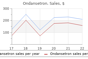
Order discount ondansetron on line
Continuous infusions of local anesthetic symptoms synonym proven ondansetron 8 mg, as might occur over several hours during labor epidural anesthesia, allow a greater total dose of anesthetic before toxic plasma levels are reached. Maximum safe dose is also influenced by vascularity of the tissue bed and whether epinephrine is added to the local anesthetic. Degree of ionization: the closer the pKa of the local anesthetic is to tissue pH, the more rapid the onset time will be. Because all local anesthetics are weak bases, those with pKa that lies near physiologic pH (7. As mentioned, the unionized form must cross the axonal membrane to initiate neural blockade. The latency of a local anesthetic can also be shortened by using a higher concentration and carbonated local anesthetic solutions to adjust the local pH. Conduction blockade proceeds from the outermost (mantle) to the innermost (core) nerve bundles. Generally speaking, mantle fibers innervate proximal structures, and core fibers innervate distal structures. This accounts for early block of more proximal areas, and muscle weakness may appear before the sensory block if motor fibers are more peripheral. Intercostal nerve block > caudal > epidural > brachial plexus > sciatic-femoral > subcutaneous. These drugs cause local tissue vasoconstriction, limiting uptake of the local anesthetic into the vasculature and thus prolonging its effects and reducing its toxic potential (see Question 14). Epinephrine, usually in 1:200,000 concentration, is also a useful marker of inadvertent intravascular injection. Epinephrine is contraindicated for digital blocks or other areas with poor collateral circulation. Systemic absorption of epinephrine may also cause hypertension and cardiac dysrhythmias, and caution is advised in patients with ischemic heart disease, hypertension, preeclampsia, and other conditions in which such responses may be undesirable. Systemic toxicity is caused by elevated plasma local anesthetic levels, most often a result of inadvertent intravascular injection and, less frequently, a result of systemic absorption of local anesthetic from the injection site. There have been multiple case reports of cardiac arrest and electrical standstill after bupivacaine administration, many associated with difficult resuscitation. It may be related to the lipid solubility of bupivacaine, which results in slow dissociation of this drug from cardiac sodium channels (fast-in, slow-out). By contrast, recovery from less lipid-soluble lidocaine is rapid (fast-in, fast-out). In an effort to minimize the risk of cardiac toxicity in the event of an accidental intravascular injection, the use of bupivacaine in concentrations greater than 0. The two classes of agents differ in their allergic potential and method of biotransformation. Lipid solubility, pKa, and protein binding of the local anesthetics determine their potency, onset, and duration of action, respectively. Bupivacaine has the highest risk of producing severe cardiac dysrhythmias and irreversible cardiovascular collapse. Short-acting muscle relaxants may be indicated for ongoing muscle activity or to facilitate intubation if necessary. Treatment includes sustained cardiopulmonary resuscitation, repeated cardioversion, high doses of epinephrine, and use of bretylium to treat ventricular dysrhythmias. Intravenous intralipid has been used successfully in cases in which conventional resuscitation measures were unsuccessful. The overall risk of permanent neurologic injury due to neurotoxicity is extremely small. These symptoms appear within 24 hours of spinal anesthesia and generally resolve within 7 days. They are seen most commonly with lidocaine spinal anesthesia and are rare with bupivacaine. Patients having surgery in the lithotomy position appear to be at increased risk of neurologic symptoms following either spinal or epidural anesthesia. The mechanism of neural injury is thought to be that nonhomogeneous distribution of spinally injected local anesthetic may expose sacral nerve roots to a high concentration of local anesthetic with consequent toxicity. Avoid injecting large amounts of local anesthetic in the subarachnoid space, especially if less than an anticipated response is obtained with the initial dose. Prilocaine is metabolized in the liver to O-toluidine, which is capable of oxidizing hemoglobin to methemoglobin. Prilocaine in a dose greater than 600 mg can produce clinical methemoglobinemia, making the patient appear cyanotic. Benzocaine, used as a spray for topical anesthesia of mouth and throat, can result in methemoglobinemia if excessive amounts are used in the form of multiple sprays or if spraying the area for a longer duration than recommended. Methemoglobin is reduced through methemoglobin reductase, and this process is accelerated by intravenous methylene blue (1 to 2 mg/kg). Cardiotoxicity from bupivacaine has almost always been fatal, often requiring placement of the patient on cardiopulmonary bypass while the drug slowly clears from the cardiac muscle tissue. Animal studies have demonstrated that lipid infusion increases resistance to local anesthetic toxicity and improves success of resuscitation from local anesthetic overdose. The mechanism of beneficial effect is thought to be a reduction in tissue binding of local anesthetic and a beneficial energetic-metabolic effect. Ropivacaine is an amide local anesthetic that is structurally and behaviorally similar to bupivacaine. When compared to bupivacaine, it is less cardiotoxic and produces less motor block, thus allowing analgesia with less motor compromise (differential blockade). However, some of these benefits may be related to somewhat lower potency of ropivacaine and may not exist when equipotent doses are compared. Because of their superior toxicity profile, both ropivacaine and levobupivacaine are suitable for situations requiring relatively large doses of local anesthetics. All of the components of organ perfusion, including preload (end-diastolic volume), afterload, inotropy, heart rate, and myocardial oxygen supply and demand, can be pharmacologically modified. Preload can be altered with intravascular volume shifts as well as with drugs that change vascular tone and, most important, the venous capacitance vessels. In addition, arterial vasodilators may shift failing myocardium to a more effective contractile state due to afterload reduction and decreased impedance to ventricular ejection. However, the intrinsic contractile state is not improved by vasodilators, in contrast to the effect of positive inotropic agents, nor are they with pure vasoconstrictors such as phenylephrine. The salutary effects of arterial vasodilators are in most cases somewhat limited by their decreased but parallel impact on venous capacitance, which decreases preload and thus cardiac output. Vascular tone can also alter the ability of the intrinsic contractile state by increasing afterload, or increased vascular tone. This leads to increased impedance to ventricular ejection by making it more difficult on ventricular contraction. Although increasing systemic vascular resistance may be improved, actual ventricular ejection may be hampered and increase ventricular wall tension. The general goal of inotropic support is increasing cardiac output by improving myocardial contractility to optimize end-organ perfusion. In addition, for enlarged hearts, a decrease in ventricular diameter, wall tension, and myocardial oxygen demand is desirable. Some inotropic agents may also serve to decrease pulmonary vascular resistance, improving right heart output and forward flow. The ideal inotrope increases contraction of cardiac muscle without changing preload or afterload, while improving pulmonary vascular resistance and myocardial oxygen demand, and has no propensity to cause arrhythmias. Amrinone and milrinone are approximately equipotent to dopamine and dobutamine in increasing cardiac output through increased inotropy and improved lusitropy (myocardial relaxation).
Generic ondansetron 8mg mastercard
Proinflammatory cytokines accumulate and treatment yeast infection male buy ondansetron 4mg with amex, even after 2 weeks, are capable of significantly priming neutrophils for an exacerbated inflammatory response. Much of the evidence for immune modulation and infection related to transfusion is retrospective in nature and, as such, suffers from a failure to control for confounding variables. There are insufficient numbers of randomized, controlled studies of sufficient power, and the studies that do exist have been conducted on critically ill patients, not in the perioperative setting (perhaps with the exception of patients having coronary bypass). The Transfusion Requirements in Critical Care trial was sufficiently powered to evaluate the impact of transfusion on outcome. The groups under study were divided into a restrictive transfusion (hemoglobin trigger of 7 g/dl, targeting a hemoglobin level between 7 and 9 g/dl) and a liberal transfusion group (hemoglobin transfusion trigger of 10 g/dl, targeting a hemoglobin level of 10 to 12 g/dl). Thirty-day mortality was lower in the restrictive transfusion group, although a statistical significance was not found. However, if the patients were subdivided by acuity of illness, results would show that fewer acutely ill patients in the restrictive transfusion group had lower 30-day mortality. Other prospective studies are less convincing in their findings, but there are overlapping transfusion triggers, and the patient populations differ. Some observational studies have found that the number of transfused units is an independent risk factor for mortality and increased length of stay. In sum, the final word on the impact of transfusion on mortality has yet to be written. Because of some associated conditions, the recipient has a high level of inflammatory mediators. The administered blood product provides the second event, through classic antibody-antigen coupling of the lipid products or other cytokines generated during storage of the blood products. The primed white blood cells are activated to release substances such as superoxides that damage the pulmonary endothelium. If the patient experiences deterioration in oxygenation during transfusion, the transfusion should be discontinued and the remainder of the transfused blood returned to the laboratory for analysis. Diuresis and steroid administration are contraindicated, but aggressive pulmonary support is necessary. If further transfusions are needed, it is wise to use blood products that have a reduced likelihood of having inflammatory mediators, including leukoreduced packed erythrocytes, packed units less than 14 days old, washed erythrocytes, or, in the case of platelets, apheresis units less than 3 days old. If an individual does not have the type A antigen, over time anti-A antibodies (also known as agglutinins) form. Individuals with type O blood have no antigen and develop both A and B antibodies Table 6-2). Acute hemolytic reactions are caused by complement activation and release of proteolytic enzymes that digest the red cell membrane. People with type O blood have neither A nor B antigens (agglutinogens) on their cell surface. There are six common antigens in the Rh system; the presence of the D antigen is what is most commonly referred to as Rh positive. The Rh blood type system is slightly different because Rh agglutinins rarely form spontaneously. Usually massive exposure, as from a prior transfusion, is necessary to stimulate their formation. An Rh-negative patient can receive Rh-positive blood in an emergency situation, although antibodies will form in some patients, and there may be a delayed, usually mild, hemolytic transfusion reaction. However, after receiving Rh-positive blood, the Rh-negative patient will be Rh sensitized and can have a more significant transfusion reaction if exposed to Rh-positive blood at a later date. Under these circumstances the fastest choice is to use type O, Rh-negative uncrossmatched blood. Type-specific, uncrossmatched blood would be the next choice, followed by type-specific, partially crossmatched blood, and finally, fully crossmatched blood. Massive transfusion is defined as the administration of more than one blood volume within several hours. Mannitol and loop diuretics are used on occasion, but caution must be used to avoid creating hypovolemia with diuresis. Follow serum potassium levels and continuously monitor the electrocardiogram for electrocardiographic signs of hyperkalemia. It should be noted that only about 55% of pre-donated units are returned to the patient. The patient scheduled for autologous transfusion still runs the risk of clerical errors and bacterial infection. Erythropoietin stimulates erythrocyte production in 5 to 7 days and has been shown to reduce use of allogeneic blood in patients with renal insufficiency and anemia of chronic disease and when transfusion is refused. The benefits of alternatives to erythrocyte transfusion include a lack of antigenicity, possible unlimited availability, no disease transmission risk, long storage life, and better rheologic properties. Such compounds are manufactured from human recombinant hemoglobin, outdated human blood, or bovine blood. The stromal components of erythrocytes are removed, and the hemoglobin molecule polymerized or liposome encapsulated to prevent rapid renal excretion and nephrotoxicity. Pulmonary hypertension and myocardial ischemia are risks; in fact, reports of death from myocardial infarction have delayed release of these solutions for general use. These solutions also result in platelet activation; release of proinflammatory mediators; methemoglobinemia; and, because of their color, interference with laboratory tests. Numerous transfusion-related reactions are possible, and vigilance while administering under anesthesia is essential because many of the classic signs and symptoms might be missed in a draped patient under general anesthesia. Preoperative evaluation includes history, physical examination, and appropriate lab testing. Prior surgery without transfusion suggests the absence of an inherited coagulation disorder. Review of medications is necessary to identify medications with anticoagulant potential. Coagulation studies may confirm a clinical suspicion that the patient has a bleeding disorder. No evidence supports the value of preoperative coagulation studies in asymptomatic patients. Three intertwined processes ensure that blood remains in a liquid state until vascular injury occurs: primary hemostasis, secondary hemostasis, and fibrinolysis. Fibrin can be formed via two pathways (intrinsic and extrinsic) and involves activation of circulating coagulation precursors. Regardless of which pathway is triggered, the coagulation cascade results in the conversion of fibrinogen to fibrin. This rigid division has lost absolute validity because of the crossover of many factors. However, the classic two-pathway model is still useful for the interpretation of in vitro coagulation studies. The fibrinolytic system is activated simultaneously with the coagulation cascade and functions to maintain the fluidity of blood during coagulation. These fibrin degradation products possess anticoagulant properties because they compete with fibrinogen for thrombin; they are normally cleared by the monocyte-macrophage system. Coagulation is limited to injured tissue by localization of platelets to the site of injury and maintenance of normal blood flow in noninjured areas. The monocyte-macrophage system scavenges activated coagulation factors in regions of normal blood flow. Normal vascular endothelium produces prostacyclin (prostaglandin I2); it is a potent vasodilator that inhibits platelet activation and helps confine primary hemostasis to the injured area. Intraoperative bleeding can be severe with counts of 40,000 to 70,000/mm3, and spontaneous bleeding usually occurs at counts <20,000/mm3. However, qualitative differences in platelet function make it unwise to rely solely on platelet count. Assessment of preoperative platelet function is further complicated by lack of correlation between bleeding time or any other test of platelet function and a tendency for increased intraoperative bleeding.
Generic ondansetron 4 mg on-line
Differential expression of cytokeratins 8 and 20 distinguishes craniopharyngioma from rathke cleft cyst medicine cat herbs buy discount ondansetron 4mg. Clinical course and surgical prognosis of 33 cases of intracranial epidermoid tumors. Pituitary-specific knockout of the Carney complex gene prkar1a leads to pituitary tumorigenesis. Telomerase activity in pituitary adenomas: significance of telomerase expression in predicting pituitary adenoma recurrence. Distinct clonal composition of primary and metastatic adrencorticotrophic hormoneproducing pituitary carcinoma. Sustained Notch signalling in progenitors is required for sequential emergence of distinct cell lineages during organogenesis. Deoxyribonucleic acid methyltransferase 3B promotes epigenetic silencing through histone 3 chromatin modifications in pituitary cells. Congenital gigantism due to growth hormone-releasing hormone excess and pituitary hyperplasia with adenomatous transformation. Rathke cleft cysts, which are topographically and pathogenically closely associated with craniopharyngiomas, are covered separately in Pituitary and Suprasellar Tumours (Chapter 41). The epithelial cysts can be roughly divided into those of ectodermal (epidermoid, dermoid), neuroectodermal (ependymal, choroid plexus) and endodermal (Rathke cleft, colloid, neurenteric) origin. Secondary changes, such as epithelial atrophy, ulceration, squamous metaplasia and cyst rupture with haemorrhage, inflammation and granuloma formation are common and may hinder precise classification. In others, both the diagnosis and patient management rely heavily on a careful radiologic workup to rule out connections between intracranial and extracranial contents. In the cranial vault, the frontal and parietal bones are most often involved and in the petrous region, they may cause facial paralysis and bone destruction. Large intradiploic variants usually break through the inner table and may also destroy the outer table to cause soft-tissue swelling under the scalp. In addition to the usual generalized signs and symptoms of increased intracranial pressure, the common cerebellopontine variety most often presents with involvement of the facial nerve followed by unilateral hearing loss and other cranial nerve palsies. They may be easily shelled out from adjacent structures or firmly anchored as a result of local inflammation. Cysts in the ventricles or in the subarachnoid space are liable to rupture and cause meningitis. The cut surface reveals an interior filled with soft, pasty to dry, flaky material. Biological Behaviour Epidermoid cysts originate from the ectoderm, in common with dermoids. Cerebellopontine epidermoids derive from the first branchial groove, presumably from entrapped or misplaced migratory cells; this is similar to the derivation of acquired cholesteatomas in the ear. The treatment of choice is surgery; complete resection is more common today because of improvements in microsurgery. This epithelial lining reproduces the normal layers of the epidermis, complete with keratohyalin granules. The progressive production and desquamation of keratin result in the formation of concentric lamellae that fill the interior of the cyst, causing gradual expansion. The lining occasionally may be papillary, but the typical squamous epithelium is maintained. They may be slightly more common in males and slightly younger patients than epidermoids. Keratinizing squamous epithelium covers collagenous connective tissue containing skin adnexal structures. Colloid cysts have been reported in association with astrocytoma,26 the naevoid basal cell carcinoma syndrome,58 nasal dermoid sinus,16 and agenesis of the corpus callosum. Familial colloid cysts are rare, although families with two or more affected members should be screened, because an autosomal dominant inheritance has been suggested. Rare examples in the lateral ventricles, fourth ventricle, and outside the ventricular system have been reported, although they are perhaps better regarded as neurenteric cysts because the histology of these two lesions is identical (discussed later). Their wall thickness varies, including occasional papillary projections and rarely calcified plaques. The cyst contains thick, cheesy, yellowish material that results from the secretory activity of sebaceous glands and from desquamated epithelium. The connection of dermoid cysts with dermal sinuses is known to occur in both intracranial and spinal examples. At the other extreme, large cysts splay the fornices causing memory deficits and occlude the foramina of Monro, causing obstructive hydrocephalus. The most common symptom is headache, which is often episodic and positional, such that it is exacerbated when the patient lies down to sleep and improves upon standing. T2-weighted images may show a reversal of the T1 pattern, although often, there is a hyperintense periphery and a Microscopy Dermoid cyst lining is similar in places to that of epidermoid cysts (see earlier), comprising simple stratified squamous epithelium supported by collagen. Bone and cartilage are rare, their presence being more typical of classic teratomas. Biological Behaviour these lesions are slow growing and benign, but are likely to recur when incompletely removed. Dermal sinuses penetrating the dura may be the route of pyogenic infection, a potentially serious complication, which can be prevented by early surgery. Rarely, chronic inflammation destroys the cyst wall, causing gliosis in the adjacent cerebral tissue. A thin rim of contrast enhancement represents the capsule and intracystic fluid levels are occasionally seen. Endoscopic removal has also gained popularity because of its reduced morbidity, intra-operative time and length of hospitalization. Unsuccessful stereotactic aspiration is usually related to high viscosity or deflection of the cyst away from the aspiration needle as a result of small size. The wall is usually thin, surrounding a homogeneous, soft, opaque or occasionally denser, hyaline-like material. The cyst may be firmly anchored to surrounding brain structures, including the foramina of Monro, the wall of the lateral ventricle or the columns of the fornix. The fibrous capsule of the lesion may be intricately associated with the connective tissue stroma of the choroid plexus of the third ventricle. Stretches of cuboidal or flattened columnar epithelium may alternate with ciliated, simple or pseudostratified columnar epithelium. A xanthogranulomatous reaction occasionally develops because of desquamation of lining cells, exposure of colloid material, and microhaemorrhages: epithelioid cells and macrophages contain refractile material and haemosiderin. Small microcysts in the surrounding fibrovascular stroma may occasionally be seen. Occasional colloid cysts may display mainly squamous differentiation and basally located cells may resemble myoepithelial cells. There are six cell types in the lining epithelium: ciliated cells with occasional abnormal cilia, nonciliated cells with microvilli coated with granulofibrillary material, goblet cells with secretory activity, basal cells with tonofilaments and desmosomes, basally located, elongated cells with scattered, membrane-bound secretory granules in the electron-lucent cytoplasm and small, undifferentiated cells poor in organelles. These cell types and their distribution within the lining are similar to normal upper respiratory epithelium and the lining of spinal neuroenteric cysts, suggesting that colloid cysts originate from the endoderm. Unlike choroid plexus tumours, they are negative for prealbumin (transthyretin), S-100 protein, glial fibrillary 1912 Chapter 42 Cysts and Tumour-like Conditions sites, Macroscopical appearances and Microscopy Most cases are encountered in the intradural extramedullary spinal compartment. Solitary cysts occur most frequently in the cervical region, whereas the lumbosacral cases are often associated with dysraphic defects. For both groups, ventral location is more common, followed by dorsal lesions and the relatively rare intramedullary examples. Histologically, neurenteric cysts may be lined by a single-layered or pseudostratified cuboidal or columnar, ciliated or non-ciliated epithelium, resembling gastrointestinal or respiratory epithelium, mounted on a basement membrane. In addition, some contain mucous or serous glands, smooth muscle, various connective tissue components, lymphoid tissue and even ganglia, while rare examples additionally show glioependymal elements. Of 57 posterior fossa neuroepithelial cysts, 32 were symptomatic and 25 were incidental. Both sexes were equally affected and the mean age of 35 patients was 30 years, ranging from 5. They may also be found in the lateral convexities, posterior fossa, brain stem, spinal cord, and within the subarachnoid space. These cysts are thought to originate from ectopic or pinched off remnants of neural tube. In subarachnoid examples, a derivation from leptomeningeal neuroglial heterotopias is favoured. Consistent with its congenital nature, a large example has been reported in a 22-week estimated gestational age fetus and mimicked holoprosencephaly on ultrasound.
Generic 8 mg ondansetron overnight delivery
Sharma medicine 2410 purchase ondansetron 8 mg mastercard, Dafin Muresanu, Aruna Sharma, and Ranjana Patnaik Cannabinoid Receptors in Brain: Pharmacogenetics, Neuropharmacology, Neurotoxicology, and Potential Therapeutic Applications Emmanuel S. Sharma Molecular Bases of Methamphetamine-Induced Neurodegeneration Jean Lud Cadet and Irina N. Schwartz Pharmacological and Neurotoxicological Actions Mediated By Bupropion and Diethylpropion Hugo R. Rodan and Adrian Rothenfluh Neural Plasticity, Human Genetics, and Risk for Alcohol Dependence Shirley Y. Hill Using Expression Genetics to Study the Neurobiology of Ethanol and Alcoholism Sean P. Miles 376 Genetic Variation and Brain Gene Expression in Rodent Models of Alcoholism: Implications for Medication Development Karl Bjrk, Anita C. Buck Glutamate Plasticity in the Drunken Amygdala: the Making of an Anxious Synapse Brian A. La Ethanol Action on Dopaminergic Neurons in the Ventral Tegmental Area: Interaction with Intrinsic Ion Channels and Neurotransmitter Inputs Hitoshi Morikawa and Richard A. Payne Characteristics and Contents of Dreams Michael Schredl Trait and Neurobiological Correlates of Individual Differences in Dream Recall and Dream Content Mark Blagrove and Edward F. Propper To What Extent Do Neurobiological SleepWaking Processes Support Psychoanalysis Buijs, and Eric Fliers Contents of Recent Volumes 377 Preparation for Awakening: Self-Awakening Vs. Forced Awakening: Preparatory Changes in the Pre-Awakening Period Mitsuo Hayashi, Noriko Matsuura and Hiroki Ikeda Circadian and Sleep Episode Duration Influences on Cognitive Performance Following the Process of Awakening Robert L. Matchock the Cortisol Awakening Response in Context Angela Clow, Frank Hucklebridge and Lisa Thorn Causes and Correlates of Frequent Night Awakenings in Early Childhood Amy Jo Schwichtenberg and Beth Goodlin-Jones Pathologies of Awakenings: the Clinical Problem of Insomnia Considered From Multiple Theory Levels Douglas E. Waddington the Trigeminal Circuits Responsible Chewing Karl-Gunnar Westberg and Arlette Kolta for Volume 98 An Introduction to Dyskinesia-the Clinical Spectrum Ainhi Ha and Joseph Jankovic L-dopa-induced Dyskinesia-Clinical Presentation, Genetics, And Treatment L. Lozano Ultrastructural Basis for Craniofacial Sensory Processing in the Brainstem Yong Chul Bae and Atsushi Yoshida Mechanisms of Nociceptive Transduction and Transmission: A Machinery for Pain Sensation and Tools for Selective Analgesia Alexander M. Lane Experiences of Contents of Recent Volumes 379 Homeostatic Control of Neural Activity: A Drosophila Model for Drug Tolerance and Dependence Alfredo Ghezzi and Nigel S. Rosser Clinical Phenomenology of Dystonia Carlo Colosimo and Alfredo Berardelli Genetics and Pharmacological Treatment of Dystonia Susan Bressman and Matthew James Experimental Models of Dystonia A. Edmondson Behavioral Outcomes of Monoamine Oxidase Deficiency: Preclinical and Clinical Evidence Marco Bortolato and Jean C. Shih Kinetic Behavior and Reversible Inhibition of Monoamine Oxidases-Enzymes that Many Want Dead Keith F. Stern Selective Inhibitors of Monoamine Oxidase Type B and the "Cheese Effect" John P. Harris and Sabine Bahn Immune and Neuroimmune Alterations in Mood Disorders and Schizophrenia Roosmarijn C. Guest, Eva Hradetzky, Wolfgang Kluge, Viktoria Stelzhammer and Hendrik Wesseling Stem Cell Models for Biomarker Discovery in Brain Disease Alan Mackay-Sim, George Mellick and Stephen Wood the Application of Multiplexed Assay Systems for Molecular Diagnostics Emanuel Schwarz, Nico J. Guest, Rauf Izmailov and Sabine Bahn Algorithm Development for Diagnostic Biomarker Assays Rauf Izmailov, Paul C. Guest, Sabine Bahn and Emanuel Schwarz Challenges of Introducing New Biomarker Products for Neuropsychiatric Disorders into the Market Sabine Bahn, Richard Noll, Anthony Barnes, Emanuel Schwarz and Paul C. Ng and Chi-Ming Lee Clinical Utility of Serum Biomarkers for Major Psychiatric Disorders Nico J. Hoogendijk the Future: Biomarkers, Biosensors, Neuroinformatics, and E-Neuropsychiatry Christopher R. Turck Imaging Brain Microglial Activation Using Positron Emission Tomography and Translocator Protein-Specific Radioligands David R. Tsuang Proteomic Technologies for Biomarker Studies in Psychiatry: Advances and Needs Daniel Martins-de-Souza, Paul C. Harris and Sabine Bahn Converging Evidence of Blood-Based Biomarkers for Schizophrenia: An update Man K. Smith and Aruna Sharma Neurovascular Aspects of Amyotrophic Lateral Sclerosis Maria Carolina O. Voltarelli and Svitlana Garbuzova-Davis Quercetin in Hypoxia-Induced Oxidative Stress: Novel Target for Neuroprotection Anand Kumar Pandey, Ranjana Patnaik, Dafin F. Muresanu, Aruna Sharma and Hari Shanker Sharma Environmental Conditions Modulate Neurotoxic Effects of Psychomotor Stimulant Drugs of Abuse Eugene A. Kiyatkin and Hari Shanker Sharma Central Nervous Tissue Damage after Hypoxia and Reperfusion in Conjunction with Cardiac Arrest and Cardiopulmonary Resuscitation: Mechanisms of Action and Possibilities for Mitigation Lars Wiklund, Cecile Martijn, Adriana Miclescu, Egidijus Semenas, Sten Rubertsson and Hari Shanker Sharma Interactions Between Opioids and Anabolic Androgenic Steroids: Implications for the Development of Addictive Behavior Fred Nyberg and Mathias Hallberg Neurotrophic Factors and Neurodegenerative Diseases: A Delivery Issue Barbara Ruozi, Daniela Belletti, Lucia Bondioli, Alessandro De Vita, Flavio Forni, Maria Angela Vandelli and Giovanni Tosi Neuroprotective Effects of Cerebrolysin, a Combination of Different Active Fragments of Volume 103 Lost and Found in Behavioral Informatics Melissa A. Baker A Survey of the Neuroscience Resource Landscape: Perspectives from the Neuroscience Information Framework Jonathan Cachat, Anita Bandrowski, Jeffery S. Martone the Neurobehavior Ontology: An Ontology for Annotation and Integration of Behavior and Behavioral Phenotypes Georgios V. Schofield, and Robert Hoehndorf Ontologies for Human Behavior Analysis and Their Application to Clinical Data Janna Hastings and Stefan Schulz Text-Mining and Neuroscience Kyle H. Cohen Applying In Silico Integrative Genomics to Genetic Studies of Human Disease: A Review Scott F. Jay 382 Model Organism Databases in Behavioral Neuroscience Mary Shimoyama, Jennifer R. Thomas Hayman, Victoria Petri, and Rajni Nigam Accessing and Mining Data from Large-Scale Mouse Phenotyping Projects Hugh Morgan, Michelle Simon, and Ann-Marie Mallon Bioinformatics Resources for Behavior Studies in the Laboratory Mouse Carol J. Mulligan Large-Scale Neuroinformatics for In Situ Hybridization Data in the Mouse Brain Lydia L. Hawrylycz Opportunities for Bioinformatics in the Classification of Behavior and Psychiatric Disorders Elissa J. Goldberg Intrinsic Mechanisms Regulating Axon Regeneration: An Integrin Perspective Richard Eva, Melissa R. Goldberg Inflammatory Pathways in Spinal Cord Injury Samuel David, Juan Guillermo Zarruk, and Nader Ghasemlou Combinatorial Therapy Stimulates Long-Distance Regeneration, Target Reinnervation, and Partial Recovery of Vision After Optic Nerve Injury in Mice Silmara de Lima, Ghaith Habboub, and Larry I. Levin Role of Electrical Activity of Neurons for Neuroprotection Takeshi Morimoto Molecular Control of Axon Growth: Insights from Comparative Gene Profiling and HighThroughput Screening Murray G. Blackmore Gatekeeper Between Quiescence and Differentiation: p53 in Axonal Outgrowth and Neurogenesis Giorgia Quadrato and Simone Di Giovanni Cyclin-Dependent Kinase 5 in Axon Growth and Regeneration Tao Ye, Amy K. Ip Volume 107 Neuromodulation: A More Comprehensive Concept Beyond Deep Brain Stimulation Clement Hamani and Elena Moro Computational Models of Neuromodulation Christopher R. Reid the Pros and Cons of Growth Factors and Cytokines in Peripheral Axon Regeneration Lars Klimaschewski, Barbara Hausott, and Doychin N. Foreman and Bengt Linderoth Magnetoencephalography and Neuromodulation Alfons Schnitzler and Jan Hirschmann Current Challenges to the Clinical Translation of Brain Machine Interface Technology Charles W. Reis Tissue Engineering and Peripheral Nerve Reconstruction: An Overview Stefano Geuna, S.
Diseases
- Chronic spasmodic dysphonia
- Dissecting cellulitis of the scalp
- Dystrophinopathy
- Levator syndrome
- Telecanthus hypertelorism pes cavus
- Progressive multifocal leukoencephalopathy
- Cor triatriatum
Generic 4 mg ondansetron fast delivery
In the older individual the image quality is degraded because of decreased perfusion and increased arterial transit times 5ht3 medications purchase ondansetron american express. Published studies to date, however, are generally small and most were performed in patients at late stages of dementia [62]. Within the white matter however, axonal boundaries and myelin sheaths form geometric restrictions. Grey matter tissue is structured differently and much less pronounced anisotropy of diffusion is observed. Assessment of anisotropy of the diffusion profile provides key insights into the geometry of local tissue structure [16]. In addition, the amount of diffusivity in each direction can be precisely estimated, such that potentially subtle changes in tissue composition can be measured. As explained previously, diffusion is measured as signal attenuation, such that for high diffusion values the signal may be as low as the noise level. From the raw data, the diffusion tensor in each voxel is computed, describing the local diffusion processes [64]. The diffusion tensor is characterized by its eigenvalues and eigenvectors, describing the orientation and shape of the diffusion profile. The principal eigenvector refers to the main orientation of the diffusion, which is assumed to mimic the local tissue structure. The eigenvalues quantify the apparent diffusion parallel and perpendicular to the principal orientation. In fact, a voxel of 2 mm isotropic may contain up to 104 axons, represented by a single fibre track [67]. Most research focuses on increasing our understanding of the pathophysiological processes that eventually result in those changes that can be appreciated on conventional imaging such as atrophy and white matter lesions. Fibre tracking can improve the accuracy of measurements by localizing the changes to specific tracts [68]. Many of the published findings, however, are at present not readily transferrable to clinical practice. It is able to measure brain function, as opposed to structural imaging that mainly relates to anatomical pathological changes. Red, green, and blue colours reflect left/right, anterior/posterior and inferior/superior orientations respectively. Oxyhaemoglobin (haemoglobin that has oxygen molecules bound to it), on the other hand, is diamagnetic, i. If the amount of deoxyhaemoglobin decreases due to an increase in perfusion, the local T2* value will increase, since less signal dephasing occurs. Furthermore, sophisticated postprocessing, in which a diversity of analysis options may significantly impact the results, is required to identify active brain regions. Initial studies have primarily focused on task-specific activation changes, such as those of memory processing. It is generally assumed that with decreased task performance, brain activation is also decreased. A popular theory, however, is that in the early stages of neurocognitive decline brain activation in fact increases, indicative of a compensatory mechanism by which the patient maintains a certain level of cognitive functioning [76]. It seems that specific neurodegenerative diseases target anatomically predictable networks [79]. The Larmor frequency of non-water protons slightly differs from that of water protons due to the so-called chemical shift phenomenon. Effectively, from one single voxel signals with different frequencies can be recorded and various metabolites can easily be discriminated from each other, as well as from the abundant water signal. The attractiveness of quantitative imaging in neurodegenerative diseases is obvious. For both diagnosis and follow-up, the accuracy of clinical quantitative assessments is expected to outperform qualitative assessments. For instance, changes in relaxation time due to changes in disease states will eventually appear as differences in image contrast, but as long as they are small they can easily remain undetected on visual inspection. Although proven highly accurate, this technique is time consuming and therefore not a serious candidate for clinical routine. This technique is very fast, but quantification is often cumbersome and inaccurate at high T1-values, hindering the direct comparison of T1 values across subjects. Relaxometry requires extensive postprocessing of the data, which is often not feasible in a clinical setting. Moreover, to use relaxometry in clinical practice, the spatial resolution of brain scans should at least be comparable to that of conventional anatomical brain scans, which-if even feasible-will add substantially to the total scan duration. Here, two or even three gradient echo acquisitions are performed with different flip angles that can subsequently be converted to a T1-value per voxel [86]. It is most popular to acquire images at multiple echo times, which in general does not add significantly to the scan time. Another option is to induce different T2-weightings of the signal during preparation and prior to the readout module of the sequence. Contrast administration may be required when inflammatory, infectious, or oncological diseases are considered as differential diagnoses. We highly recommend the use of 3D sequences, allowing for reformatting in any plane at any desired resolution. The latter would include focal abnormalities consistent with for instance subdural haematoma, brain tumour, hippocampal sclerosis, herpes encephalitis, or alcoholic encephalopathy. Separately report the focal regions of atrophy as well as the degree of asymmetry. Diffusion-weighted and fluid-attenuated inversion recovery imaging in Creutzfeldt-Jakob disease: high sensitivity and specificity for diagnosis. Magnetic resonance imaging in the clinical diagnosis of Creutzfeldt-Jakob disease. Susceptibility-weighted imaging: technical aspects and clinical applications, part 2. Susceptibility-weighted imaging: technical aspects and clinical applications, part 1. Clinical diagnosis of cerebral amyloid angiopathy: validation of the Boston criteria. Prevalence and risk factors of cerebral microbleeds: an update of the Rotterdam scan study. The measurement of diffusion and perfusion in biological systems using magnetic resonance imaging. Voxel-based correlation between coregistered single-photon emission computed tomography and dynamic susceptibility contrast magnetic resonance imaging in subjects with suspected Alzheimer disease. Note areas of increased mineral deposition such as the globus pallidus and substantia nigra. Finally, the conclusion needs to reflect the interpretation of all imaging findings and should ideally include a most likely and differential diagnosis. Magnetic field and tissue dependencies of human brain longitudinal 1H2O relaxation in vivo. Fast spin echo sequences with very long echo trains: design of variable refocusing flip angle schedules and generation of clinical T2 contrast. Structural magnetic resonance imaging in the practical assessment of dementia: beyond exclusion. Optimal strategies for measuring diffusion in anisotropic systems by magnetic resonance imaging. Spin diffusion measurements: spin echoes in the presence of a time-dependent field gradient. Diffusion-based tractography in neurological disorders: concepts, applications, and future developments.
8mg ondansetron overnight delivery
The pars tuberalis is the superior portion of the adenohypophysis that wraps itself around the neural stalk medicine just for cough buy discount ondansetron 8mg online. It is composed primarily of gonadotrophs that with age undergo squamous metaplasia. The superior hypophysial arteries flow through the infundibulum of the neurohypophysis and form the portal vessels that transport regulatory hormones from the hypothalamus to the pituitary gland. The middle hypophyseal arteries supply blood directly to the adenohypophysis, whereas the inferior hypophyseal arteries supply the pars nervosa. Venous blood from the pituitary gland drains mainly into the internal jugular veins; however, there is evidence that reverse flow in the short portal vessels allows adenohypophyseal secretion to affect neurohypophyseal and hypothalamic function. They show a wide range of biological behaviour, ranging from microscopic incidental findings to small lesions with severe hormonal manifestations, to large invasive neoplasms (Box 41. The anatomic/ radiologic classification categorizes pituitary adenomas based on size and degree of invasion. Silent ectopic adenomas usually present with mass effects and their diagnosis depends on careful examination of resected tissue specimens. Although classified as benign because 1876 Chapter 41 Pituitary and Suprasellar Tumours Table 41. In adults, after fusion of the epiphyseal plates, the hormone excess causes acral enlargement and prominence of facial bones, resulting in prognathism and facial deformities. Prolactin hypersecretion causes gonadotropin insufficiency and sexual dysfunction; in women this is manifested as amenorrhea. Prolonged oestrogen or androgen deficiency results in osteopenia and osteoporosis. Thyrotropin excess is rare and manifests as clinical or subclinical hyperthyroidism. It is imperative to distinguish primary from secondary forms of thyroid hormone excess. Patients with this disorder also develop osteoporosis, diabetes and immunosuppression. Tumours of any type that destroy a significant proportion of pituitary parenchyma result in hypopituitarism. Destruction of the posterior pituitary can result in diabetes insipidus but this is rarely evident in patients with primary pituitary adenomas; instead, it usually indicates the presence of a more aggressive infiltrative tumour or an alternative diffuse tumour-like or inflammatory process. With suprasellar extension, the optic chiasm can become involved, resulting in visual field deficits, usually initially a bitemporal hemianopsia. Extension well outside the sella can result in cranial nerve defects or, rarely, cavernous sinus syndrome; these are unusual features of primary pituitary adenomas (Box 41. Pituitary apoplexy Pituitary apoplexy constitutes a true endocrine emergency in which acute haemorrhagic infarction of a sellar tumour (usually an adenoma), results in rapid expansion with symptoms and signs of elevated intracranial pressure. These features are common as focal changes in many pituitary tumours; however, true pituitary apoplexy refers to those extreme cases where haemorrhagic infarction of the pituitary is accompanied by the appropriate clinical features. T2-weighted imaging identifies heterogeneous areas within large lesions; these usually correspond to regions of haemorrhage or necrosis. Following gadolinium administration, the normal gland shows increased contrast uptake that delineates it from the adenoma that shows no or minimal contrast enhancement. The uses of lateral polytomography, air encephalography and/ or carotid angiography are largely of historical interest only. The demonstration of somatostatin receptors in pituitary tumours has led to the use of scintigraphic visualization of radiolabelled somatostatin analogues to localize some adenomas. However, in addition to identifying primary pituitary adenomas, this imaging technique has localized other lesions, such as metastatic deposits, and is therefore not as specific as originally postulated. Ultrastructurally,115 corticotroph cells are large and polygonal with ovoid or irregular nuclei that harbour nucleoli in contact with the inner nuclear membrane. The secretory granules range in size from 150 to 450 nm in diameter and are distinctive because of their marked variability in shape and electron density. Sparsely granulated corticotroph adenomas are less common than the densely granulated variant. The perinuclear ring of pale hyaline material represents the accumulation of cytokeratins 7 and 8 that are intermediate filaments on electron microscopy. In patients with tiny microadenomas, occasionally no diagnostic adenoma tissue is included in the pathology specimen. This may be attributed to loss of the adenoma during suction of a bloody operative field. Grossly, these tumours are usually well demarcated and are located in the lateral wing of the adenohypophysis. They tend to be soft lesions that rarely cause compression of the surrounding gland. There is variable cytoplasmic immunoreactivity for -subunit of glycoprotein hormones. By electron microscopy,115 the tumour cells resemble nontumourous somatotrophs; they have spherical nuclei with prominent nucleoli, parallel arrays of rough endoplasmic reticulum and well-formed Golgi complexes. Secretory granules are numerous, homogeneous, dense and spherical with diameters ranging from 150 to 600 nm. Sparsely granulated somatotroph adenomas are composed of solid sheets of poorly cohesive chromophobic cells. Unlike other pituitary adenomas, they can exhibit striking nuclear pleomorphism and have been misdiagnosed as metastatic carcinomas. In contrast to densely granulated somatotroph adenomas, they have marked reduction of 1880 Chapter 41 Pituitary and Suprasellar Tumours (a) (b) (c) 41. There is marked nuclear atypia and these lesions can be mistaken for metastatic carcinoma. Immunohistochemical stains for low molecular weight cytokeratins reveal the characteristic feature of this tumour type, the fibrous body, which manifests as juxtanuclear globular reactivity. Ultrastructurally,115 the tumour cells are irregularly shaped with eccentric, pleomorphic and multilobulated nuclei. The characteristic fibrous body is a juxtanuclear, spherical mass composed of intermediate filaments. They are the most frequent findings in patients with gigantism and in young patients with acromegaly. Microscopically, these tumours are composed of acidophilic cells arranged in a diffuse or solid pattern, resembling densely granulated somatotroph adenomas. Ultrastructurally, the tumour cells resemble densely granulated somatotrophs;115 however, secretory granules have mottled cores, are variably pleomorphic and can measure up to 1000 nm. Sparsely granulated somatotroph adenomas are unusual in that they exhibit significant nuclear pleomorphism and atypia that is not found in most pituitary adenomas or carcinomas. There is no consistent morphologic alteration attributable to medical administration of somatostatin analogues66 that are being increasingly offered as preoperative medical therapy to reduce the surgical complications of acromegalic patients. Although almost half of adenomas found incidentally at autopsy are of this type,70 the incidence is much lower in surgical series, probably because these tumours are often treated medically. Prolactinomas are more common in females, who tend to present at a younger age with hormonal disturbances. In contrast, men tend to present later, with larger tumours that more often result in mass effects and hypopituitarism secondary to adenohypophyseal destruction. Whereas this has been attributed to sexual dimorphism of the perception of the relevant symptoms, recent evidence suggests that the tumours grow faster in men. Microadenomas are most commonly located in the posterolateral portions of the gland. Psammoma bodies may occur in pituitary adenomas and are most common in prolactinomas; they result in a gritty consistency. Sparsely granulated lactotroph adenomas are the most common variant of adenoma arising from lactotrophs. Chromophobic tumour cells are arranged in papillae, trabeculae or solid sheets; tumour cells may form pseudorosettes around vascular spaces. Densely granulated lactotroph adenomas are much less common than the sparsely granulated variant. Ultrastructurally, densely granulated cells have abundant rough endoplasmic reticulum; secretory granules are numerous and can measure up to 700 nm.
Buy generic ondansetron on-line
The pulse specific relaxation time that it takes for the spin to restore its orientation along the B 0-field after the B1 magnetic pulse is called the T1 relaxation time symptoms type 2 diabetes purchase discount ondansetron line. It should be noted that the spin relaxation time is also sensitive to inhomogeneities (non-uniform distribution) in the magnetic field. The relationship describing spin relaxation taking into account these effects is called T2* (pronounced T2 star). During their relaxation the spins emit electromagnetic radiation, which can be imagined as an orthogonal combination of electric and magnetic waves travelling with a specific frequency in the radio range. Correspondingly, the measured electromagnetic radiation can be assigned to the particular slice. In 2003 both researchers received the Nobel Prize for their pioneering work on this topic. An atom consists of two main parts-the nucleus and a cloud of negatively charged electrons. Two types of particles-positively charged protons and electrically neutral neutrons-constitute the nucleus. The only exception from this basic principle is the hydrogen atom, which has only one proton and no neutrons. Without the presence of a strong magnetic field the spins are oriented in all possible directions. After its removal the spins are reoriented back towards the principle orientation of the B0 field (T1 relaxation). In parallel, a dephasing of the spin in the transverse plane to the B 0 field takes place (T2 relaxation). Using this knowledge we can apply Fourier transformation to reconstruct the final image from k-space. In contrast, most recent developments such as relaxometry-based multiparameter mapping make it possible to delineate and quantify the specific contribution of particular brain tissue properties. The valence number of the isotope indicates the nuclear mass of a nucleus of its atoms. These isotopes have a decreased nuclear gradient assigning a different excitation frequency to each column of the selected slice. The third step, phase encoding, additionally introduces a time gradient in the excitation of different rows of the selected slice. This gradient represents the time shift between the excitation of different rows of the slice and leads to a phase shift in the excitation and correspondingly in the relaxation frequency of each row. The nuclei of the radioactive isotopes are unstable and break down with time to reach the state of the corresponding stable chemical element. During its decay a radioactive nucleus emits a positron (a positively charged antimatter counterpart of the electron). On its way through the tissue (typically about 1 mm) the positron loses kinetic energy until it slows down so far that it can interact with an electron. When both emitted photons collide with detector plates they induce an electric current. By computing the time lag between their arrival at the detector plates and using the knowledge that both photons are travelling in opposite directions from the point of their origin, it is possible to reconstruct the origin at which this pair of photons was emitted. The resulting count map shows the concentration of tracer decay at each particular location. To attain higher precision of the maps of radioactive decay, we need to adjust for these effects. The interaction of both leads to a deflection of the photon from its initial direction of travel. Another source of noise, attenuation of the signal, results from the absorption of photons by atoms of the tissue it is travelling through. Correspondingly, photons having a longer path through the object tissue have a higher probability of being deflected (scattering) or absorbed (attenuation). Higher intensities indicate a higher glucose consumption in the corresponding regions. A colliding positron and electron annihilate each other and produce two photons travelling into opposite directions on a line of response. Both photons hit two detector plates (indicated in yellow) positioned in a detector ring around the event. The origin of both photons can be computed from the time lag between their arrivals at the corresponding detector plates. The widespread application of neuroimaging techniques in this field is supported by the fact that the majority of recent diagnostic criteria for neurodegenerative diseases require the inclusion of imaging biomarkers as supportive criteria [10,11]. This paradigm shift is mainly due to the vast amount of knowledge that neuroimaging has contributed to the field of neurodegeneration research in the past two decades. Despite this progress the inclusion of imaging biomarkers is still only a supportive feature, not a core diagnostic measure, in most of these proposed criteria. It is important to note that the extraction of features from both techniques requires multiple sophisticated preprocessing and statistical analysis steps. Imaging studies provide strong evidence for involvement of distinct anatomical regions in each of the three subtypes [34,35]. Another technical development in the field, which is particularly noteworthy in the context of studying the mechanisms behind neurodegeneration, is the development of tracers targeting specific transmitter systems. Studying the temporal dynamics of neurodegenerative changes and the ability of neuroimaging to capture subtle disease progression-related changes is a necessary requirement from both a clinical and a basic neuroscience perspective. With time these accumulative pathological changes spread over the whole brain and lead to synaptic dysfunction and to subsequent cell death mirrored in the specific pattern of brain atrophy [31]. Its clinical core features comprise progressive mental impairment mainly characterized by attentional, problem solving, and visuospatial deficits [37]. Histopathologically, it is characterized by the presence of Lewy bodies-abnormal aggregates of -synuclein and ubiquitin in neurons. However, there is much controversy on the anatomical pattern of changes associated with the syndrome. Immunopositive Lewy neurites and Lewy bodies are therefore essential for the diagnosis [45]. In particular, glucose hypometabolism was observed in occipito-parietal and inferior lateral prefrontal regions. Some studies also reported glucose metabolism increases in basal ganglia, ventral thalamus, motor cortex, and cerebellum [54]. Histopathologically, it is mainly characterized by loss of neurons in the putamen and caudate nucleus [60] accompanied by the loss of D1 and D2 receptor binding in the same regions [61]. It is characterized by selective neuronal loss and gliosis both focally restricted to the cornu ammonis 1 and subiculum part of the hippocampus. In addition, the asymmetry in diffusion properties successfully differentiated between both types of atypical parkinsonism [77]. Future perspectives Although clinical routine still mostly relies on evaluation of acquired images by radiologists and nuclear medicine specialists, advances in machine learning now make it possible to apply automated image evaluation algorithms for detection and differentiation of most neurodegenerative diseases. This type of algorithm requires a training dataset of images (from any imaging modality) with known diagnostic labels. A pattern recognition algorithm is then applied to these images to detect characteristic patterns that are distinct between different diagnostic labels. The trained algorithm can be then applied to new imaging data and provides a decision in favour of one of the previously learned diagnostic labels. The decision is thus based on the similarity of the new image to one of the previously learned disease patterns. This type of machine learning algorithm has been successfully shown to differentiate between most common dementia syndromes with reasonable accuracies of more than 90% [82,87,88]. As compared to evaluation by radiologists and nuclear medicine specialists these approaches have several advances: for instance, higher reliabilities and the possibility to integrate several imaging and clinical modalities into a single diagnostic framework.
Buy cheap ondansetron 4 mg line
His first recommendation was for its stimulant effects "in those functional states which we now cover by the name neurasthenia" (wording that offers medications on airline flights buy ondansetron 4 mg line, perhaps, an early indication of the scale of his ambition). His second was for the treatment of indigestion, and his third "in the withdrawal of morphine. But the suggestion that it offered a cure for morphinism, which had not originated with Freud but would remain obstinately attached to him, exposed its dangers. But it was not long before Fleischl began to increase his doses of cocaine and switched to subcutaneous injection, revealing that large doses of the pure drug led to powerful cravings and tolerance, inability to eat or sleep, and paranoid hallucinations. Virtually overnight, Fleischl had gone from being one of the first patients cured of a morphine habit with cocaine to one of the first to present the terrifying symptoms of a full-scale addiction to it. He himself had never taken it in anything other than small amounts, and never by the more compulsive methods of sniffing or injecting. Cocaine made a rapid transit from a nostrum of dubious efficacy to , in the words of the leading German psychiatrist Albrecht Erlenmeyer, "the third scourge of mankind," after alcohol and opium (quoted in Freud, 1887, p. In Phantastica, his influential compendium of 1924, Louis Lewin would remind his readers that he had "at once objected" in 1885 to "the unfortunate theory that morphinism could be cured by cocaine," predicting a "twofold craving": "this, and worse, is what in fact happened. Yet Lewin in 1887, on a steamer bound for New York, recorded finding himself a little dizzy and with a headache: "Ah! The distinctions between medicine and pleasure, use and abuse, feeling better and feeling better than well, would remain difficult to draw. By 1887, as the British Medical Journal noted, an "undeniable reaction against the extravagant pretensions advanced on behalf of this drug has already set in" (British Medical Journal, 1887, Vol. William Hammond replied to the alarmists with a forceful essay on Cocaine and the So-Called Cocaine Habit. Hammond had been using cocaine medicinally for a spinal irritation and also as a general tonic; he had drunk it, injected it, experimented with large doses (a gram injected over 20 min) and prescribed large doses to others. His conclusion was that the cocaine habit was "similar to the tea or coffee habit" (Hammond, 1887, pp. Unlike opiates, to which patients developed a physical dependence, Hammond maintained that there was not "a single instance of a well-pronounced cocaine habit" where the addict was unable to renounce the drug by simple willpower (Hammond, 1887). The debate was necessarily entangled with those surrounding the newly minted diagnosis of addiction, which was variously 34 Mike Jay construed as a physical disease, a moral weakness, a hereditary taint, or the affliction of a pathological "type. Between 1884 and 1887, the wholesale price in the United States dropped from around 15 dollars a gram to 30 cents (Spillane, 1999, p. Parke, Davis, in their promotional brochure of 1885, offered cocaine in powders, solutions, tablets, lozenges, even cigars, and cheroots claiming the drug to be "the most important therapeutic discovery of the age, the benefits of which to humanity will be simply incalculable," and "as the facts recorded would now indicate, the long sought for specific for the opium habit" (Parke, Davis and Company, 1885, pp. Assurances that cocaine "can supply the place of food, make the coward brave, the silent eloquent" (Parke, Davis and Company, 1885) ran alongside ads for smart pocket-sized steel cases containing cocaine, morphine, and miniature needles. Gone were the Incas and conquistadors: cocaine had escaped its perception as an exotic leaf and become a pure white drug, an exemplar of the miracles of modern pharmacy. During this height of the cocaine boom-in retrospect, the euphoric high before the crash-the public was bombarded with miraculous claims and dire warnings. Each new shift in medical opinion was read as a sign of the times, in which some saw a new age of scientific marvels and others a chaos of dangerous fads and overstimulated neurotics. Consumers were obliged to negotiate an unregulated market of products, most of them without any reliable indication of dosage, and to discover by trial and error, the difference between strong and weak preparations, when and how they were best taken, and the consequences of taking too much. This process of negotiation can be observed in the two enduring literary characters to emerge from the cocaine boom, one dealing with it implicitly and the other explicitly: Dr. I felt younger, lighter, happier in body; within I was conscious of a heady recklessness" (Stevenson, 1886, p. Whether fable, metaphor or disguised confession, Jekyll and Hyde was prophetic of the ways in which the cocaine debate would be framed. Jekyll and Hyde would come to characterize the essential danger of cocaine: that in feeding egotism and the urge for instant gratification, it makes evil feel good. I can dispense then with artificial stimulants" (Doyle, Sir Arthur Conan, 1928a,b, p. What motivates him, and distinguishes him from the vast majority of subsequent fictional detectives, is his primary interest in pleasing himself. Ultimately, the only reason he bothers to solve crimes is to keep his mind active enough to dispense with his 7% solution. The Sign of Four emerged in 1889, and it is this first period of Sherlock Holmes stories that is most liberally spiked with drug references. In the first published short story, A Scandal in Bohemia, we hear that Holmes "had risen 36 Mike Jay out of his drug-created dreams, and was hot on the scent of some new problem" (Doyle, 1928, p. He had met Oscar Wilde at the famous dinner at the Langham Hotel in 1890 when the Picture of Dorian Gray was commissioned, and it is likely that he had Wilde partly in mind while conceiving his "pallid," "languid" detective. His cocaine habit is not intended to be representative of its time, but to stand apart from it. Readers might imagine his supply of the drug being dispatched in sealed vials from Germany, and his hypodermic kit the bespoke creation of an exclusive pharmacist in Piccadilly or Mayfair. The inner Holmes as well as the outer was faithfully conceived around the bohemian stereotype. He is solitary, haunted by an existential darkness: the "black moods" that come over him, his swings from insomnia or obsessive round-the-clock work to days and weeks "in the dumps," when he does not "open my mouth for days on end" (Doyle, Sir Arthur Conan, 1929a,b, p. For a late Victorian doctor like Conan Doyle, these were the traits of the highly strung, "neurasthenic type," the febrile "brainworkers" who were identified as a high-risk group for drug addiction. In the Sign of Four, Doyle mirrors these mood swings by giving Holmes a dual dependence on morphine and cocaine, but morphine is never subsequently mentioned: perhaps, he felt that it carried rather too strong a whiff of the pathological addict, while cocaine remained an intriguing if reprehensible vice. Concern about its dangers rose sharply throughout the 1890s, and by 1900 serious lobbying to control and prohibit it had begun. The cocaine addict-gaunt, unwashed, and manic, obsessively filling a syringe or poised on the edge of violence-was becoming familiar from sensation novels, lurid true-crime tales, and images of big-city vice. These anxieties were sharpest in the United States, where the "cocaine fiend" had an all too recognizable face: that of the young black male, denizen of the jazz club and after-hours bar, high on his "five-cent sniff," and, like Mr. Watson from the "drug mania" that had threatened to check his remarkable career (Doyle, 1928, p. It was a narrative twist that undermined the coherence of his hero, requiring him to forget why he had become a detective in the first place. The consumer of the late nineteenth century was presented with coca and cocaine preparations in a bewildering variety of forms and potencies, accompanied by a clamor of contested information and opinion, medical claim and counter claim. Though typically framed in medical terms, its use spanned strict medical applications, a general health tonic, an accessory to recreation, and, in its later stages, the untrammeled and self-destructive pursuit of pleasure. Although coca promoters such as Angelo Mariani were tireless in maintaining that the dangers were confined to the pure drug, the benefits and risks of cocaine and the milder herbal preparations were routinely conflated. The image of the "cocaine fiend" drew on wider anxieties that were also explored in popular fiction: the free availability of powerful stimulants in an anonymous and atomized urban society, and the disruptive subcultures that might coalesce around them. Coca and cocaine are now subsumed within a materia medica that encompasses pure caffeine powder and off-label modafinil, street methamphetamine and Ritalin, khat and mephedrone, and to which dozens of new "legal highs" are added each year. Frome and London: John Murray and Jonathan Cape (in Sherlock Holmes: Long stories). Sacred gifts, profane pleasures: A history of tobacco and chocolate in the Atlantic world. Muller,1, Malgorzata Filip,,2 Contents Introduction Human and Animal Effects of Psychostimulants Chemistry and Pharmacology of Psychostimulants Psychostimulant Use Versus Abuse 4. Strategies for the Treatment of Psychostimulant Use Disorder: Clinical and Nonclinical Approaches, Evolving Targets 5. The psychostimulants include several chemical classes, being derivatives of benzoylecgonine, phenethylamine, phenylpropanolamine, or aminoaryloxazoline. Psychostimulant drugs activate the brain reward pathways of the mesoaccumbal system, and continued use leads to persistent neuroplastic and dysfunctional changes of a variety of structures involved in learning and memory, habit-forming learning, salience attribution, and inhibitory control. There are a variety of neurochemical and neurobehavioral changes in psychostimulant addiction, for 1 Contributed equally.
