

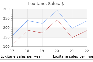
Discount 25mg loxitane otc
Colposcopic examination Colposcopy is particularly sensitive for subtle genital injuries symptoms weight loss loxitane 25 mg with visa. Some colposcopes have cameras attached, making it possible to detect and photograph injuries simultaneously. Using colposcopy, it has been found that the injury to the posterior fourchette is the most commonly seen in women after rape. A cotton swab inserted through the hymeneal orifice may also be used to look at the hymeneal rim. In this way, apparent folds and indentations smooth out and small nicks and tears can be easily identified. Hymen: Laceration of hymen occurs with the first intercourse and in a virgin, this is the principal evidence of the same. The annular hymen which nearly closes the vaginal orifice may suffer several tears. Even when examined after 3-4 days of offence, the edges are swollen, congested and smaller. Posterior commissure: the posterior commissure may be ruptured, especially if there is disparity in size between the male and the female organs. The presence of spermatozoa in the vagina is proof of connection, but not of rape; their absence is no proof that connection has not taken place. Sometimes, the history and examination suggests sexual intercourse, but evidence is often absent or inconclusive. There may be number of explanations besides the obvious suggestion of a false complaint (Table 25. Evidence becomes weaker or disappears as time passes, particularly after > 36 h; mechanical elimination (drainage, hygiene), biological degradation and physiologic dilution may yield negative results. Swabbing of mouth, vagina and anus for sperm detection should always be performed on rape victims. Evidence from Vaginal Discharge Vaginal discharge may arise from local infection, worms or uncleanliness. An initial negative reaction may be of value, if a positive reaction is obtained after 6 weeks. Wounds: Age of abrasions and contusions should corroborate with the alleged time of assault. Seminal fluid: Survival time of spermatozoa in vagina of living individual is quite variable. In the later case, it is probable that the specimen was obtained from cervical mucus. Venereal disease: Development of venereal disease may be helpful in estimating the time of assault. However, mark of genital injury should be looked for, as rape is generally associated with greater violence than consensual sexual intercourse. The majority of adult rapes are associated with a sudden forcible dilation of vagina resulting in some degree of local or general injury. Bruising, abrasion or lacerations are at all times consistent with forcible intercourse with a consenting woman and do not necessarily indicate rape. A second examination of the victim would be made, for bruising may take a little time to come to the surface, especially in the lower vagina. The vagina may show laceration or bruising with effusion of blood, and swelling and inflammation of the vulva, even when no marks of violence indicating a struggle may be found externally. In case of older women, senile atrophy and friability of their genitalia results in extensive vaginal lacerations and perineal trauma. In women who have been used to sexual intercourse, injuries from rape normally disappear or become obscure in 3-4 days. The presence of violence in other parts of the body is the chief evidence of the crime. All injuries of the labia and vagina found in cases of sexual assault are not due to rough manual and penile contact. Tears in the deeper part of vagina and gross lacerating wounds of the vault are not likely to occur during sexual intercourse, but are often caused by sexual perverts using instruments. The presence of spermatozoa in the vaginal secretion is a positive sign of sexual intercourse. It is important when searching for motile sperms in an individual of alleged raped only few hours before to obtain the specimen from the vaginal pool and not from the cervix, since sperm seen on a cervical swab may not be caused by the rape, but by sexual intercourse 2-3 days before (if history of consensual intercourse is present). In dead, the sperm are destroyed by decomposition and not by drainage or by the action of vaginal secretions. Sperms that are deposited on materials like cotton, cloth or paper and air dried can be identified years after the event. If however, sexual intercourse is still strongly suspected or if acid phosphatase test was weakly positive, an assay for prostate specific antigen (p30) should be performed. The highest levels are within the first 12 h with gradual disappearance by 48-72 h. Because it usually disappears in the first 24 h after intercourse, it is most useful as an indicator of recent intercourse, compared with non-motile sperm which can be identified upto 2-3 days after intercourse. Rape on Children A small child must never be held down during examination of the genital area, this is equivalent to sexually assaulting the child and will intensify the trauma. When indicated, the child should be taken to the operating room and anesthetized so that proper assessment and treatment can be done. As such, the Rape on Deflorate/Sexually Active Woman In deflorate women, even without childbirth, the hymen is completely destroyed, the vaginal orifice dilated and the mucous membrane wrinkled and thickened with complete loss of rugosity. Complete penetration can occur in such women and leaves no evidence, except semen. The only proof that the penetration has occurred is presence of spermatozoa in the vagina. The absence of injury Natural Sexual Offences hymen is usually intact and there may be little redness and tenderness of the vulva. Further penetration forces the penis backwards (symphysis pubis prevents its anterior movement) and the hymen is torn posteriorly. If the penis advances into the vagina, the hymenal tear extends into or through the perineal body and often involves the anterior wall of the ano-rectal canal. Full penile penetration produces bruising of the vaginal walls and frequently tears of the anterior and posterior vaginal walls. Vaginal vault may rupture and there may be vaginal herniation of abdominal viscera. The hymen shows a linear tear in the posterior or posterio-lateral quadrant which may extend into the posterior vagina and on to the skin of the perineum. Any attempt to separate the thighs for examination causes great pain because of the local inflammation. The absence of marks of violence on the genitals of the child when an early examination is made, is strong evidence that sexual intercourse has not taken place. Swabs from any soiled area of skin, bite marks and swabs from mouth, pharynx, vagina and anus for spermatozoa, microorganisms, P30 glycoprotein and sexually transmitted diseases. The examination should be tailored to the requirements of the particular case and collection of all samples may not be necessary. Thus, specimens are placed in individual packages, labeled, dated, sealed and held until handed over to police personnel after receiving a receipt. After the evaluation, the patient is provided with facilities to wash, change clothing, use mouthwash and urinate or defecate, if needed. Rape kit: It is a set of items used by medical personnel for gathering and preserving physical evidence following an allegation of sexual assault.
Diseases
- Syndactyly Cenani Lenz type
- Erythrokeratodermia progressive symmetrica ichthyosis
- Spondyloepiphyseal dysplasia nephrotic syndrome
- Arterial dysplasia
- Marfan Syndrome type IV
- Chromosome 8, trisomy
- 3 hydroxyisobutyric aciduria, rare (NIH)
- Noise-induced hearing loss
Discount 25 mg loxitane fast delivery
Other neurological manifestations Various other neuropsychiatric manifestations have been described: i medications removed by dialysis order 25mg loxitane amex. Sympt oms include mood change, cognitive deficit, memory loss, lethargy, autonomic dysfunction, peripheral neuropathy and extrapyramidal symptoms. Extrapyramidal manifestations include dystonias, resting tremor, cog-wheel rigidity and choreoathetosis. Neuro-ophthalmological sequelae including optic atrophy, degeneration of retina, myopia owing to spasm or paresis of accommodation. The clinician must keep in mind that misdiagnosis is a potential medico-legal pitfall. Three types of paralyses are recognized based on the time of occurrence, and differ in their pathophysiology. Chlorinated Hydrocarbons the chlorinated hydrocarbons can be divided into four categories: 53 0 Fundamentalsof Forensic Medicine and Toxicology Death is due to respiratory failure. Decontamination of the body should be carried out and the airway cared for, similar to organophosphosphate insecticides. Cholestyramine (non-absorbable bile acid binding anion exchange resin which increases the fecal excretion of organochlorines) is given in a dose of 16 g/day in divided doses for few days. Recovery is likely, if onset of convulsions is delayed by more than an hour or if convulsions can be controlled readily. Kerosene-like smell from the mouth and nostrils, may be found even in decomposed bodies. Mucosa of the esophagus, stomach and intestine is congested, and emits a kerosene-like smell. Cyclodienes and related compounds: Endrin, aldrin, chlordane, chlordecone, dieldrin, endosulfan, hepatachlor, isobenan and mirex. Endrin Physical properties: It is the most toxic of all the chlorinated insecticides. The preparations available in market contain endrin in 20-50% concentration mixed with 50-80% of a solvent, such as aromax, a petroleum hydrocarbon smelling like kerosene. It is extensively used in India, and in Andhra Pradesh the poisoning is occurring at an alarming rate, both in urban and rural populations. Metabolism: Endrin is partially metabolized in the liver and directly excreted in the urine, feces and milk; it is rapidly metabolized and eliminated, and does not persist in body tissues. Signs and Symptoms Toxic effects rapidly follow ingestion, inhalation or skin contamination. Headache, giddiness, restlessness, irritability, dilated pupils, incoordination, ataxia, mental confusion, tremors, tonic and clonic convulsions, coma. Chronic poisoning: Long-term exposure to some of these compounds results in cumulative toxicity characterized by weakness, loss of weight, ataxia, tremors, mental changes, oligospermia, increased tendency to leukemia, purpura, aplastic anemia and liver carcinoma. Sodium bicarbonate should be administered to maintain an alkaline urine so as to prevent the precipitation of acid hematin crystals and blocking of the renal tubules. If cyanosis is present, methemoglobin is suspected and treated with methylene blue. Naphthalene Physical properties: It is a solid volatile substance obtained from the middle fraction of coal-tar distillation and has chemical properties similar to benzene. Uses: Deodorant in lavatories, as a pesticide in moth balls and in the dye industry for the manufacture of indigo and certain azo dyes. Action It causes hemolysis with subsequent blocking of renal tubules and hepatic necrosis. The symptoms include pallor, mild jaundice, burning sensation in the urethra, and pain in the bladder and loins. Severe poisoning may damage the liver and kidneys, and result in cyanosis, profuse perspiration, convulsions, coma and death. Rarely, homicide is possible and the poisoning may be mistaken for viral pneumonia. Absorption and excretion: Absorption through inhalation, skin or eye contact is minimal. It is distributed in all the organs, but highest concentrations are found in kidneys and lungs, followed by muscles. More than 90% of the absorbed paraquat is excreted unchanged in the urine within the first 24 h, but can be detected in urine upto 3 weeks after ingestion. Ulceration and corrosion of mouth, oropharynx, and esophagus; nausea, vomiting, hematemesis, diarrhea, dysphagia. Pyrethrins and Pyrethroids Pyrethrins are extracted from Cr ysant hemum ci ner ari aefol i um plant. For example, d-allethrin, pyrethrum, allethrin, deltamethrin, decamethrin, cypermethrin and fenvalerate. Action: They prolong the inactivation of the sodium channel by binding it in the open state. On ingestion, there is nausea, vomiting, headache, vertigo, restlessness, paraesthesias, fasciculations, muscular weakness, hyperthermia, altered mental state, convulsions, pulmonary edema and coma. Postmortem Findings External: There may be ulceration around the lips and mouth due to dribbling. A 5-year-old child presents with confusion, increased salivation, fasiculations, miosis, tachycardia and hypertension. A farmer visiting an orchard gets unconscious, excessive salivation, constricted pupils and fasciculation of muscles. A patient is admitted with acute organophosphorus insecticide poisoning, develops ptosis, inability to lift the head and difficulty in breathing on the third day. The non-toxic residues left in grains are phosphite and hypophosphite of aluminum which is harmless. This process is fully reversible and full recovery occurs in patients who survive without any residual effect. Headache, dizziness, altered mental state, restlessness, convulsions, acute hypoxic encephalopathy, coma. Cause of death: Metabolic acidosis or mixed metabolic acidosis and respiratory alkalosis, and acute renal failure are frequent. The most specific and sensitive method for detecting phosphine is gas chromatography with a nitrogenphosphorous detector. It has an antiperoxident effect and it combats free radical stress due to phosphine. There may be capillary dilation, paucity of glial cells, degenerated Nissel granule in the cytoplasm and deeply stained eccentric nucleus, degeneration of neurons and appearance of necrotic patches. A case of poisoning was brought to the causalty, gastric lavage turned black when it was heated after being treated with silver nitrate. Metabolism: After absorption, it is metabolized by glucuronidation and sulfation and by cytochrome P450 oxidase system. Oral methionine is an alternative, but is unreliable in patients who are vomiting.
Buy cheap loxitane on-line
There is o en associated emotional lability or obsessive-compulsive traits treatment 02 discount loxitane, which may last longer than the chorei orm movements (which usually resolve within 6 weeks but sometimes may take up to 6 months). Where possible, age-speci c re erence ranges should be determined in a local population o healthy people without a recent group A streptococcal in ection. Duckett Jones in 1944 to develop a set o criteria (subsequently known as the Jones criteria) to aid in the diagnosis. For this group, there is a set o "low-risk" criteria; or all others, there is a set o more sensitive criteria. Echocardiography should be per ormed on all possible cases to aid in making the diagnosis and to determine the severity at baseline o any carditis. Additionally, joint mani estations can only be considered in either the major or minor categories but not both in the same patient. At higher doses, the patient should be monitored or symptoms o salicylate toxicity such as nausea, vomiting, or tinnitus; i symptoms appear, lower doses should be used. Fever, joint mani estations, and elevated acute-phase reactants sometimes recur up to 3 weeks a er the medication is discontinued. Instead, bed rest should be prescribed as needed while arthritis and arthralgia are present and or patients with heart ailure. Many clinicians treat cases o severe carditis (causing heart ailure) with glucocorticoids in the belie that they may reduce the acute in lammation and result in more rapid resolution o ailure. There is recent evidence that corticosteroids are e ective and lead to more rapid symptom reduction in chorea. Once the acute episode has resolved, the priority in management is to ensure long-term clinical ollow-up and adherence to a regimen o secondary prophylaxis. Patients and their amilies should also be educated about their disease, emphasizing the importance o adherence to secondary prophylaxis. Note that some organizations recommend a minimum o 10 years o prophylaxis a ter the most recent episode, or until 21 years o age (whichever is longer), regardless o the presence o carditis with the initial episode. It can be given every 3 weeks, or even every 2 weeks, to persons considered to be at particularly high risk, although in settings where good compliance with an every-4-week dosing schedule can be achieved, more requent dosing is rarely needed. Oral penicillin V (250 mg) can be given twice daily instead but is less e ective than benzathine penicillin G. Registries improve the ability to ollow patients and identi y those who de ault rom prophylaxis and to institute strategies to improve adherence. The mitral valve regurgitation is moderate with a typical posterolaterally directed regurgitant jet o rheumatic carditis. During diastole, the motion o the anterior mitral valve lea et tip is restricted with doming o the body o the lea et toward the interventricular septum. The ailure o coaptation o the mitral valve lea ets is the result o chordal elongation and annular dilatation. Over time, un ti na an stru tura a terati ns in mu tip e vas u ar be s an pr gressive vis era rgan ys un ti n ue t br sis minate the inia pi ture. The inf ammatory and immune responses initiate and sustain broblast activation and di erentiation, resulting in pathologic brogenesis and irreversible tissue damage. V ascular damage results in tissue ischemia that urther contributes to progressive brosis and atrophy. Mi r vesse s sh w enhan e permeabi ity an transenthe ia euk yte iape esis, abn rma a tivati n agu ati n as a es, e evate thr mbin pr u ti n, an impaire brin ysis. Un er n rma n iti ns, these br ti resp nses nstitute se - imite physi gi rem e ing ne essary r tissue repair an regenerati n. A b an n nin ammat ry b iterative vasu pathy as a ate n ing is pr minent in the heart, ungs, ki neys, an intestina tra t. C agen ber a umu ati n is m st pr minent in the reti u ar ermis, an the br ti pr ess inva es the subja ent a ip se ayer with entrapment a ip ytes. Pu m nary br sis is hara terize by expansi n the a ve ar interstitium, with a umu ati n agen an ther matrix pr teins. Intima thi kening the pu m nary arteries, best seen with e astin stain, un er ies pu m nary hypertensi n. The wer es phagus sh ws pr minent atr phy the mus u ar ayers an hara teristi vas u ar esi ns; striate mus e in the upper thir the es phagus is genera y spare. Rep a ement the n rma intestina tra t ar hite ture resu ts in iminishe perista ti a tivity, with gastr es phagea re ux, ysm ti ity, an sma -b we bstru ti n. The hara teristi arteri ar esi ns are n entri intima hypertr phy an umina narr wing, a mpanie by ntra ti n ban ne r sis re e ting is hemia-reper usi n injury an my ar ia br sis. The wrists, e b ws, sh u ers, hip gir es, knees, an ank es be me sti ue t br sis the supp rting j int stru tures. In s me patients, i use tanning in the absen e sun exp sure is a very ear y mani estati n skin inv vement. Skin thi kening in mbinati n with br sis the subja ent ten ns a unts r ntra tures the wrists, e b ws, an knees. T inning the ips with a entuati n the entra in is r teeth an ne wrink es (ra ia urr wing) ar un the m uth mp ete the pi ture. These esi ns, reminis ent here itary hem rrhagi the angie tasia, are pr minent n the a e, han s, ips, an ra mu sa. With isease pr gressi n, angina, exerti na near-syn pe, an sympt ms an signs right-si e heart ai ure appear. The path gi eatures atr phy sm th mus e, inta t mu sa, an b iterative sma vesse vas u pathy are simi ar thr ugh ut the ength the gastr intestina tra t. En s py may be ne essary t ru e ut pp rtunisti in e ti ns with Candida, herpes virus, an yt megavirus. The path genesis inv ves b iterative vas u pathy an umina narr wing the rena ar uate an inter bu ar arteries. Pa pab e ten n ri ti n rubs, peri ar ia e usi n, new unexp aine anemia, an thr mb yt penia may be harbingers impen ing s er erma rena risis. In s me ases, s er erma rena risis is misiagn se as thr mb ti thr mb yt peni purpura r ther rms thr mb ti mi r angi pathy. An asi na ra i gi n ing is pneumat sis yst i es intestina is ue t air trapping in the b we wa that may rare y rupture an ause benign pneum perit neum. The requen y ma r vasu ar inv vement, in u ing periphera vas u ar an r nary artery isease, may be in rease. Whereas the entra nerv us system is genera y spare, sens ry trigemina neur pathy ue t br sis r vas u pathy an ur, presenting with gra ua nset pain an numbness. Furtherm re, ar i pu m nary inv vement may w rsen uring pregnan y, an new nset s er erma rena risis has been es ribe. O asi na y, u thi kness bi psy the skin is require r estab ishing the iagn sis s ere ema, s er myxe ema, r nephr geni systemi br sis. The n ing igita tip pitting s ars an ra i gi evi en e pu m nary br sis in the wer bes are parti u ar y he pu iagn sti a y. In r er t minimize irreversib e rgan amage, the management i ethreatening mp i ati ns must be pr a tive, with regu ar s reening an initiati n appr priate interventi n at the ear iest p ssib e pp rtunity. We en urage patients t be me ami iar with the spe trum p tentia mp i ati ns an un erstan therapeuti pti ns an natura hist ry, an emp wer them t partner with their treating physi ians. This requires a ng-term re ati nship between patient an physi ian, with ng ing unse ing an en uragement. In retr spe tive stu ies, d-peni i amine stabi ize an impr ve skin in urati n, prevente new interna rgan inv vement, an impr ve surviva. Patients sh u ress warm y, minimize exp sure r stress, an av i rugs that pre ipitate r exa erbate vas spasti epis es.
Purchase 25 mg loxitane with amex
Small arrows indicate dissipation of the compressive forces to the anulus fibrosus medications you cannot crush discount loxitane american express. On the other hand, the nucleus pulposus that is responsible for dissipating the compressive forces on the disc by exerting a hydrostatic pressure on the anulus fibrosus consists of up to 50 % of proteoglycans (percent wet weight), whereas the anulus fibrosus only contains 20 % proteoglycans. These differences in proteoglycan content are also reflected by the water content of the two tissues (80 % in the nucleus pulposus and 70 % in the anulus fibrosus). The exact role of these additional matrix proteins and glycoproteins is not completely clear [55, 87]. It is important to keep in mind that the disc matrix is not a static but a dynamic structure. The components of the matrix are continuously degraded and replaced by newly synthesized molecules. Degradation of matrix components is the anulus resists high tensile forces the collagen and proteoglycan interplay influences disc functions In the normal disc, matrix degradation and synthesis are in balance Table 1. The balance between synthesis, degradation and accumulation of matrix molecules determines the quality and integrity of the disc matrix and is also prerequisite for adaptation/ alteration of the matrix properties to changing environmental conditions. The blood vessels closest to the disc matrix are therefore the capillary beds of the adjacent vertebral bodies and small capillaries in the outermost part of the anulus fibrosus [24, 46]. The blood vessels present in the longitudinal ligaments running adjacent to the disc and in young cartilage endplates (less than 12 months old) are branches of the spinal artery [49, 50, 142]. As a consequence of the avascularity, the nutrient supply to the disc cells and removal of metabolic waste products is entirely dependent on diffusion mainly from or to the capillary beds of the adjacent vertebrae [49]. Animal experiments indicated that the role of the peripheral small capillaries for the nutrient supply is only of minor importance [102]. The dependency of nutrient supply to the inner parts of the disc on diffusion together with the poor diffusion capacity of the disc matrix severely limits nutrient and waste exchange. As a result, a gradient between the inner parts and the peripheral regions of the disc builds up with very low levels of glucose and oxygen and high levels of the waste product lactic acid on the inside [49]. These gradients are even further aggravated by the disc cells using oxygen and glucose and producing lactic acid [49, 56]. The restricted nutrient supply and the increasing acidic milieu, due to the accumulation of lactic acid, are considered the main factors limiting cell viability and therefore the integrity of the disc matrix. Macroscopic Disc Alterations Onset and progression of age-related alterations of the disc can be determined with various techniques. Disc nutrition Glucose and oxygen concentration were found to drop steeply from the endplate towards the inner part of the nucleus pulposus (glc glucose, O2 oxygen). Lactate concentration displayed the opposite course, with highest levels in the inner region (lac lactate). This profile reflects the nutrient limitations in the inner disc and the lower pH values on the inside due to the acidic waste product lactate. The sagittal section through an intervertebral disc shows the region of the determined concentrations (adapted from [143]). However, more detailed information has been gained from macroscopic postmortem analysis of intervertebral disc tissue from individuals of various ages [92]. These studies have led to grading systems that on one hand allow the evaluation of stages of disc degeneration, but also illustrate the process of age-related degeneration. The original grading system was established by Friberg and Hirsch (and propagated by Nachemson) and has been further refined by Thompson et al. Intervertebral Disc) chondrocyte proliferation (increasing cell clusters due to reactive proliferation)) mucous degeneration (accumulation of mucous substances)) cell death) tear and cleft formation) granular changes: increasing accumulation of granular tissue Cartilage Endplate) cell proliferation) cartilage disorganization) presence of cracks in the cartilage) presence of microfractures) formation of new bone) bony sclerosis First signs of tissue degradation are seen between 10 and 16 years of age when tears in the nucleus pulposus occur along with focal disc cell proliferation and granular matrix transformation [17]. In parallel, the amount and extent of acidic mucopolysaccharides in the matrix increase. The general structure of the nucleus pulposus and the anulus fibrosus, however, is preserved in this age group. The nucleus is accordingly transformed by multiple large clefts and tears and the matrix shows significant granular changes. In this age group particularly the anulus fibrosus Chondrocyte proliferation is the first sign of disc degeneration 102 Section Advanced disc degeneration is indicated by a loss of nuclear/annular distinction Basic Science Disc degeneration exhibits a spatial heterogeneity is more and more affected, resulting in a loss of the clear distinction between nucleus and anulus. Huge clusters of proliferating cells are observed near clefts and tears that are filled with granular material. In individuals older than 70 years, the structural abnormalities change more to scar-like tissue and large tissue defects. Therefore, histological features can hardly be determined and characterize a "burned-out" intervertebral disc. The histological approach, although it largely parallels the macroscopic classification proposed by Thompson et al. Whereas macroscopic and histological approaches concur in the progressive loss of structure in all anatomical regions of the intervertebral disc, the microscopic approach revealed an earlier occurrence of nuclear clefts already in the second decade of life. In addition, the histologic approach revealed the heterogeneity of the alteration within the disc, indicating relevant spatial differences with more alterations usually present in the posterolateral aspects of the disc. In addition, the microscopic approach underlined the importance of nutritional supply to the disc cells for the maintenance of a healthy disc and the lack thereof for the onset and progression of disc degeneration. Since vascularization was seen to disappear from the disc during the first decade, nutritional supply to the disc cells becomes severely impaired during the subsequent phase of growth [17]. Age-Related Changes in Vascularization and Innervation the disc is the largest avascular structure of the human body Vascular changes in the endplate play a key role in the nutritional supply Calcification of the endplates and occlusion of the vascular channels are detrimental to the disc Although there is still some debate over the presence of blood vessels and nerve endings in the inner portions of pathologic discs, there is consensus that the healthy adult disc is the largest avascular and aneural tissue in the human body [61, 88]. This absence of significant vascular supply to the intervertebral disc matrix has important consequences for the maintenance of discal structures as discussed above [17, 88]. In fetal and early infantile intervertebral discs blood vessels penetrate both the endplate and the peripheral region of the anulus fibrosus. However, by late childhood the blood vessels disappear, leaving only small capillaries accompanied by lymph vessels that penetrate up to 2 mm into the outer anulus fibrosus [46, 124]. Since the importance of this peripheral vascularization for the nutrient supply of the disc is not known in detail, the consequences of its disappearance are also unknown. More important for the blood supply to the inner regions of the disc and therefore better described is the vascularization of the interface between adjacent vertebral bodies, cartilage endplate and the disc. The vertebral bodies are supplied by different arteries that are either responsible for the outer regions, the mid-anulus region, or the central core [23, 116]. These arteries of the vertebral body feed capillaries that, after penetrating channels in the subchondral plate, terminate in loops at the bone-cartilage interface [143]. The channels penetrating the subchondral plate are present in the fetus and infants, but disappear during childhood, compromising the blood supply to the inner disc [22]. Later during aging, sclerosis of the subchondral plate is observed and the cartilage endplates undergo calcification followed by resorption and finally replacement by bone [14, 28]. These age-related changes at the bone-disc interface restrict blood supply to the disc even further, finally cutting off nutrient supply to the inner parts of the disc [13, 96]. So far, it is not entirely clear whether calcification of the endplates causes disc degeneration or if age-related changes during degeneration in the environment of the endplates lead to calcification. However, it is thought that the impairment of the already critical supply of the disc cells with nutrients might be a major cause of disc degeneration. Age-Related Changes of the Spine Chapter 4 In contrast to fetal discs, the adult disc is aneural 103 Distribution of nerve fibers is very similar to the occurrence of blood vessels, as they are only, if at all, detectable in the outermost zone of the anulus fibrosus of healthy adult discs. In contrast, fetal and infantile discs contain small nerve structures adjacent to vessels also in central portions of the disc, i. From adult age on, the intervertebral disc remains avascular and aneural until advanced age. Only in those rare cases where the disc is completely destroyed and fibrously transformed may the ingrowth of blood vessels be associated with innervation of this fibrous tissue. Accordingly, this pattern is restricted to those cases where the original disc structure is completely lost. Molecular Changes of the Extracellular Matrix During Aging the structure and composition of the extracellular matrix are of fundamental significance for the biomechanical properties of the intervertebral disc.
Kankol (Cubebs). Loxitane.
- What is Cubebs?
- Are there safety concerns?
- How does Cubebs work?
- Dosing considerations for Cubebs.
- Are there any interactions with medications?
- Increasing urination, amoebic dysentery, gas, gonorrhea, loosening of mucous, and cancer.
Source: http://www.rxlist.com/script/main/art.asp?articlekey=96521
Order loxitane once a day
Family education regarding the need medicine cabinet purchase loxitane from india, procedure, and time frame for the use of the restraint is required. If possible, maintain a list of the weights of common restraint materials in use when weighing infants for monitoring daily growth. Evaluate the patient and proper use, placement, and position of restraint according to patient need, hospital policy, and regulatory agency requirement. Ensure that the infant is in a proper and functional position that promotes flexion and midline positioning of upper and lower extremities. Rationale:Prevention of contractures and support of self-calming techniques of neonates (prone, sidelying). Prone positioning during procedures and at rest provides for improved breathing and sleep, lower expenditure of energy, and more stable physiologic functioning. Side-lying positioning is the best alternative to prone for procedures and sleeping. This position allows for more midline positioning of the upper and lower extremities. Rationale: Prevents contractures and neurovascular injury; preserves skin integrity; reduces friction and pressure to skin from restraint material (1) When utilizing tape for securing an extremity to a board, use transparent tape when possible to allow for careful and complete assessment of the underlying skin. Rationale: Constriction from a tight restraint can cause neurovascular injury and impede circulation. Specific assessments related to oxygenation, musculoskeletal system, and cardiorespiratory conditions need to be performed in relation to the restraint device and its usage (1). Attach restraint to a fixed location on bed (if necessary), maintaining the opportunity for quick release and regular vascular checks (safety pin, secure tucking, etc. Do not attach restraint to equipment that can be moved (crib side rails, incubator doors), as injury may occur. Limb injury (fracture or dislocation) from movement of infant without release of secured restraint or from securing restraint to movable object. Impairment or compromise of medical state, including oxygenation, musculoskeletal system, and cardiorespiratory conditions (1) 7. Extravasation injury leading to impairment of skin integrity, tissue necrosis, infection, and/or nerve and tendon damage (6) G. A temporary alternative to restraint usage during procedures is therapeuticholding. This is defined as the "use of a secure, comfortable, temporary holding position that provides close physical contact with the parent or caregiver for 30 minutes or less" (2). Staff must properly prepare the parent or caregiver and provide proper supervision throughout the procedure. The American Academy of Pediatrics has outlined recommendations addressing infant sleep positioning to reduce the risk of sudden infant death syndrome. In terms of positioning the infant, they should be placed in a "supine position (wholly on their back)" (7). Therefore, when returning the patient to a sleep and/ or recovery position following a procedure, health care professionals should endorse and model this behavior for parents and caregivers whenever possible. Policy Statement: the changing concept of sudden infant death syndrome: diagnostic coding shifts, controversies regarding the sleeping environment, and new variables to consider in reducing risk. Failure of restraint resulting in self-injury and/or interference with treatment 2. Reduces but does not eliminate bacterial counts on the skin (1) Has an immediate effect (2) Has variable residual activity by binding to the stratum corneum of the skin 2. Organisms, usually of low virulence, which survive and multiply on skin and can be cultured repeatedly. Can be eradicated completely by hand washing with antiseptic solutions leads to a decrease in the unnecessary use of antibiotics and the potential for antibiotic resistance. Hospital managers continuously develop and update strict policies and regulations (3) as well as quality improvement projects aimed to promote adherence to aseptic technique and hand hygiene (4). Iodine solutions for preparation of skin in premature and low-birthweight infants (may cause skin and thyroid problems in high concentrations) (5) 2. Background Bloodstream bacterial infection is an extremely common complication of prematurity. The majority of etiologic pathogens are nosocomial, most often transmitted by health care personnel. Use of aseptic technique is critical in reducing the number of bloodstream infections as well as in decreasing the number of contaminated blood cultures, which in turn E. Indications: Reasonably anticipated risk of skin, eye, mucous membrane, or parenteral contact with 33 34 Section I Preparation and Support blood or other potentially infectious materials, including semen, vaginal secretions, cerebrospinal fluid, synovial fluid, pleural fluid, pericardial fluid, peritoneal fluid, amniotic fluid, saliva, and any body fluid that is visibly contaminated with blood. Major components (1) Use gloves when touching blood, body fluids, mucous membranes, or nonintact skin and when handling items or surfaces soiled with blood or body fluids. Contamination of instruments with antiseptic is undesirable and may invalidate specimens taken for culture. If hand disinfectants are not allowed to dry, alcoholbased disinfectant vapors can accumulate inside incubators (11). After the procedure, remove iodophor from all but immediate area of procedure to prevent absorption through skin (5,12,13). Use hexachlorophene for skin preparation in newborns only as recommended by the American Academy of Pediatrics (15). Use only in term infants during outbreak of Staphylococcus aureus infection if other infectioncontrol measures have been unsuccessful. Reapply alcohol prior to each attempt at procedure or with any delay, as efficacy is short-lived and flora will regenerate quickly. The warm, wet skin surface under gloves offers an ideal environment for bacterial multiplication. Latex and vinyl gloves offer comparable permeability, but vinyl gloves leak more readily. In medical emergencies, aseptic technique should be used as allowed by the situation, with at least antiseptic skin preparation of the patient, use of gloves, and a sterile field as large as possible under the circumstances. Personnel suffering from allergies to antimicrobial soaps may wash thoroughly for 3 to 5 minutes with plain soap or 70% isopropanol with glycerin prior to gloving (8). Personnel suffering from skin cracking due to frequent use of antiseptic soaps may use moisturizing skin products or barrier creams after hand washing. Products with a bacteriostatic ingredient, such as gels containing 60% ethanol, and emollients are safe and effective in reducing skin problems (8). Routine hand decontamination can be done with soap and water or alcohol-based hand rubs (8). Non-latex-containing gloves should be available for staff with latex allergy and to avoid allergic reactions in the patient, particularly in susceptible patients such as those with myelomeningocele (16). Technique (See Procedures Website for Video) A 3- to 5-minute "scrub" (vigorous washing up to the elbows) is necessary when entering the nursery. Subsequently, a 15to 30-second hand washing is indicated prior to and after each patient contact. Preparation of personnel (1) Wear cap/beard cover if hair is likely to contaminate the field. Iodophor preparations appear to be equally effective when applied with disposable sponges or brushes. Vigorous scrubbing with a brush leads to skin breakdown and possible contamination and is contraindicated. Be sure to include between the fingers and the lateral surface of the fifth finger. In clinical situations where traditional hand-washing facilities are unavailable, such as during patient transport, alcohol-based hand rinses, foams, or wipes may be used for hand cleaning. When an alcohol solution is used, make three to five applications of 3 to 5 mL each and rub hands well until completely dry. Preparation of patient skin (1) If necessary, cut hair in area of procedure with small scissors, taking care not to nick skin.
Cheap loxitane online
In his text On the Anatomy of the Spinal Nerves (Anatome Medullae Spinalis et Nervorum indeprovenientium) (1666) treatment alternatives for safe communities order loxitane 25mg on line, Blasius was the first to provide a demonstration of the origin of the spinal nerve roots and a differentiation between the gray matter of the spinal cord [6]. He illustrated the blood supply of the spinal cord with an accuracy that is still unsurpassed. Weitbrecht is also credited with providing a very concise description of the intervertebral disc for his time. At the beginning of the 19th century, it was still believed that some parts of the spinal cord contained the "centers of feeling". Furthermore it was believed that the spinal cord consisted of bundles of nerve fibers grouped into columns. After the microscope entered clinical and pathological practice, the cellular contents of the gray matter were identified, and since then there have been steady advances in our understanding of the spinal cord. Anesthesia and Supportive Techniques An invasive and effective spinal surgery would not have been possible without major advances in anesthesia and supportive techniques such as antisepsis, antibiotics and diagnostic imaging. He tried the effect of this substance first on himself and recommended that nitrous oxide ("laughing gas") could be useful for narcotizing patients during operations. On 16 October 1846, Morton presented his narcotizing method to the public in the operating theater of the Massachusetts General Hospital in Boston. Further improvements were made by Sir James Simpson, an English gynecologist and obstetrician, who introduced chloroform as a narcotizing agent after a large series of heroic self-experiments. Antisepsis and Antibiotics Infections were thought to be a divine punishment For a long period of history, infections were thought to be a divine punishment. On Infection, Infectious Diseases and Their Cure (De Contagiosis Morbis Eorumque Curatione) that infections are not only transmitted by air but also by human contact. Therefore, he proposed irrigation and disinfection of the operation field by using a weak solution of carbolic acid [71]. After several experiments he was able to extract a liquid substance, which he called penicillin, because of the name of the mold, Penicillium notatum, and he published his results in 1929. Scoliosis Since the beginning of written history, scoliosis has been a major concern in medical texts. The clinical image of scoliosis very much impressed ancient physicians and treatment remained poor for centuries. Even today, treatment is unsatisfactory since correction of scoliosis is not possible without spinal fusion. History of Spinal Disorders Chapter 1 9 Pathogenesis During antiquity and the Middle Ages, the pathogenesis of scoliosis was not clear and it has still not been unraveled today. It was often supposed that the spinal deformities were caused by luxation of spinal elements. When research on scoliosis started, it was commonly believed that muscle dysfunction was the cause. During the second half of the 19th century, research focused on the spinal osseous changes in patients suffering from scoliosis. The measuring machine consisted of a glass plate with engraved squares on which a sheet of paper was fixed. This apparatus allowed the depiction of a three-dimensional representation of the scoliosis [107]. Schulthess also invented stereotactic machines to produce calibrated corrections and to measure rotation. In 1906, he published a very comprehensive book on scoliosis, which served for many years as a reference textbook [108]. He also thought that it was better to operate on patients at an early age rather than waiting for the development of large curves. Non-operative Treatment Probably the first description of the treatment of spinal deformity is recorded in the Srimad Bhagwat Mahapuranam, an ancient Indian epic written between 3500 and 1800 B. There, the Indian god Lord Krishna cures the hunchback of one of his female devotees named Kubja by applying axial traction. The state of the art medical textbook of antiquity On Articulation (part of the monumental and famous Corpus Hippocraticum) was probably written by the Greek physician Hippocrates.
Loxitane 10mg lowest price
Introduction of improvised drugs symptoms 0f brain tumor buy loxitane once a day, devices, techniques and previous experience have also contributed to safety during anesthesia. Deaths which are the direct result of administration of an anesthetic Death during Administration of Anesthesia (not due to anesthesia) i. The injury or disease process which necessitated surgical intervention is serious enough, the anesthetic may have only precipitated the death. Surgical shock and exhaustion: When surgery has been unduly delayed and preoperative condition of patient is poor, shock and exhaustion may be major factors responsible for causing the death of the patient or the patient has been unable to bear the stress of anesthesia and surgery. Inability to take precautions and corrective measures when required is commonly observed. This problem is now rare due to advent of newer anesthetic agents which do not form explosive mixtures. Respiratory failure: Death occurs due to an inadequate supply of oxygen to tissues. Nitrous oxide used during general anesthesia leads to a rapid expansion of the pneumothorax. If pneumothorax is significant, gaseous exchange is affected leading to hypoxic injury and death. Neurogenic cardiovascular failure: It is the most common cause of sudden death under general anesthesia. It usually occurs when some intervention is done at a time when the depth of anesthesia is still inadequate. Malignant hyperthermia: When it occurs, it is usually seen with halogenated anesthetics and succinylcholine. Local anesthetics: Toxicity results from overdose or allergic reactions, hypersensitivity and idiosyncrasy. Depressive: Causing respiratory paralysis Very rarely, the heart may be affected directly, or when an abnormally high concentration is injected into a nerve, permanent loss of function may occur. Spinal anesthesia: During spinal anesthesia (block), sympathetic blockade occurs along with sensory and motor blockade. This sympathetic blockade leads to varying degrees of hypotension which may be fatal, if not detected and corrected early. These include-hypoxemia, atelectasis of lungs, pneumonia, pulmonary edema, pneumothorax, bronchospasm, oxygen toxicity and aspiration of gastric contents, blood or foreign bodies. Neurological sequelae of these complications can be blindness, paraplegia, paraesthesia, vegetative state and death. Postmortem Examination Most deaths concerning anesthesia are unlikely to be evident at autopsy. Surgical mistakes being anatomical, may be observable at the postmortem and anesthetic mistakes being physiological, are usually not appreciable after death, except where overdose with specific drug is involved. Findings of the autopsy surgeon alone will not be sufficient to explain death and therefore, it is advisable to hold a discussion across the autopsy table involving forensic expert, anesthetist and the surgeon/ clinician concerned. In case of death following anesthesia/surgery, the forensic pathologist must answer the following questions: i. Was the death due to the effects of the operation or anesthesia or is it due to the disease for which operation was being carried out Would the patient have died, if he has not undergone through the anesthesia or operation Was the patient suffering from any predisposing condition that made him more susceptible to death from anesthetic or operative procedure Was the death due to some unsuspected natural disease, directly unrelated to the disease for which surgery was being performed During postmortem examination, the following are to be taken into consideration: i. Detailed hospital record of the patient, including full clinical and pre-anesthetic checkup. Postmortem changes need to be differentiated from abnormalities existing during life. Presence of pre-existing natural disease, such as heart disease or respiratory insufficiency and their contribution to the cause of death must be evaluated. Surgical and anesthetic devices, such as airways, endotracheal tubes, needles or catheters should not be removed prior to autopsy. In esophageal intubation, a radiograph will show a ring of edema of esophageal mucosa at the level of the tube along with distention of stomach and intestines. Chloroform and halothane are hepatotoxic and chloroform may cause ventricular fibrillation sometimes. A full range of specimens for histological, toxicological and bacteriological examinations and those required to exclude hazards associated with blood or fluid transfusions, must be collected. In case of inhaled anesthetic, specimens should be kept in containers of appropriate size to avoid empty space, and are sealed and refrigerated/frozen. Alveolar air should be collected with needle and syringe by puncturing the lung underwater before the chest is opened. While opioids produce analgesia, muscle relaxants cause paralysis of muscles including those of the diaphragm. But in India, independent viability of fetus is taken as more than 28 weeks of gestation. Abortifacient drugs: Most of them have no effect on the uterus or fetus, unless given in toxic doses and often sold to exploit distressed woman. Abrus precatorius, Calotropis, seeds of cutard apple and carrots, and unripe fruit of papaya or pineapple. Unexplained (40%): In spite of the numerous factors mentioned, it is sometimes difficult to pinpoint exact cause of abortion. Artificial or Induced Abortion It means willful termination of pregnancy before viability. It is usually induced before the 3rd month, and causes infec tion and inflammation of the endometrium. Severe pressure on abdomen by kneeling, blows, kick, tight bandage and massage of uterus through abdominal wall. Cupping: A mug is turned upside down over a lighted wick and placed on the hypogastria. Accidental: A general shake-up in advanced pregnancy can produce abortion, but if the fetus is healthy, abortion will not occur. Syringing: Ordinary enema syringe with a hand bulb is commonly used to inject fluid into uterus, the hard nozzle being inserted into cervix. Syringe aspiration: Large syringe with a plastic cannula is inserted into cervix; develops suction which ruptures early gestational sac and leads to aspiration and expulsion of contents. Vacuum aspiration: the cervix is dilated and a tube attached to a suction pump extracts the fetus. Rupturing of membranes: the membranes are ruptured by introduction of an instrument, like probe, stick, uterine sound, catheter, pencil, pen holder, knitting needle and hairpin. Abortion stick: It is a wooden or bamboo stick, 12-18 cm long, wrapped at one end with cotton, wool or piece of cloth and soaked with juice of marking nut, calotropis or paste made of arsenious oxide or lead. It is introduced into the vagina or os by dais (traditional birth attendants) and retain there, till contraction starts. Dilation of cervix: Foreign bodies are introduced and left in cervical canal, like pessaries, laminaria (a dried seaweed) or sea tangle tent which dilate the cervix, irritate uterine mucosa and produce marked congestion and uterine contractions with expulsion of fetus. Cervical canal may be dilated by introducing a compressed sponge into the cervix and leaving it there. Slippery elm bark (Ulmus fulva) obtained from tree in Central America, is inserted into cervical canal in portions of 1-3 inches long. Diagnosis is established by demonstration of mucin, lanugo hair, vernix caseosa, fat globules, meconium and squamous cells in cut sections of the lung.
Purchase loxitane 25mg amex
Collagen represents the main structural component of the discal extracellular matrix with variable compositions of isoforms seen in the different anatomic subsettings medications band cheap loxitane 10 mg on-line. While the overall collagen content in the nucleus pulposus remains fairly constant over the years, that of the anulus fibrosus decreases with advancing age. In a healthy intervertebral disc, aggrecan is present in the nucleus pulposus as large aggregates with hyaluronan. During degeneration aggrecan molecules are increasingly subjected to proteolytic cleavage. Cleavage of aggrecan has severe consequences for the healthy disc:) smaller aggrecan fragments are generated that diffuse more easily from the disc matrix) loss of aggrecan resulting in decreasing osmotic pressure) dehydration of the disc matrix) increased outflow of matrix molecules) increased inflow of mediators such as growth factor complexes and cytokines Taken together, changes in the composition of the disc matrix often result in a loss of disc height. This rapid loss of disc height puts the apophyseal joints to abnormal loads, predisposing to osteoarthritic changes. Loss of disc height also allows the ligamentum flavum to thicken, leading to a narrowing of the spinal canal. Age-related changes of collagen are predominantly qualitative Aggrecan loss significantly compromises biomechanical properties 104 Section Basic Science Disc collagens are degraded by various matrix metalloproteinases the observed changes in the molecular composition of the disc matrix are mainly due to degradation of the existing matrix components and synthesis of new matrix components. During degeneration the balance between degradation and synthesis is disturbed, leading to increased degradation and therefore resulting in loss of tissue from the disc. This loss of tissue due to proteolytic destruction of the matrix components goes along with the occurrence of clefts and tears, which in turn leads to biomechanical instability and thus to a loss of functional properties of the disc. Therefore, the proteolytic matrix destruction holds a central role in disc degeneration [98]. Additionally, there is evidence that increased activity of proteolytic enzymes has to be noted in regions of clefting and tissue disruption. These aggrecanases differ in their specificity for parts of the aggrecan molecule. The combined action of various proteinases and the ratio between these degradative processes and the synthesis of new matrix components are responsible for the remodeling of the disc matrix during degeneration. Modulation of Cells and Matrix by Cytokines and Growth Factors Cytokines and growth factors modulate disc matrix Many studies have analyzed the ability of disc cells to either produce or respond to cytokines and growth factors (Table 3). There is more and more evidence that Age-Related Changes of the Spine Chapter 4 105 Table 3. However, for most factors it is difficult to distinguish if they are part of the normal, age-related degeneration process or mainly important during pathological changes of the disc. Therefore, the mechanism of cytokine action is of major importance for the understanding of disc degeneration and also represents a potential target for therapeutic interventions. Despite this importance, only little is known about the age-related changes in cytokine and growth factor expression patterns. Recent studies provide evidence that factors of both classes are induced during age-related degeneration. This may consequently be responsible for the matrix disarrangement, including the formation of granulation tissue, characterized by changes to collagen and proteoglycan synthesis and also changes to the collagen composition of the matrix. Taken together, alterations to the expression of catabolic and anabolic factors during degeneration might disturb the delicate balance between matrix synthesis and degradation that is essential for the maintenance of a healthy disc matrix. Once this balance is disturbed, degeneration progresses together with matrix degradation or alteration. Etiology of Disc Degeneration Although the etiology of disc degeneration is far from being understood, there is consensus that not a single factor can be held responsible for the complex phenomenon of disc degeneration. Rather a multitude of exogenous and endogenous factors, each contributing individually, might influence the progress of degenerative changes of the discs. These factors can be divided into three main groups:) nutritional effects) genetic predisposition) mechanical load Insufficient nutritional supply of the disc cells is thought to be a major problem contributing to disc degeneration. Since the intervertebral disc is the largest avascular tissue in the human body, its cells are facing the precarious situation of having to maintain a huge extracellular matrix with a "fragile" supply of nutrients that is easily disturbed. Whereas the cells in the outer anulus fibrosus may be supplied with nutrients from blood vessels in the adjacent longitudinal ligaments, the supply of the nucleus pulposus cells is almost completely dependent on the capillary network in the vertebral bodies. Due to the size of the intervertebral disc, the nutrients need to diffuse from the capillaries through the endplate and the disc matrix to the cells in the nucleus of the disc. With the originally cartilaginous endplates becoming calcified when degeneration progresses, the supply of disc cells with nutrients will become even more restricted. This will consequently lead to:) limited nutrient supply (glucose and oxygen) particularly in the disc center) accumulation of waste products. Since lactic acid is not only the major waste product of disc cells but also an acid, its accumulation results in a lowered pH inside the disc. In vitro experiments have shown that low oxygen concentrations and acidic pH significantly affect the synthetic activity and especially proteoglycan synthesis rates of disc cells, which might lead to a fall in proteoglycan content and therefore to disc degeneration in vivo. Several recent studies have reported a strong Failure of disc nutrient supply primarily causes disc degeneration the accumulation of lactic acid is detrimental to the disc Genetic predisposition has a major impact on disc degeneration Age-Related Changes of the Spine Chapter 4 107 familial predisposition for disc degeneration and herniation [48, 83, 84, 144]. If mutations in these genes occur, normally innocuous conditions or forces might lead to accelerated or enhanced degenerative changes, suggesting that disc degeneration may be explained primarily by genetic influences and that environmental factors have only modest effects. However, it is important to keep in mind that despite the dominating role of genetic predisposition, injuries can occur when normal forces are applied to abnormally weak tissues, or when abnormally high forces are applied to normal tissues [2]. Considering the influences of the genetic predisposition discussed above, the impact of mechanical forces on disc degeneration is only minor. Therefore, it is not surprising that several studies carried out in humans did not provide a strong causal link between occupational exposures and disc degeneration [146]. Even well-controlled animal experiments did not provide a conclusive connection between mechanical load and degeneration. However, it is conceivable that abnormal loads might cause damage to the adjacent vertebral bodies, especially the bony endplates, which in turn might contribute to the initiation of disc degeneration [3]. Environmental factors have only modest effects on disc degeneration Abnormal mechanical loads contribute secondarily to disc degeneration the Cartilage Endplate Normal Anatomy and Composition A morphological distinction of the disc and bone interface is the thin cartilage endplate. The collagen fibers within it run horizontal and parallel to the vertebral bodies along with the fibers continuing into the disc [120]. At birth, the human cartilage endplates make up approximately 50 % of the intervertebral space Cartilage endplates are mechanically important and influence nutritional pathways and growth 108 Section Basic Science the endplate is important for the mechanical support and nutritional supply of the disc (compared with approximately 5 % in the adult) and have large vascular channels running through them. Soon after birth, the vascular channels of the cartilage endplate fill in with extracellular matrix such that no channels remain by the end of the first life decade. The cartilage endplate in humans functions in early life as a growth plate for the adjacent vertebral body; its structure is typical of that seen in the epiphyseal growth plate of long bones. By adulthood, the cartilage endplate is a layer of hyaline cartilage (approximately 0. The endplate occupies the central 90 % of the interface between the disc and the vertebral body, encompassed by a ring of bone that forms via the epiphysis fusing with the vertebral body in the rim region. Functionally, the endplate is involved in two important mechanical functions [19]:) preventing the nucleus pulposus from bulging into the vertebral bodies) partially absorbing the hydrostatic pressure dissipated by the nucleus pulposus under loading Similar to the disc, the ability of the endplate to withstand mechanical forces depends on the structural integrity of the matrix. Analyses on the microscopic level revealed that the abundance of obliterated blood vessels in the endplate gradually increases between 1 month and 16 years of age. The decrease in blood vessels [17] is paralleled by:) an increase in cartilage disorganization) a decrease in endplate cell density) cartilage cracks) microfractures Endplate calcification/ ossification obstructs nutritional pathways these changes, especially the loss of blood vessels, can cause nutritional consequences for the intervertebral disc. With advanced degeneration and markedly reduced disc height, further changes of the endplate are induced resulting in:) complete endplate disarrangement) dense sclerosis of the adjacent vertebral bodies Age-Related Changes of the Spine Chapter 4 109 the Facet Joints Normal Anatomy the facet joints, also called zygapophyseal joints, are paired diarthrodial articulations between the posterior elements of adjacent vertebrae. The joints exhibit the features of typical synovial joints and are an essential part of the posterior support structures of the spine consisting of:) pedicles) lamina) spinous and transverse processes Anatomically, the facet joints are responsible for restraining excessive mobility and for distributing axial load over a broad area. Adams and Hutton have found that the facet joints resist most of the intervertebral shear force [4]. The posterior anulus is protected in torsion by the facet surfaces and in flexion by the capsular ligaments. The earlier described "menisci" in the joints were found to be rudimentary fibrous invaginations of the dorsal and ventral capsule. They are basically fatfilled synovial reflections, some of which contain fibrous tissue probably as a result of mechanical stress. At the posterolateral aspect of the facet joint, a fibrous capsule composed of several layers of fibrous tissue and a synovial membrane is present. It has been shown that the synovial lining (small C-type pain fibers) and the capsules are richly innervated [16, 133].

Buy on line loxitane
Enzymes secreted by the synovium digest cartilage matrix that has been released rom the sur ace o the cartilage treatment 5th disease loxitane 25 mg amex. Additional pathologic changes occur in the capsule, which stretches, becomes edematous, and can become brotic. T eir role in osteoarthritic cartilage is unclear, but their release rom cartilage into the joint space and joint uid likely triggers synovial in ammation, which can, in turn, produce release o enzymes and trigger nociceptive stimulation. Mechanical symptoms, such as buckling, catching, or locking, could also signi y internal derangement, such as meniscal tears, and need to be evaluated. In ammatory arthritis is likely i there is prolonged morning sti ness and many other joints are a ected. A physical examination should ocus on whether tenderness is over the joint line (at the junction o the two bones around which the joint is articulating) or is outside o it. Anserine bursitis, medial and distal to the knee, is an extremely common cause o chronic knee pain that may respond to a glucocorticoid injection. Innervated structures in the joint include the synovium, ligaments, joint capsule, muscles, and subchondral bone. Some diseased joints have no synovitis, whereas others have synovial in ammation that approaches the severity o joints with rheumatoid arthritis (Chap. Increased ocal loading as part o the disease not only damages cartilage but probably also injures the underlying bone. For an older person whose daily constitutionals up and down hills bring on knee pain, routing the constitutional away rom hills might eliminate symptoms. Weight loss may have a commensurate multiplier e ect, unloading both knees and hips and probably relieving pain in those joints. Weight-bearing joints such as knees and hips can be unloaded by using a cane in the hand opposite to the a ected joint or partial weight bearing. A physical therapist can help teach the patient how to use the cane optimally, including ensuring that its height is optimal or unloading. Note the narrowed joint space on medial side o the joint only (white arrow), the sclerosis o the bone in the medial compartment providing evidence o cortical thickening (black arrow), and the osteophytes in the medial emur (white wedge). Further, radiographs may be normal in early disease as they are insensitive to cartilage loss and other early ndings. Fourth, "arthrogenous inhibition" may occur, whereby contraction o muscles bridging the joint is inhibited by a nerve a erent eedback loop emanating in a swollen and stretched joint capsule; this prevents maximal attainment o voluntary maximal strength. The degree o weakness correlates strongly with the severity o joint pain and the degree o physical limitation. Most e ective exercise regimens consist o aerobic and/or resistance training, the latter o which ocuses on strengthening muscles across the joint. Exercises are likely to be e ective, especially i they train muscles or the activities a person per orms daily. Activities that increase pain in the joint should be avoided, and the exercise regimen needs to be individualized to optimize e ectiveness. Range-o -motion exercises, which do not strengthen muscles, and isometric exercises that strengthen muscles, but not through range o motion, are unlikely to be e ective by themselves. Low-impact exercises, including water aerobics and water resistance training, are o en better tolerated by patients than exercises involving impact loading, such as running or treadmill exercises. A patient should be re erred to an exercise class or to a therapist who can create an individualized regimen, and then an individualized home-based regimen can be cra ed. Physicians should reinorce the exercise prescription at each clinic visit, help the patient recognize barriers to ongoing exercise, and identi y convenient times or exercise to be done routinely. The combination o exercise with calorie restriction and weight loss is especially e ective in lessening pain. Malalignment in the rontal plane (varus-valgus) markedly increases the stress across the joint, which can lead to progression o disease and to pain and disability. Correcting malalignment, either surgically or with bracing, may relieve pain in persons whose knees are malaligned. Malalignment develops over years as a consequence o gradual anatomic alterations o the joint and bone, and correcting it is o en very challenging. One way is with a tted brace, which takes an o en varus osteoarthritic knee and straightens it by putting valgus stress across the knee. Un ortunately, many patients are unwilling to wear a realigning knee brace; in addition, in patients with obese legs, braces may slip with usage and lose their realigning e ect. They are indicated or willing patients who can learn to put them on correctly and on whom they do not slip. Pain rom the patello emoral compartment o the knee can be caused by tilting o the patella or patellar malalignment with the patella riding laterally or medially in the emoral trochlear groove. Using a brace to realign the patella, or tape to pull the patella back into the trochlear sulcus or reduce its tilt, has been shown, when compared to placebo taping in clinical trials, to lessen patello emoral pain. Commercial patellar braces may be a solution, but there is insuf cient evidence on their ef cacy to recommend them. Although their e ect on malalignment is questionable, neoprene sleeves pulled to cover the knee lessen pain and are easy to use and popular among patients. I occasional medication use is insuf ciently e ective, then daily treatment may be indicated, with an anti-in ammatory dose selected (able 19-1). Patients should be reminded to take low-dose aspirin and ibupro en at di erent times to eliminate a drug interaction. Patients at high risk or gastrointestinal side e ects should also take either a proton pump inhibitor or misoprostol. Trials have shown the e cacy o proton pump inhibitors and misoprostol in the prevention o ulcers and bleeding. Certain oral agents are sa er to the stomach than others, including nonacetylated salicylates and nabumetone. When absorbed through the skin, plasma concentrations are an order o magnitude lower than with the same amount o drug administered orally or parenterally. However, when these drugs are administered topically in proximity to a super cial joint (knees, hands, but not hips), the drug can be ound in joint tissues such as the synovium and cartilage. Large publicly supported trials have ailed to show that, compared with placebo, these compounds relieve pain in persons with disease. Each surgery may provide the patient with years o pain relie be ore a total knee replacement is required. These are highly ef cacious operations that relieve pain and improve unction in the vast majority o patients, although rates o success are higher or hip than knee replacement. The chance o surgical success is greater in centers where at least 25 such operations are per ormed yearly or with surgeons who per orm multiple operations annually. I the patient su ers or many years until their unctional status has declined substantially, with considerable muscle weakness, postoperative unctional status may not improve to a level achieved by others who underwent operation earlier in their disease course. Polarized light microscopy alone can identi y most typical crystals; apatite, however, is an exception. Apart rom the identi cation o speci c microcrystalline materials or organisms, synovial uid characteristics in crystal-associated diseases are nonspeci c, and synovial uid can be in ammatory or nonin ammatory. Without crystal identi cation, these diseases can be con used with rheumatoid or other types o arthritis. A list o possible musculoskeletal mani estations o crystalassociated arthritis is shown in Table 20-1. Usually, only one joint is a ected initially, but polyarticular acute gout can occur in subsequent episodes. The metatarsophalangeal joint o the rst toe o en is involved, but tarsal joints, ankles, and knees also are a ected commonly. The rst episode o acute gouty arthritis requently begins at night with dramatic joint pain and swelling. Joints rapidly become warm, red, and tender, with a clinical appearance that o en mimics that o cellulitis.
