

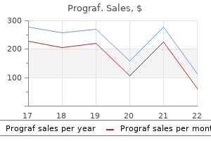
Cheap prograf 1 mg on line
Lipomatous glioneurocytoma of the posterior fossa with divergent differentiation: case report antiviral untuk chicken pox discount prograf 0.5 mg online. A rosette-forming glioneuronal tumor of the spinal cord: the first case of a rosetteforming glioneuronal tumor originating from the spinal cord. Extracranial metastasis of anaplastic ganglioglioma through a ventriculoperitoneal shunt: a case report. A case report with immunohistochemical, ultrastructural and proliferation studies. Intraventricular neurocytoma: a clinical and pathological study of three cases and review of the literature. Evidence for developmental precursor lesions in epilepsy-associated glioneuronal tumors. Papillary glioneuronal tumour: clinicopathological and biochemical study of one case with 7-year follow up. Tau-associated neuropathology in ganglion cell tumours increases with patient age but appears unrelated to ApoE genotype. Gliofibromas (including malignant forms), and gliosarcomas: a comparative study and review of the literature. Dysembryoplastic neuroepithelial tumors located in the caudate nucleus area: report of four cases. Cytokeratin expression in adrenal phaeochromocytomas and extraadrenal paragangliomas. Darwish B, Arbuckle S, Kellie S, Besser M, Chaseling R, Desmoplastic infantile ganglioglioma/astrocytoma with cerebrospinal metastasis. Malignant supratentorial ganglioglioma (ganglion cell-giant cell glioblastoma): a case report and review of the literature. Dysembryoplastic neuroepithelial tumour: a surgically curable tumour of young patients with intractable partial seizures. NeuN expression correlates with reduced mitotic index of neoplastic cells in central neurocytomas. A Golgi and electron microscopic study of a dysplastic gangliocytoma of the cerebellum. Studies with the Golgi method in central gangliogliomas and dysplastic gangliocytoma of the cerebellum. Genetic differences between neurocytoma and dysembryoplastic neuroepithelial tumor and oligodendroglial tumors. Ependymoma with neuropil-like islands: a case report with diagnostic and histogenetic implications. Immunocytochemical detection of calcineurin and microtubule-associated protein 2 in central neurocytoma. Anti-Hu immuno labelling as an index of neuronal differentiation in human brain tumors: a study of 112 central neuroepithelial neoplasms. Transcallosal resection of hypothalamic hamartomas in patients with intractable epilepsy. A review of the histology, ultrastructure, immunohistology, and molecular biology of extra-adrenal paragangliomas. A rosette-forming glioneuronal tumor of the fourth ventricle: infratentorial form of dysembryoplastic neuroepithelial tumor Mixed conventional and desmoplastic infantile ganglioglioma: an autopsied case with 6-year follow-up. Desmoplastic infantile astrocytoma and ganglioglioma: a search for genomic characteristics. Dysembryoplastic neuroepithelial tumors in two children with neurofibromatosis type 1. Decreased expression of neuropeptides in malignant paragangliomas: an immunohistochemical study. A histologic, immunohistochemical, and ultrastructural study and review of the literature. Desmoplastic cerebral astrocytomas of infancy: a histopathologic, immunohistochemical, ultrastructural, and molecular genetic study. Central neurocytoma: histologic atypia, proliferation potential and clinical outcome. Hypothalamic neurocytoma with vasopressin immunoreactivity: immunohistochemical and ultrastructural observations. In vitro neuronal and glial production and differentiation of human central neurocytoma cells. Intramedullary spinal cord gangliocytoma: case report and a review of the literature. Central neurocytoma: morphological, flow cytometric, polymerase chain reaction, fluorescence in situ hybridization, and karyotypic analyses. Cerebellar liponeurocytoma with an unusually aggressive clinical course: case report. Allelic losses in oligodendroglial and oligodendrogliomalike neoplasms: analysis using microsatellite repeats and polymerase chain reaction. Rosette forming glioneuronal tumor in association with Noonan syndrome: pathobiological implications. Rosette-forming glioneuronal tumour of the lateral ventricle in a patient with neurofibromatosis 1. Malignant glial tumour arising from the site of a previous hamartoma/ ganglioglioma: coincidence or malignant transformation Primary spinal paragangliomas: a clinicopathological and immunohistochemical study of 30 cases. Atypical extraventricular neurocytoma with oligodendroglioma-like spread and an unusual pattern of chromosome 1p and 19q loss. Papillary glioneuronal tumors: a review of clinicopathologic and molecular genetic studies. Tumours of the lateral ventricular wall, especially the septum pellucidum: clinical presentation and variations in pathological features. Immunohistochemical expression of tyrosine kinase (Trk) receptor proteins in mature neuronal cell tumors of the central nervous system. Dysembryoplastic neuroepithelial tumors in childhood: long-term outcome and prognostic features. Mutations of the p53 tumour suppressor gene in neoplasms of the human nervous system. Cerebral ganglioglioma and neurofibromatosis type I: case report and review of the literature. Factors contributing to resectability and seizure outcomes in 44 patients with ganglioglioma. Composite pleomorphic xanthoastrocytoma and ganglioglioma: report of four cases and review of the literature. Oligodendrogliomas with neurocytic differentiation: a report of 4 cases with diagnostic and histogenetic implications. Desmoplastic infantile astrocytoma: recurrence with malignant transformation into glioblastoma: a case report. A report of a desmoplastic ganglioglioma in a 12-year-old girl with review of the literature. Composite ganglioglioma/dysembryoplastic neuroepithelial tumor: a clinicopathologic study of 8 cases. Synaptophysin staining in normal brain: importance for diagnosis of ganglioglioma. Paraganglioma of the cauda equina: report of 2 cases and review of 59 cases from the literature.
Cheap prograf on line
Initially antiviral neuraminidase inhibitor order prograf 0.5mg, they were based purely on clinical assessment, with laboratory data, i. Periventricular frontal (white arrows) and occipital (blue arrows) plaques appear as regions of hyperintensity on the proton density (a) and T2-weighted (b) scans. Demyelination is evident as a loss or absence of signal within both lesions on the myelin water map (short-T2 component) (e). Both also have a long-T2 component (f), the histopathological basis of which is not yet defined but has been postulated to originate from extracellular fluid. Multiplicity, gadolinium enhancement and anatomical localization of lesions contribute to the assessment. Multiple sclerosis is most often characterized by relapses and remissions of neurological dysfunction, often with a subsequent gradual accumulation of residual impairment following incomplete remissions. The latter is the least common of these forms, occurring in approximately 5 per cent of patients. The length of the illness can vary; in some patients there may be an asymptomatic or minimally symptomatic period of survival until death from another cause many years after the initial manifestation. Accumulating evidence suggests that the incidence in different regions is dynamic and may be influenced by migration patterns, seasonal variations and diverse genetic and environmental factors in different populations. Moreover, increasingly complex global migration patterns have complicated these analyses further and additional studies point to intraregional variations. For those who emigrated after adolescence from high-prevalence areas in northern Europe, the risk was high, whereas in those emigrating in childhood, the risk was low. Most likely, they are an infectious agent or agents (see under Virology and Microbiology, p. There is evidence to suggest this may be linked to low levels of vitamin D, the synthesis of which is linked to ultraviolet light exposure. In these models, immune responses to defined antigens, usually peptides of either a myelin protein or a virus, are induced by immunization or infection. Furthermore, once the disease is initiated, it might progress as a consequence of additional sensitization and increased pathogenetic responses to these potential autoantigens, i. Early inflammatory responses might represent true autoimmunity, direct responses to as yet unidentified infectious agents (as suggested by epidemiological studies; see Epidemiology, p. Multiple responses might also expand, contract and evolve in a single patient over time, thereby obscuring the distinction between autoimmune and infectious aetiologies. The clinical and pathological disease may, therefore, result from a combination of multiple distinct immunopathogenetic mechanisms. Over the years, a variety of viruses have been implicated as the environmental agent responsible for the disease. However, it has also been found in other inflammatory disorders but less frequently in controls. Although such a notion would be considered heretical only a few years ago, there is increasing evidence that this concept should be given proper attention and investigation. There are also viruses that induce an autoimmune demyelinating in animals of predisposing genetic background (see under Pathogen-induced Models, in Animal Models of Human Demyelinating Diseases, p. As could be deduced from the earlier discussions, there are two main schools of thought, namely immunological/autoimmunological and infectious (see Box 23. Several professional societies, clinicians and radiologists have weighed in on the issue and have expressed caution in the use of these invasive interventions. It may refer to the presence of inflammatory infiltrates or to the presence of ongoing demyelination, but because inflammation and demyelination are virtually inseparable pathogenetically, it usually implies both. The precise temporal sequence of these events and the extent of inflammatory cell infiltration necessary for initiating them are, however, unclear. Small numbers of plasma cells may also be seen, but they are generally more abundant in chronic lesions. The early inflammatory events and the specific interacting cell types in the inflammatory process in typical lesions are now considered. When this event occurs, the activated lymphocyte acquires its functional phenotype (Th1, Th2, Th17 or T reg), downregulates molecules required for lymph node entry and upregulates those that allow it to enter the systemic circulation and interact with endothelial cells. Much of the knowledge in this area has been gleaned from in vitro studies or in animal models, examining particularly T-cells. Multiple Sclerosis 1311 the first interaction of activated T-cells with endothelial cells of most vascular beds is via glycoprotein ligands on the lymphocyte surface binding to selectins on the endothelial cell surface. In the early phases of the interaction of lymphocyte and endothelium, chemokine receptors on the lymphocyte interact with chemokines on the endothelial surface. This initiates, via G-protein signalling, configurational changes in integrins on the lymphocyte such that they have a much greater avidity for their respective endothelial adhesion molecule receptors. Of interest, lymphocytes seem to scan the vessels preferentially against the blood flow. This chemotactic process is mediated by chemokine gradients, because of positioning of chemokines on the abluminal aspect of the vessel. At the ultrastructural level in areas with active disease, they show increased numbers of pinocytotic vesicles110 and reduced numbers of mitochondria in more chronic stages. Experimental data involving 2-photon in vivo imaging indicate that the perivascular space is constantly scanned by T-cells in the search for antigen. Multiple Sclerosis (a) 1313 of a potential autoimmune repertoire (see earlier under Peripheral Immune Organs, p. Their activities, therefore, generally dampen the cell-mediated immunopathological processes. Dendritic cells, which are professional antigen-presenting cells of myeloid origin, also populate the perivascular inflammatory cuffs where they are in close proximity to individual T-cells, a physical relationship that would facilitate early events in the immune response. Multiple Sclerosis (a) (b) 1315 Furthermore, recent studies indicate that these two pools of macrophages respond in different ways when subjected to conditions that drive them toward either an M1 or an M2 phenotype. They are the most numerous cells in active lesions and have proliferative capacity. Microglial cells that have not transformed into macrophages retain their ramified morphology. The latter is an exquisitely sensitive immunohistochemical marker for microglial activation and it persists when they transform into macrophages. Monocytes are attracted from the circulation to endothelial cell luminal surfaces and then migrate through micro-vessel walls and accumulate in perivascular inflammatory cuffs. These processes result from an array of specific adhesion molecule- and chemokine-mediated recognition mechanisms. Thus, this change has been interpreted as due to the effect of enzymes or other myelinotoxic species produced by the macrophage. A newly forming spinal cord lesion shows macrophages containing phagocytosed myelin particles aligned alongside demyelinated and partially demyelinated axons. Insert of the area indicated by the arrow shows myelin entering the cell in the form of two major dense lines (arrowheads) that are separated by a constant gap from the plasma membrane of the macrophage (small arrows). Sectioned longitudinally, such fibres have been shown to consist of a mixture of remyelinating and partially demyelinated axons. Intact white matter with scattered reactive astrocytes is on the left side of the field; the active plaque centre is to the right side of the field. Astrocytes also secrete proteases that catabolize myelin and regulate microglial myelin phagocytosis. An oligodendrocyte contacts a demyelinated axon within a chronic plaque in which there is no widening of the extracellular compartment. An area in a newly forming lesion shows microglia/macrophages (arrowheads) that stain for immunoglobulin G (IgG).
Best purchase prograf
Rods may also occur in normal eye muscles how soon after hiv infection symptoms cheap prograf american express, at myotendinous junctions, in ageing muscle and occasionally in a variety of disorders. However, there is considerable variability and overlap in Inherited Muscle Disorders 1583 25 25. Recessive mutations associated with core-like lesions occur in all parts of the gene. We have, however, retained the two historical categories of central core disease and multi-minicore disease because these are familiar terms to pathologists, but emphasize the overlap between them. Central Core Disease In 1956, Magee and Shy published details of muscle biopsies from a family in which fibres showed amorphous central areas. In contrast, some of these patients may show considerable improvement, and it may be possible to wean them off ventilation; one reported child eventually became independently ambulant. Weakness is more pronounced in the pelvic girdle and axial muscles than in the upper limbs. Facial involvement is usually mild, and lack of complete eye closure may be the only finding. Contractures, other than tendon Achilles tightness, are rare, but many affected individuals have marked ligamentous laxity, occasionally associated with patellar instability. Apart from the most severe neonatal cases and some patients with congenital dislocation of the hips, most patients achieve independent walking. The core usually extends a considerable way down the fibre, and associated myofibrils are often hypercontracted (structured) or, in some cases, very disrupted (unstructured). Additional proteins such as B-crystallin, small heat-shock proteins, myotilin and filamin C also accumulate within cores. Clearly demarcated cores are not always evident, and some cases may show only subtle unevenness of oxidative enzyme stains or multiple focal areas of disruption, which resemble minicores. Some biopsies may show rods and cores, and occasionally rods may be an obvious feature. In (a) no cores are visible but in (b) there are typical large cores; in (c) there are multiple large cores; and in (d) minicores and uneven staining. In such cases, the separation of fascicles by adipose tissue and fibrous tissue has caused diagnostic confusion with a muscular dystrophy. Some of these samples may show only subtle unevenness of oxidative enzyme stains, whereas others show large classic cores or multiple small cores. There have been various reports of this disorder encompassing a broad range of clinical phenotypes, but with similar histopathological features. Four clinical categories of patients with minicores have been identified, and their molecular defects are being defined. Pregnancy is complicated by polyhydramnios, and there is often a history of miscarriages and neonatal death in the maternal line. Features include marked neonatal hypotonia, a variable degree of external ophthalmoplegia, feeding difficulties and respiratory failure at birth, which is often fatal. Some affected infants may survive if the respiratory problems in the neonatal period can be managed. It is a dual-specificity phosphatase that dephosphorylates phosphatidylinositol 3-phosphate and phosphatidylinositol (3,5)-bisphosphate. The number of fibres with central nuclei can vary between muscles and may not be numerous at birth. As in many congenital myopathies, type 1 fibres may be predominant, and most fibres are small in diameter (hypotrophic and/or atrophic), particularly type 1 fibres. Note in transverse section not all fibres show a central nucleus that probably relates to the spacing of the nuclei down the fibre as seen in (b). Necklace fibres have a basophilic loop internally within the fibre and near the sarcolemma. The presence of abundant desmin and vimentin has been put forward as evidence of a developmental defect,379 but it is not a feature of all fibres with central nuclei. The immunolabelling of myosin isoforms shows that fibres with central nuclei have the fast or slow isoform of mature muscle, without fetal myosin, indicating that maturation, at least with regard to myosin isoforms, does occur in myotubular myopathy. Autosomal Centronuclear Myopathies Several cases of centronuclear myopathy not linked to Xp28 have been identified, some of which have treatable myasthenic-like symptoms. Inheritance may be recessive or dominant and some causative genes have been identified (see Table 25. Dynamin 2 is involved in membrane remodelling, endocytosis and membrane trafficking, actin assembly and centrosome cohesion and it interacts with amphiphysin 2. Distal involvement precedes involvement of the limb girdles, trunk and neck muscles. Bilateral ptosis is almost invariable, and involvement of extraocular muscles is common. Muscle imaging shows a pattern distinct from other neuromuscular disorders, which may help to direct molecular analysis. It is characterized predominantly by involvement of lower leg muscles, with mild involvement of the posterior thigh and gluteus maximus. The pathological features seen in muscle biopsies include multiple internal nuclei, type 1 fibre uniformity, cores of varying size devoid of oxidative enzyme staining, and sometimes internal basophilia in occasional fibres. Immunohistochemistry shows an absence of titin only with an antibody specific to the M-line C-terminal domain and immunoblots show a secondary absence of calpain-3. Type 1 hypotrophy has also been reported in some patients with congenital myasthenia, but these were not molecularly confirmed and myasthenic symptoms can occur in association with defects in several genes. Other Early Onset Myopathies with Structural Defects A number of other structural features are associated with early onset disorders resembling congenital myopathies, the molecular causes of which are now known. Some disorders were classified as congenital myopathies before identification of the defective gene, but they have now been reclassified as part of the clinicopathological spectrum of other disorders. A number of other cases have been reported with unusual ultrastructural features that include cylindrical spirals, fingerprint bodies, hexagonal arrays. Central basophilia has been observed in the rare cases with mutations in the slow myosin-binding protein-C. In this section we discuss myopathies caused by defects in myosins and titin (see also Congenital Myopathies and Allied Disorders, p. With electron microscopy the accumulated material has a granular and slightly filamentous appearance. Muscle pathology in these can include small type 1 fibres (sometimes resembling fibre type disproportion), predominance of type 1 or type 2 fibres, occasional necrotic and regenerating fibres, mild fibrosis, increased internal nuclei, core-like areas, lobulated fibres, rimmed vacuoles, tubulofilamentous inclusions and ring fibres. The dominant form was identified in a large Swedish family with congenital joint contractures but no hypotonia. Weakness is predominantly proximal with atrophy of the quadriceps femoris muscle, which may affect ambulation. Histopathology in dominantly inherited cases shows mild changes in young cases with focal core-like areas in type 2A fibres, which are reduced in number and size. In adult cases pathological features are more pronounced and rimmed vacuoles containing p62 are present. Recessively inherited cases show an absence of 2A fibres and non-specific myopathic changes. Talipes, hip dislocation and knee flexion contractures can be present in the most severe cases. The areas of myosin aggregation Disorders Associated with Defects in Titin Titin is a giant protein that stretches from the M-line to the Z-line. Defects in only some domains have been identified, but next-generation sequencing is increasing the number of mutations detected. The phenotype of some is defined as a limb-girdle dystrophy (see Limb-Girdle Muscular 25. Immunohistochemistry using exon-specific antibodies to the C-terminal M-line M8/M9 region shows an absence of titin, but titin is detected with antibodies to other domains. Eosinophilic inclusions or deposits are a typical feature but not present in all biopsies. These inclusions are reddish or dark green with Gomori trichrome staining and some of them have the appearance of cytoplasmic bodies. The cytoplasmic body-like inclusions stain positively for filamentous actin using fluorescent-labelled phalloidin. Defective proteins that cause a myofibrillar myopathy Desmin is a highly conserved intermediate filament of skeletal, cardiac and smooth muscle. It localizes to the extramyofibrillar space around the Z-line where it forms a peripheral lattice with plectin and links the myofibrils to the nuclei, mitochondria and sarcolemma.
Cheap prograf 5mg with visa
Increased iron content in the putamen of patients with striatonigral degeneration antiviral ganciclovir buy prograf uk. Unclassifiable parkinsonism in two European tertiary referral centres for movement disorders. Dramatic tissue-specific mutation length increases are an early molecular event in Huntington disease pathogenesis. Preferential neurodegeneration in the cervical spinal cord of progressive supranuclear palsy. Novel histopathologic findings in molecularly-confirmed pantothenate kinase-associated neurodegeneration. Ultrastructure and biochemical composition of paired helical filaments in corticobasal degeneration. Ultrastructural instability of paired helical filaments from corticobasal degeneration as examined by scanning transmission electron microscopy. Preliminary report on geographic distribution, with special reference to Mariana Islands, including clinical and pathologic observations. Morphogenesis of Lewy bodies: dissimilar incorporation of alpha-synuclein, ubiquitin and p62. Prominent psychiatric features and early onset in an inherited prion disease with a new insertional mutation in the prion protein gene. A comparative clinical, pathological, biochemical and genetic study of fused in sarcoma proteinopathies. Membrane-bound alpha-synuclein has a high aggregation propensity and the ability to seed the aggregation of the cytosolic form. Direct transfer of alpha-synuclein from neuron to astroglia causes inflammatory responses in synucleinopathies. The gene for paroxysmal non-kinesigenic dyskinesia encodes an enzyme in a stress response pathway. Cerebellar degeneration in neuroleptic malignant syndrome: neuropathologic findings and review of the literature concerning heat-related nervous system injury. Conserved C-terminal charge exerts a profound influence on the aggregation rate of alpha-synuclein. Abnormal involuntary movements induced by subthalamic nucleus stimulation in parkinsonian patients. Pallidonigral pigmentation and spheroid formation with multiple striatal lacunar infarcts. Expression of the ceruloplasmin gene in the human retina and brain: implications for a pathogenic model in aceruloplasminemia. Presentation, diagnosis and management of multiple system atrophy in Europe: final analysis of the European multiple system atrophy registry. Lewy body disease with and without dementia: a clinicopathological study of 35 cases. Neuropathological features of corticobasal degeneration presenting as corticobasal syndrome or Richardson syndrome. Ballooned neurons in several neurodegenerative diseases and stroke contain alpha B crystallin. Biochemical increase in phosphorylated alpha-synuclein precedes histopathology of Lewy-type synucleinopathies. Intracerebral inoculation of pathological alpha-synuclein initiates a rapidly progressive neurodegenerative alpha-synucleinopathy in mice. Hereditary ferritinopathy: a novel mutation, its cellular pathology, and pathogenetic insights. Collaborative analysis of alpha-synuclein gene promoter variability and Parkinson disease. The relationship between encephalitis lethargica and influenza: a critical analysis. Neuronal intranuclear inclusion disease without polyglutamine inclusions in a child. Chronic traumatic encephalopathy in athletes: progressive tauopathy after repetitive head injury. Corticobasal degeneration: a disease with widespread appearance of abnormal tau and neurofibrillary tangles and its relation to progressive supranuclear palsy. Pathologic and biochemical studies of juvenile parkinsonism linked to chromosome 6q. Hereditary ceruloplasmin deficiency with hemosiderosis: a clinicopathological study of a Japanese family. Increased dopaminergic cells and protein aggregates in the olfactory bulb of patients with neurodegenerative disorders. Ueber eigenenartige Krampfkrankheit des kindlichen und jugendlichen Alters (Dysbasia lordotica progressiva, Dystonia Musculorum Deformans). Distinct pathological features of the gallyas- and tau-positive glia in the parkinsonism-dementia complex and amyotrophic lateral sclerosis of Guam. Substantia nigra in progressive supranuclear palsy, corticobasal degeneration and parkinsonism-dementia complex of Guam: specific pathological features. Morphological substrate of autonomic failure and neurohormonal dysfunction in multiple system atrophy: impact on determining phenotype spectrum. No mutation in the entire coding region of the alpha-synuclein gene in pathologically confirmed cases of multiple system atrophy. The spectrum of pathological involvement of the striatonigral and olivopontocerebellar systems in multiple system atrophy: clinicopathological correlations. Mutations in two genes encoding different subunits of a receptor signalling complex result in an identical disease phenotype. Accumulation of tubular structures in oligodendroglial and neuronal cells as the basic alteration in multiple system atrophy. The distribution of oligodendroglial inclusions in multiple system atrophy and its relevance to clinical symptomatology. Applicability of current staging/categorization of alpha-synuclein pathology and their clinical relevance. Consensus statement on the definition of orthostatic hypotension, pure autonomic failure, and multiple system atrophy. Accumulation of phosphorylated alpha-synuclein in the brain and peripheral ganglia of patients with multiple system atrophy. A quantitative investigation of neuronal cytoplasmic and intranuclear inclusions in the pontine and inferior olivary nuclei in multiple system atrophy. Identification and characterization of epsilon-sarcoglycans in the central nervous system. Rapidly progressive parkinsonism in a self-reported user of ecstasy and other drugs. Genetic heterogeneity in familial idiopathic basal ganglia calcification (Fahr disease). Frontotemporal lobar degeneration with ubiquitin-only-immunoreactive neuronal changes: broadening the clinical picture to include progressive supranuclear palsy. Neuronal intranuclear inclusion disease: report on a case originally diagnosed as dopa-responsive dystonia with Lewy bodies. Multiple system atrophy-parkinsonism with slow progression and prolonged survival: a diagnostic catch. Mapping, cloning and genetic characterisation of the region containing the Wilson disease gene. Primary dystonia and dystonia-plus syndromes: clinical characteristics, diagnosis and pathogenesis.

Buy prograf australia
The myonuclei around neuromuscular junctions are specialized and have a role in the transcription of the specific proteins of the neuromuscular junction antiviral for influenza discount 1mg prograf free shipping. Most of the crucial features of the neuromuscular junction are not visible with routine histological stains, but sites of innervation can be demonstrated with specific histochemical stains, antibodies and fluorescently labelled neurotoxins, Normal Muscle: Structure and Function (a) 1529 (b) 25 25. The fibre type profile varies between muscles and even across different regions of the same muscle. The overall properties of any given muscle result largely from the proportion of its different fibre types, which are influenced by innervation/neuromuscular activity, exercise/training, mechanical loading/unloading, hormones and ageing. Alterations in fibre type profile may also be influenced by various pathological processes (see General Histological and Histochemical Abnormalities, p. The high oxidative metabolism of type 1 fibres is also reflected in a higher lipid content, which can be demonstrated with oil red O, Sudan black and Nile red dyes. Three fibre types can thus be identified in normal muscle (types 1, 2A and 2B), with an additional 25. However, the identification of these fibres is now 1530 (a) Chapter 25 Diseases of Skeletal Muscle (a) 2B 1 2A 2C (b) (b) 2B 1 2A 2C (c) 2B 25. In practice, the most important diagnostic distinction is between type 1 and all type 2 fibres (slow versus fast myosin). Adult human fibres, however, in common with a few other large species, do not show the fast 2B myosin protein, although the gene is present. Studies with antibodies specific to fast 2X myosin are limited, and it has not been extensively studied in pathological samples. Fibres expressing only 2X myosin are usually identified by exclusion, and their histochemical equivalents in human muscle have not been elucidated fully. In general, type 1 fibres have wider Z-lines and more mitochondria and lipid, but a less extensive sarcoplasmic reticulum, T-tubule system, triads and glycogen. The appearance of the M-line is also characteristic of the fibre type, muscle and species from which the section has been taken. Ultrastructural differences are less distinct in human muscle than in other species, but the Z-line and M-line, either alone or in combination, can be good indicators of fibre type in the human tibialis anterior, where type 1 fibres were shown to have broader Z-lines and five strong M-bridge lines; type 2A fibres to have intermediate Z-lines, three strong M-bridge lines and two weak M-bridge lines; and type 2B fibres to have narrow Z-lines and three strong M-bridge lines, with the two outer ones being very weak or absent. Fibre nuclei align with their longitudinal axes parallel to that of the fibre, and in longitudinal section they can be seen to be elliptical and display dense peripheral heterochromatin, together with a prominent nucleolus and finely stippled nucleoplasm. A double nuclear membrane surrounds the nucleus, the outer of which is continuous with the endoplasmic reticulum. Emerin is a component of the inner nuclear membrane itself, whereas lamin A/C, along with other lamins and related proteins, is localized to the nuclear lamina beneath the nuclear membrane. Metabolically active chromatin (euchromatin) is in the pale internal areas along with the nuclear matrix, which cannot be distinguished by electron microscopy. All nuclei are basophilic, stain blue with haematoxylin after an alkaline rinse, and usually stain red with the acid pH of the Gomori trichrome stain. The nuclei of the syncytial adult muscle fibres are post-mitotic and unable to undergo division. Muscle fibre number in mammals is normally determined prenatally or soon after (up to 4 months in humans), and any increase in muscle size is due to increases in the size, not number, of the individual fibres. Postnatally, fibre size is regulated by both the number of nuclei incorporated into each fibre and the volume of cytoplasm that each nucleus supports, which varies between different fibre types. The main source of all new nuclei added to the fibre during growth and regeneration is the satellite cell population, where stem cell activity is thought to reside. Ultrastructurally, satellite cells have nuclei with dense peripheral heterochromatin and a small volume of cytoplasm that contains few organelles, free ribosomes, rough endoplasmic reticulum, glycogen, microtubules and intermediate filaments. Most muscles, with the exception of some craniofacial and oesophageal muscles, are derived from the somites. Genetargeting experiments in the mouse define a transcriptional hierarchy in which the paired-domain transcription factors 25 25. These myotubes have large central nuclei with a prominent nucleolus, and scattered myofibrils. Early primary myotubes are initially clustered within a common basal lamina, but as differentiation continues each becomes surrounded by its own basal lamina. Secondary myotubes arise from successive waves of fusion of post-mitotic myoblasts, along the surface of the primary myotubes. These initially form within the vicinity of innervation sites on the primary myotube, and early secondary myotubes are at first encased within the same basal lamina as the parent primary myotube. With increased maturation, these secondary myotubes separate and attain their own basal lamina. Myosin heavy chain isoforms are expressed sequentially during development and are influenced by both innervation and hormones. Primary myotubes are innervated by pioneering axons at an early phase of myogenesis, and most express slow myosin and are destined to become slow, type 1 fibres. It has been proposed that these constitute the fundamental motor units of the developing neuromuscular system and are responsible for early slow movements. Secondary myotubes, however, are hybrid fibres and can express various combinations of fetal/neonatal, fast and slow myosin. These become organized into large, fast motor units later in development, eclipsing the original slow response. At birth, a number of fibres stain histochemically as 2C fibres and coexpress fetal myosin with fast or slow myosin. The embryonic isoform of myosin is not usually detected in neonatal human muscle, except in pathological situations, preterm cases or cases with delayed maturation. During the neonatal period, some fibres with a particularly large diameter stain intensely with most histological stains and have properties of type 1 fibres. This flow of ions induces a voltage change, which results in the opening of voltage-gated sodium channels in the plasma membrane of the sarcolemma, followed by depolarization. This allows a large influx of positive ions, resulting in depolarization of the postsynaptic membrane and muscle fibre. Cholinesterase is secreted by the muscle cell and is anchored to the basal lamina that lies between the nerve terminal and the muscle end plate. The calcium is released from the lateral sacs of sarcoplasmic reticulum adjacent to the T-tubules, and muscle contraction is initiated within milliseconds of depolarization of the motor end plate. Both genetic defects of various proteins that form the neuromuscular junction and acquired autoimmune variants are known, (see Myasthenic Syndromes, p. According to the sliding filament model of muscle contraction, shortening of the sarcomere is achieved via the action of (thin) actin filaments sliding (or being pulled) along the (thick) myosin filaments. During this process, sarcomere length and the H-zone (the area where thin filaments do not overlap with the thick filaments) length decrease, whereas A-band length (the area containing thick filaments) remains constant at 1. Few are pathognomonic of a particular disease, but a combination present in a sample, and assessed in the context of the clinical features, usually leads to identification of the type of disorder, if not an accurate diagnosis. Multidisciplinary meetings between the pathologist and clinicians play an essential part in directing further investigations. Every pathologist tends to have a favoured panel of methods to apply to a muscle biopsy, neuromuscular transmission and Muscle contraction Neuromuscular transmission is the process by which an action potential generated in motor neurons passes down General Histological and Histochemical Abnormalities box 25. More details in relation to specific disorders and gene defects are described in the appropriate sections. Sample handling and freezing technique, however, can influence the shape and degree of separation of the individual fibres.
Discount prograf 0.5 mg with visa
Hereditary motor and sensory neuropathy type I: clinical and neurographical features of the 17p duplication subtype antiviral side effects proven prograf 0.5 mg. Multiple mononeuropathy as the initial presentation of systemic lupus erythematosus: nerve biopsy and response to plasma exchange. The American College of Rheumatology 1990 criteria for the classification of vasculitis: introduction. A myelin galactolipid, sulfatide, is essential for maintenance of ion channels on myelinated axon but not essential for initial cluster formation. Ultrastructural changes in the dorsal root and trigeminal ganglia of rats poisoned with methyl mercury. Different types of chronic inflammatory demyelinating polyneuropathy have a different clinical course and response to treatment. Pathologic changes and their distribution in peripheral nerves in lepromatous leprosy. Clustering of voltagedependent sodium channels on axons depends on Schwann cell contact. Expression and possible function of nerve growth factor receptors on Schwann cells. Mechanisms of toxic injury in the peripheral nervous system: neuropathologic considerations. Paraproteinaemia in neurological disease: incidence, associations, and classification of monoclonal immunoglobulins. Morphological progression of myelin abnormalities in IgM-monoclonal gammopathy of undetermined significance anti-myelin-associated glycoprotein neuropathy. Transthyretin quaternary and tertiary structural changes facilitate misassembly into amyloid. Experimental diabetic neuropathy with spontaneous recovery: is there irreparable damage Advances in understanding and treatment of immune-mediated disorders of the peripheral nervous system. Attempts to establish the armadillo (Dasypus novemcinctus) as a model for the study of leprosy: I. Adult polyglucosan body disease: core description of an expanding genetic and clinical syndrome. Quantitation of pseudomotor innervation in skin biopsies of patients with diabetic neuropathy. The pathology of neuropathies with focal thickening of the myelin sheath (tomaculous neuropathy). Peripheral nerve involvement in ataxia telangiectasia: histological and ultrastructural studies of peroneal nerve biopsy in two cases. Microangiopathy in human diabetic neuropathy: relationship between capillary abnormalities and the severity of neuropathy. Sural nerve pathology in diabetic patients with minimal but progressive neuropathy. The effect of acrylamide and other sulfhydryl alkylators on the ability of dynein and kinesin to translocate microtubules in vitro. Expression and functional roles of neural cell surface molecules and extracellular matrix components during development and regeneration of peripheral nerves. Epidemiological correlates of diabetic neuropathy: report from Pittsburgh Epidemiology of Diabetes Complications Study. The ubiquitin proteasome system in synaptic and axonal degeneration: a new twist to an old cycle. Pyridoxine megavitaminosis: an analysis of the early changes induced with massive doses of vitamin B6 in rat primary sensory neurons. Neuropathological alterations in diabetic truncal neuropathy: evaluation by skin biopsy. Plasma exchange and intravenous immunoglobulins: mechanism of action in immune-mediated neuropathies. Immune mechanisms in acquired demyelinating neuropathies: lessons from animal models. Peripheral sensorimotor and autonomic neuropathy associated with systemic lupus erythematosus. Autonomic and peripheral neuropathy in endstage liver disease and following liver transplantation. Experimental diphtheritic neuropathy in the mouse: a study in cellular resistance. Polyneuropathy and IgM monoclonal gammopathy: studies on the pathogenetic role of anti-myelin-associated glycoprotein antibody. Familial systemic paramyloidosis with lattice dystrophy of the cornea, progressive cranial neuropathy, skin changes and various internal symptoms: a previously unrecognized heritable syndrome. Recent advances in the genetics of distal hereditary motor neuropathy give insight to a disease mechanism involving copper homeostasis that may extend to other motor neuron disorders. Antimyelin- associated glycoprotein antibodies predict the development of a neuropathy in asymptomatic patients with IgM monoclonal gammopathy. Motor neuron disease and adult hexosaminidase A deficiency in two families: evidence for multisystem degeneration. Juvenile Sandhoff disease: a Japanese patient carrying a mutation identical to that found earlier in a Canadian patient. Spatial distribution of nerve fiber pathology and vasculitis in microscopic polyangiitis-associated neuropathy. Clinical manifestations And management of antiretroviral nucleoside Analog-related mitochondrial toxicity. Polyneuropathy in hypothyroidism: clinical, electrophysiological and morphological findings in four cases. Peripheral neuropathy in hepatitis C virus infection with and without cryoglobulinemia. A mutation in apolipoprotein A-I in the Iowa type of familial amyloidotic polyneuropathy. The frequency of undiagnosed diabetes and impaired glucose tolerance in patients with idiopathic sensory neuropathy. Clinicopathological study of an autopsy case with sensory-dominant polyradiculoneuropathy with antiganglioside antibodies. Cerebrotendinous xanthomatosis: defective liver mitochondrial hydroxylation of chenodeoxycholic acid precursors. Multifocal demyelinating motor neuropathy: pathologic evidence of inflammatory demyelinating polyradiculoneuropathy. Motor and sensory demyelinating mononeuropathy multiplex (multifocal motor and sensory demyelinating neuropathy): a separate entity or a variant of chronic inflammatory demyelinating polyneuropathy Sensory neuropathy with monoclonal IgM binding to a trisulfated heparin disaccharide. Early gene responses of trophic factors in nerve regeneration differ in experimental type 1 and type 2 diabetic polyneuropathies. Insulin deficiency rather than hyperglycemia accounts for impaired neurotrophic responses and nerve fiber regeneration in type 1 diabetic neuropathy. Peripheral nerve changes in thiamine-deficient rats: an electron microscope study. Occlusive microangiopathy by immunoglobulin (IgM-kappa) precipitation: pathogenetic relevance in paraneoplastic cryoglobulinemia neuropathy. A role for mitogen-activated protein kinases in the etiology of diabetic neuropathy. Prognosis of patients with primary systemic amyloidosis who present with dominant neuropathy. Neural targeting of Mycobacterium leprae mediated by the G domain of the laminin-alpha 2 chain.
Discount 0.5 mg prograf
Deaths are often unwitnessed cities with highest hiv infection rates buy generic prograf 5mg line, often nocturnal, occurring during or just after a seizure, with the body found prone in or close to bed. Clinical evidence of a seizure is often present (bitten tongue or urinary incontinence). Neuropathological examination of the fixed whole brain may reveal mild degrees of cerebral oedema and congestion, but no significant swelling. As well as excluding drug overdose, toxicological investigations at autopsy can also give some indication of antiepileptic drug compliance. In paediatric series, temporal lobectomies represent around 25 per cent of cases, with Surgical Pathology 691 hemispherectomies, multilobar and frontal lobe resections more commonly performed. Various semi-quantitative and quantitative scoring methods have been reported over the years in an aim to categorize the patterns of cell loss, as well as identify cases with more subtle neuronal loss Table 11. Residual hilar neurons may show enlargement, coarse cytoplasmic staining with cresyl violet, argyrophylia on silver stains, accumulation of microtubules and neurofilaments and increased dendritic complexity. This is followed by a latent interval (often several years) before the emergence of habitual seizures. Clinical risk factors include longer duration of epilepsy and preoperative history of secondary generalized seizures; the surgical approach and extent of resection may also contribute. Differences in incidence between series may be accounted for by different definitions of subfield anatomical regions, different methodologies (quantitative versus qualitative evaluation in determination of neuronal loss) and different thresholds for cut-off points for significant neuronal loss compared to control values. The rate of adult neurogenesis in the subgranular zone dentate gyrus is altered under various pathological conditions, with normal physiological roles in learning and memory. Indeed, granule cell loss in the internal limb of the dentate gyrus in epilepsy322 and loss of hippocampal regenerative capacity in the dentate gyrus, have been correlated with memory dysfunction. Epileptogenesis in Hippocampal Sclerosis Challenges in addressing epileptogenesis in advanced-stage human pathology is to distinguish contributing processes that are pre-existing abnormalities from maladaptive reorganizational alterations, promoting hyperexcitable networks. Both mechanisms may be equally relevant in hippocampal epileptogenicity and have implications for preventive or treatment strategies. Interneurons in the hippocampus are diverse in morphology and classified in tissue sections according to their location, dendritic and axonal projections, and protein content,157 which closely correlates with their functional characteristics. Animal models of epilepsy can inform the time course of interneuronal changes as well as functional alterations. However, there are important differences between hippocampal interneuronal populations in humans and animals and direct comparison is not always possible. Easily recognized in tissue sections, it can occur in the absence of cell loss, is significant to network changes260 and is a useful tool in diagnostic practice. This may represent a neuroprotective strategy, with the buffering properties of calcium-binding proteins protecting cells from excitotoxic insults. Recent studies have highlighted the endogenous anticonvulsant properties of neuropeptides and their protective effects against epilepsy, mainly through inhibition of glutamate release, making these potential agents for the pharmacomodulation of seizure activity. These multipolar cells, with their characteristic proximal thorny spine excrescences (as visualized on Golgi stain or biocytin injection) represent a significant proportion of all hilar neurons. Mossy cells are excitatory local neurons and receive afferent input from mossy fibre collaterals of granule cells and, in turn, form extensive axonal networks in the inner molecular layer of the dentate gyrus, synapsing with granule cell apical dendrites. In addition to this positive feedback loop, mossy cells also innervate inhibitory basket cells of the dentate gyrus. Thus they are unique cells in that they have both excitatory and inhibitory effects on granule cells. Mossy cells are highly excitable neurons and considered to be particularly vulnerable to excitotoxic injury following a variety of cerebral insults including ischaemia, mild trauma and seizures. This diagram illustrates some of the major changes reported that are detailed in Table 11. In particular, the granule cells display remarkable plasticity including transcriptional dysregulation, changes in receptor density and subunit composition, resulting in altered kinetics, affinity and pharmacology. These changes can occur in the latent period in experimental systems and precede the onset of chronic seizures. In addition, endocannabinoid presynaptic receptor system alterations have been found in epilepsy. Normally fewer than 1 per cent of mossy fibres possess a recurrent axonal branch into the molecular layer. The majority (over 90 per cent) of these sprouted mossy fibres appear to make synaptic contact (excitatory asymmetric synapses)84 with apical dendrites and spines of granule cells in the inner molecular layer. In the normal hippocampus, dense Timm staining is seen in the hilus but not in the supragranular region. The Timm granules correspond to mossy fibre terminals on ultrastructural examination380 and several granules may be present in a single mossy fibre synaptic terminal. In one study however, pathological correlation disclosed frequent underlying pathologies, including low grade tumours and cortical dysplasias. The glial fibrillary acidic protein (b,d,f) and Luxol fast blue (a,c) sections are shown. Studies support that the extent of parahippocampal resection (which includes the entorhinal cortex in its anterior part) dictates outcome following temporal lobe surgery. The subiculum, between the hippocampus proper and the parahippocampal cortex, appears generally well-preserved in surgical material, with no detectable neuronal loss22,117 or alterations in synaptic density4 in quantitative studies. In general, temporal lobe volume reduction or atrophy is more often identified ipsilateral to the seizure focus, whereas extratemporal atrophy is more often bilateral. Extratemporal cortical changes can involve the cingulate, insular, occipitotemporal, orbitofrontal, parietal and dorsal frontal cortices,59 with some variation in distribution dependent on the side of seizure onset. Subtle traumatic damage (as a consequence of head injury during seizure) rather than trans-synaptic hippocampal pathway degeneration, was proposed as the most likely mechanism. Some imaging series suggest that it is more prevalent in epilepsy patients28,167 but others show a similar frequency in healthy volunteers. Genetic studies have advanced our understanding of the aetiology of these lesions in the light of normal cortical development. Various classification schemes have evolved over the years, which segregate dysplasia into subtypes based on their histological features with the aim to improve consistency in reporting among centres Table 11. There is a wide variability in associative cognitive impairment, with several series reporting it as a rare finding. The extent of the abnormality on imaging (and reflected in histological specimens) is variable; it can be limited to one gyrus, with a predilection for the bottom of the sulcus in some cases,198 or show more extensive involvement of one lobe over several gyri. The microscopic features that characterize cortical dysplasia in conventionally stained sections (H&E, cresyl violet/Luxol fast blue and silver stains) were first described by Bruton and Corsellis in a series of 10 patients undergoing surgery in 1971,391 and remain the key diagnostic criteria. There is loss of the normal six-layered neocortical cytoarchitecture, with layer I remaining relatively cell free, although it may be broader than normal. The junction between the deep cortical layers and white matter is often ill defined. A lack of the normal radial, columnar organization of cortical neurons is also apparent. These cortical cytoarchitectural abnormalities are strikingly visualized with NeuN staining. They mostly resemble pyramidal neurons but display abnormal dendritic patterns with tortuous, thick dendrites with decreased spine density. In cresyl violet stained sections, the Nissl substance appears abnormally clumped and eccentric thickening of nuclear membranes can be seen. Dysmorphic neurons are present throughout the cortex (but usually not layer I) and often trail into the underlying white matter. The cortical neuronal density may be altered and neurons with a normal appearance are interspersed among dysmorphic cells. Abnormal polarity of dysmorphic neurons ranges from slight rotation to complete inversion in relation to the pial surface.
Purchase prograf on line amex
These are highly specific for the classical presentation stages of hiv infection by who buy discount prograf 5 mg online, but have relatively low sensitivity. Atrophy of the superior cerebellar peduncle is common,546 although it is preserved in some of the variants. Mild generalized or predominantly posterior frontal atrophy with involvement of the precentral and postcentral gyri can occur. There is marked pallor of the substantia nigra (b) and the outlines of the cerebellar dentate nucleus are blurred (d). Of the cortical regions, tufted astrocytes are most frequent in the primary motor and premotor cortices, less so in the superior and middle frontal gyri and parietal cortex and scanty in temporal and limbic cortices. Abnormal tau protein in neurons and glia contributes to widespread neuropil threads Table 12. Swollen, achromatic ballooned neurons may be seen in limbic areas and are indicative of coexistent argyrophilic grain disease Table 12. Tau-positive astrocyte processes or astrocyte cell bodies (tufted astrocytes) in the areas of neurofibrillary tangles and neuropil threads confirm the diagnosis. In this variant, the astrocytes show punctate or multiple small globular tau deposits and are Gallyas-negative. Apraxia of leg movement together with pyramidal deficits causes difficulty with walking. Severely affected cortex shows loss of laminar architecture, transcortical microvacuolation and marked astrocytosis. Neuropathology Macroscopic Findings Macroscopically, there is cortical atrophy of the posterior frontal and parietal regions with involvement of the precentral and postcentral gyri. The cortical atrophy may show a parasagittal distribution and the temporal and occipital lobes are macroscopically unremarkable. Both of these genetic similarities reinforce commonality in the molecular pathology of the two conditions. The posterior putamen tends to be more affected, which is particularly apparent in early disease stages. The superior cerebellar peduncle (arrow) is well preserved, as is the height of the pontine tegmentum. In white matter, they are most numerous in the internal and external capsules, corpus callosum, corticospinal tracts, middle cerebellar peduncle, cerebellar hemisphere and beneath the motor cortex. Neurons may contain both cytoplasmic and intranuclear inclusions, the latter having the appearance of a web of fine fibrils (d). It may be divided into two main groups, hereditary and sporadic, with a wide range of causes Table 12. The mean age of onset is 40 years, but some patients have onset in infancy or old age. The responsible gene, which codes for a protein called huntingtin, is located on chromosome 4p16. Certain populations, such as those of African and of Japanese ancestry, have a significantly lower rate of disease. Patients with onset before age 20 years are more likely to have hypokinesia and rigidity at an early stage. Neuropsychological problems can antedate the onset of the movement disorder, especially in patients with late-onset disease. Generally, the disease progresses more quickly with an early onset than with a late onset. In the late stages of disease patients have cognitive decline, are increasingly rigid, dystonic and frequently have dysphagia. Microscopic Findings A scheme for grading the severity of striatal pathology has been proposed and correlates with clinical severity Table 12. The pattern of striatal degeneration is stereotypical with neuronal loss progressing in a caudal to rostral direction in both caudate and putamen, dorsomedially to ventrolaterally in the caudate nucleus and dorsally to ventrally in the putamen. This instability is particularly notable with paternal transmission, where the expansion is presumed to be responsible for anticipation with successive generations having an earlier disease onset. This reflects a negative correlation between repeat length and age of onset, and a positive correlation with severity of disease. Note the severe atrophy of the caudate and putamen as well as the discolouration of the grey matter. The hemisphere on the right is from a person of a similar age without neurological disease. Early-stage cases may have no discernable gross changes, whereas late-stage cases often have severe atrophy, with brain weights ranging from 800 g to 1000 g. This can be demonstrated by immunohistochemistry using antibodies against huntingtin (especially against the amino-terminal region), ubiquitin or expanded polyglutamine tracts. These inclusions tend to be present in the cerebral cortex, the hippocampus and, to a lesser extent, the neostriatum, amygdala, dentate and red nuclei. Nuclear inclusions are more prevalent in patients with large repeat region expansion, whereas neuritic aggregates appear to be an age-related phenomenon. However, there are several settings in which histopathological diagnosis remains important. Where possible, post-mortem studies should include the brain, spinal cord, peripheral nerve, skeletal muscle, liver, adrenal gland and bone marrow. This scenario is more likely in patients in whom psychiatric symptoms or dementia overshadow any movement disorder. Some cases of frontotemporal lobar degenerations may have significant atrophy of the caudate, but cortical atrophy is more pronounced and there are characteristic inclusions. The severity of the neurological problems and the degree of acanthocytosis are not correlated. All of these subtypes have variable manifestations of dyskinesia, cognitive decline and progressive neurodegeneration primarily in the basal ganglia, but they are differentiated by genetic testing. Neuropathological data are sparse for neuroacanthocytosis and not well correlated with genetics. Typically, gross atrophy of the neostriatum, with significant loss of smalland medium-sized neurons and accompanying astrocytosis, is present. Pathological examination shows atrophy of the basal ganglia, variable cortical atrophy and prion-specific changes, including typical prion plaques. Striatal neurodegeneration shows a dorsal-to-ventral gradient, and there are neuronal intranuclear inclusions that stain for both ubiquitin and expanded polyglutamine tracts,352 but not for huntingtin. An autopsy of one patient found mild-to-moderate neuronal loss and gliosis in the striatum with gliosis and decreased volume of the cerebral hemispheric white matter. Palatal myoclonus (or tremor) occurs in lesions of the central tegmental tract or dentate nucleus and may be associated with hypertrophy of the inferior olive. Such lesions may be degenerative or due to a range of pathologies, including infarction, neoplasia and demyelination. Segmental myoclonus is associated with inflammatory, traumatic or neoplastic diseases of the spinal cord. Brain stem myoclonus has been described in adults with infective disorders and cerebral lymphoma. Most cases are caused by damage to the subthalamic nucleus or its outflow tracts, most commonly through infarcts or small haemorrhages, but rarely, infection, metastasis, demyelination or head injury may be responsible. Elucidation of the disease gene underlying many dystonias has facilitated accurate molecular classification Table 12. It is characterized by bilateral or unilateral involuntary movements, dysarthria, affective changes, decreased tone and, less commonly, headache, seizures, weakness and sensory abnormalities. Imaging studies suggest signal abnormalities in the basal ganglia, which sometimes persist. Focal myoclonus (rhythmic myoclonus) occurs in Primary Dystonias Primary dystonias include dystonias that are predominantly generalized and those with a tendency to remain focal.
