

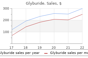
Purchase glyburide 2.5 mg with mastercard
Phlyctenular conjunctivitis is a delayed hypersensitivity response to antigens of bacteria such as Mycobacterium tuberculosis and Staphylococcus aureus diabetes in dogs and ketones order cheap glyburide line. It is characterized by an elevated, hard, red tri- Pinguecula is a common degenerative disease caused by ultraviolet solar radiation and is similar to solar-induced changes in the skin, with epithelial atrophy, degeneration of collagen, and hyalinization of elastic tissue (see Chapter 61). The incidence of secondary infection, ulceration, and epithelial dysplasia is low. This appears as a pearly white plaque on the conjunctiva, sometimes called leukoplakia. Areas of squamous metaplasia are more often subject to infection and ulceration but are not precancerous. The more superficial lesions can be treated by local excision and have a good prognosis. With deeper invasion, lymphatic and vascular involvement commonly occurs, and the prognosis is guarded even following radical exenteration of orbital contents. Because the lesion is behind the conjunctival sac, special techniques are required to obtain tissue for diagnosis. Histologically, there is edema, hyperemia, and infiltration of the orbital soft tissue with neutrophils, eosinophils, lymphocytes, and plasmacytes. The diagnosis is usually made when orbital exploration in a patient suspected clinically of having a neoplasm shows only nonspecific chronic inflammation and fibrosis. Most cases are believed to be the result of exposure to ultraviolet radiation, complicating the actinic lesions pinguecula and pterygium. The neoplastic process progresses through increasing grades of dysplasia to carcinoma in situ and then invasive squamous carcinoma. Conjunctival squamous carcinoma usually invades superficially and almost never metastasizes. The condition is usually bilateral and is believed to be caused by an autoantibody (exophthalmos-producing factor) that may persist even after the hyperthyroidism is treated (Chapter 58). It may occur (1) de novo, (2) in relation to a preexistent melanocytic nevus, or (3) in relation to an acquired melanocytic hyperplasia (lentigo). It presents clini- the lacrimal gland is situated in the lateral wall of the orbit and is rarely the site of pathologic processes. The autoimmune process usually causes atrophy of the gland; more rarely, the lymphocytic infiltration may produce a mass lesion (benign lymphoepithelial lesion). Primary neoplasms of the lacrimal glands are very rare and are similar to salivary gland neoplasms. Inflammatory lymphoid proliferations (pseudotumors) may be distinguished from lymphomas by immunologic methods (polyclonal versus monoclonal; Chapter 29). The diagnosis is made by demonstrating an undifferentiated neoplasm in which primitive rhabdomyoblasts may be identified. With chemotherapy and radiation, orbital embryonal rhabdomyosarcomas can be controlled and sometimes cured. The retina is the inner light-sensitive layer and is composed of modified neurons, the axons of which form the optic nerve. The choroid, which is pigmented, and the fibrous sclera are the outer layers of the eyeball. The lens is attached to the sclera by the ciliary muscle, contraction of which controls the focal length of the lens. The lens and ciliary muscle separate the anterior part of the eyeball, filled with aqueous humor, from the posterior part, which is filled with vitreous. The iris projects into the aqueous humor in front of the lens, partially separating the aqueous humor into anterior and posterior chambers. The iris, by its contraction, controls the amount of light entering the eye and also gives the eyes their color. The circular black opening in the center of the iris through which light passes into the eye is the pupil. It is commonly a well-differentiated, very low grade, fibrillary astrocytoma that grows slowly over several years. Acute endophthalmitis or panophthalmitis results, with swelling and a severe neutrophil infiltration. Untreated, there may be severe destruction with softening and collapse of the eyeball (phthisis bulbi). Toxoplasma gondii infection involves the choroid and retina and occurs either as a congenital transplacental infection or as an acquired infection. Congenital toxoplasmosis may cause neonatal or intrauterine death from encephalitis; survivors frequently show chorioretinitis, sometimes as the only manifestation of congenital toxoplasmosis. The organism persists in the choroid as pseudocysts, causing symptoms in childhood and early adult life. Pathologically, there is focal coagulative necrosis of the retina and choroid, with granulomatous inflammation and fibrosis. Toxoplasma can be identified as small crescent-shaped trophozoites and as larger pseudocysts. Ocular larva migrans is usually caused by larvae of Toxocara canis (a dog nematode) that reach the interior of the eye through the uveal or retinal blood vessels. It causes a granulomatous endophthalmitis with large numbers of eosinophils around the larvae. Rheumatoid arthritis typically produces scleritis and uveitis, in which foci of necrotic collagen surrounded by palisading histiocytes resemble ill-defined rheumatoid nodules. Sympathetic ophthalmia is an uncommon diffuse granulomatous uveitis that affects both eyes after a penetrating injury (or surgery) to one eye. It is believed to be the result of an immunologic reaction against antigens released or altered in some unknown way by the injury. The entire uveal tract is infiltrated by lymphocytes and plasma cells and may show illdefined epithelioid cell granulomas. It has no blood supply and derives its nutrition from the aqueous humor of the anterior chamber. Despite the fact that the oldest epithelial cells become compressed centrally throughout life, the lens normally remains transparent. Pathologically, visible cataracts may occur with minimal changes in water content of cortical cells. With advanced or mature cataracts, the epithelial cells break down, fragment, and undergo dissolution. In diabetes, high glucose levels cause excess production of sorbitol within the lens. Sorbitol is not diffusible and exerts a strong osmotic effect, leading to water imbibition and ultimately to cell degeneration. In galactosemia, galactose is metabolized in the lens to dulcitol with similar results. Current treatment methods, which include extraction of the cataract and implantation of a prosthetic lens, are very successful. The lens is derived from surface ectoderm and consists of Glaucoma is defined as an increase in intraocular pressure sufficient to cause degeneration of the optic disk and optic nerve fibers. Normal intraocular pressure as measured by tonometry (which measures the pressure required to cause flattening of the cornea to a specified amount) is 10-20 mm Hg; elevations in pressure are thought to have a dual effect, inducing deformational changes in the optic disk plus decreased retinal blood flow. However, the correlation between intraocular pressure and optic nerve damage is not exact. Glaucoma is the result of an abnormality in the dynamics of aqueous humor circulation. Aqueous humor production occurs at the ciliary body, partly by diffusion from plasma and partly by ^active secretion by the epithelium of the ciliary processes. Absorption of aqueous humor occurs at the iridocorneal angle by the trabecular meshwork and canal of Schlemm. Glaucoma is a common disorder, with about 2% of all people over 40 years of age affected. Glaucoma may occur as a complication of other diseases affecting the eye (secondary glaucoma) or as a primary disease.
Order glyburide 2.5 mg online
If the disease is recognized early and the patient removed from the source of antigen diabetes test devices glyburide 2.5 mg on-line, the disease is reversible. With continued exposure, diffuse interstitial fibrosis occurs, leading to end-stage honeycomb lung. Clinical Features Patients present with acute dyspnea, fever, and cough 4-6 hours after exposure to the antigen. As pulmonary fibrosis ensues, the disease goes into its chronic phase, with all the features of diffuse interstitial lung disease. Systemic lupus erythematosus may also be complicated by immune complex deposition in the alveoli, transient patchy lung infiltrates, vasculitis, and acute alveolar damage. In the lung, hemorrhage into the alveoli is the dominant feature; hemosiderincontaining macrophages may be found in the sputum. In the chronic phase, the lung is firm because of marked interstitial fibrosis, brown because of hemosiderin, and may show changes of honeycomb lung. Eosinophilic pneumonias are a group of diseases characterized by eosinophilic pulmonary interstitial infiltrates and peripheral blood eosinophilia. In the chronic phase, there is progressive dyspnea, cough, and right heart failure due to pulmonary fibrosis. Patients almost invariably have evidence of glomerulonephritis, most frequently microscopic hematuria. Treatment by plasmapheresis (to remove the antibody) and steroids has proved effective in a minority of cases. Changes that occur in the lung vary with the type and amount of dust inhaled, particle size, and the presence of other lung diseases, most importantly those associated with cigarette smoking. Some dusts such as coal dust do not evoke a fibrous response (noncollagenous pneumoconioses), whereas others such as silica do (collagenous pneumoconioses). In some patients, inhalation of several different kinds of dust results in mixed disease (eg, anthracosilicosis). There is a variable latent period between exposure to dust and onset of clinical disease that may be as long as 20-30 years. Cobalt Tin1 Kaolin or porcelain clay Ceramics manufacture Diatomaceous earth pneumoconiosis Diatomaceous earth these dusts in pure form tend not to cause fibrosis (noncollagenous pneumoconioses) and have minimal clinical effects. Other dusts, especially silica and asbestos, may lead to severe fibrosis (collagenous pneumoconioses). There may also be insignificant dilation of the respiratory bronchiole ("focal dust emphysema"). Rarely, coal workers develop progressive massive fibrosis when heavy exposure is coupled with a complicating factor such as infection with Mycobacterium tuberculosis (found in 40% of patients with massive fibrosis), significant silica contamination (silica induces fibrosis), or development of allergic responses to various proteins that have passively adsorbed onto the coal dust. Progressive massive fibrosis is characterized by the presence of multiple irregular, firm, homogeneous black fibrous masses in both lungs. More than 1 million workers in the United States are at risk for developing silicosis. Significant pulmonary disease usually occurs with 10-15 years of exposure but may rarely occur after as little as 1 year. Larger crystals (> 5 (im) are caught in the bronchial mucus layer and wafted upward by the ciliary action to be expelled; particles less than 1 jum remain airborne and are exhaled. Silica is toxic to the internal organelle membranes of the macrophages and causes phagolysosomal disruption, cell death, and liberation of free silica particles. Silica crystals are also carried in lymphatics to the hilar lymph nodes, where similar silicotic nodules form. One hypothesis suggests that fibrosis is the result of a fibroblast-stimulating factor liberated by macrophages upon phagocytosis of silica particles. A second hypothesis attributes fibrosis to a lymphokine produced by silica-activated T lymphocytes. Grossly, the silicotic nodule is gray-black (due to associated carbon pigment), hard, and brittle and has concentric rings of hyalinized collagen in cross sec- Silicosis is caused by inhalation of crystalline silicon dioxide (silica) dust particles in the range of 1-5 jum. Nodules are found mainly along lymphatic pathways, especially around the hilum and in the upper lobes. Silica particles are recognized as birefringent needle-shaped crystals in the nodules when examined by polarized light. Clinical Features Silicosis is often asymptomatic, being found incidentally at chest x-ray or histologic examination of lungs and hilar lymph nodes removed for an unrelated reason. Rarely, when patients are exposed to massive amounts of dust, acute lung disease may occur, with alveolar thickening and accumulation of proteinaceous material in the alveoli (acute silicotic proteinosis). More often, there is chronic pulmonary fibrosis with a mild restrictive ventilatory defect, slowly progressive dyspnea, and pulmonary hypertension (cor pulmonale). Complications Progressive massive fibrosis may complicate chronic silicosis, particularly when the level of exposure to dust is high. The disorder is characterized by confluence of silicotic nodules into large masses of fibrous tissue that cause obliteration of vessels and bronchioles. Central necrosis and cavitation may occur in these masses as a result of ischemia. Progressive massive fibrosis commonly involves the upper lobes and is associated with a significant ventilatory defect and respiratory failure. Patients with silicosis have a greatly increased incidence of tuberculosis, believed to be due to the adverse effects of silica dust on macrophage function. Tuberculosis causes extensive necrosis in the nodules, and large numbers of tubercle bacilli can be found in such lesions. Silicosis is also associated with an increased incidence of autoimmune disease, especially progressive systemic sclerosis. It is present in such diverse components of the modern environment as insulation, flame retardants, flooring and roofing materials, water and sewage pipes, and brake linings in vehicles, making low-grade exposure almost universal among urban dwellers. It is estimated that up to 11 million workers in the United States have had significant asbestos exposure since 1940. Asbestos-related disease was first recognized in those with the highest levels of exposure, ie, workers in shipyards and the construction industry. It is becoming clear, however, that lower levels of exposure are also associated with significant risk. Asbestos-related neoplasms occur in families of shipyard workers- due presumably to the presence in the home of contaminated clothing-and in communities with asbestos-based industries (air pollution by asbestos dust). It is estimated that about 10,000 deaths every year in the United States are due to asbestos-related diseases. Pathology One of the most common changes associated with asbestos exposure is thickening of the parietal pleura by a plaque-like deposition of hyalinized collagen, maximal in the lateral and diaphragmatic pleura. This change on chest x-ray provides epidemiologic evidence of significant asbestos exposure. Pleural fibrosis does not cause symptoms and does not fall within the definition of asbestos pneumoconiosis because it does not involve lung parenchyma. Asbestos fibers, when inhaled into the alveoli, are taken up by macrophages and evoke a diffuse interstitial fibrosis. The mechanism of stimulation of pulmonary fibrosis by asbestos is poorly understood. Asbestos, unlike silica, is not cytotoxic to macrophages; there is evidence of activation of macrophages by asbestos. Asbestos, when added to in vitro cultures of fibroblasts, stimulates increased collagen synthesis by these cells. Initially, fibrosis occurs around bronchioles but eventually extends into the alveolar interstitium. Ferruginous bodies are best seen in sections that have been stained for iron with Prussian blue. While ferruginous bodies are most commonly seen in asbestosis, they are not diagnostic because a similar iron-glycoprotein coat may form on other types of inhaled fibers. Asbestos fibers that do not have the iron-glycoprotein coat are not visible microscopically, but they outnumber coated fibers 10:1. The amount of asbestos in the lung thus cannot be accurately estimated by microscopy. A quantitative evaluation of asbestos is best made by chemical analysis of lung tissue. The fibrosis progresses even after the drug is withdrawn and may cause death if excessive dosages of the drug have been used. Paraquat, a commonly used herbicide, causes a severe toxic reaction in the lung when ingested or inhaled.
Buy glyburide online
This occurs early in idiopathic hemochromatosis and later in secondary forms of hemochromatosis diabetes mellitus type 2 effects generic 2.5 mg glyburide with amex. It is believed that free iron accumulates in the cytoplasm when the capacity for storage as ferritin and hemosiderin is exhausted. Free ferric iron undergoes reduction^ causing abnormal electron transfers and the formation of toxic oxygenbased free radicals. Over a long period of time, there is progressive loss of liver cells accompanied by fibrosis-leading to cirrhosis, which is commonly macronodular. Hepatic hemochromatosis progresses rapidly and may be complicated by hepatocellular carcinoma. Increased pigmentation of the skin and pancreatic islet destruction result in bronze diabetes, which is an alternative name for hemochromatosis. The primary defect is in the liver cell and is corrected by liver transplantation. Intracellular copper produces an injury manifested as a progressive microvacuolar fatty change and focal liver cell necrosis. It may present, usually in late childhood, as acute hepatitis clinically resembling viral hepatitis. With continuing liver cell necrosis, it progresses to fibrosis and finally to cirrhosis with chronic liver failure. Histochemical methods (rubeanic acid stain) are not always positive because of the abnormal protein binding of the copper in liver cells. Microscopically, the neurons are decreased in number, representing chronic cell necrosis. This is not seen with the naked eye until a late stage of the disease but can be recognized early by slit-lamp examination. The abnormal gene results in hepatic synthesis of an abnormal ccj-antitrypsin molecule that accumulates in the liver cell cytoplasm, appearing as eosinophilic globules. They also stain by immunoperoxidase methods using antibody against ocj-antitrypsin. The demonstration of these globules establishes the diagnosis, which can be further confirmed by absence of oCj-antitrypsin in the blood. Affected patients present in early infancy following feeding of milk (which contains lactose, a disaccharide of glucose and galactose). Diagnosis in a neonate followed by administration of a diet that contains no milk products prevents liver damage. Cavernous Hemangioma Hemangiomas are common incidental findings at surgery, radiologic examination, and autopsy. Histologically, they are composed of large endotheliumlined spaces filled with blood. Peliosis Hepatis Peliosis hepatis is a rare degenerative condition associated with multiple blood-filled spaces in the liver, many of which lack an endothelial lining. Sclerosing Bile Duct Adenoma Bile duct adenoma is uncommon and usually presents as an incidental finding at surgery. Histologically, bile duct adenoma is composed of irregular glands surrounded by collagen. Liver Cell Adenoma Liver cell adenoma is a rare benign neoplasm that occurs mainly in women taking oral contraceptives and athletes taking anabolic steroids; some tumors have regressed when these drugs were withdrawn. Microscopically, liver cell adenomas are composed of cytologically benign hepatocytes arranged in thickened cords. The distinction from a well-differentiated hepatocellular carcinoma may be difficult. Clinically, patients may present with a mass, sudden pain due to infarction, or hemorrhage due to rupture through the liver capsule. Focal Nodular Hyperplasia Focal nodular hyperplasia is another mass lesion of the liver that has been etiologically related to oral contraceptives. Focal nodular hyperplasia most frequently presents as a solitary solid mass lesion, usually subcapsular, well circumscribed, and only rarely larger than 5 cm. On cut section, it is gray-white and typically has a central scar with bands of fibrosis radiating to the periphery. Microscopically, nodules of liver tissue are separated by fibrous bands in which portal tracts containing bile ductules can be identified. It is uncommon in Western Europe and North America (about 8000 cases a year in the United States). Etiology the cause is unknown, but several factors have been implicated: (1) Aflatoxin, a product of the fungus Aspergillus flavus, which grows on improperly stored grain and nuts (including peanuts), is toxic to liver cells. It is present in high levels in grain in Africa and Asia, leading to the suggestion that chronic ingestion of aflatoxin may be at least partially responsible for the high incidence of liver cell carcinoma in these areas; (2) Hepatitis B virus infection is strongly suspected of causing hepatocellular carcinoma. African and Far East countries where hepatocellular carcinoma is common have high rates of hepatitis carriers, probably with vertical transmission of the virus from generation to generation; and (3) Hepatitis C virus infection is also associated with hepatocellular carcinoma. Presentation and risk factors: Hepatocellular carcinoma, cholangiocarcinoma, carcinoma of gallbladder or biliary tract. The increased cell turnover in regenerative nodules of cirrhosis is associated with cytologic abnormalities that have been interpreted as premalignant dysplastic changes. While all types of cirrhosis may be complicated by carcinoma, the association is greatest with hemochromatosis, virus-induced cirrhosis, and alcoholic cirrhosis. Microscopically, the neoplasm is composed of abnormal liver cells of variable differentiation. The cells have enlarged nuclei that show prominent nucleoli and hyperchromatism and may contain bile in the cytoplasm. Invasion of hepatic venous radicles is a typical feature that permits differentiation from adenoma. It may be difficult to distinguish a poorly differentiated hepatocellular carcinoma from metastatic carcinoma. Well-differentiated hepatocellular carcinoma, showing trabeculae of malignant hepatocytes separated by sinusoidal spaces. This is characterized by absence of portal areas and greatly expanded trabeculae composed of several layers of malignant hepatocytes. A rare variant, fibrolamellar carcinoma, has a better prognosis than the usual hepatocellular carcinoma. The entity is defined by the presence of large polygonal cells with abundant eosinophilic cytoplasm separated by broad fibrous bands. Hepatocellular carcinoma, showing a large solitary nodule that is grossly encapsulated except in one area. Rarely, hepatocellular carcinoma may secrete an ectopic hormone, causing hypoglycemia (insulinlike polypeptide), polycythemia (erythropoietin), or hypercalcemia (parathyroid hormone-like polypeptide). Surgical resection is rarely undertaken for the treatment of hepatocellular carcinoma, and chemotherapy and radiotherapy are not very effective. The median survival after diagnosis is 2 months; the 5-year survival rate is almost nil. Europe but has a relatively high incidence in the Far East, where infection with the liver fluke, Clonorchis sinensis, is thought to be a predisposing factor. Grossly, Cholangiocarcinoma presents features indistinguishable from those of hepatocellular carcinoma. The presence of cytoplasmic mucin permits differentiation from hepatocellular carcinoma, which does not secrete mucin. Differentiation from metastatic adenocarcinoma is almost impossible on histologic grounds alone. The progress of disease is often slow, but bloodstream spread ultimately occurs, and the prognosis is poor. Thorium was deposited as refractile crystals in the portal tracts and acted as a carcinogen. The tumor appears as a solid, often very large hemorrhagic mass composed histologically of intercommunicating vascular spaces lined by malignant endothelial cells. Epithelioid Hemangioendothelioma this rare malignant neoplasm of endothelial cells has a slower rate of progression than angiosarcoma. Virtually any malignant neoplasm in the body can metastasize to the liver; those from the gastrointestinal tract (via the portal vein), breast, and lung and malignant melanoma are most common. However, differentiation of hepatocellular carcinoma from metastatic carcinoma is sometimes very difficult. If metastatic spread is from an adenocarcinoma, distinction from primary cholangiocarcinoma of the liver may be impossible unless a primary adenocarcinoma is found elsewhere in the body.

Discount glyburide 5 mg overnight delivery
The disease in children and adults simulates either salmonellosis diabetes symptoms numbness and tingling order glyburide 5mg on-line, when the small intestine is maximally involved, or Shigella dysentery when colitis predominates. It is characterized by a small focus in the intestine and large mesenteric lymph nodes, analogous to the primary complex in the lung. Secondary Intestinal Tuberculosis this form of tuberculosis still occurs as a result of swallowing of infected sputum by patients with active pulmonary disease or reactivation of a dormant intestinal focus, usually in the terminal ileum or cecum. The organisms spread locally in the intestinal lymphatics, resulting in ulcers that are transverse because the intestinal lymphatics pass circumferentially. Involvement of the serosa results in fibrous adhesions between loops of intestine. Clinically, intestinal tuberculosis is a chronic illness characterized by low-grade fever and diarrhea, which may be tinged with blood. Complications of intestinal tuberculosis include intestinal obstruction, caused by strictures, fistulas, and tuberculous peritonitis. Intestinal tuberculosis, chronic phase, showing formation of a stricture secondary to fibrosis of the circumferential ulcers. The intestine proximal to the stricture is dilated secondary to intestinal obstruction. The organisms accumulate in large numbers in macrophages in the mucosa, and there is little or no inflammation. The diagnosis may be established by biopsy or identification of the organism in stools by direct examination (using acid-fast stain) or culture. Entamoeba histolytica is a common pathogen of the colon in underdeveloped countries. Ingested cysts release active amebas (trophozoites) that invade the large intestinal mucosa and enter the submucosa, which is the site of maximal involvement. Amebic colitis, showing typical flask-shaped ulcers with maximal involvement of the submucosa. The mucosal surface-as seen at colonoscopy- shows multiple ulcers separated by healthy-appearing mucosa which is, however, undermined by the submucosal abscesses. Confluence of mucosal ulcers results in large areas of denuded mucosa covered by a necrotic base. Hemorrhage and toxic megacolon may also occur with severe infection, and venous spread to the liver may occur (Chapter 42). Clinically, patients with amebic colitis present with bloody and mucous diarrhea accompanied by low-grade fever. The diagnosis is made by the finding of trophozoites of E histolytica in the stools or in a biopsy specimen. Giardia attaches itself to the surface of the small intestinal mucosal cells by its ventral sucker. In heavy infections, a large part of the mucosal surface area may be occupied by parasites, causing mechanical interference with absorption. Giardiasis also causes partial villous atrophy, which contributes to malabsorption. Clinically, infected individuals develop cramping abdominal pain, with diarrhea and steatorrhea due to malabsorption. Crypto sporidium and Isospora species may rarely infect healthy individuals, causing a mild self-limited acute diarrhea. High-magnification photograph of the edge of an ulcer, showing trophozoites of Entamoeba histolytica. They are minute organisms that are best seen by electron microscopy and silverstained sections. Intestinal helminthiasis is extremely common in underdeveloped countries and has been estimated as afflicting about 25% of the world population. In many of these diseases, the worm lives in the lumen without causing major symptoms; in others, significant clinical manifestations occur (Table 40-4). Cryptosporidium is present in large numbers, usually attached to the surface of the epithelial cell. The diagnosis is established by identifying the organism in stool smears stained with acid-fast stains. Cryptosporidium is 2-4 (Jin in diameter, round, and seen only in acid-fast stained smears. They are recognized as distinct entities with distinct clinical and pathologic features (Table 40-5). These cases are characterized as indeterminate idiopathic inflammatory bowel disease. However, there are so many differences that it is probably best to regard them as two diseases until their cause (or causes) is determined. The diagnosis of idiopathic inflammatory bowel disease is suspected clinically (chronic or recurrent diarrhea) and confirmed by colonoscopy and biopsy. The mucosal features of idiopathic inflammatory bowel disease on histology include distortion of crypt architecture, crypt destruction and loss, and a marked increase in lymphocytes and plasma cells in the lamina propria. These changes distinguish idiopathic inflammatory bowel disease from acute infectious colitides and from other causes of chronic colitis such as collagenous disease (where the crypts are normal and there is a layer of collagen under the epithelial basement membrane), ischemia, and radiation injury. In other cases, the differentiation is made on clinical and radiologic grounds (see Table 40-5). There is no inheritance pattern, and the familial tendency is likely to be the result of a shared, common environment. Sites of Involvement: Combined ileal (most commonly terminal ileum) and colonie disease is most common (50%). Involvement of the oral cavity, larynx, esophagus, stomach, and perineum are rare. Perianal disease occurs in both ileal and colonie disease and is not dependent on the presence of rectal involvement. The mucosa shows diffuse hyperemia, acute inflammation, and shallow, aphthous ulceration. In involved ileal segments, the mesenteric fat creeps from the mesentery to surround the bowel wall (creeping fat). The mucosal surface shows longitudinal serpiginous ulcers separated by irregular islands of edematous mucosa. The ulcerated, inflamed mucosa is at the top; the muscle fibers in the wall at center are separated by the inflammatory cell infiltrate. These features include lymphedema of the submucosa, lymphoid follicles at all levels of the bowel wall, and marked fibrosis. The regional mesenteric lymph nodes are frequently enlarged and may contain noncaseating granulomas. In the acute phase, fever, diarrhea, and right lower quadrant pain may mimic acute appendicitis. Chronic disease is characterized by remissions and relapses over a long period of time. Diagnosis Diagnosis is based on a combination of clinical, radiologic, and pathologic findings. Fistulas between the ileum and the colon result in malabsorption as a result of colonization of the ileum with colonic bacteria; those between ileal loops may short-circuit the bowel, again resulting in malabsorption. Enterovesical fistulas lead to urinary infections and passage of gas and feces with urine. Malabsorption syndrome may also follow disease in the terminal ileum, in which there may be failure of absorption of vitamin B12 and bile acids, resulting in megaloblastic anemia and fat malabsorption. Iron deficiency anemia may occur as a result of chronic occult bleeding, and protein-losing enteropathy as a result of loss of protein from the inflamed mucosa. Incidence In the United States, about 400,000 patients suffer from ulcerative colitis, and there are about 25,000 new cases every year. In the United States, whites are more often affected than blacks, and Jews more often than nonJews. Worldwide, the disease is most prevalent in North America and Western Europe and less prevalent in Asia, Africa, and South America. Antibodies that cross-react with intestinal epithelial cells and certain serotypes of Escherichia coli have been demonstrated in the serum of some patients with ulcerative colitis.

Purchase glyburide 5mg fast delivery
Histologically diabetes type 1 disability cheap glyburide 5 mg fast delivery, the involved ducts are distended by malignant cells that may be arranged in cribriform, papillary, or solid patterns. The cells are large and uniform, with well-defined cell membranes and nonoverlapping, round nuclei. Axillary lymph node dissection is not indicated if there is no invasion, particularly in lesions smaller than 2. Invasive ductal carcinoma-Invasive ductal carcinoma is the most common type of breast cancer, comprising 75% of all cases. Yellowish-white chalk streaks are characteristic and correspond to a peculiar deposition of elastic tissue (elastosis) around ducts in the area of involvement. Microscopically, highly pleomorphic ductal epithelial cells infiltrate the fibrous stroma. Infiltrating lobular carcinomas-Infiltrating lobular carcinomas constitute 5-10% of all breast carcinomas. Morphologic variants of breast carcinoma-Variant forms of breast carcinoma have been recognized (Table 56-3). Some of them-like medullary carcinoma, mucinous (colloid) carcinoma, and tubular carcinoma-are important to recognize because they have a better prognosis than the usual infiltrating ductal carcinoma. Medullary carcinomas tend to be large, soft, and very well circumscribed, consisting of sheets of large polygonal cells associated with a marked lymphocytic infiltrate (which may contribute to the good prognosis). Mucinous carcinomas form gelatinous lakes of mucoid material in which cancer cells are suspended. Infiltrating lobular carcinoma of the breast, showing tumor cells arranged in single rows (Indian file appearance) and fibrosis. Skin and nipple retraction and ulceration are late features with an unfavorable prognosis. Carcinoma may present in pregnancy, when diagnosis is often delayed because of overall breast enlargement and nodularity. Early detection of breast carcinoma is very important because the smaller the lesion, the greater the likelihood of cure. Self-examination of the breast is strongly recommended at monthly intervals for all women. At present, the majority of breast cancers are discovered by self-examination and screening mammography. Small masses or speckled areas of calcification are visible, and biopsy is directed by a needle placed un- Table 56-4. B: Cut surface of the same breast, showing a large infiltrative mass extending from the skin almost to the deep surface (arrows). Initially, the mass may be small and movable, but typically it enlarges, sometimes Local edema and inflammation Metastatic disease in lymph nodes, bone, brain, lung, or pleura 1 the percentage of asymptomatic patients detected by mammography screening is proportionate to the number of women screened. Annual mammograms are recommended for women at increased risk, including all women over 40 years of age. Mammography is an effective screening technique that is currently recommended for high-risk groups such as patients with a family history, a previous breast biopsy showing atypical hyperplasia, or a previous history of breast carcinoma. There is a trend to increase the age at which routine mammography begins to age 50 years for reasons of cost benefit and risk of radiation associated with mammography. The cells are large, with abundant cytoplasm that stains positively for mucin and resembles the cells of ductal carcinoma of the breast. The underlying breast shows diffuse induration, frequently without a definite breast mass. Mode of Spread Direct spread occurs along the ductal system at an early stage, often before invasion has occurred. Such intraepithelial spread may result in involvement of multiple ducts and lobules (cancerization of lobules). Local invasion may also occur into the breast stroma and then into overlying skin and underlying pectoralis major. Lymphatic spread follows predictable routes according to the site of the primary lesion. The nodes along the internal mammary artery may be involved in carcinomas located in the medial half of the breast. Spread beyond the axillary node into supraclavicular and cervical nodes is evidence of advanced disease. Bloodstream spread, with metastatic deposits in bone, liver, and lungs, occurs in the later stages in almost all cases not cured by initial treatment. Entry of cancer cells into the bloodstream probably occurs early in the course of invasive breast carcinoma, but most of these cells are either killed by the immune system or remain dormant in distant organs. The mechanisms underlying dormancy of metastatic cancer cells and the reasons for their later activation to cause clinically detectable tumor masses are unknown. Dormancy and activation of cancer cells are necessary to explain the occurrence of metastases many years after treatment of the primary tumor. Inflammatory breast carcinoma, showing a dermal lymphatic containing carcinoma cells (arrows). Diagnosis Histologic examination of a biopsy of the mass is the definitive diagnostic method. Immediate diagnosis of a biopsy specimen by frozen section examination has a high degree of accuracy in experienced hands. A complete pathologic diagnosis of breast carcinoma should provide the following information: (1) the histologic type of carcinoma; (2) the size of the tumor; (3) the stage of disease (Table 56-5); and (4) the estrogen and progesterone receptor status. Receptor status is currently established by bioassay, for which a specimen from the tumor must be removed for freezing by the pathologist immediately after excision. Immunohistochemical techniques are available for receptor determination on fixed tissue. Cytologic diagnosis utilizing a specimen obtained by fine-needle aspiration is increasing in popularity because it is rapid and cost-effective. Definitive diagnosis of the histologic type of carcinoma still requires histologic examination of tissue sections. Surgery: Surgery has been the mainstay of treatment of breast cancer for the past several decades. The standard treatment was radical mastectomy, which involves removal of the breast along with the pectoral muscles and axillary contents. The realization that this type of surgery may be too extensive led to new approaches. These are (1) modified radical mastectomy, which includes axillary node dissection but preserves the pectoralis muscle; and (2) complete excision with clear margins (lumpectomy), with axillary node dissection followed by radiation. There is a trend toward breastconserving surgery for treatment of breast carcinoma. Radiotherapy is indicated when breast-conserving surgery has been performed and in patients who develop locally recurrent disease in the chest wall. Chemotherapy: Chemotherapy has increased the disease-free survival periods in breast carcinoma but is not curative. The rationale for chemotherapy after successful surgical treatment (adjuvant chemotherapy) is that it removes microscopic foci of neoplastic cells in distant sites, thus complementing the role of surgery. Adjuvant chemotherapy is indicated in all but small, well-differentiated, nodenegative cancers with no adverse prognostic indicators. Prognosis Infiltrating carcinoma of the breast has a 5-year survival rate of about 70%. Recurrences of breast carcinoma have been recorded as late as 25 years after the primary tumor was successfully treated. The Clinicopathologic Stage: Staging of breast carcinoma is based on defined criteria relating to the primary tumor, lymph nodes, and distant metastasis (Table 56-5). Absence of steroid hormone receptors indicates a poor prognosis quite apart from the lack of response to hormonal therapy that is associated with absence of receptors. The lack of progesterone receptors has a greater value in predicting poor prognosis than lack of estrogen receptors. High proliferative activity of the cancer cells, as indicated by a high (> 12 %) S-phase fraction on flow cytometry or high expression of the proliferative antigen Ki67 indicates a poor prognosis. Aneuploidy in the cancer cells, as shown by flow cytometry, indicates a poor prognosis.
Biotin. Glyburide.
- Treating and preventing biotin deficiency.
- Is Biotin effective?
- Hair loss, diabetes, diabetic nerve pain, brittle fingernails and toenails, and others.
- How does Biotin work?
- Skin rash in infants.
- Dosing considerations for Biotin.
- Are there safety concerns?
- What is Biotin?
Source: http://www.rxlist.com/script/main/art.asp?articlekey=96334
Order 2.5 mg glyburide fast delivery
The disease is transmitted by ixodid ticks that become infected by biting deer and mice diabetes type 2 and 1 purchase glyburide 2.5 mg amex, which are the common reservoirs of infection. Lyme disease is characterized by development of a distinctive papular skin rash (erythema migrans) at the site of inoculation 1-4 weeks after the tick bite. The rash lasts several months and may be associated with spirochetemia and systemic disease. Migratory acute arthritis is one of the most common manifestations of systemic disease and may be followed by chronic arthritis. The synovial membrane shows thickening, with a lymphocytic and histiocytic infiltrate. Organisms are present in the walls of small blood vessels, blood, and synovial fluid. Demonstration of the spirochete in blood or infected tissues is rarely successful. Treatment with penicillin or tetracycline is successful if started early in the acute phase and prevents chronic arthritis and other complications. Less frequently, Streptococcus pyogenes, Streptococcus pneumoniae, Neisseria gonorrhoeae, and Haemophilus influenzae are responsible. The route of infection is hematogenous, and in most patients the primary access site of the pathogen is unknown. Pathology & Clinical Features Pyogenic arthritis is an acute inflammation that commonly involves a single large joint such as the knee or hip and is characterized by severe pain, tenderness, redness, swelling, and local warmth. High fever, often with chills and a neutrophil leukocytosis, is present in most cases. Rheumatoid factor-an autoantibody (usually IgG)-is present in the plasma of about 90% of patients with rheumatoid arthritis, but its presence is not specific for the disorder because it is present in other autoimmune diseases and in 5% of healthy persons. Immune complexes composed of rheumatoid factor and IgG have been found in the sy no vial fluid of some patients with rheumatoid arthritis. Complement levels are also frequently decreased in active disease, suggesting that complement activation by deposited immune complexes may play a role. Incidence Rheumatoid arthritis is common in the United States and Western Europe, affecting 1-2% of the population. This is followed by proliferation of granulation tissue containing numerous lymphocytes and plasma cells (this fleshy tissue is termed pannus). Local production of interleukins, tumor necrosis factor, and other cytokines accounts for many features of synovitis. The pannus eventually erodes articular cartilage, subchondral bone, and periarticular ligaments and tendons. Progressive destruction of the joint follows, with fibrosis, increasing deformity, and restriction of movement. The mechanism of destruction of cartilage and bone is not known but is probably related to synthesis of collagenase and other proteases in the pannus. Involvement of larger joints is the initial manifestation in a minority of patients. Stiffness is maximal in the morning after the joint has been inactive during the night. Swelling of the proximal interphalangeal joints of the fingers produces a typical spindled appearance of the fingers. Many patients have systemic symptoms such as low-grade fever, weakness, and malaise. Restriction of movement may cause rapid disuse atrophy of muscles around the joint. In a minority of patients, tissues other than joints show significant pathologic change (Table 68-2). Subcutaneous rheumatoid nodules are granulomas 1-2 cm in diameter seen commonly around the elbow, usually in patients with severe disease. Most other patients develop a chronic disease characterized by relapses and remissions, with slowly progressive disability from joint destruction. After 10 years of disease, about 10% of patients are severely disabled while about 50% are still fully employed. Poor prognostic factors include a classic pattern of disease with high levels of rheumatoid factor in the serum, the presence of rheumatoid nodules, and onset of disease before age 30 years. Rheumatoid arthritis (chronic phase, severe disease), showing symmetric involvement and severe deformity. It is characterized by acute onset with high fever, leukocytosis, splenomegaly, arthritis, and skin rash. Growth abnormalities may occur if the disease strikes before the age of epiphysial closure. Pathology & Clinical Features Ankylosing spondylitis maximally affects the sacroiliac joints. Subcutaneous rheumatoid nodule, showing a central area of necrosis of collagen surrounded by palisading histiocytes. Involvement of the costovertebral joints and thoracic spine may result in restriction of chest expansion and rarely produces respiratory failure. Extra-articular Manifestations Patients with ankylosing spondylitis may show degeneration of the wall of the aorta, with dilation and incompetence of the aortic valve. Course & Prognosis Ankylosing spondylitis is a slowly progressive disease that causes increasing disability from pain and stiffness of the low back. Respiratory dysfunction and aortic disease represent life-threatening complications. When assessed radiologically, changes of osteoarthrosis are present in over 40% of individuals over the age of 50 years of age. Although only a few of these patients are symptomatic, osteoarthrosis is the most common cause of joint disability. When a younger individual develops osteoarthrosis, it is almost always secondary to a predisposing abnormality in the joint. Its clinical features are very different from those of rheumatoid arthritis (Table 68-3). The latter is an inaccurate term because it implies the presence of joint inflammation, which is not present. Abnormalities in the ground substance, collagen, increased activity of matrix-degrading enzymes such as collagenase and proteoglycanases, and changes in water content have all been demonstrated in the articular cartilage in patients with osteoarthrosis, but their role in pathogenesis is unknown. Fever, malaise in some Rheumatoid factor; Terythrocyte sedimentation rate; anemia, leukocytosis, hyperglobulinemia Clear; low viscosity, high protein; neutrophils, some lymphocytes; immunoglobulins, complement, rheumatoid factor No None None None Clear, normally viscous; no inflammatory cells trauma. A few cases of osteoarthrosis occur secondary to articular cartilage diseases (eg, alkaptonuria) and severe trauma (eg, in football players). Continued loss of articular cartilage leads to exposure of subchondral bone, which appears as shiny foci on the articular surface (eburnation). Fibrosis, increased bone formation, and cystic change frequently occur in the underlying bone. The loss of articular cartilage stimulates new bone formation, usually in the form of nodules (osteophytes) at the bone edges. Clinical Features There is pain, stiffness, and swelling of affected joints, with no evidence of acute inflammation. Crepitus is a characteristic feature-a grating sound produced by friction between adjacent areas of exposed subchondral bone. They may cause compressive symptoms, most notably in spinal osteoarthrosis, in which nerve and spinal cord compression may occur. Course & Prognosis Osteoarthrosis is a slowly progressive, chronic joint disability. Eventually, elderly sufferers may be- come confined to wheelchairs; recent advancements in the technique of joint replacement with prostheses have improved the outlook of these patients considerably. The lack of pain sensation deprives the joint of its normal protective muscle and postural responses when exposed to abnormal forces. The affected joint is swollen, unstable, and frequently shows an abnormally increased range of motion resulting from destruction of intra-articular ligamentous restraints.
Syndromes
- Neck x-ray
- High levels of the protein that carries T4 in the blood (can occur with pregnancy, use of birth control pills or estrogen, liver disease, and as part of an inherited condition)
- Foamy urine
- External ear infection - chronic
- Incoordination
- Age 4-8 years: 3* mg/day
- Glucose tolerance test
- Blood or urine tests to detect histoplasmosis proteins or antibodies
- Chest pain
- Reduced movements
Buy glyburide visa
There are virtually no topics in medicine for which every article/expert agrees diabetes questionnaire quality glyburide 2.5mg, unless clinicians have the luxury of finding a large systemic review or meta-analysis that can be decisive alone. For instance, over the last decade the pendulum concerning isotretinoin inducing inflammatory bowel disease has swung multiple times. Keeping up to date on the most recent literature benefits the patients and helps protect the provider. That being said, be aware of susceptibility to thinking the last article is always right. It is imperative to look at the medical literature as a whole before forming an opinion. Being aware and critical of the available literature will help clinicians avoid these pitfalls. There are exceptions though if, because of disease severity, initial management requires multiple interventions. Clinicians can speculate with some degree of certainty when drug initiation controls the disease, that this drug provided benefit. This argument is strengthened if the patient flares when the dose is decreased and improves when dose is once again increased. Keep up to date on the medical literature: There are many topics in medicine that are still incompletely understood and most topics are not universally agreed upon. Try to make your decisions evidence based but realize that there are many decisions that cannot be based, on optimal data. This is especially true in dermatology where much of the data are limited to retrospective case series and case reports. If a provider pays relentless attention to evidence-based medicine, this may lead to inaction. Evaluating the strength and carefully interpreting available data is critical when formulating a plan. Nursing Research: Methods and Critical Appraisal of Evidence-Based Practice, 8th ed. For example, the lymphoma risk with immunosuppressive drugs will be higher in transplant patients versus psoriasis patients. In addition, providers are tasked with the challenge of interpreting whether a study result is statistically significant or the result was due to chance alone. Separate the P-value thresholds versus the actual P-value in the study, as these may be different. This is especially important with subjective or semiobjective topics such as data with patient input. A confidence interval of 95% says that you are 95% confident that the mean of the whole population is in a certain range. To determine if the 95% confidence interval is significant, it must not include the number 1. For example, if the incidence of cutaneous squamous cell carcinoma is 1/100 in the general population but 10/100 in patients taking azathioprine, the attributable risk of azathioprine causing squamous cell carcinoma is 9/100. When deciding to take on risk while making a medical decision, agreement between the prescribing physician with both the patient and collaborating physician(s) is optimal: Unfortunately, most, if not all, medical interventions carry some degree of risk. As clinicians, it is our responsibility to weigh the risks with the benefits and to then relay this information to the patient with our recommendations. These decisions should not be unilateral though, and consideration needs to be given to patient preferences. Thorough discussion with the patient as well as with collaborating physicians (when pertinent) should always be completed in these situations. If any provider does not feel comfortable, then the selected intervention will not proceed. Such examples would include consulting an obstetrician when using biologic therapy during pregnancy or consulting an ophthalmologist when considering de-escalating immunosuppression in an ocular pemphigoid patient. Of course, the patient gets to make the ultimate decision if they do not want to take on the proposed risk. The therapeutic efficacy of a treatment must be scrutinized against the safety profile of that treatment modality: the benefit of a treatment is measured in terms of therapeutic efficacy whereas the risk of a treatment is conveyed in its safety profile. When making medical decisions, both the potential benefits and risks of treatments are considered. In certain situations, physicians may choose a potentially less effective treatment modality to achieve a more favorable safety profile. For instance, many physicians will opt to start phototherapy as an initial treatment for psoriasis even though it is often less efficacious than many other therapeutic options. Only if the patient has a less than ideal response would there be consideration to escalate to systemic medications such as methotrexate or biologics, as these carry increased risk compared with phototherapy. For instance, when treating Stevens-Johnson syndrome, cyclosporine can be the best therapeutic option despite it having increased risk compared with other treatment modalities. In these situations, the most appropriate medication may be the one that is most efficacious despite the potential risk associated with it. Once initial control is obtained, the clinician can de-escalate to therapy with less risky medications. Be aware of the cost of your chosen treatment to both the patient and the healthcare system: Although cost should not be the most important factor in your medical decision, it certainly requires strong consideration. As the physician, it is important to take into account both the out-ofpocket costs that the patient will be incurring as well as the cost for the healthcare system. It can be tempting for a clinician to prescribe a newer agent expecting improved efficacy and decreased laboratory monitoring; however, this is frequently inappropriate if a reasonably effective, older, and less expensive treatment option is available. The authors frequently see this with psoriasis treatment as therapies such as methotrexate and phototherapy are avoided in many patients without cause, solely to start newer biologic agents. Prescribers should not discount these older therapies because of safety profiles, because older therapies have had more post-marketing surveillance, and thus have more-established safety profiles. The percentage of the United States economy spent on healthcare has increased from 8% in 1980 to 17% in 2011. In some ways these restrictions decrease the autonomy of the physician; however, it is an understandable strategy to try to contain healthcare cost. The therapeutic efficacy of a treatment must be scrutinized against the safety profile of that treatment modality. Suboptimal efficacy and treatment failure are different and must be distinguished. This is an undeniably remarkable advance for our field, but does this now mean that a patient obtaining 50% clearance is a treatment failure Although the role of acitretin for plaque psoriasis has diminished, methotrexate, phototherapy, and the older biologic agents should not be overlooked as treatment options. Initial therapy should often start with the aforementioned therapies and be selected based on favorable risk profiles, patient convenience, and insurance coverage. It is also prudent to consider the impact on labeling medications as treatment failures. The authors have witnessed many patients that have been labeled as treatment failures on medications when they were never treated with an adequate dose or were not on the medication for a long enough period to observe the desired response. Starting treatment before disease diagnosis is appropriate in many instances: Physicians may consider performing the initial biopsy and laboratory evaluation as well as initiating treatment at the same visit. This can be done to relieve symptoms and in an attempt to have the disease process enter remission. Physicians may also trial empiric treatment instead of pursuing additional diagnostic workup if there is high enough clinical suspicion for a certain disease process. If this is the case, the physician needs to be bold and prescribe an adequate dose. Compliance: Prescribers can be quick to blame patient compliance when a patient does not seem to be improving as completely or as quickly as desired. Although this can at times be the case, it is important that we do not default to blaming the patient. When noncompliance is suspected, it is important for the prescriber to clarify exactly how the patient is taking medication, as partial compliance seems to be more common than complete noncompliance. Ways to increase compliance include providing simple written instructions, asking the patient to repeat instructions back to you and making sure to leave the door open for any questions.
Generic 5mg glyburide overnight delivery
Apart from minor abnormalities in liver function tests (eg diabetic log printable purchase glyburide 2.5mg fast delivery, slight elevation of transaminases, bilirubin, or alkaline phosphatase), these nonsuppurative infections do not usually cause any clinical features. Chronic active hepatitis, showing marked lymphocytic infiltration and fibrosis of the portal areas. The lymphocytes extend into the peripheral part of the lobule through the limiting plate. There is ongoing necrosis of hepatocytes in the peripheral part of the lobule (piecemeal necrosis). Hyperimmune gamma globulin provides passive protection against hepatitis A and can be used to prevent a clinical attack of hepatitis A after exposure to the virus. The use of pooled hyperimmune gamma globulin (a blood product) itself carries a risk of hepatitis B and C transmission. Screening of blood donors for hepatitis B and C has virtually eradicated transmission of viral hepatitis via blood transfusion. In areas where Entamoeba histolytica is not prevalent, most liver abscesses are caused by pyogenic organisms. Many different bacteria may be involved, most commonly Escherichia coli, other gram-negative bacilli, anaerobic bacilli, Staphylococcus aureus, and streptococci. Culture of pus is necessary for etiologic diagnosis and often reveals a mixed flora. Liver abscesses are walled-off collections of pus with liquefactive necrosis of liver cells and neutrophil accumulation. Clinically, patients present with high fever, rightsided upper abdominal pain, and hepatomegaly. Pyogenic abscess is a focal lesion and not usually associated with abnormalities in liver function tests except elevation of serum alkaline phosphatase. Treatment consists of drainage of the abscess followed by antibiotic therapy directed by culture and antibiotic sensitivity of the bacteria isolated from the pus. Hepatic infection usually occurs in patients with subclinical or chronic intestinal amebic infection and very rarely during an attack of acute amebic colitis. Pathogenesis of pyogenic liver abscess, showing the three main routes of bacterial infection, via the bile duct (1), the portal vein (2), and the systemic circulation (3). About half of patients with hepatic amebiasis give no history suggestive of preceding amebic colitis. When they reach the liver, the amebas cause focal enzymatic necrosis of hepatocytes. In the early stage of the disease, there are multiple microabscesses throughout the liver (Note: Although the term "abscess" is used, amebic liver abscesses are not true abscesses because they contain few neutrophils and are composed of liquefied liver cells. Diagnosis is based on clinical findings, which include fever, pain in the lower right chest, with hepatomegaly and marked tenderness. Liver function tests are usually normal except for an elevated serum alkaline phosphatase level. Deaths are due to (1) rupture into the free peritoneal cavity; (2) rupture into the pleural cavity and lung; (3) rupture into the pericardial sac (in left lobe abscesses), causing acute pericardial tamponade; and (4) systemic spread of trophozoites, resulting in amebic abscesses in the brain and lung. Schistosoma mansoni, which causes colonic infection in the Middle East, and Schistosoma japonicum, which causes small intestinal infection in the Far East, are the species involved. The adult worms live in the intestinal venous plexuses and produce eggs that are carried via the portal vein to the liver, where they are deposited in the portal areas. They produce granulomas iri the acute phase followed by pipestem fibrosis of the portal areas in the chronic phase. Hepatic schistosomiasis causes portal hypertension and ascites and is an important cause of these conditions in endemic areas. Aspirate of bile in oriental cholangiohepatitis, showing ova of Clonorchis sinensis. During surgical removal, care must be taken to avoid spillage of cyst contents into the peritoneal cavity, since the cyst fluid is highly antigenic and may lead to anaphylactic shock. The liver is the most common site for hydatid cysts, which may reach a large size and may be multiple. Histologic examination shows a thick, acellular laminated eosinophilic wall with an inner surface lined by the germinal epithelium of the larva. The cysts are filled with a granular fluid that is characterized by numerous small larval capsules containing scoleces (brood capsules). The Infection of the bile ducts with Clonorchis sinensis is common in eastern Asia. The flukes attach with their suckers to the bil-duct wall and cause inflammation and strictures of the bile ducts and fibrosis of the surrounding liver. The dilated bile ducts proximal to the narrowed segments contain numerous crumbling black calculi. Alcoholic liver disease is most common in middle-aged men, but there is an increasing incidence among women and in the young. The greater the amount and the longer the duration of alcohol consumption, the greater the risk of liver disease. Most patients with chronic alcoholic liver disease have consumed about 150 g or more of ethyl alcohol daily for over 10 years (a standard 750 mL bottle of 80-proof whisky contains about 300 g of alcohol). About 50% of alcoholics have no detectable liver disease, 30% have alcoholic hepatitis, and 20% develop cirrhosis. Acetaldehyde or a related substance is believed to exert a toxic effect on liver cells. Malnutrition, which frequently coexists with alcoholism, may aggravate the liver injury. It is the result of decreased fatty acid oxidation, increased synthesis of triglycerides, and impaired secretion of lipoproteins by the liver cell. Fat accumulates first as small globules that coalesce, increasing in size and pushing the hepatocyte nucleus to one side. In developed countries, it has been estimated that about 10% of the population consume potentially harmful amounts of ethyl alcohol. B: Acute alcoholic hepatitis (acute sclerosing hyaline necrosis), C: Chronic alcoholic liver disease, precirrhotic. Clinically, patients present with an acute onset of fever, jaundice, tender enlargement of the liver, and ascites, commonly after a recent bout of heavy drinking. Symptoms and most of the pathologic features resolve with cessation of drinking, but the fibrosis increases progressively with each episode. Chronic Alcoholic Liver Disease: Chronic ingestion of alcohol is associated with progressive fibrosis in the centrizonal region of the liver and distortion of liver architecture by fibrous bands that may connect portal areas and central veins; this differs from cirrhosis in the absence of true regenerative nodules. Alcoholic Cirrhosis: Cirrhosis of the liver is discussed on p 653 and alcoholic cirrhosis on p 655. Unpredictable (Idiosyncratic) Toxicity Some hepatotoxic drugs cause liver injury in an unpredictable manner, usually unrelated to dose and in only a small percentage of susceptible individuals. Massive Hepatocellular Necrosis: Isoniazid, halothane, and methyldopa may cause submassive or massive liver necrosis, with acute liver failure. Lesser degrees of liver necrosis have been reported following exposure to other antituberculous drugs and antidepressants. Isoniazid-induced necrosis is important because of the recommendation that this drug be used for chemoprophylaxis in all patients who give a positive tuberculin skin test. Predictable (Dose-Related) Toxicity Agents in this group cause liver cell necrosis in all individuals at a predictable dose level. Acetaminophen, which is normally metabolized by the glutathione reductase system, is an example of this phenomenon. In overdosage, the glutathione becomes exhausted, and alternative metabolic pathways yield toxic products. Salicylates in high doses and cytotoxic anticancer drugs such as methotrexate also cause liver cell necrosis. Note the large pale zone around the central vein composed of necrotic liver cells. Malnutrition (kwashiorkor) is associated with fatty liver and nutritional cirrhosis.
Discount glyburide online american express
The muscular paralysis is associated with absence of atrophy (amyotrophic) diabetes symptoms high sugar levels cheapest generic glyburide uk, hypertonia, and exaggerated deep tendon reflexes. Neuronal degeneration is associated with irregular neuronal discharge, leading to muscle fasciculations, which is a feature of the disease. Note that overlapping clinical features appear as the disease progresses toward the end stage, with severe deficits of both upper and lower motor neurons. Death usually occurs in 1-6 years from bronchopneumonia associated with respiratory muscle paralysis. Myelin-stained transverse section of the spinal cord, showing areas of demyelination involving the posterior and lateral columns. Peripheral neuropathy or optic neuropathy may also occur as isolated lesions in patients with vitamin B12 deficiency. The diagnosis is important, because vitamin B12 therapy will prevent progression- although it does not repair the demyelinated fibers. It causes involvement of the floor of the third ventricle and the periaqueductal region of the midbrain. The early lesion is characterized by petechial hemorrhages and capillary proliferation. Histogenetic Classification: Classification on a histogenetic basis has great theoretical value and provides a means of logically remembering all the different kinds of intracranial neoplasms (Table 65-1). Topographic Classification: When a patient presents with an intracranial neoplasm, its location can usually be ascertained by clinical examination and radiologic studies. According to their location, intracranial neoplasms may be classified as supratentorial or infratentorial. Further subdivisions in these main compartments are recognized (Table 65-2), leading to a topographic classification. For example, if a child presents with a neoplasm in a cerebellar hemisphere, it is most likely a juvenile pilocytic astrocytoma (Table 65-2). Classification According to Biologic Potential: the criteria used to determine malignancy in neoplasms are somewhat different from those used elsewhere in the body: 1. Metastasis within the craniospinal axis via the cerebrospinal fluid does occur, most commonly with medulloblastoma, pineoblastoma, malignant ependymoma, pineal germinoma, and glioblastoma multiforme. Destructive infiltration of the brain is the major criterion of malignancy for intracranial neoplasms, and infiltration of brain substance usually prevents complete removal at surgery. Neurologic deficits resulting from destructive invasion by malignant neoplasms are irreversible. Benign neoplasms, on the other hand, cause neurologic deficits due to compression; these often reverse when the neoplasm is removed. Intracranial and spinal neoplasms may be primary or metastatic; in most autopsy series, metastatic tumors are more common. Primary intracranial neoplasms number about 13,000 new cases per year in the United States and represent about 2% of deaths from malignant neoplasms. They are the second most common group of neoplasms in children, after leukemia and lymphoma if considered as one group. Taken overall, 65% of primary intracranial neoplasms are of glial origin (gliomas), 10% meningiomas, 10% acoustic schwannomas, 5% medulloblastomas, and 10% others. Cell Type Cellular derivatives of the neural tube Glial cells Astrocytes Oligodendroglia Ependymal cells Neurons Mixed glial and neuronal Pinealocyte Cells derived from the neural crest Schwann cell Arachnoid cell Other cells Connective tissue cells Lymphoid cells Vascular cells Pituicytes Embryonic remnants Ectodermal derivatives Notochordal remnants Germ cells Melanocytes Adipocytes Metastatic neoplasms Tumors of bone (skull and vertebrae) Neoplasm Glioma1 Astrocytoma Glioblastoma multiforme Oligodendroglioma Ependymoma Subependymoma Choroid plexus papilloma Medulloblastoma Ganglioglioma Pineocytoma Pineoblastoma Schwannoma Neurofibroma Meningioma Sarcomas Malignant lymphoma Hemangioblastoma Pituitary adenoma Craniopharyngioma Epidermoid cysts Dermoid cysts Chordoma Teratoma Germinoma Melanoma Lipoma 5. Benign intracranial neoplasms frequently produce extremely serious clinical disease and may cause severe neurologic deficits and death unless treated. The term thus does not mean that these neoplasms are harmless but implies rather that they are slow growing and do not infiltrate the brain substance. In general, intracranial neoplasms cause the following clinical and pathologic changes: A. Compression: Compression of adjacent neural tissues occurs with all expanding neoplasms. When the rate of growth is slow, compression leads to atrophy, which may cause symptoms of dysfunction-eg, atrophy of the motor cortex adjacent to a meningioma causes upper motor neuron paralysis; compression of a cranial nerve may cause cranial nerve palsy. In general, relief of compression is followed by significant recovery of function. Destruction: Destruction of neural tissues by direct infiltration with a malignant neoplasm produces an irreversible deficit. Cerebral Edema: Cerebral edema is commonly present around infiltrative neoplasms and may be severe. It is believed to result from the neovascularization that accompanies malignant neoplasms. The new vessels have a poorly developed bloodbrain barrier that permits exit of proteins and fluids more easily than from normal vessels. Cerebral edema causes elevation of intracranial pressure that is additive to the mass effect of the tumor. Irritative Effects: Irritation of neural tissues may occur with both compressing and infiltrating neoplasms. Abnormal stimulation is usually manifested as either simple or complex partial focal epilepsy. A neoplasm near the motor cortex may generate an abnormal electrical potential that causes motor stimulation of the entire contralateral half of the body (jacksonian epilepsy). Up to 5% of individuals with intracranial neoplasms experience one or more seizures. Note that although only a minority of cases of epilepsy are due to tumors, seizures should be fully investigated for cause due to tumor or other treatable disease, particularly when the onset is in adult life or when the seizure is focal rather than generalized (see Chapter 62). Hydrocephalus: Neoplasms in the region of the third ventricle or in the posterior fossa may cause obstructive hydrocephalus. In its narrowest usage it is synonymous with astrocytomas; in its broadest usage it includes Oligodendroglioma and ependymal neoplasms. Rapidly growing neoplasms such as glioblastoma multiforme and medulloblastoma are highly malignant. Low-grade malignant neoplasms such as well-differentiated astrocytoma and Oligodendroglioma grow slowly. Recurrence after treatment is almost invariable with malignant intracranial neoplasms. Recurrence also occurs with many benign neoplasms such as meningioma and Craniopharyngioma, and therefore recurrence of itself is not a criterion of malignancy. Common intracranial neoplasms classified by location of lesion and age of patient. Location Supratentorial Cerebral hemisphere 30% Rare 1 Children 70% Glial neoplasms Meningiomas Metastases 1 Adults Suprasellar Craniopharyngioma Juvenile pilocytic astrocytoma Pineoblastoma Germ cell tumor (teratoma) 70%1 Medulloblastoma Ependymoma Pituitary adenoma Craniopharyngioma Glial neoplasms Pineocytoma Germ cell tumor (germinoma) 30%1 Brain stem glioma Pineal Infratentorial (posterior fossa) Midline Cerebellar hemisphere Cerebellopontine angle Spinal cord Epidural Intradural but extramedullary Juvenile pilocytic astrocytoma Epidermoid cyst Rare Bone tumors Rare Metastases Hemangioblastoma Schwannoma (acoustic neuroma) Meningioma Common Metastases Bone tumors Neurofibroma Schwannoma Meningioma Ependymoma Astrocytoma Intramedullary Ependymoma Percentages refer to frequency of intracranial neoplasms within each category; ie in children, 70% of neoplasms are infratentorial and 30% are supratentorial, while in adults 70% of neoplasms are supratentorial and 30% are infratentorial. Increased Intracranial Pressure: Intracranial neoplasms cause increased intracranial pressure due to (1) the mass effect of the neoplasm itself, (2) cerebral edema, or (3) hydrocephalus. Many patients with intracranial neoplasms present with the effects of increased intracranial pressure-headache, vomiting, papilledeina, and false localizing signs due to displacement of the brain and to herniations (see Chapter 62). Shift of structures can often be detected radiographically and provides a clue to the site of intracranial neoplasms. Microscopically, they show slightly increased cellularity and astrocytes with cytologic features that deviate very slightly from normal. Neurofibrillary processes are present and, often, abundant (fibrillary astrocytoma). Well-differentiated astrocytomas, although low-grade malignant neoplasms, are practically impossible to excise surgically because of irregular extension be- Diagnosis Clinical examination and radiologic imaging provide excellent localization of mass lesions of the nervous system. Specific diagnosis, however, is based on microscopic examination of a sample from the tumor. Stereotactic biopsy and open resection are the methods available for obtaining tissue samples. Ex; animation of cerebrospinal fluid is rarely useful because (1) lumbar puncture is not usually performed in the presence of mass lesions, and (2) the yield of neoplastic cells in cerebrospinal fluid is low even with highly malignant neoplasms. Microscopically, it is composed of astrocytes that show greater cellularity, more pleomorphism, neovascularization with proliferation of endothelial cells, and an increased mitotic rate. Anaplastic astrocytomas show a higher rate of cell proliferation than well-differentiated astrocytoma. The proliferative rate may be measured by (1) the percentage of cells in the S phase of the cell cycle and (2) the percentage of cells expressing proliferation-associated antigens such as Ki67. Grossly, glioblastomas appear as large infiltrative masses that typically extend across the midline as the so-called "butterfly" tumor. Microscopically, they are composed of highly pleomorphic astrocytic cells with frequent mitotic figures.
