

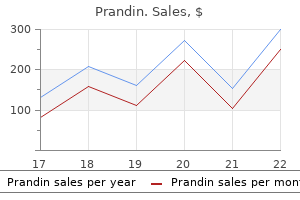
Buy prandin with paypal
The sequences of events are: An unknown antigen is taken up by antigen-presenting cells blood sugar 20 buy prandin discount. Polyclonal hypergammaglobulinemia (another manifestation of helper T-cell dysregulation). Epithelioid cells are modified macrophages and characteristically have abundant eosinophilic cytoplasm and vesicular nuclei. Inset shows giant cell with asteroid body (upper right) and Schaumann body (lower right) 344 Exam Preparatory Manual for Undergraduates-General and Systemic Pathology Other findings: Two other microscopic findings of sarcoidosis include: Sarcoidosis: Schaumann bodies-laminated Schaumann bodies: these are laminated concretions of calcium and proteins. Sarcoidosis: Asteroid bodies the granulomas may resolve completelyStellate (star shaped) As the lesion become chronic, proliferation of collagen forms concentric laminated layer inclusions within giant giving them an onion-skin appearance and later may form nodular hyaline scars. Fate of Granuloma Sarcoidosis: Diagnosis is by exclusion of other granulomatous diseases. Histologic diagnosis is made after exclusion of organisms that are known to be associated with granuloma formation. Noncaseating (hard) granuloma is not specific for sarcoidosis, and may be seen in 1) mycobacterial infections, 2) fungal infections, 3) malignancy, and 4) environmental agents. In advanced cases the coalescence of granulomas produces small palpable or visual nodules measuring 1 to 2 cm. Microscopy: the granulomas are found in the pulmonary interstitium along the lymphatics, around bronchi and blood vessels, although alveolar lesions are also seen. Lymph Nodes They are involved in almost all cases, particularly the hilar and mediastinal nodes. Insidious onset of respiratory symptoms (shortness of breath, cough, chest pain, hemoptysis) or of constitutional signs and symptoms (fever, fatigue, weight loss, anorexia, night sweats). May be detected on routine chest films as bilateral hilar lymphadenopathy or may present with peripheral lymphadenopathy, skin lesions, eye involvement, splenomegaly, or hepatomegaly. Recovery with resolution of the disease: Occurs either spontaneous or induced by steroid therapy in about 65% to 70% of patients. Chronic or progressive course: It may lead to permanent loss of some lung function or some permanent visual impairment in about 25% of patients. Originally, the term pneumoconiosis (or dust diseases of the lung) was used for nonneoplastic lung reaction to inhalation of mineral dusts. Pneumoconioses: Lung diseases produced by organic as well as inorganic particulates and chemical fumes and vapors. Pathogenesis Only a small percentage of exposed people develop pneumoconioses, and indicates a genetic predisposition. Amount of dust retained in the lung and airways: It depends on the concentration of dust in air, the duration of exposure, and the efficiency of host clearance mechanisms. Size, shape, and floating capacity of the particles: Most dangerous particles range in size from 1 to 5 m in diameternay reach the terminal small airways and air sacs and settle in their linings. Solubility and physiochemical reactivity of the particles: It influenced by their size. Smaller particles which are soluble in pulmonary fluids can reach toxic levels rapidly and produce acute lung injury. Larger particles which are not soluble persist within the lung parenchyma for years produce pneumoconioses. Group of lung diseases caused by coal mine dust is commonly referred to as black lung. Lung lesions due to progressive accumulation of carbon particles can be divided into 3 stages. Pulmonary asymptomatic anthracosis It is a harmless coal-induced lesion in lungs of coal miners and also seen in persons residing in urban areas and tobacco smokers. Inhaled carbon pigment is engulfed by macrophages present in the alveoli or interstitium of lung accumulate in the connective tissue also carried by the lymphatics to the regional lymph nodes. Gross:Coal macule: Characteristic nonpalpable lesion that measure 1 to 2 mm in diameter. As the lesion progresses dilation of adjacent alveoli may produce centrilobular emphysema. Microscopy:Coal macule: It consists of focal collections of many carbon-laden macrophages surrounding respiratory bronchioles. Silicosis Definition: Silicosis is a parenchymal lung disease associated with inhalation of crystalline silicon dioxide (silica). Most prevalent slowly progressive pneumoconiosis and usually present many years after exposure. Susceptible individuals: Sandblasters, showing two large black scars in the upper part of lung stone cutting, polishing and sharpening of metals, ceramic manufacturing, foundry work, tunneling through rock with high quartz content and the cleaning of boilers. The crystalline forms disease due to inhalation of silica include quartz, cristobalite, and tridymite are much more toxic and fibrogenic. After inhalation, the particles reach the of silica are mos toxic and alveoliinteract with alveolar epithelial cells ingested by alveolar macrophages fibrogenic. Morphology Chronic silicosis occurs in two forms, namely, simple form and progressive massive fibrosis. The periphery of the lymph node may show thin rim of calcification and are seen radiographically as eggshell calcification. These lesions are hard and some of them may undergo central softening and cavitation. Polarized light microscopy may show birefringent silica particles in the center of silicotic nodule. Clinical Course In simple nodular silicosis, lung functions are either normal or only moderately affected. Chest radiographs typically show a fine nodularity in the upper zones of the lung. The patients have increased susceptibility to lung infections such as Mycobacterium tuberculosis, atypical mycobacteria and fungi. Progressive massive fibrosis shows scarring at the upper lobe Silicosis: Extensive nodular pulmonary fibrosis. Asbestosis: Interstitial fibrosis of the lung caused by exposure to asbestos dust. Asbestosis and Asbestos-Related Diseases Definition: Asbestosis is defined as interstitial fibrosis of the lung caused by exposure to asbestos dust. It does not include asbestos-induced pleural diseases and carcinoma of lung that are found in asbestos-exposed workers. Asbestos Use and Exposure Asbestos is a family of crystalline hydrated silicates that form fibers. They have unique physical-chemical properties that make them effective for insulation, reinforcing materials, and friction products. Asbestos is used for fireproofing, in heat and sound insulations and for strengthening plastics and cement. Asbestos Types Asbestos is a generic term used for naturally occurring fibrous silicates. Serpentine form: Chrysotile (white asbestos) is the most important serpentine form asbestos used in industry. Chrysotiles consists of long, curly, flexible structure are likely to become impacted in the upper respiratory passages and removed by the mucociliary action. They are less readily carried to the lungs and even if trapped in the lungs, they fragment into short particles and easily ingested by macrophages. Amphibole form: the main amphibole forms are crocidolite (blue asbestos) and amosite (brown asbestos). Amphibole form consists of straight, rigid, brittle fibers that may align themselves in the airstream and remain stable in the lung. They neither fragment nor are soluble and thus reach deeper into the lungs, where they can penetrate epithelial cells and reach the interstitium. Though they are not commonly used, they are more pathogenic than chrysotiles particularly with respect to induction of malignant pleural tumors (mesotheliomas). The initial injury occurs at bifurcations of small airways and ducts, where the asbestos fibers land and penetrate.
Diseases
- Myopathy congenital multicore with external ophthalmoplegia
- Branchio-oto-renal syndrome (BOR syndrome)
- Bacterial pneumonia
- Mental retardation epilepsy bulbous nose
- Rhizomelic pseudopolyarthritis
- Usher syndrome, type IA
- Lissencephaly, isolated
- T-Lymphocytopenia
- Thoracic dysplasia hydrocephalus syndrome
- Epidermolysis bullosa, junctional
Buy generic prandin 2 mg
There are two basic types of bone: compact (or cortical) and cancellous (or trabecular) diabetes mellitus type buy prandin with a mastercard. The interior part contains the cancellous bone, which has the appearance of sponge-like or honeycombed structures. Because of its large surface area, cancellous bone contains bone-forming cells and is the site of mineral-requiring bone formation. Contrary to its appearance, bone is a dynamic tissue, and calcium and phosphate are continuously deposited and released. Bone is a modified connective tissue consisting of a cellular component, an organic matrix, and an inorganic (mineral) phase. The last are a type of mesenchymal cell that can differentiate into any of the other three types and to which the other types can revert. During bone formation, osteoblasts secrete tropocollagen, mucopolysaccharides, sialoproteins, and lipids to form the organic matrix. When this matrix matures into an insoluble, fibrillar network (osteoid), mineralization begins with a nucleation step, followed by precipitation of calcium and phosphate from the surrounding interstitial fluid. The incorporation of fluoride ions into bones and teeth increases the ratio of crystalline to amorphous calcium phosphate, which increases the hardness of the mineral. Alkaline phosphatase may regulate bone mineralization by hydrolysis of pyrophosphate, which is a potent inhibitor of mineralization in vitro. This enzyme is localized in osteoblasts, and its activity is increased in sera of patients afflicted by rickets, osteomalacia, and hyperparathyroidism, all of which are associated with increased osteoblastic activity. As mineralization progresses, osteoblasts become surrounded by growing bone and differentiate into osteocytes, which reside in individual lacunae in the bone. These lacunae communicate with each other via canaliculi, exchanging substrates and metabolites. G Mineral Metabolism Chapter 35 663 Bone turnover (remodeling) is a dynamic, continuous process. The remodeling process is tightly regulated and is coupled to the resorption of old and defective bone and formation of new bone. The remodeling is accomplished by the formation of temporary anatomical structures known as basic multicellular units. In these units, multinucleated osteoclasts located in the front resorb the existing bone, and the osteoblasts coming from the rear carry out bone formation. The attachment of osteoclasts to the bone surface occurs at specific target sites consisting of integrin receptors that recognize specific bone matrix proteins. Osteoclast attachment is mediated by mechanical stimuli or release of chemotactic substances from the damaged bone. Resorption of the bone requires hydrogen ions, lysosomal enzymes, and collagenase, which are secreted through the ruffled borders containing microvilli of osteoclasts. The low pH is responsible for the solubilization of the mineral component of the bone. The importance of carbonic anhydrase in the production of H1 and bone resorption is evident in osteopetrosis (discussed later). During bone matrix synthesis, osteoblasts become lining cells or osteocytes, and some undergo apoptosis. Thus, in bone remodeling, regulators of apoptosis of osteoclasts and osteoblasts play a major role. For example, increased production of cytokines, namely interleukin-1, interleukin-6, and tumor necrosis factor that occurs due to estrogen deficiency, leads to increased bone resorption to a level greater than that of bone formation and causes osteoporosis (discussed later). The osteocyteanalicular system plays an important role in activating the bone remodeling process by functioning as a transducer that detects microfractures or other flaws in the bone structure. Both osteoblasts and osteoclasts are derived from osteoprogenitor cells originating in the bone marrow. Osteoblast precursors are pluripotent mesenchymal stem cells, and the osteoclast precursors are hematopoietic cells of the monocyteacrophage lineage. The development of osteoblasts and osteoclasts is regulated by several growth factors and cytokines whose responsiveness, in turn, is modulated by systemic hormones. This protein is a specific ligand (L) that binds to receptors located on the osteoclast precursor cells, initiating the formation of multinucleated osteoclasts. Calcium and Phosphate in the Diet Sufficient dietary calcium acid phosphate must be absorbed to support growth (including during pregnancy) and replace minerals lost from the body. Phosphates are present in adequate quantities in a wide variety of foods, and it is very unlikely that hypophosphatemia results from inadequate dietary phosphorus. Phosphate is absorbed from the small intestine with Ca21 as a counterion, and by an independent process that requires vitamin D metabolites. Serum phosphate concentration is maintained within a narrow range (reference interval: 2. Rickets and postmenopausal osteoporosis are related to inadequate intestinal absorption of calcium. In the adult, an unavoidable loss of about 300 mg of Ca21 per day occurs in urine, feces, and sweat. During pregnancy and lactation, there is calcium deposition in the fetus and calcium loss in milk. About 80% of the 25 g of calcium present in a full-term fetus is deposited during the last trimester of pregnancy. Increased calcium intake is recommended for older women because of the occurrence of osteoporosis. Administration of vitamin D metabolites together with calcium is indicated for older women. Calcium is absorbed both actively and passively throughout the small intestine and, to a small extent, in the colon. The active transcellular transport occurs in the duodenum and the passive paracellular process takes place in the jejunum and ileum. The chemical gradient and the sojourn time of the food passing through the intestine determine the movement of calcium that occurs by a passive process. The absorption of calcium in the colon becomes nutritionally significant under conditions of small intestine resection. The active, saturable calcium transport consists of three steps: uptake by the brush-border cell membrane, diffusion through the cytoplasm, and extrusion at the basolateral surface where calcium is transferred to the portal blood circulation. Binds to cell surface receptors and activates Gs-proteincoupled adenylate cyclase and Gq-protein coupled to phospholipase C; increases bone mineralization and activity of renal 1-hydroxylase; in kidney, reabsorption of Ca21 increases and reabsorption of phosphate decreases. Inadequate Ca21 intake produces rickets or osteomalacia; hypophosphatemia rarely (if ever) results from dietary inadequacy; excess dietary Ca21 causes hypercalciuria and risk of urolithiasis. Estrogens increase renal 1-hydroxylase activity; promote osteoblast differentiation. Results are from animal studies; some of these mechanisms may also explain effects observed in humans. These hormones have other major roles in the body, and their effects on calcium and phosphate homeostasis are generally of secondary importance. Increased physiological need during pregnancy, lactation, and growth enhances calcium absorption. Phosphate is also absorbed in the small intestine by an active process, with maximal absorption occurring in the middle of the jejunum. Lactose and other sugars increase water absorption, thereby enhancing passive calcium uptake. The effect of lactose is especially valuable because of its presence in milk, a major source of calcium. This effect may contribute to the incidence of lead poisoning (plumbism) among young inner-city children exposed to high dietary levels of both lead and lactose. Calcium absorption is reduced by high pH; complexing agents such as oxalate, phytate, free fatty acids, and phosphate; and shortened transit times. These factors are probably of clinical importance only when associated with vitamin D deficiency, marginal calcium intake, or malabsorption disorders. Absorption is also reduced by increased intake of protein, fat, and plant fiber; increasing age; stress; chronic alcoholism; immobilization. As intestinal absorption of calcium increases, urinary calcium excretion also increases. When the latter exceeds 300 mg/day, formation of calcium phosphate or calcium oxalate stones (urolithiasis) may occur. Hypercalciuria may result from decreased reabsorption of calcium due to a renal tubular defect or from increased intestinal absorption of calcium.
Discount prandin online
Disteche diabetes in dogs type 1 buy prandin with visa, X chromosome regulation: diverse patterns in development, tissues and disease, Nat. For the leading strand, elongation occurs by continuous replication; for the lagging strand, elongation occurs by discontinuous replication. Small-scale mutations include base substitution, base deletion, and base insertion. Large-scale mutations include chromosomal mutations such as translocations, inversions, deletions, and nondisjunctions. The Ames test is used to determine whether a certain chemical is potentially carcinogenic. There are three broad categories for repair types: direct reversal of damage, excision of damaged region followed by precise replacement, and damage tolerance. Excision repair includes mismatch repair, base excision repair, and nucleotide excision repair. In the case of human beings, approximately 6 3 109 base pairs in each diploid cell must be copied prior to its undergoing mitosis (somatic cell) or meiosis (germ cell). It has three sites, which provide three distinct catalytic activities: 30 to 50 exonuclease, 50 to 30 exonuclease, and 50 to 30 polymerase. This activity is directed against a base-paired strand and consists of stepwise removal of nucleotides one by one from the 50 -terminus. Furthermore, the nucleotides removed can be either of the deoxyribonucleotides or ribonucleotides. Under certain conditions, the displacement reaction does not occur and instead the 50 to 30 exonuclease acts on the strand that would otherwise be displaced, removing one downstream nucleotide for each nucleotide added to the 30 side of the nick. In nick translation, a nucleotide is removed by an exonuclease activity for each nucleotide added. Alpha subunits catalyze the formation of the phosphodiester bond, and the subunits are responsible for the 30 to 50 exonuclease activity and are required for proofreading. Pol is a nuclear polymerase that is involved in base excision and repair, analogous in function to E. Pol, which is a heterotetrameric enzyme, is the major polymerase of mammalian cells; it is found in the nuclei and is analogous to E. As a replication fork moves along a circular helix, coiling of the daughter molecules around one another causes the individual polynucleotide strands of the unreplicated portion of the molecule to become wound more tightly, i. Thus, advancement of the replication forks causes positive supercoiling (Chapter 21) of the unreplicated portion. This additional coiling can be removed by a topoisomerase that introduces negative supercoiling. However, the lagging-strand elongation requires many enzymes working in coordination to extend the strand 30 to the 50 direction. The Okazaki fragments in prokaryotes are 1000000 bases long, whereas in eukaryotes they are only 10000 bases long. The high error rates of viral reverse transcriptases provide selective advantage for their survival in the host system. Unlike prokaryotes, most eukaryotes are multicellular organisms, except for the unicellular eukaryotes such as yeast, flagellates, and ciliates. External signals are delivered to cells during the G1 phase of the cell cycle and activate the synthesis of cyclins. Polymerase is the major polymerase in leading-strand synthesis; polymerases and are the major polymerases in lagging-strand synthesis. In eukaryotes, Okazaki fragments generated during lagging-strand synthesis are shorter than those in E. At a higher level of organization, chromosomes are divided into regions called euchromatin and heterochromatin. The lagging strand remains free of histone complexes while new histones are made and assembled. The extension of telomere sequences by telomerases in these cells contributes to their immortality. Since up to 90% of tumors contain telomerases, which confer their immortality, telomerase inhibitors are being tested as a cancer therapy. Those that affect either the template or the priming ability of the growing strand; and 3. Next, in the translocation step, the telomerase complex moves by six nucleotides along the extended telomere sequence for another round of telomere sequence extension. The telomere synthesis terminates when the enzyme dissociates from telomere sequence. Harley, Telomerase and cancer therapeutics, Nature Reviews Cancer 8 (2008) 16779. Bleomycin is used to treat squamous cell carcinoma, lymphomas, and testicular carcinoma. The most common changes are a substitution, an addition, a rearrangement, or a deletion of one or more bases. Mutations introduced into somatic cells can cause disease or cell death, but these are confined to the affected individual. However, mutations introduced into germline cells can result in much more serious consequences, as they can be the causes of inheritable diseases. Types of Mutations There are two different categories of mutations: largescale and small-scale. Deletions of chromosomal segments could also be caused by chromosomal rearrangements during meiosis. Mutagen and Mutagenesis Physical agents and chemical reagents that cause mutations are called mutagens and include environmental chemicals and ionizing radiations. A biological assay called the Ames test is used widely to assess mutagenic potential of chemical substances. This test is named after its developer, Bruce Ames, and is based on the assumption that any substance that is mutagenic for the test strain of bacteria may also be a carcinogen that causes cancer in eukaryotes. In the Ames test, a strain of Salmonella typhimurium with a mutated histidine gene, for which the amino acid histidine has to be supplied in culture media for survival, is cultured with the suspected mutagen to be tested. If the suspected mutagen reverses the mutations on the defective histidine gene and produces a normally functioning gene (back mutation), thus enabling the strain to survive in histidine-deficient culture medium, the suspected chemical compound is deemed to be a carcinogen. Although some chemicals that passed the Ames test have caused cancers in laboratory animals, its low cost and ease in testing make it an invaluable tool for screening substances in our environment for possible mutagenicity/ carcinogenicity. The codon frame change is induced, and the change in amino acid sequence read from the upper strand in groups of three bases is also shown. Base substitution mutations are further divided into transition and transversion mutations, depending on the types of base changes. Purine to purine changes are called transition, and purine to pyrimidine or pyrimidine to purine mutations are called transversion. Also, depending on the consequences of a mutation, base substitutions are classified as silent, missense, and nonsense mutations. Silent mutation refers to base changes that do not induce amino acid sequence changes in the affected gene product. Missense mutation refers to changes that convert an amino acid codon to a different amino acid codon. Nonsense mutation or chain termination mutation refers to the base changes that result in termination of protein synthesis by introducing a new stop codon. Deletion and insertion of bases cause codon frame shifts, which give rise to changes in the reading frame of the base sequence of a gene and result in the synthesis of a completely different protein. Large-scale mutations usually refer to chromosomal mutations, which include translocations, inversions, deletions, and nondisjunctions of chromosomes. Photoreactivation is a light-induced (30000 nm) enzymatic cleavage of a thymine dimer to yield two thymine monomers. Photolyases are found in bacteria, fungi, plants, and many vertebrates, but are not found in placental mammals. All organisms employ at least three types of excision repair systems: mismatch repair, base excision repair, and nucleotide excision repair. The linked MutH protein nicks the opposite strand of the methylated A base on the parental strand. However, the counterpart of prokaryotic MutH protein has not been identified in eukaryotes. UvrA dissociates & UvrB forms complex with UvrC at the damaged site alkylation, and deamination.
2mg prandin visa
For significant shortening to occur by this mechanism diabetes otc medications cheap 1mg prandin free shipping, cross-bridges must repeatedly form, create tension, be broken, and reform. The speed of sarcomere shortening, then, depends on the mean crossbridge cycling rate. Mechanism of Contraction: Excitation/ Contraction Coupling the primary intracellular event in the activation of contraction is an increase in [Ca21]i in the sarcoplasm, from about 0. In skeletal muscle, nerve action potentials release acetylcholine (Ach) onto the postsynaptic membrane, depolarizing it and initiating an action potential that propagates along the sarcolemma and down the T-tubules. The connection between depolarization and Ca21 release is called excitationontraction (or E) coupling. There are circumstances in which contraction occurs in the absence of action potentials. These are named for their binding to dihydropyridines and the plant alkaloid ryanodine, respectively. It comprises a voltage sensor (the 1 subunit), which is similar to the voltage-dependent fast Na1 channel, and four other proteins. These have been shown to be ligand-gated Ca21 channels, for which an operative ligand is moderate [Ca21]i. Since the resulting increase in [Ca21]i can open other RyR channels, this produces a surge of Ca21 release. In both cases, relaxation requires resequestering of the Ca21, as described later. Since Ca21 entry through these channels is not required for E coupling in skeletal muscle, these drugs have little effect in this tissue. Vascular smooth muscle, however, is very dependent on entry of extracellular Ca21 for contraction, and in this tissue these drugs significantly reduce tension. Three RyR genes are expressed in humans; RyR1 predominates in skeletal muscle and RyR2 in heart. Ryanodine at low concentration (,10 M) binds to RyR in the open state and holds these channels open, producing sustained contraction, but at high concentration, ryanodine binds to a lower affinity site that blocks the channels. Dantrolene sodium blocks RyR1 channels in skeletal muscle and is a direct-acting muscle relaxant, while producing little effect on the RyR2 channels in cardiac muscle. There are three other proteins with lower capacity but higher affinity for Ca21 than calsequestrin. Mechanism of Contraction: Activation of Contraction Ca21 binding to troponin C is the key event in activation of contraction. The troponin complex controls the behavior of tropomyosin in skeletal muscle, and as TnC saturates with Ca21, it reverses the tropomyosin inhibition of myosin binding to actin. The result is that myosin heads are able to contact actin, with formation of active cross-bridges and generation of tension. The initiation of cross-bridge formation by calcium is called activation of contraction. Cooke, Modulation of the actomyosin interaction during fatigue of skeletal muscle, Muscle Nerve 36 (2007) 75677. When the tropomyosinmediated inhibition of contraction is reversed, the myosin heads interact with actin. Myosin binds initially via loop regions near the catalytic site, followed by progressively more extensive hydrogen bonding. In this process, a large surface area on each protein is removed from interaction with cell water during formation of the bond between them. In cases in which proteins bind without changes in conformation, such interaction would be associated with an energy change of 600 kJ/mol of bonds, a number roughly twice as great as that actually observed for binding of myosin to actin. There must necessarily be metabolic specializations to meet this peak demand, and to do so quickly. Energy metabolism is discussed in Chapters 13 to 16, and some muscle-specific aspects are discussed later, under "Energy Supply in Muscle. Diversity and Plasticity in Skeletal Muscle Skeletal muscle fibers have differing mechanical and metabolic properties, and there are fiber classification schemes based on these differences. However, even within a muscle unit, there may be appreciable inter-fiber differences in metabolic profile. Consequently, the following classifications should be viewed as simplified categories, not precise descriptions of populations of fibers. However, fiber histology and myosin gene expression have some controlling influences in common, so that speed of contraction and twitch duration tend to vary inversely. When not inhibited by phospholamban, it has very high affinity for Ca21 on its sarcoplasmic face, allowing this protein to pump the sarcoplasmic [Ca21] to the 0. This staining pattern is 348 Essentials of Medical Biochemistry reversed at pH,4. Metabolic Profile Muscle fibers can be classified as high-glycolytic, highoxidative, or intermediate. Microanalytic techniques enable assessment of activity of enzymes in the energy pathways of cells, such as succinate dehydrogenase, glyceraldehyde-3-phosphate dehydrogenase, and adenylate kinase (myokinase). However, the Brooke and Kaiser typing and energy pathway profiles reflect quite different specializations of the cell, and there is heterogeneity of enzyme profile within a muscle unit. Multigene Families Encoding Muscle Proteins Muscle fiber diversity is mainly due to the existence of multiple genes for contractile and regulatory proteins. Expression of these genes tends to be tissue-specific or fiber-type-specific, and for many there are fetal, adult, and (for some) neonatal isoforms. The significance of having multiple genes is that muscle can alter its functional characteristics to adapt to changing functional demand by selectively altering which genes are expressed. Differentiation of Fiber Types and Muscle Plasticity the fiber type distribution within a muscle is determined by a combination of genetic and developmental factors, and by the pattern of recruitment of the muscle unit. Due to this latter influence, the twitch and enzyme characteristics of muscle fibers are somewhat malleable, being influenced by training (especially endurance training) or by detraining (as in immobilization). An example of developmental influence is the faster speed of shortening (when normalized for fiber length) in muscles of the head and neck derived from the mesoderm of the pharyngeal arches compared to those derived from the mesoderm of the somites. When normalized for fiber length, the extraocular muscles have the highest speed of shortening. Low-volume high-intensity training tends to do the opposite, albeit less markedly. Spinal cord injury, however, eventually results in almost complete disappearance of type I fibers. Control of the metabolic gene program is less clear, but studies reported in the last few years have provided some insight. Type I fibers usually have only modest activity of glycolytic enzymes and myokinase, and normally do not produce pyruvate at rates much in excess of the rate at which they can oxidize it. This process is also partially dependent on Ca21 (via CaM kinases), which increases immediately on stimulation. Most prominently, oxidative metabolism can utilize lipid and amino acids in addition to the glucose/glycogen on which anaerobic glycolysis depends. This enables a vastly greater volume of work to be performed aerobically than anaerobically. A typical value for glucose stored as glycogen in muscle is about 430 g, and about 70 g in liver, equivalent to 2100 kcal total. Aerobic fiber types have high activities of the enzymes for -oxidation of fatty acids, and their mitochondria also have abundant carnitine palmitoyl acyltransferases and associated substances. Most of the fatty acid oxidized is derived from lipolysis in adipose tissue during exercise. Adipocyte lipolysis depends, at least in part, on activation of lipoprotein lipase by catecholaminergic stimulation and other hormonal changes accompanying exercise, and takes many minutes (20 to 40) to fully accelerate. Utilization of lipid already present in the plasma as protein-bound fatty acids or as triglyceride can begin promptly, and is not dependent on hormonal mechanisms. The rate of lipid oxidation as a function of exercise intensity is an inverted "U"-shaped curve, increasing to a maximum at 60%0% of maximum aerobic power, and decreasing to about 75% of this rate at 85% of maximum aerobic power. As the power requirement approaches the limits of -oxidation, the flux through glycolysis increases rapidly. In these fibers, glucose derived from muscle glycogen and from glycogenolysis and gluconeogenesis in liver is split to pyruvate at rates far greater than the rate at which pyruvate can be oxidized. Intramuscular [lactate] as high as 450 mMol/kg of cell water has been reported in humans. Lactate efflux from muscle occurs mainly by carriermediated lactate-proton cotransport and by simple diffusion of undissociated lactic acid.
American Pawpaw. Prandin.
- How does American Pawpaw work?
- What is American Pawpaw?
- Are there safety concerns?
- Dosing considerations for American Pawpaw.
- Fever, vomiting, swelling of the mouth and throat, and other conditions.
Source: http://www.rxlist.com/script/main/art.asp?articlekey=96301

Discount 0.5mg prandin overnight delivery
Cardiovascular Syphilis Most frequently involves the aorta and known as syphilitic aortitis diabetes insipidus in young adults generic prandin 1mg line. Saccular aneurysm and aortic valve insufficiency: Occlusion of the vasa vasorum due to endarteritis leads to necrosis and scarring of the aortic media, causing a loss of elasticity, strength and resilience. Gradual weakening and slow progressive dilation of the aortic root and arch, causes aortic valve insufficiency and aneurysms of the proximal aorta. Syphilitic aneurysms are saccular and seen in the ascending aorta, which is unusual site for the more common atherosclerotic aneurysms. On gross examination, the aortic intima appears rough and pitted (tree-bark appearance). Myocardial ischemia: Narrowing of the coronary artery ostia (at the origin from aorta) caused by subintimal scarring may lead to myocardial ischemia/infarction. Cardiovascular syphilis: Involves proximal aorta and lead to saccular aneurysm of aorta and aortic valve incompetence. Chronic meningovascular disease: Chronic meningitis involves base of the brain, cerebral convexities and spinal leptomeninges. Tabes dorsalis: It is characterized by demyelination of posterior column, dorsal root and dorsal root ganglia. General paresis of insane: Shows generalized brain parenchymal disease with dementia; hence called as general paresis of insane. Benign Tertiary Syphilis It is characterized by the formation of nodular lesions called gummas in any organ or tissue. Bone and joints: It causes local pain, tenderness, swelling, and sometimes pathologic fractures. In the liver, scarring due to gummas may cause a distinctive hepatic lesion known as hepar lobatum. Syphilitic gumma: Central area of coagulative necrosis surrounded by plump, palisading macrophages, fibroblasts and plenty of plasma cells. Congenital syphilis: CausedTransmission occurs, when mother is suffering from primary or secondary syphilis (when by maternal transmission the spirochetes are abundant. Early (infantile) syphilis: It occurs in the first 2 years of life and often manifested by nasal discharge and congestion (snuffles). Destruction of the vomer causes collapse of the nasal bridge produces characteristic saddle nose deformity. Late (tardive) syphilis: Manifests 2 years after birth, and about 50% of untreated children with neonatal syphilis will develop late manifestations. Congenital syphilis: May lead to intrauterine death or infantile syphilis with widespread injury to skin, liver, bone and lungs. Laboratory DiagnosisImmunofluorescence of exudate from the chancre is important for diagnosis in primary syphilis. Jarisch-Herxheimer reaction:Treatment of syphilitic patients having a high bacterial load, by antibiotics can cause a massive release of endotoxins, and cytokine that may manifest with high fever, rigors, hypotension, and leukopenia. Hyperemia Hyperemia: Active process whereas congestion is a Definition: Hyperemia is an active process in which arteriolar dilation leads to increased passive process. Congestion Definition: Congestion is a passive process resulting from reduced venous outflow of blood from a tissue/organ. Local: examplesCongestion of leg veins due to deep venous thrombosis edema of the lower extremity. Chronic passive congestion: It usually produces edema in the organ/tissue in which the venous outflow is reduced. MechanismChronic left ventricle failure reduces the flow of blood out of the lungs leads to chronic passive pulmonary congestion increases pressure in the alveolar capillaries and they become excessively filled with blood. Hemosiderin-ladenPulmonary edema: It is due to forced movement of fluid from congested vessels into the macrophages are known alveolar spaces. TheFibrosis: It develops due to increased fibrous tissue in the interstitium of lung. Congestion is most prominent around terminal hepatic venule (central veins) within hepatic lobules. CausesChronic obstruction to the outflow of venous blood from spleen leads to higher pressure in the splenic vein. Obstruction of the extrahepatic portal vein or splenic vein: Due to spontaneous portal vein thrombosis, which is usually caused by intrahepatic obstructive disease, or inflammation of the portal vein (pylephlebitis). Thrombosis of the splenic vein can also develop by infiltrating tumors arising in neighboring viscera, such as carcinomas of the stomach or pancreas. Gamna-Gandy bodies: Iron-containing, fibrotic, and calcified foci of old hemorrhage. In long-standing chronic splenic congestion, spleen is markedly enlargement (1000000 g). Definition: An abnormal accumulation of fluid in the interstitial space within tissues is called Edema: Excess fluid in the interstitial spaces within edema. Transudate: It is protein-poor fluid caused by increased hydrostatic pressure or reduced plasma protein. Exudate: It is protein-rich fluid produced due to increased vascular permeability and is Q. Lymphatic obstruction Inflammation Sodium retention Anasarca: Severe form of generalized edema. Generalized EdemaIt is systemic in distribution and affects visceral organs and the skin of the trunk and lower extremities. Heart failure Nephrotic syndrome (renal diseases with massive loss of serum proteins into the urine) Cirrhosis of the liver Q. Normal Fluid BalanceNormally the fluid flows out from the arteriolar end of the microcirculation into the interstitium. Mechanism of EdemaAny mechanism, which interferes with the normal fluid balance, may produce edema. Increased Hydrostatic Pressure Hydrostatic pressure at the capillary end of microcirculation drives the fluid out of the capillary into the interstitial tissue space. Examples,Deep venous thrombosis in a lower extremity may produce localized edema in the affected leg. Decreased Plasma Osmotic Pressure Plasma osmotic pressure normally tends to draw the fluid into the vessels. The plasma osmotic pressure is dependent on plasma proteins, mainly on albumin (major plasma protein). Decreased plasma osmotic pressure may be due to:Reduced albumin synthesis: Occurs in severe liver diseases. Malabsorption and protein losing enteropathy are characterized by loss of protein in the stool. Consequences of decreased plasma osmotic pressureDecreased plasma osmotic pressure increased movement of fluid from circulation into the interstitial tissue spaces reduced intravascular volume decreased renal perfusion activates increased production of renin, angiotensin, and aldosterone results in salt and water retention. Sodium and Water Retention Increased retention of sodium salt is invariably associated with retention of water. Lymphatic Obstruction Lymphatic obstruction causes impaired drainage of lymph and produces localized edema, called as lymphedema. Causes of lymphatic obstruction:Chronic inflammation of lymphatics associated with fibrosis. If the block is in the inguinal region, it can produce edema of the external genitalia and lower limbs (upper arm if axillary region is involved) which may be massive and resemble the leg of an elephant and is known as elephantiasis. Lymphedema: Lymphatic obstruction after modified radical mastectomy, radiation and filariasis. So, Lymphatic edema: Fluid edema fluid produced due to lymphatic obstruction has a high protein concentration. It may involve any organ or tissue, but is most common in subcutaneous tissues, the lungs, and the brain. Microscopically, edema appears as a clear space, which separates the extracellular matrix. In most cases, the distribution of edema is dependent on gravity and is termed dependent edema. Thus, it is prominent in the legs when standing, and in the sacrum when recumbent. If pressure is applied by a finger over substantially edematous subcutaneous tissue, it displaces the interstitial fluid and leaves a depression. Initially, it is observed in tissues with loose connective tissue matrix, such as the eyelids and scrotal region.
Buy genuine prandin line
Dipeptidases and tripeptidases are associated with the brush-border membranes diabetes type 1 what is it buy prandin overnight delivery, but their functions are not clearly understood. The major products of intraluminal digestion of protein are mixtures of amino acids (30%40%) and small peptides (60%0%). Absorption of Amino Acids and Oligopeptides Dipeptides and tripeptides that escape brush-border membrane peptidases are actively transported against a concentration gradient by Na1-dependent mechanisms. Free amino acids are transported into enterocytes by four active, carrier-mediated, Na1-dependent transport systems remarkably similar to the system for glucose. These systems transport, respectively, neutral amino acids; basic amino acids (Lys, Arg, His) and cystine; aspartic and glutamic acids; and glycine and imino acids. Entry of amino acids into cell compartments elsewhere in the body may require different transport systems. Glutamate, glutamine, aspartate, and asparagine are metabolized in the enterocyte (Chapter 15). Absorption of food proteins (or their partially digested antigenic peptides) can cause allergic manifestations, whereas bacterial and viral antigens stimulate immunity by production and secretion of secretory IgA (Chapter 33). Digestion Protein digestion begins in the stomach, where protein is denatured by the low pH and is exposed to the action of pepsin. This endopeptidase hydrolyzes peptide bonds that involve the carboxyl group of aromatic amino acid residues, leucine, methionine, and acidic residues. Chyme contains potent secretagogues for various endocrine cells in the intestinal mucosa. Activation begins with the conversion of trypsinogen to trypsin by enteropeptidase (previously called enterokinase) present in the brushborder membranes of the duodenum. Enteropeptidase cleaves between Lys-6 and Ile-7 to release a hexapeptide from the N-terminus of trypsinogen. The importance of the initial activation of trypsinogen to trypsin by enteropeptidase is manifested by children with congenital enteropeptidase deficiency who exhibit hypoproteinemia, anemia, failure to thrive, vomiting, and diarrhea. The combined action of these enzymes produces oligopeptides having two to six amino acid residues and free amino acids. Hydrolysis of oligopeptides by the brush-border aminopeptidases releases amino acids. Leucine aminopeptidase, a Zn21-containing enzyme, is an integral transmembrane glycoprotein with a carbohydrate-rich Disorders of Protein Digestion and Absorption the principal causes of protein maldigestion and malabsorption are diseases of the exocrine pancreas and small intestine. Primary isolated deficiency of pepsinogen or pepsin, affecting protein assimilation, has not been described. Gastrointestinal Digestion and Absorption Chapter 11 153 Defects in neutral amino acid transport (Hartnup disease), in basic amino acids and cystine (cystinuria), dicarboxylic aminoaciduria, and aminoglycinuria have been reported. The clinical severity of these disorders is usually minimal and relates to the loss of amino acids or relative insolubility of certain amino acids in the urine. In cystinuria, for example, cystine can precipitate in acidic urine to form stones. In Hartnup disease, severe nutritional deficiencies are uncommon, since the essential amino acids are absorbed as dipeptides or oligopeptides. Skin and neuropsychiatric manifestations characteristic of nicotinamide deficiency that occur in Hartnup disease respond to oral nicotinamide supplementation. Lipids Dietary fat provides energy in a highly concentrated form and accounts for 40%5% of the total daily energy intake (100 g/day in the average Western diet). Lipids contain more than twice the energy per unit mass than carbohydrates and proteins (Chapter 5). The efficiency of fat absorption is very high; under normal conditions, almost all ingested fat is absorbed, with less than 5% appearing in the feces. The predominant dietary lipid is triacylglycerol (a triglyceride), which contains three long-chain (l6-carbon or longer) fatty acids (Chapter 16). The dietary lipids include essential fatty acids (Chapter 16) and the lipid-soluble vitamins A, D, E, and K (Chapters 35 and 36). Digestion and absorption of lipids involves the coordinated function of several organs but can be divided into three phases: luminal, intracellular, and secretory. However, lipases secreted by lingual glands at the base of the tongue are active at acid pH and initiate the hydrolysis of triacylglycerol without a requirement for bile acids. The free fatty acids also stabilize the surface of triacylglycerol particles and promote binding of pancreatic colipase. This phase aids in the optimal action of pancreatic lipase and is particularly important in disorders of pancreatic function or secretion. The major functions of the stomach in fat digestion are to store a fatty meal, to contribute to emulsification by the shearing actions of the pylorus, and to gradually transfer the partially digested emulsified fat to the duodenum by controlling the rate of delivery. The hydrolysis of triacylglycerol in the duodenum and jejunum requires bile and pancreatic juice. Bile acids are powerful detergents that, together with monoacylglycerol and phosphatidylcholine, promote the emulsification of lipids. The products of digestion are relatively insoluble in water but are solubilized in micelles. Micelles also contain lipid-soluble vitamins, cholesterol, and phosphatidylcholine. Pancreatic lipase functions at the lipidater interface, its activity being facilitated by colipase (M. B10,000), also secreted by the pancreas as procolipase activated by tryptic hydrolysis of an Argly bond in the N-terminal region. Colipase anchors the lipase to the triacylglycerol emulsion in the presence of bile salts by forming a 1:1 complex with lipase, and protects lipase against denaturation. Colipase deficiency (with normal lipase) is accompanied by significant lipid malabsorption, as is pancreatic lipase deficiency. Pancreatic juice contains esterases that act on short-chain triacylglycerols and do not require bile salts, as well as cholesteryl esterase. Phosphatidylcholine in the diet (4 g/day) and in bile secretions (172 g/day) is hydrolyzed to lysophosphatidylcholine and fatty acid by phospholipase A2, a pancreatic enzyme with an absolute requirement for Ca1 ions and for bile acids. The secreted form, pro2 phospholipase A2, is activated by tryptic hydrolysis of an Argla bond in the N-terminal region. Phospholipase A2 also hydrolyzes fatty acids esterified at the 2-position of phosphatidylethanolamine, phosphatidylglycerol, phosphatidylserine, phosphatidylinositol, and cardiolipin, but has no effect on sphingolipids. Lipid absorption in the duodenum and jejunum appears to be a passive diffusion process. Lipid-laden micelles migrate to the microvilli, and the fatty acids, monoacylglycerols, and lysophosphoglycerols are transferred across the membrane according to their solubility within the lipid bilayer of the cell surfaces. Bile acids are not absorbed into the enterocyte but migrate to the ileum, where they are actively absorbed and transferred to the liver via the portal venous system. The bile acid pool is recycled several times daily (enterohepatic circulation) to meet the demands of lipid digestion, and disorders that interfere with this process lead to malabsorption of lipids. A cytoplasmic fatty acid-binding protein with high affinity for long-chain fatty acids transports fatty acids to the smooth endoplasmic reticulum for resynthesis of triacylglycerol. Medium-chain triacylglycerols are partly water-soluble, are rapidly hydrolyzed by lingual and pancreatic lipases, and do not require the participation of bile acids. Thus, medium-chain triacylglycerols can be digested and absorbed in the presence of minimal amounts of pancreatic lipase and in the absence of bile salts. For this reason, they are used to supplement energy intake in patients with malabsorption syndromes. Coconut oil is rich in trioctanoylglycerol (8-carbon) and tridecanoylglycerol (10-carbon). Intracellular (Mucosal) Phase Fatty acids (long-chain) are activated, and monoacylglycerols are converted to triacylglycerols at the smooth endoplasmic reticulum. Esterification of monoacylglycerol to diacylglycerol and triacylglycerol catalyzed by monoacylglycerol transacylase and diacylglycerol transacylase, respectively. In a minor alternative pathway, triacylglycerol is synthesized from glycerol-3-phosphate and acyl-CoA by esterification at the 1,2-positions of glycerol, removal of the phosphate group, removal of the phosphate group, and esterification at C3 (Chapter 17). Mg21;K1 the triacylglycerols are incorporated into a heterogeneous population of spherical lipoprotein particles known as chylomicrons (diameter 7500 nm) that contain about 89% triacylglycerol, 8% phospholipid, 2% cholesterol, and 1% protein. Phospholipids of chylomicrons arise by de novo synthesis (Chapter 17) or from reacylation of absorbed lysolecithin. The protein apolipoprotein B-48 (apo B-48) forms a characteristic protein complement of chylomicrons and is synthesized in enterocytes. Absence of apo B-48 synthesis, as in the rare hereditary disease abetalipoproteinemia, leads to fat malabsorption. Secretion Vesicles that contain chylomicrons synthesized within the endoplasmic reticulum and the saccules of the Golgi apparatus migrate toward the laterobasal membrane, fuse with it, and extrude the chylomicrons into the interstitial fluid, where they enter the lymphatic vessels through fenestrations.
Effective prandin 0.5mg
The reaction product of one step in a metabolic pathway is transformed in the second step to another product diabetes mellitus ogtt generic 0.5mg prandin overnight delivery, thus providing a direction and progressive movement from one substrate to the next until the pathway is complete. The ability of a cell to produce a given amount of product by an enzymatic reaction during its lifespan is proportional to the turnover number and the number of molecules of that enzyme in the cell. The activity of an enzyme is expressed practically as specific activity, defined as micromoles (mol) of substrate converted to product per minute per milligram (mg) of enzyme protein. Historically, a unit of activity is usually defined as that quantity of enzyme which catalyzes the conversion of 1 mol of substrate to product per minute under a defined set of optimal conditions. This unit, referred to as the International Unit (U), is expressed in terms of U/mL of biological specimen. The International Union of Biochemistry recommends use of a unit known as katal (kat); one katal is the amount of enzyme that converts one mole of substrate to product per second. As noted earlier in point 3, units of activity and katals are dependent on the pH, temperature, electrolyte concentration, and other properties of the solution in which the reaction occurs. In clinical disorders, the activity of a variety of enzymes is measured in biological fluids. Elevated activities of enzymes originating from the liver and myocardium are indicative of damage to these organs and elevated concentrations of substrates. The algebraic expressions by which Km and kc are obtained from their respective graphs are noted within the figures. Although hydrolytic reactions are bisubstrate reactions in which water is one of the substrates, the change in water concentration is negligible and has no effect on the rate of reaction; thus, hydrolytic reactions are indistinguishable from single-substrate reactions. The general two-substrate reaction can be most simply written as follows: Substrate A 1 Substrate B"Product C 1 Product D (6. If a particular substrate must bind first with the enzyme before the second substrate can bind, the reaction is known as an ordered single-displacement reaction. The values for Linear Plots for Michaelisenten Kinetic Behavior Before computers became widespread, particularly personal computers, the Michaelisenten equation was algebraically rearranged to linear forms to facilitate estimation of values for Km and kc. Straight-line plots, much easier to evaluate than curves, also provide some useful simplifications for estimating values of Km and kc, when data are not available to fully describe the entire rectangular hyperbola. Two such reformulations are historically important: the Lineweaverurk plot, which is a double-reciprocal plot (Equation 6. In a double-displacement reaction, initially only one substrate is bound; this is followed by the release of one product. In competitive inhibition, the inhibitor is commonly a structural analogue of a substrate and competes with the substrate for binding at the active site. Inhibitor studies have contributed much of the available information about enzyme mechanisms. In the body, some of the processes controlled by enzyme inhibition are blood coagulation (hemostasis), blood clot dissolution (fibrinolysis), complement activation, connective tissue turnover, and inflammatory reactions. Reversible inhibitors bind noncovalently; irreversible inhibitors commonly form covalent bonds with the enzyme or react with residues involved in catalysis and modify them chemically. Many irreversible inhibitors are similar to substrates, but do not dissociate from the enzyme active site; they act as suicide substrates. In this relationship, Km is multiplied by a term that includes the inhibitor concentration, [I], and the inhibitor constant, (1 1 [I]/Ki). Km because S and I compete for binding at the active site, and thus a higher concentration of S is required to achieve half-maximal velocity. The Lineweaverurk plot is diagnostically useful for distinguishing types of reversible inhibition. For competitive inhibition, the lines for [S] and [I] at any [I] intersect at the same point on the ordinate but have different slopes and different intercepts on the abscissa (because of differences in Km(app)). In noncompetitive inhibition, the inhibitor does not usually bear any structural resemblance to the substrate, and it binds to the enzyme at a site distinct from the substrate-binding site. No competition exists between the Reversible Inhibition In reversible inhibition, which is further subdivided into competitive, noncompetitive, uncompetitive, and mixed types, the activity of the enzyme is fully restored when the inhibitor is removed from the system in which the enzyme functions. In reversible inhibition, equilibrium exists between the inhibitor, I, and the enzyme, E (Equation 6. Note: Ki 5 70 Essentials of Medical Biochemistry inhibitor and the substrate, and the inhibition cannot be overcome by increase of substrate concentration. An inhibitor may bind either to a free enzyme or to an enzymeubstrate complex; in both cases, the complex is catalytically inactive. Noncompetitive inhibition is relatively rare except for certain heavy metal inhibition of enzymes in which a reactive is involved. The apparent Vmax and the apparent Km are both divided by a factor of (1 1 [I] /Ki) Table 6. A noteworthy example in clinical enzymology is the inhibition of intestinal alkaline phosphatase by L-phenylalanine. Uncompetitive inhibition is more common in two-substrate reactions with a double-displacement reaction mechanism. Competitive inhibition occurs in several different circumstances, which depend on the mechanism of the enzyme-catalyzed reaction; four are illustrated next. Lead poisoning causes anemia (low levels of hemoglobin), owing to inhibition of heme synthesis at two sites at least. Porphobilinogen synthase and ferrochelatase, both of which contain sulfhydryl groups (Chapter 27), are thus inhibited. Enolase catalyzes a step in the metabolism of glucose, a reaction that has an absolute requirement for a divalent metal ion. Thus, addition of fluoride ions inhibits the breakdown of glucose in the glycolytic pathway. Mixed inhibition shows more complicated behavior, and the intersections or the parallelism of the Lineweaverurk plots is not seen. Microorganisms susceptible to sulfonamides are those that synthesize their own folic acid or that cannot absorb folic acid derived from the host. Sulfonamides, however, have no effects on host cells (or other mammalian cells) that require preformed folic acid. Dihydrofolate Reductase Folate-dependent reactions in the body are inhibited by folate analogues (or antagonists. Xanthine Oxidase Uric acid, the end product of purine catabolism in humans, is formed by the sequential oxidation of hypoxanthine, and xanthine is catalyzed by xanthine oxidase: Hypoxanthinexanthine Allopurinol, a structural analogue of hypoxanthine, is a competitive inhibitor as well as a substrate for xanthine oxidase. This type of inhibition, described as mechanism-based enzyme inactivation, depends on both the structural similarity of the inhibitor to the substrate and the mechanism of action of the enzyme. The substrate analogue is a suicide substrate because the enzyme is inactivated in one of the steps of the catalytic cycle. The suicide substrate, by virtue of its high selectivity, provides possibilities for many in vivo applications. Allopurinol, which affects both the penultimate and ultimate steps in the production of uric acid, is used to lower plasma uric acid levels in conditions associated with excessive urate production. Sodium urate has a low solubility in biological fluids and tends to crystallize in derangements of purine metabolism that result in hyperuricemia. The crystalline deposits of sodium urate are responsible for recurrent attacks of acute arthritis or of renal colic (pain in kidney(s) due to either stone formation or acute inflammation; see also discussion of purine catabolism in Chapter 25). Reversible Inhibition: Example in a Two-Substrate Reaction In two-substrate enzyme-catalyzed reactions with a double-displacement reaction mechanism, high concentrations of the second substrate may compete with the first substrate for binding. For example, in the reaction catalyzed by aspartate aminotransferase L-Aspartate 1 -ketoglutarate " L-glutamate 1 oxaloacetate (6. Although allopurinol is transformed into alloxanthine, it remains tightly bound to the active site of the enzyme by chelation with Mo41. Xanthine oxidase uses molybdenum in a catalytic cycle that requires the reversible oxidation and reduction of Mo41 to Mo61. In the presence of alloxanthine, the reoxidation of Mo41 to Mo61 is very slow, and thus, the rate of the overall catalytic process is slowed. In this type of inhibition, an inhibitor bearing a particular structural similarity to the substrate binds to the active site of the enzyme and, through the catalytic action of the enzyme, is converted to a reactive compound that can form a covalent (or coordinate covalent) bond with a functional group at the Reversible Inhibition by Reaction Products Competitive inhibition can occur in freely reversible reactions owing to accumulation of products. Even in reactions that are not readily reversible, a product can function as an inhibitor when an irreversible step precedes the dissociation of the products from the enzyme.
Prandin 0.5 mg for sale
Inadequate blood flow leads to deprivation of oxygen and other nutrients to the tissue cells diabetes type 2 in elderly prandin 1mg with visa, as well as to the removal of waste products. Lactic acidosis is also associated with thiamine deficiency, malignancy, hepatic toxins, and some therapeutic agents. The primary treatment of lactic acidosis involves correcting the underlying cause, such as reversal of circulatory failure. D-Lactic Acidosis Lactic Acidemia and Lactic Acidosis As discussed earlier, some tissues produce lactate as an end product of metabolism [2]. As we will see in the following discussion, D-lactate is also produced under certain pathological conditions, which presents a unique clinical problem. Under normal conditions, lactate is metabolized in the liver, and the blood lactate level is between 1 and 2 mM. The unusual form of lactic acidosis known as D-lactic acidosis is due to increased production and accumulation of D-lactate in circulation. The normal isomer synthesized in the human body is L-lactate, but the D-lactate isomer can occur in patients with jejunoileal bypass, small bowel resection, or other types of short bowel syndrome. In these patients, ingested starch and glucose bypass the normal metabolism in the small intestine and lead to increased delivery of nutrients to the colon, where gram-positive, anaerobic Carbohydrate Metabolism I: Glycolysis and the Tricarboxylic Acid Cycle Chapter 12 177 bacteria. A limited quantity of D-lactate is converted to pyruvate by the mitochondrial flavoprotein enzyme D-2hydroxy acid dehydrogenase. Thus, the development of D-lactate acidosis requires excessive production of D-lactate and impairment in its metabolism. The clinical manifestations of D-lactic acidosis are characterized by episodes of encephalopathy after ingestion of foods containing carbohydrates. The diagnosis of D-lactic acidosis is suspected in patients with disorders of the small intestine causing malabsorption, and when the serum anion gap (Chapter 37) is elevated in the presence of normal serum levels of L-lactate and other organic acids. The treatment of D-lactic acidosis consists of oral administration of antibiotics, limitations of oral carbohydrate intake, and recolonization of the colon by bacterial flora that does not produce D-lactate. A kinase and a phosphatase are tightly bound to the pyruvate dehydrogenase subunit and participate in the regulation of the activity. A limited passive transport of pyruvate across mitochondrial membrane can also occur. Defects in the mitochondrial pyruvate transport system can lead to severe clinical manifestations such as dysmorphism and lactic acidemia [6]. Inside mitochondria, pyruvate undergoes oxidative decarboxylation by three enzymes that function sequentially and are present as a complex known as the pyruvate dehydrogenase complex. Conversion of pyruvate to acetyl-CoA requires four vitamins: thiamine, pantothenic acid, riboflavin, and niacin. Dihydrolipoyl transacetylase plays a central role in transferring hydrogen atoms and acetyl groups from one enzyme to the next in the pyruvate dehydrogenase complex. This is made possible by the lipoyl-lysyl swinging arm of the transacetylase, which is about 1. These enzyme complexes differ in specificity of E1 and E2, but all contain the same E3 (the dihydrolipoyl dehydrogenase). An autoimmune mitochondrial antibody-mediated destruction of the intrahepatic bile ducts, known as primary biliary cirrhosis, is directed against the E2 subunit of pyruvate dehydrogenase and -ketoglutarate dehydrogenase. Some bacterial enzymes exhibit structural homology with human E2 enzyme, and this molecular mimicry is suggested as the trigger for the autoimmune destruction in primary biliary cirrhosis. The antimitochondrial antibodies in serum and elevated liver enzymes are used in the diagnosis of primary biliary cirrhosis. The phospho enzyme is converted to the active dephospho enzyme by a pyruvate dehydrogenase phosphatase, an Mg21/Ca21stimulated enzyme that is also stimulated by insulin in adipocytes. For example, fatty acid oxidation (Chapter 17) provides all three metabolites and thus decreases the need for pyruvate oxidation. In experimental animals and in humans with lactic acidemia due to a variety of causes, dichloroacetate administration lowers the concentration of lactate through promotion of pyruvate oxidation. Compounds like dichloroacetate have potential applications in disorders associated with lactic acidemia. Abnormalities of Pyruvate Dehydrogenase Complex Thiamine deficiency causes decreased pyruvate oxidation, leading to accumulation of pyruvate and lactate, particularly in the blood and brain, and is accompanied by impairment of the cardiovascular, nervous, and gastrointestinal systems. Inherited deficiency of pyruvate dehydrogenase complex is accompanied by lactic acidemia and abnormalities of the nervous system. In pyruvate dehydrogenase complex deficiency, diminished levels of acetyl-CoA cause decreased production of acetylcholine; in pyruvate carboxylase deficiency, decreased production of oxaloacetate may lead to deficiency of amino acid neurotransmitters. Although no particular therapeutic method is well established in the treatment of inherited disorders of pyruvate metabolism, ketogenic diets have been beneficial in pyruvate dehydrogenase complex deficiency, since they provide the product of the deficient reaction (acetyl-CoA). Administration of large doses of thiamine may be of benefit because mutations of pyruvate dehydrogenase complex may give rise to decreased affinity for thiamine pyrophosphate. In one patient, administration of dichloroacetate was beneficial even in the absence of a ketogenic diet. In pyruvate carboxylase deficiency, administration of diets supplemented with aspartate and glutamate demonstrated sustained improvement in neurological symptoms. These two amino acids cross the bloodrain barrier after amidation to asparagine and glutamine in non-neural tissues. One important target is the dithiol form of the lipoyl group of pyruvate dehydrogenase and -ketoglutarate dehydrogenase complexes. In earlier times arsenicals were used in the treatment of syphilis; however, due to their toxicity, they have been replaced with better drugs such as penicillin. Since the intermediates in the cycle are not formed or destroyed in its net operation, they may be considered to play catalytic roles. However, several intermediates are consumed because they are biosynthetic precursors of other metabolites (amphibolic role) and hence may become depleted. They are replenished (anaplerotic process) by other reactions to optimal concentrations. Acetyl-CoA also serves as the precursor in the synthesis of fatty acids, cholesterol, and ketone bodies. All enzymes of the cycle are located in the mitochondrial matrix except for succinate dehydrogenase, which is embedded in the inner mitochondrial membrane. Thus, the reducing equivalents generated in the cycle have easy access to the electron transport chain. Formation of citrate is the committed step of the cycle and is regulated by allosteric effectors. Citrate formation is also regulated by availability of substrates, and citrate is an allosteric inhibitor. It regulates glycolysis by negative modulation of phosphofructokinase activity (see glycolysis section). All of the preceding reactions occur in the cytoplasm, and citrate exits from mitochondria via the tricarboxylate carrier. Isomerization of Citrate to Isocitrate In the isomerization reaction of citrate to isocitrate, the tertiary alcoholic group of citrate is converted to a secondary alcoholic group that is more readily oxidized. The cis-aconitate may not be an obligatory intermediate, since the enzyme presumably can isomerize citrate to isocitrate directly. Aconitate dehydratase has an ironsulfur center, the exact function of which is not known. The reaction is considered to be near-equilibrium, even though under physiological conditions the ratio of citrate to isocitrate is about 9:1 owing to rapid conversion of isocitrate to subsequent products of the cycle. A three-point attachment of the enzyme to the substrate accounts for the asymmetrical nature of the reaction. The cofactor, prosthetic groups, and coenzyme requirements are identical to those of pyruvate oxidation. Thus, under conditions of abundance of energy, the enzyme is inhibited, and under O C -Ketoglutarate the reaction is of the nonequilibrium type, with a G00 of 28 kcal/mol (233. The mutant forms generate an oncometabolite 2-hydroxyglutarate (see text for details). The enzyme is stereospecific for the trans hydrogen atoms of the methylene carbons of the substrate, so that only the trans isomer is produced (the cis isomer is maleate). Succinate dehydrogenase is competitively inhibited by malonate, the next lower homologue of succinate (Chapter 6). This reaction takes place in the cytoplasm and is a source of acetyl-CoA for fatty acid biosynthesis. This reaction is involved in the hydroxylation of prolyl and lysyl residues of protocollagen, a step in the synthesis of collagen.
Discount prandin uk
The acidase disorders are assessed by measuring arterial blood gases and pH values diabetes test glucose tolerance order discount prandin on line, venous blood electrolyte concentrations, and serum and urinary anion gap values. In a lean person, it accounts for a larger fraction of the body mass than in an overweight person. Since most biochemical reactions take place in an aqueous environment, the control of water balance is an important requirement for homeostasis. Water permeates cell membranes through water channels consisting of integral membrane proteins known as aquaporins. Solute concentrations are regulated because of barriers imposed by membrane systems. Intracellular fluid makes up 30%0% of the body weight, or about two-thirds of total body water. The anions are mainly proteins and organic phosphates, with chloride and bicarbonate at low concentrations. Extracellular fluid contains sodium as the predominant cation and accounts for 20%5% of body weight, or one-third of total body water. It makes up vascular, interstitial, transcellular, and dense connective tissue fluid pools. Vascular fluid is the circulating portion, is rich in protein, and does not readily cross endothelial membranes. Their composition differs considerably from that of the rest of the extracellular fluid, with which they rapidly exchange contents under normal conditions. Dense connective tissue (bone, cartilage) fluid exchanges slowly with the rest of the extracellular fluid and accounts for 15% of total body water. Movements of water occur mainly via aquaporins affected by osmosis and filtration. Thus, active movement of salts into an area creates a concentration gradient down which water flows passively. In filtration, hydrostatic pressure in arterial blood moves water and nonprotein solutes through specialized membranes to produce an almost protein-free filtrate: this process occurs in the formation of the renal glomerular filtrate. Filtration also accounts for movement of water from the vascular space into the interstitial compartment, which is opposed by the osmotic (oncotic) pressure of plasma proteins. Cells move ions (especially Na1 and K1) against a concentration gradient by a "sodium pump" that actively transports sodium across the plasma membranes (Chapter 11). The kidneys are the major organs that regulate extracellular fluid composition and volume via their functional units known as nephrons. The average number of nephrons present in adult per kidney is about 1 million, and this number declines with the normal aging process. Low birth weight infants due to intrauterine growth retardation have a decreased number of nephrons. Active reabsorption (principally in the proximal tubule) of solutes from the glomerular filtrate; and 3. Approximately 80% of the water is reabsorbed in the proximal tubule, a consequence of active absorption of solutes. The facultative absorption of water depends on the establishment of an osmotic gradient by the secretion of Na1 from the ascending loop and uptake by the descending loop in the loop of Henle. As a result, the proximal end of the loop is hyperosmotic (1200 mosm/kg), and the distal end is hypo-osmotic with respect to blood. The osmolality of extracellular fluid is due mainly to Na1 and accompanying anions. Volume receptors sense the effective circulating blood volume, which, when decreased, stimulates the reninngiotensinldosterone system and results in retention of Na1 (Chapter 30). They both have a single disulfide linkage and are derived from precursors by intracellular proteolysis. A soluble cytosolic guanylyl cyclase that is activated by nitric oxide binding to the heme group of the enzyme causes vascular relaxation (Chapter 15). A combination of inhibitors of two systems that promote optimal cardiovascular function, which ameliorates neurohormonal overactivation in subjects with congestive heart failure, has been valuable as 704 Essentials of Medical Biochemistry synergistic therapeutic agents. Body water is derived from 2 L of water consumed daily in food and drink and 300 mL of metabolic water formed daily by oxidation of lipids and carbohydrates. Water loss occurs by perspiration and expiration of air (B1 L/day), in feces (B200 mL) (Chapter 11), and in urine (1 L/day). This osmolality is directly related to the number of particles present per unit weight of solvent. Conversely, the osmotic pressure of an osmolal solution (1 mol of particles/kg of water) is 22. In this sense, "number of particles" is roughly defined as the number of noninteracting molecular or ionic groups present. Since glucose does not readily dissociate, 1 mol dissolved in 1 kg of water (a molal solution) produces 1 mol of "particles" and has an osmolality of 1. Sodium chloride dissociates completely in water to form two particles from each molecule of NaCl so that a molal solution of NaCl is a 2 osmolal solution. With aqueous solutions, osmolarity is sometimes used interchangeably with osmolality. Although this practice is not strictly correct (moles of particles per liter of solution versus moles of particles per kilogram of solvent), in water at temperatures of biological interest, the error is fairly small unless solute concentrations are high. Thus, with urine, the approximation is acceptable, whereas with serum it is not, because of the large amount of protein present. Although osmolarity is more readily measured, it is temperature-dependent, unlike osmolality. In terms of vapor pressure (Pv), the osmotic pressure is defined as follows: 5 Pv solvent 2 Pv pure solution As defined here, osmolality 5 /22. In one instrument, solution and solvent vapor pressures are measured by the use of sensitive thermistors to detect the difference in temperature decrease caused by evaporation of solvent from a drop of pure solvent and a drop of solution. Because the rate of evaporation (vapor pressure) of the solution is lower, the temperature change will be less, and the vapor pressure difference can be calculated. Instruments that measure the freezing point of a sample are used in clinical laboratories to determine serum and urine osmolality. Since water passes freely through most biological membranes, all body fluids are in osmotic equilibrium; consequently, the osmolality of plasma is representative of the osmolality of other body fluids. Because of their size and general inability to pass through biological membranes, proteins are important determinants of fluid balance between intravascular and extravascular spaces. That portion of the osmotic pressure which is due to proteins is often referred to as the oncotic pressure. Since many molecules in plasma interact, the measured osmolality of a sample is an effective osmolality and is lower than the value calculated from the concentrations of all the ions and molecules it contains. A solution that has the same effective osmolality as plasma is said to be isotonic. If a solute can permeate a membrane freely, then a solution of that solute will behave like pure water with respect to the membrane. Thus, a solution of urea will cause red cells to swell and burst as pure water does, because urea moves freely across erythrocyte membranes. The osmolality of urine can differ markedly from that of plasma because of active concentration processes in the renal tubules. The membranes of renal collecting ducts show varying degrees of water permeability and permit removal of certain solutes without simultaneous uptake of water. Plasma osmolality can be calculated from the concentrations of plasma Na1, glucose, and serum urea nitrogen: Osmolality 5 1:86a1 mEq=L1 1 glucoseg=dL18 serum urea nitrogeng=dL2:8 the numerical denominators for glucose and urea nitrogen convert the concentrations to moles per liter. Such an estimated osmolality is usually 6 mosm less than the value determined by freezing point or vapor pressure measurements. If the latter value is much greater than the estimated value, molecules other than Na1, glucose, and urea must account for the difference. Such "osmolal gaps" occur in individuals suffering from drug toxicity (alcohol, barbiturates, salicylates), acute poisoning due to unknown substances, and acidosis (keto-, lactic, or renal). Determination of osmolality is helpful in the management of patients with fluid and electrolyte disorders. Changes of about 2% or more are detected by hypothalamic osmoreceptors (Chapter 29), which elicit a sensation of thirst and production of hypertonic urine. Under conditions of fluid restriction, urine osmolality can reach 800,200 mosm/kg (normal is 390090 mosm/kg), or three to four times the plasma levels. Decrease in plasma osmolarity (as in excessive water intake) produces urine with decreased osmolality.
