

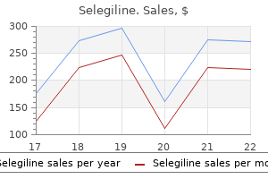
Cheap 5 mg selegiline fast delivery
Have the patient follow your hands as your encourage lateral tilting symptoms of strep throat purchase selegiline 5mg fast delivery, with no rotation or anterior/posterior tilting, of the pelvis. This can also be done in standing to facilitate dissociation of the pelvis/hip and lumbar spine. The principles for release are the same as those described for releasing the superficial fibers of multifidus; the response of the hypertonic point in the relevant muscle is monitored as the lumbar spine is positioned such that the hypertonic fascicle/muscle is shortened. Cues to release/relax are given, the response noted, and the muscle is subsequently taken into a full stretch. Passive segmental flexion and sideflexion of the lumbar spine should be improved and the release of these muscles now allows further assessment of the articular system of the lumbar spine (mobility of the zygapophyseal joints (Chapter 8)). The thorax is posteriorly tilted (extended), the pelvis is anteriorly tilted, and the lumbar spine is compressed between the two. Unilateral hypertonicity of these muscles can create a long multisegmental curve in the thorax and lumbar spine and an intrapelvic torsion. When combined with poor intrapelvic control, unlocking of the pelvis can occur (thorax-driven pelvic impairment, see Case report Julie G). This young woman habitually overactivates her erector spinae to position her thorax relative to her pelvis. Her abdominal wall is lengthened, her lumbar spine is shortened, and her thorax is posteriorly tilted. The resultant force vector has produced a rotoscoliosis in her thorax and lumbar spine and an intrapelvic torsion. Capture the cranial and caudal end of the hypertonic fascicle(s) with the heels of your hands and interlock your fingers. Ensure that your elbows are aligned and do not press any further anteriorly into the chest for the rest of the technique. Immediately follow this technique with a simultaneously come off the chest (arrows) (see second part of Video 10. Instruct the patient to let their chest soften into the ball/ pillow and slowly let the air leave their chest. Once maximum shortening/release is obtained, quickly adduct your shoulders, approximate your wrists, and come off the chest (see case report). With the patient sitting with their arms lightly crossed, palpate the cranial end of the hypertonic fascicle. Once the barrier is reached, instruct the patient to gently rotate either towards or away from you. Once the neural component has been released, lengthen the specific fascicle and/or the entire erector spinae manually (myofascial component) with a moderately strong stretch. If the technique has been successful, the thorax and lumbar spine will flex further and lateral bending to the opposite side will be improved. After the fascicles have been released stretch the myofascia in either (A) sidelying or (B) sitting. With one hand, palpate the intercostal spaces above and below the rib of interest and be sure to direct the needle towards the rib. For longissimus thoracis pars thoracis, insert the needle directly over the transverse process of the thoracic vertebra. Unless you are very confident of your surface anatomy, all needling in the thorax should be contained to between the transverse and spinous processes. More home practice and cues for restoring optimal diaphragmatic breathing and releasing the chest are described in Chapter 11. It is common to find this muscle hypertonic when it is used as part of a non-optimal strategy for transferring loads between the thorax and pelvis. It is often hypertonic in women with stress urinary incontinence (Chapter 6), although hypertonicity of this muscle is not exclusive to this group. This has significant implications for tasks requiring rotation of the thorax, as well as for function of the diaphragm and respiration. Remind the patient to use any cues previously found helpful to optimize self-release. Monitor the tender point in the external oblique and shorten the hypertonic fascicle by approximating the specific rib(s) towards the linea alba. Shorten the fascicle by approximating the associated rib obliquely towards the linea alba and contralateral side of the pelvis. Hold the fascicle in this shortened position and wait for the resultant dampening of tone to occur (positional release). Cue the patient by saying `let my fingers sink into your abdomen, let your rib cage relax and widen. Continue to use words to encourage relaxation/ release and monitor the fascicle for any recurrence of hypertonicity. The lengthening of this anterior oblique sling can be taken further to include the contralateral adductor. If the technique has been successful, the trunk (thorax and lumbar spine) will rotate further and the infrasternal angle will have widened. For releasing the chest unilaterally, have the patient in sidelying with the side to be released uppermost. Remind them to check their ability to rotate/translate their chest relative to the pelvis frequently during the day and to use their breath to release any acquired increase in resting tone. It is often useful to combine the specific cues for individual muscle release into an integrated cue for lengthening of the sling. For example, to lengthen the left anterior oblique sling from the foot to the rib cage, have the patient support the right lower extremity in a strap with the left hand. Then, while maintaining this relaxed state, have them gently reach the heel long without losing control of either the pelvis or the thorax. With the patient either (A) supine or (B) sidelying, use your hands to manually cue where they are to send their breath for home practice. In order for the chest to expand laterally, the brain must somehow release/relax the external oblique. Have them take the lower extremity further into the stretch, breathe into any tensed/tight spots, and repeat three to four times. The low horizontal fibers are released with an intramuscular technique, whereas the upper and middle fibers are released with a positional technique. In this home practice, the patient is using a yoga strap to support her right leg as she abducts the lengthened lower extremity to a position where a comfortable stretch is felt in the monitored muscle group, the adductors. This is an illustration of the myofascial stretch portion of this release technique. Approximate the medial end towards the iliac crest (towards its origin) and hold the fascicle in this shortened position. Wait for the resultant dampening of tone to occur and then add awareness by cuing the patient to `allow the belly to soften, to let my fingers sink into your abdominal wall. Once the neural component has been released, lengthen the fascicle manually (myofascial component). Recheck the response to verbal cue to isolate a contraction of transversus abdominis. If the technique has been successful, the optimal isolated response of transversus abdominis to a verbal cue is often, but not always, restored. Prior to training the deep system (Chapter 11), the patient should release the low horizontal fibers of the internal oblique (if hypertonic) by repeating this technique. Teach them how to find the hypertonic fascicle and how to feel for its release/relaxation through touch, fascicular shortening, and imagery. The middle and upper fibers of the internal oblique are often hypertonic and when they are dominant during an automatic task, such as a curl-up, the rib cage and the infrasternal angle tends to widen. These fibers tend to hold the lower ribs in posterior rotation and restrict expiration and contralateral rotation of the lower thorax. Palpate the hypertonic fascicle and shorten it by rotating/sideflexing the thorax. Hold this position and wait for the resultant dampening of tone to occur (positional release). Cue the patient to `let the muscle in your waist soften, let your pelvis roll backwards, let my fingers sink into your waist, let your ribs relax into my hand.
Selegiline 5mg amex
Lindqvist and associates in 1992127 and Westermark and coworkers in 2006137 found that in cases of condylar element replacement alone symptoms torn meniscus 5mg selegiline sale, there was severe erosion of the fossa with significant heterotopic bone formation. All such maneuvers can lead to component material fatigue or overload, promoting early failure with functional loading. More significantly, these manipulations can lead to "stock" component micromotion that will interfere with osseointegration. Micromotion leads to the formation of a fibrous connective tissue interface between the altered component and the host bone, resulting in early loosening of that component and potential early catastrophic or certain premature device failure in the future. In addition, there are patient-fitted or custom-made devices that are designed and manufactured for each specific anatomic situation, can conform to any unique anatomic situation, and require no alteration or supplementation to achieve initial implantation stability. Because the components interface precisely with the host bone, the screw fixation secures the components to the host bone, mitigating micromotion and maximizing the opportunity for osseointegration of the components and fixation screws. It was noted that both groups showed good skeletal and occlusal stability over the follow-up period. Returning the joint(s) and muscles of mastication to function as soon as possible postoperatively enhances healing and decreases the development of intra-articular scar tissue that will compromise mandibular range of motion. When calculating cost of a procedure or device, it is important that not only is the cost of the device considered but also all factors associated with its placement, including overall treatment time and patient morbidity. The quality of life in these patients remains poor and possible revision procedures may be necessary in the life-long management of this patient group. Considerations for the use of alloplastic temporomandibular joint replacement in the growing patient. On costochondral grafts replacing mandibular condyles in juvenile chronic arthritis: a clinical, histologic and experimental study. Cartilaginous growth centers transplanted to replace mandibular condyles in monkeys. Presented at the 67th Annual Scientific Session of the American Association of Oral and Maxillofacial Surgeons. Transactions of the Third Congress of the, International Association of Oral and Maxillofacial Surgeons; 1970; pp. Adaptation of autogenous costochondral grafts used for temporomandibular joint reconstruction. The costochondral graft: a solution or a source of facial asymmetry in growing children Growth of the mandible after replacement of the mandibular condyle: an experimental investigation in Macaca mulatta. Long-term radiological findings following reconstruction of the condyle with fibular free flaps. Costochondral graft/reconstruction of the condyle/ramus unit: Long-term follow-up. Costochondral grafts to replace mandibular condyles in juvenile chronic arthritis patients: long-term effects on facial growth. Wen-Ching K, Huang C-S, Chen Y-R: Temporomandibular joint reconstruction in children using costochondral grafts. The effect of a unilateral costochondral graft on the growth of the marmoset mandible. Interrelationship between size and tissue-separating potential of costochondral transplants. Simultaneous mandibular distraction and arthroplasty in a patient with temporomandibular joint ankylosis and mandibular hypoplasia. Correction of micrognathia attributable to ankylosis of the temporomandibular joint using a gradual distraction technique: case report. Intraoral mandibular distraction osteogenesis in facial asymmetry patients with unilateral temporomandibular joint ankylosis. Simultaneous mandibular and maxillary distraction in hemifacial microsomia in adults: Avoiding occlusion disasters. Experimental study of reconstruction of the temporomandibular joint using bone transport technique. Reconstruction of mandibular condyle by transport distraction osteogenesis: experimental study in Rhesus monkey. Abukawa H, Terai H, Hannouche D, et al Formation of a mandibular condyle in vitro by tissue engineering. Osseointegrated implants in the treatment of the edentulous jaw: experience from a ten year period. Referat Uber die Durch das Moderne Chirurgische Experiment Gewonnenen Positiven Resultate, Betreffenddie Naht 33. Initial vascularization and tissue differentiation are influenced by fixation stability. Effect of partial immobilization on reconstruction of ankylosis of the temporomandibular joint with autogenous costochondral graft. Abstracts of the Fourth Congress of the European Association for Maxillofacial Surgery; 1978; p. Reconstruction of the mandibular condyle using transport distraction osteogenesis. Management of severe maxillary deficiency in childhood and adolescence through distraction osteogenesis with an external, adjustable, rigid distraction device. Arthroplasty of the temporomandibular joint in children with interpositional tantalum foil. Immediate prosthesis following radical resection in advanced primary malignant neoplasm of the mandible. A new surgical procedure in bilateral reconstruction of condyles utilizing iliac crest bone grafts and creating new joints by means of non-electric metal: a preliminary report. The use of zirconium metal plate in arthroplasty of temporomandibular joint ankylosis. Prosthetic restoration of the left temporomandibular joint in a case of partial ankylosis. Transactions of the 2nd Congress of the International Society of Plastic Surgeons. Temporomandibular joint ankylosis corrected by creating a false stainless steel fossa. The correction of mandibular ankylosis by arthroplasty and insertion of a cast vitallium glenoid fossa. Mandibular joint arthrosis corrected by insertion of cast vitallium glenoid fossa prosthesis. Temporomandibular joint reconstruction with a custom total temporomandibular joint prosthesis: use in the multiply operated patient. Teflon and Silastic for mandibular replacement: experimental studies and reports of cases. Temporomandibular joint ankylosis corrected by creating a false Silastic sponge fossa. Use of a Silastic testicular implant in reconstruction of the temporomandibular joint of a 5-year-old child. Surgical implant replacement of the fractured displaced mandibular condyle: report of three cases. Development of alloplastic materials for temporomandibular joint prosthesis: a historical perspective with clinical illustrations. Use of a biocompatible interface for binding tissues and prosthesis in temporomandibular joint surgery. Experience with a polymer glenoid fossa prosthesis for partial or total temporomandibular joint reconstruction. Arthroplasty of the temporomandibular joint with the use of a vitallium condyle prosthesis: report of three cases. Condylar replacement alone is not sufficient for prosthetic reconstruction of the temporomandibular joint. Treatment outcomes for temporomandibular joint reconstruction after Proplast-Teflon implant failure. Temporomandibular joint reconstruction of the complex patient with the Techmedica custom-made total joint prosthesis. Subjective and objective outcomes in patients reconstructed with a custom-fitted alloplastic temporomandibular joint prosthesis. Outcomes of total alloplastic replacement with peri-articular autogenous fat grafting for management of re-ankylosis of the temporomandibular joint.
Purchase selegiline without a prescription
Designating focused training time (where the therapist provides 1:1 feedback and cuing for optimal performance of the task) thus consolidates new maps and builds precision and confidence medicine you can take during pregnancy selegiline 5 mg for sale, so that the patient can continue to use the new networks as they walk out of the treatment session and go about the rest of their day. Part of the process of creating new strategies for function is to address any deficits in the deep muscle system, and to use specific training tasks to start coordinating the deep and superficial muscles; this was covered in Chapter 11. The intent of this chapter is to further develop strategy and meaningful task analysis (advanced assessment) and to provide specific principles and techniques to train new postural and movement strategies for functional tasks that require integrated total body movement. It is essential to explain to the patient that the aim of their home program is to practice the building blocks and use the new neural networks, which are component skills necessary to learn a new strategy for function and 368 performance. Thus, these building blocks are concurrently incorporated into all activities of daily life, and most importantly into their meaningful tasks. This is quite different to having a routine of exercises that exists as a separate activity that is then forgotten during other activities. Successful training of new strategies for function and performance requires awareness and mindful practice, until the new strategies are fully integrated and become a part of the person in the middle of the puzzle. Before we discuss the specific techniques and examples of functional progressions, we need to consider some additional assessment tools. Advanced assessment Finding the driver for the whole body Any functional task, whether it be sustained postural positions or dynamic activities, requires integration of all regions of the body. This is an essential process that enables the therapist to determine how all areas of the body are linking and interacting with each other during total body function. Note that the primary driver(s) for the whole body strategy will relate to all of these problems. That is, treating the key driver(s) should positively impact all problems; if it does not, then there are other underlying impairments that are concurrent drivers that need to be addressed. As noted in Chapter 8, the strategy analysis tests previously described do not encompass all of the possible screening tests for automatic strategy analysis. Assess the level of commitment to the current strategy: (a) first in standing, and then in positions related to the meaningful task. Attempt to manually correct the alignment in the area of failed load transfer (use cues to release if needed), and determine which region of the body is more resistant to correction (pelvis/hips, thorax, foot). Continued needs further assessment to determine why; further assessment will direct what to release (Chapter 10) and then the task can be reassessed to determine how this area relates to the pain experience and total body strategy. However, as noted in Chapter 9 and illustrated by the case reports, non-optimal strategies relate to both mechanical and nonmechanical pain presentations, and treatment based on the Integrated Systems Model is effective for patients with pain driven by multiple mechanisms, in both acute and chronic states. This is because the approach addresses cognitive and emotional features of the meaningful task, as well as mechanical features, and how they relate to the person in the middle of the puzzle. Assessment of function and performance must include other measurable parameters such as speed and accuracy, as well as those that reflect quality of movement. When multiple joints/regions fail to transfer load optimally, it is imperative to find the primary driver (the one that has the biggest impact on the others) for the meaningful task being assessed. It is very common for all three regions to fail; the first one to do so is identified as the primary driver. Manual or verbal cues for correction are then applied to the primary driver and the impact of this correction on all joints being assessed is noted. In advanced task analysis and consideration of total body function 372 and performance, both objective biomechanical outcomes as well as the subjective and qualitative components of the total body strategy are evaluated. Patients are amazed by the ease they feel in their body, and the increased energy and reduction in fatigue levels they experience. Whole body meaningful task analysis Once a hypothesis is made about where the driver is, the next step is to test this hypothesis in a task that most closely resembles the meaningful task. Depending on the complexity of the task, this step may be as simple as adding load to the whole body screening task already assessed (for example as would be case for the). Firstly, if you have truly identified the driver, correcting the driver will positively impact the strategy used for the aggravating and goal-related tasks, providing further confirmation and support for your hypothesis. Correcting the driver should also positively impact all features of a multisystem presentation; for example,if a patient with pelvic girdle pain has coexisting complaints of stress urinary incontinence and/or difficulty breathing in certain tasks, correcting the driver should improve these symptoms, or improve impairments related to these symptoms as well. If correcting the driver makes these symptoms worse, this suggests that you are correcting a compensatory component and that the driver may lie in one of the other systems. Thus, meaningful task analysis provides further information for testing your hypothesis (confirmation or rejection, Chapter 9). It helps patients understand how their experience of the problem 373 the Pelvic Girdle and your evaluation of the driving cause for the problem are related. However, when gait is more closely simulated (allowing thoracopelvic rotation, and movement through the hip, knee, and foot for the whole stance phase from heel strike to toe off), the more differences there are in the biomechanical requirements of the two tasks (step forward versus gait). Combining resisted arm movements with relevant postures provides specific information about the level and direction of the poorly controlled segment. For example, a performer from the Cirque du Soleil reported that his pain was provoked only when he repetitively moved his legs over his body in multiple directions during a sustained onearm handstand. In standing, an area of loss of control was noted during lateral bending of the trunk, but no pain 374. The direction of the load can be varied (flexion, extension, rotation) and the segmental response noted. This test is indicated if the patient reports difficulty with arm loading in the seated position (narrative reasoning and hypothesis development). However, the same control deficit was amplified, and more obvious, when he performed the aggravating task, lateral bending of his lower body while sustaining a onearm handstand. For example, if a runner does not experience problems until 45 minutes into a run, the primary driver may be easier to observe if you assess the patient soon after they have done a 45-minute run to compare their strategy from a rested start point and at a more fatigued point. If the meaning perspective (beliefs, expectations, motivations, attitudes; see Chapter 9) or emotional context is a significant contributor to the non-optimal strategy, it is essential to try to replicate these dimensions in meaningful task analysis. Again, this is a situation where meaningful task analysis provides key information and should be performed in the first one or two appointments. Finally, assessing strategies during tasks that closely simulate meaningful tasks provides a basis for designing a treatment program tailored specifically to the patient. Although is it usually not possible to simulate every aspect (biomechanical, environmental, social, emotional) of the goal-related functional task, a creative approach to this challenge can often simulate the key aspects. The more specific the information elicited from the patient about aggravating activities and tasks that they have difficulty performing, the easier it will be to identify which features are key to replicate. Asking these questions often leads the patient to reflect on, and realize, subtleties related to context or environment that are relevant. This patient reported that in her yoga practice performing the triangle pose was more difficult to the left (not painful). Sometimes, patients cannot identify consistent features related to mechanical stressors that aggravate their pain. For example, a patient may report pain with sitting at work, but note that `some days it is fine, other days it is not. Appropriate space for movement, as well as equipment such as treadmills, wind trainers. Visualization and imagery can be used to simulate different environments and contexts. For example, an elite mountain bike racer presented with low back pain only experienced during races. This mountain biker (who also runs) complained of persistent low back pain that was aggravated by cycling and not running. To determine the primary driver for this whole body task, several areas require assessment of the strategy he uses during his meaningful task, cycling on his bike. Here, the therapist is monitoring his (A) hips, (B) thorax, and (C) knee and foot, specifically paying attention to regional/segmental position, mobility, and control as well as activation of the superficial muscle systems for synergy. Screening tests of simple tasks revealed his areas of failed load transfer and a hypothesis of the source of his problem was made.
Selegiline 5 mg overnight delivery
Table 9: Glucagon Doses Product Glucagon GlucaGen Body weight (kg) <20 20 <25 25 Dose (mg) 0 97140 treatment code order selegiline 5 mg without prescription. In fact, the best predictor of whether or not someone will experience this is a high incidence of antecedent hypoglycemia. Hypoglycemia awareness training can also improve patient recognition of early manifestations of hypoglycemia and prevent episodes of severe hypoglycemia. Ask patients to note any results outside of the target range in their log and follow the previously developed action plan (see "Correction Insulin Doses"). Continuous glucose monitoring systems have been developed that can provide patients with a glucose value in real time as well as warn the patient or his or her caregiver when glucose levels are too high or too low. Such devices can be very helpful for patients with diabetes, but their considerable expense often necessitates preauthorization processes with health insurers. Stress to your patients that testing, record keeping, healthy eating, and exercise are for their benefit, not yours. Healthy living with diabetes is directly due to their success at diabetes self-management. A wide variety of patient education materials are available from the American Diabetes Association. Vial stoppers will maintain a sufficient seal for about 100 punctures, and once opened, insulin vials can be stored at room temperature for about a month. The dawn phenomenon may be a result of growth hormone and cortisol, insulin agonists, which begin to rise at 4:00 a. Thus, in the early morning, insulin levels must rise to keep blood glucose levels normal. B: During the day, the basal insulin level is a result of stress, illness, activity, and body weight. However, the increased insulin requirement is an individualized response, and therefore therapy must be tailored to each patient. Lilly manufactures a regular human insulin that is available by prescription only in a U-500 strength (500 units/ml), which is reserved for special circumstances, such as severe insulin resistance. Insulin-treated patients who are traveling to other countries should be aware that insulin obtained there may be a different strength than U-100. Regular insulin, insulin lispro, insulin aspart, insulin glulisine, insulin detemir, and insulin glargine preparations are clear. The use of an insulin pump requires considerable patient education and office staff support until mastered. Patients using a pump must be motivated and committed to meticulous self-management and monitoring. Also, patients with a strong dawn phenomenon or with severe hypoglycemia may have better control of these problems with pumps. Any patient starting insulin pump therapy requires education and support above and beyond the usual until he or she becomes skilled at pump use and troubleshooting. Consider referring patients for education sessions with a health care team experienced in pump therapy. Because interstitial fluid glucose correlates highly with blood glucose, this allows patients to evaluate their glucose values in real time. Rapid-acting insulins-lispro, aspart, and glulisine-are well suited for use in pumps. Requirement 50% less basal Rationale Anti-insulin hormones (growth hormone and cortisol) at lowest effectiveness Largest effects of antiinsulin hormones Basal dose is affected by stress, exercise, illness, etc. However, other patients have a different ratio or may have a 1:10 ratio except when eating certain foods. Insulin-resistant patients may require much more insulin per carbohydrate serving. Example 1: A patient with premeal glucose levels in the target range is experiencing consistently elevated postprandial blood glucose levels. Example 2: Same meal composition, different patient, within target premeal glucose level but a 2-h postprandial glucose level of 240 mg/dl. This gene was closely related to the previously described viral sarcoma (v-src) gene discovered by Francis Rous in 1911 [3]. However, while the v-src oncogene was a virus, the c-src gene was a normal cellular gene that became misregulated during cancer formation. This discovery highlighted the understanding of cancer as a disease in which normal cellular function goes wrong. These findings provided evidence that the presence of a single oncogene within a normal cell was not sufficient to cause tumor formation. These results indicated that at least one additional "hit" or alteration of the genome was necessary for the tumors to form. A group of genes that influence the development of cancer through a different mechanism of action was also discovered. This class of genes appeared to protect cells from the development of cancer and as a result was named tumor-suppressor genes. If these genes are inactivated, either through mutation or by other means, the risk of cancer development is increased. Cancer can be broadly described as the result of genetic and other changes that lead to alterations in normal cellular function. The effect of these alterations is unlimited growth and expansion that occurs both locally and at distant sites within the body. It is estimated that in 2010, 1,529,560 new cases of cancer will be diagnosed in the United States, with 562,340 deaths [1]. The majority of all cancers occur sporadically, and only 5% are hereditary, although enhanced risk for some cancers may be familial. A variety of causes can contribute to malignant transformation and the development of a tumor. These can include genetic changes that either actively contribute to the development of cancer when altered (oncogenes) or lead to cancer when their protective effect is removed (tumor suppressor genes). The development of cancer is also influenced by the normal cells surrounding the tumor cells, as the tumor cells interact with such "stromal" cells to create conditions that are favorable for their continued growth and survival. Once tumors reach a critical size, this interaction with the "tumor microenvironment" enables the tumors to coopt the process of normal blood vessel development resulting in the formation of new blood vessels to further feed the tumor in a process known as angiogenesis. The first findings that linked a genetic cause to cancer came with the discovery of cellular oncogenes. This experiment involved the formation of cellular hybrids of tumor cells with normal cells. To his surprise, the resulting cell fusions were nontumorigenic, meaning that the genes of the normal cells that were important to normal cell functioning and proliferation were dominant to those genes of the tumor cells. In 1971, Knudson [10] used statistical analysis to show that retinoblastoma is a cancer caused by two mutational events. This gene is a transcription factor that normally functions as a checkpoint to stop the progress of the cell cycle once it is activated by stress signals within the cell. However, when this gene is mutated, its function is altered, predisposing cells to erroneous division cycles and higher mutation rates [12]. As a consequence, the p53 gene has been shown to be mutated in over 50% of all human cancers, reflecting its important function in regulating the normal growth of cells. Individuals who harbor inherited mutations in the p53 gene have a much higher propensity for developing cancer during their lifetimes [13]. There are nongenetic causes of cancer including exposure to high or chronic levels of a chemical or physical carcinogen and chronic inflammation that can contribute to tumor formation. Additionally, heterocyclic amine mutagens, created by the cooking of red meat at high temperatures, have also been identified as causative agents in colon cancers. Another important nongenetic influence that can lead to the formation of tumors is chronic inflammation, caused either by exposure to something in the environment or by an infectious agent. Cancers of hematopoetic origin include cancers of the blood cells and are known as leukemias, while the cancers of the lymphatic system are known as lymphomas.
5 mg selegiline with visa
Early detection and analysis of tumor characteristics are vital in achieving disease control medications 512 selegiline 5 mg on line. In locally advanced breast cancer patients, the current standard of care often involves neoadjuvant chemotherapy [27,28]. The presence of disease in four or more axillary lymph nodes prompts the addition of a third radiation field to treat the high axilla and supraclavicular nodes. Finally, the use of antiangiogenic agents in treating breast cancer is currently being investigated. One caveat is that it is difficult to predict disease activity in patients with mucinous histology; measurement errors were caused by the presence of both mucinous tumors and rectal wall fibrosis. As dose levels are increased, the use of new imaging methods to more accurately target the prostate and the accuracy of dose delivery become crucial. This, in theory, could provide tumor dose escalation while simultaneously lowering toxicity. In fact, in follow-up, one patient has shown marked improvement from baseline pretreatment symptoms of urinary frequency. However, two substantial hurdles remain prior to wide implementation of this approach. The second is the widespread use of image-fusion capabilities in radiation facilities. It is well established that cervical tumors are characteristically hypoxic, which renders them more aggressive and more likely to metastasize [65,66]. Additionally, hypoxia also increases the resistance of tumors to radiation therapy [67], and an increased frequency for local failure and lower overall survival is seen in patients with highly hypoxic tumors [68,69]. It is thus important that a noninvasive technique to assess the extent of hypoxia in cervical tumors is established. Additionally, an improved local control was observed in patients with tumors exhibiting high signal intensity [71]. It was hypothesized that this high signal intensity is due to greater blood perfusion, which results in increased oxygenation and subsequent radiosensitivity. It was found that the fraction of radiobiologically hypoxic cells inversely correlated with Ktrans. It also has been proven that tumor microenvironment and hemodynamics play a major role in how neoplasms can be imaged and followed during the course of therapy. Integration of preoperative anatomic and metabolic physiologic imaging of newly diagnosed glioma. Vandecaveye V, De Keyzer F, Verslype C, Op de Beeck K, Komuta M, Topal B, Roebben I, Bielen D, Roskams T, Nevens F, Dymarkowski S. Vandecaveye V, De Keyzer F, Vander Poorten V, Dirix P, Verbeken E, Nuyts S, Hermans R. The contribution of 18F-fluoro-2-deoxy-glucose positron emission tomographic imaging to radiotherapy planning in lung cancer. Nomori H, Shibata H, Uno K, Iyama K, Honda Y, Nakashima R, Sakaguchi K, Goya T, Takanami I, Koizumi K, Suzuki T, Kaji M, Horio H. Choice of technique, image interpretation, diagnostic accuracy, and transfer to clinical practice. Dynamic magnetic resonance imaging in determining histopathological prognostic factors of invasive breast cancers. Montemurro F, Martincich L, Sarotto I, Bertotto I, Ponzone R, Cellini L, Redana S, Sismondi P, Aglietta M, Regge D. Preoperative chemotherapy: updates of National Surgical Adjuvant Breast and Bowel Project protocols B-18 and B-27. Sequential preoperative or postoperative docetaxel added to preoperative doxorubicin plus cyclophosphamide for operable breast cancer: National Surgical Adjuvant Breast and Bowel Project protocol B-27. A prospective randomized pilot study to evaluate predictors of response in serial core biopsies to single agent neoadjuvant doxorubicin or paclitaxel for patients with locally advanced breast cancer. Neoadjuvant chemotherapy for breast carcinoma: multidisciplinary considerations of benefits and risks. Chakravarthy A, Nicholson B, Kelley M, Beauchamp D, Johnson D, Frexes-Steed M, Simpson J, Shyr Y, Pietenpol J. Functional magnetic resonance imaging in oncology for diagnosis and therapy monitoring. Early changes in functional dynamic magnetic resonance imaging predict for pathologic response to neoadjuvant chemotherapy in primary breast cancer. Relation of number of positive axillary nodes to the prognosis of patients with primary breast cancer. Predicting nodal status using dynamic contrast-enhanced magnetic resonance imaging in patients with locally advanced breast cancer undergoing neoadjuvant chemotherapy with and without sequential trastuzumab. Clinical and molecular prognostic factors in sphincter-preserving surgery for rectal cancer. Distal rectal cancer: sphincter-sparing is also a challenge for the radiation oncologist. Operative versus nonoperative treatment for stage 0 distal rectal cancer following chemoradiation therapy: long-term results. Prognostic implications of response to preoperative infusional chemoradiation in locally advanced rectal cancer. Tumor downstaging and sphincter preservation with preoperative chemoradiation in locally advanced rectal cancer: the M. Endorectal ultrasonography versus phased-array magnetic resonance imaging for preoperative staging of rectal cancer. Dose escalation with 3D conformal treatment: five year outcomes, treatment optimi- 57. Dose escalation with three-dimensional conformal radiation therapy affects the outcome in prostate cancer. Prostate cancer treatment with radiotherapy: maturing methods that minimize morbidity. Debois M, Oyen R, Maes F, Verswijvel G, Gatti G, Bosmans H, Feron M, Bellon E, Kutcher G, Van Poppel H, Vanuytsel L. The contribution of magnetic resonance imaging to the three-dimensional treatment planning of localized prostate cancer. Static field intensity modulation to treat a dominant intra-prostatic lesion to 90 Gy compared to seven field 3-dimensional radiotherapy. Tumour hypoxia and vascular density as predictors of metastasis in squamous cell carcinoma of the uterine cervix. Tumor size and oxygenation are independent predictors of nodal diseases in patients with cervix cancer. Oxygenation predicts radiation response and survival in patients with cervix cancer. The prognostic value of pimonidazole and tumour pO2 in human cervix carcinomas after radiation therapy: a prospective international multi-center study. Yamashita Y, Baba T, Baba Y, Nishimura R, Ikeda S, Takahashi M, Ohtake H, Okamura H. Angiogenic activity of cervical carcinoma: assessment by functional magnetic resonance imaging-based parameters and a histomorphological approach in correlation with disease outcome. Tumour oxygenation levels correlate with dynamic contrast-enhanced magnetic resonance imaging parameters in carcinoma of the cervix. Assessment of hypoxia in human cervical carcinoma xenografts by dynamic contrastenhanced magnetic resonance imaging. Positron emission tomography for target volume definition in the treatment of non-small cell lung cancer. As there is a substantial leap from in vitro to in vivo testing, as the experimental conditions change drastically between test tubes and a living organism, we will restrict the discussion to those agents with demonstrated in vivo applications. Solid tumors are composed of cancer cells that divide at an accelerated pace and that do not (typically) naturally undergo apoptosis. This is a tremendous advantage over neighboring normal cells, as cancer cells are able to proliferate more efficiently, eventually outgrowing, invading, and even replacing normal tissues. These processes require energy, which is obtained from nutrients in the blood stream. Those nutrients are available to the first few layers of cells around a blood vessel, which would in principle limit the development of tumors. However, cancer cells have the ability to recruit new blood vessels (neovasculature) that allow them to develop further [2].
Zhi Gan Cao (Licorice). Selegiline.
- Are there any interactions with medications?
- Are there safety concerns?
- Upset stomach (dyspepsia), when a combination of licorice and several other herbs is used.
- What is Licorice?
- How does Licorice work?
Source: http://www.rxlist.com/script/main/art.asp?articlekey=96849
Cheap 5 mg selegiline free shipping
The otic medicine valley high school generic selegiline 5mg visa, optic, and olfactory placodes develop in association with the forebrain neuroectoderm. Longitudinal section of amnion in a 23-day-old embryo shows fusion of the neural folds and initial formation of somites. The Cranial Neural Crest the neural crest is a multipotential cell population derived from the lateral edges of the neural plate during the fourth week of embryonic life. It was first described in 1868 as a band of cells between the surface epithelium and the neural tube in chicken embryos that appears at the dorsolateral edge of the closing neural folds along nearly the entire length of vertebrate neuraxis. Each pharyngeal arch is delineated by discrete swellings that are produced by the proliferative activity of these crest cells. These ectodermally derived cells contribute extensively to the formation of mesenchymal structures in the head and neck as they migrate. All of the skeletal and connective tissue of the face, with the exception of dental enamel, is derived from neural crest cells, whereas skeletal and connective tissue of the trunk is mesodermal in origin. Rather, these progenitor cells must be instructed by signals from other tissues to generate craniofacial skeletal elements of appropriate shape and size. Tissues that provide the instructive signaling for cranial neural crest fate specification include, but are not limited to , the isthmic organizer behind the midbrain region, cranial placode, and the pharyngeal arch ectoderm and endoderm. Fortunately, a two-component genetic system for indelibly marking the progeny of neural crest cells has significantly improved our ability to analyze the fate of the cranial neural crest during normal as well as abnormal craniofacial embryogenesis. Early facial development is remarkably similar between different vertebrate species. In mice, cranial neural crest cells start to migrate soon after gastrulation, and the entire delamination takes place between the stages of 3 to 16 somites. Lateral view of a 31-day-old embryo shows somites along the back and development of the pharyngeal arches and limb buds. These facial prominences, each of whose outgrowth is composed of different neural crest cells and regulated by different genes, are covered by a thin epithelium derived from the ectoderm. Frontal view of the developing face in 4-, 5-, 6-, and 7-week-old embryos shows merging of the facial prominences (primordia or processes). The medial nasal prominences grow further, moving toward each other and eventually merge in the midline (globular prominence) early in the sixth week to form the central part of the upper lip and the primary palate. There still is some controversy regarding the origin of the central part of the upper lip, which some believe is of frontonasal prominence origin. At this same time, the maxillary and mandibular prominences merge laterally, determining the width of the mouth. The groove between the prominences is gradually filled out by proliferation of the mesenchyme so that the prominences appear to merge. Facial clefting is a result of failure of epithelial disintegration and lack of merging. The nasal placodes that formed at about 5 weeks each are separated inferiorly by a nasal groove. With continued proliferation of mesenchyme, the placodes submerge to form the nasal pits, the precursors to the anterior nares. As the nasal pits continue to submerge with the proliferating mesenchyme, they are eventually separated from the stoma- todeum by only a thin oronasal membrane. This membrane will rupture at the beginning of the seventh week, forming a continuous nasal and oral cavity. Genetic Analysis of Craniofacial Development Our fundamental understanding of normal and abnormal human craniofacial development has been enormously advanced by gene targeting in mice in which more than 90 knockout mutants have been created to date. Although this large number reflects both the genetic and the morphogenetic complexity of head development, many of these malformations cluster into a few groups that can be characterized by the age of onset and the pattern of malformations observed in humans. The following is a summary of what is known at present of the genetics of the major human craniofacial malformations gained from homozygous knockout mouse studies. A number of these genes are expressed in the anterior visceral endoderm, which may act as an independent head organizer during early mammalian development. An unusual case is a homozygous knockout of Hoxa2 (normally expressed in the second pharyngeal arch), which produces a homeotic respecification into first archlike structures. Clefts of the secondary palate frequently occur with this group, as an outcome of disturbed facial structural anatomic relationships. The cartilaginous neurocranium (chondrocranium) that will form the cranial base is cartilage from neural crest origin. The membranous viscerocranium that will give rise to the maxilla, zygomatic bone, squamous temporal bone, and mandible is derived from the neural crest. The cartilaginous viscerocranium that will form the middle ear ossicles, styloid process of the temporal bone, hyoid bone, and laryngeal cartilages is from neural crest ectoderm. Endochondral ossification centers occur in the cartilaginous components and intramembranous ossification sites form in the membranous components of the neurocranium and viscerocranium. Osteoblast differentiation with the onset of mineralization results from a rapid angiogenic process with vascular ingrowth closely surrounding the centers and sites of ossification. The earliest ossification of the craniofacial bones begins in the seventh and eighth weeks of gestation. Lateral view of a 20-week-old embryo illustrates initial development of the cartilaginous and membranous neurocranium and viscerocranium. Ossification the onset of ossification generally follows the chronologic sequence of mandible, maxilla, palatine, cranial base, and cranium, with intramembranous sites usually preceding endochondral centers. Endochondral ossification of the mandibular condylar cartilage does not begin until 15 weeks. Maxillary intramembranous ossification begins in the area of the infraorbital foramen lateral to the nasal capsule. The vertical portion of the palatine bone then begins intramembranous ossification in the region of the palatine nerve, followed by ossification of the anterior, then the posterior borders of the incisive foramen, spreading through the hard palate from the canine area. Following ossification of the main portion of the mandible and maxilla, during the sixth week of gestation, endochondral ossification of the cranial base occurs in the midline from the foramen magnum to the nasal bone, and intramembranous ossification occurs laterally. Apposition of bone on the alveolar margins of the maxilla and mandible in the presence of developing tooth germs forms the initial alveolar processes. As this occurs, the palatal shelves become elongated and elevate medially toward each other, beginning fusion at the end of the eighth week and completing in the ninth week of gestation. Frontal view of face, coronal section of stomodeum, and inferior view of palate in 7- and 12-week-old embryos. The first is a small "midgrowth" spurt that occurs in many children at 6 to 8 years old that has been attributed to increased adrenal secretion of androgenic hormones. The second is a dramatic endocrine-mediated "pubertal growth" spurt during adolescence. Growth of the craniofacial complex during the fetal period is characterized by a constant rate during the second trimester. At the start of the fetal period, the head is half the length of the fetus, and by birth, it is one quarter the length. This results in an early proportional predominance of the neurocranium over the face that only reduces to an 8:1 proportion by birth. The formation and maintenance of cranial sutures are regulated by tissue interactions with the underlying dura mater as the brain develops. The frontal bones are derived from neural crest, whereas the parietal and occipital bones are of mesoderm origin. The metopic suture is the only human cranial suture not formed between neural crest and mesoderm, being entirely of neural crest origin. The cranial base growth parallels the rapid growth of the cranial vault during the fetal period. The anterior cranial base grows sevenfold whereas the posterior cranial base increases fivefold. The ossification centers that begin the formation of the facial bones late in the embryonic period at 6 to 8 weeks enlarge during the early fetal period until most of the bones have developed into a definitive shape by 14 weeks.
Discount generic selegiline uk
This period medicine you can give dogs order selegiline online pills, known as the "dwell time" is equal to the inverse of the readout bandwidth. Many techniques have been developed to increase the speed of acquisition or to achieve a particular result using nonrectangular strategies for filling k-space. This section will discuss two ways to fill k-space using rectangular sampling strategies. Once k-space is filled, the 2D Fourier transform reconstructs grayscale images of the distribution of spins in the imaged slice. It can be shown that the pixel size in the x direction is inversely proportional to two times k xmax. The goal of the imaging process is to use the chosen pulse sequence to fill this space as efficiently as possible. However, in order to sample the signal that evolves, the readout gradient, Gx, is turned on prior to the maximum amplitude of the echo signal. This approach that fills k-space from the outer edge at maximum Gy to the opposite edge at minimum Gy is known as a "top-to-bottom sequential" filling scheme [5]. There are, however, numerous other ways to use the phase-encoding gradients to acquire the necessary ky data to fill k-space in a different order simply by controlling the amplitude of the phase-encoding gradient at a fixed turn-on time [5]. Because the echo is formed by a gradient reversal, the initial polarity of the gradient can be either negative or positive, followed by the reversal and readout of the signal. Starting at t1 when the first readout gradient prephasing pulse begins, the trajectory moves from t1 to t2 as shown in the bottom of the figure. Depending on the phase-encoding gradient strength, the readout of the signal begins at t3 and proceeds through k-space from -k xmax to +k xmax at t4, when the readout of the signal is complete. This phase-stepping process with alternating readout directions continues through the number of phase-encoding steps specified by the pulse sequence until k-space is fully covered. The spiral sequence has the advantage that it does not so sparsely sample the lower spatial frequency areas of k-space as happens with radial pulse sequences where there can be a loss of low frequency information. Other approaches can involve incomplete filling of k-space in which zero-padding fills unsampled parts of k-space with zeros to decrease the acquisition time for the sequence. Other methods incorporate acquisitions that use the property of symmetry for the spatial frequency data to omit collection of a complete set of k-space data. In this design, slightly more than half of k-space plus data at zero spatial frequency are collected and the missing data synthesized from the collected data. The trade-off is that the signal-to-noise level is inferior to a full k-space acquisition. These and other approaches have been developed to move beyond the simple pulse sequence methods [5]. The most common method used now is to apply the 2D or 3D Fourier transform to the 2D or 3D k-space data that have been acquired in a rectilinear fashion to reconstruct the image information. However, other methods of reconstruction can be used to address particular ways in which k-space has been filled. One example is the use of projection reconstruction techniques similar to those used in computed tomography when the k-space data appear as a series of radial lines in k-space. All of these methods are of interest, but most reconstruction takes place using the Fourier transform. In some cases an interpolation process is used when the acquisition is not rectilinear to create a rectilinear mapping of k-space from the original nonrectilinear data. However the reconstruction process is performed, the key information is that the reconstruction algorithm should be appropriate for the way the data were originally placed into k-space. Other ways of filling k-space, such as the radial path described earlier can use a filtered back projection method for reconstruction, if some interpolation is performed to place the data in rectangular form. The procedure for implementing the reconstruction requires that there be some preparations to the data before the actual transformation step. This extended padding can enhance the resolution of the resulting reconstructed image by providing a smoothing of the data in the image due to a reduction in the partial volume effect [5,9]. Another processing step that is also performed is using a windowing function to control the presence of ringing and data overshoot artifacts due to truncation of the k-space data. It is equivalent to multiplying the k-space data by a rectangular function, which leads to a characteristic artifact because of the sharp transition from data at the edge of k-space to zero. These effects can be reduced using a nonrectangular window function, a filter that reduces the higher spatial frequencies in some sort of smooth fashion at the edges of the defined k-space data. The process is called apodization and can use a variety of window functions to reduce the Gibbs artifact at the expense of some loss of resolution [5]. In many cases, rectangular fields of view are used to shorten the acquisition time by reducing phase-encoding steps. A zero-filling process makes up for fewer phase-encoding steps followed by an interpolation in image space to achieve a square matrix. In some systems it is necessary to accommodate the fact that the gradients that are supposed to be linear over their range are not, leading to some geometric and intensity changes that have to be corrected. Additionally, there often are some scaling processes so that the images that are stored fall within the range of the display system. Each of these multiple coils is coupled to a separate receiver so that the excitation and reception processes are separated in what is termed a "phased array. This technique has been known for some time when it was introduced as a way to improve the coverage of large areas of anatomy while improving the signal-to-noise ratios over that of a large single element coil [11]. The resulting images can be assembled using a square-root-of-the-sum-of-the-squares formulation to create magnitude images from the multiple coils. There are two basic approaches that have been developed for using coil arrays to improve acquisition speed. The idea of using multiple coils to increase the speed of acquisition is to take fewer lines of k-space in the phase-encoding direction by increasing the distance between the phase-encoding lines. This distance is sometimes given the name R to indicate a "reduction factor" or "acceleration factor" [5]. The reconstruction process takes these aliased images and prescan data from the coil sections to reconstruct the correct image. The prescan data come from a 3D low resolution acquisition, which can be run very quickly, that provides the spatial sensitivity maps for all of the coils with the patient in place. If the index k goes from 1 to Nc, where Nc refers to the number of coil elements in the array, one can rewrite Equation 4. Since this is a (linear formulation with the linear weights, nkm), as the unknowns, a linear least-squares fitting approach can be used to fit the coil sensitivity profiles, Ck, to the mth order spatial harmonic repreimk sented by e y. The 1D signal, Sk(ky), from the phase-encoding direction in a coil k can be shown to be the Fourier transform of the spin density with coil sensitivity profile weighting: for l = 1 to R, which gives the locations of the pixels. C is the coil sensitivity matrix for each coil at the R superimposed positions with dimensions of number of coils times R. The ability to perform this inversion is based in part on there being fewer R pixels than the number of coils in the array. An added consideration is the ease with which the inversion can be performed, which is based on the geometry of the coil array in use during the acquisition. This factor, called the geometry factor, or g-factor, is a measure of the difficulty of inverting the matrix in Equation 4. The general idea is to approximate the complex exponential functions that would have been present in the missing phase-encoding lines of k-space using linear combinations of coil sensitivity information. Extra calibration lines acquired during data acquisition from each coil are used as a reference to determine what combination of adjacent lines in data from each coil most closely approximates the calibration line in each single coil. In this approach, a series of coefficients are developed that can be used to generate the missing k-space lines in data from each coil. Once this is accomplished, each full k-space data set for each coil of the array can be Fourier-transformed into image space where all of the coil images are combined. However, both k-space and image space implementations of parallel acquisition technology have been highly successful, with continuing development of systems having larger numbers of parallel channels demonstrating concomitant increases in imaging performance.
Order selegiline overnight
Of critical importance is the number of sites for water to access the inner sphere of the ion hb treatment discount selegiline 5mg amex, but in general, the stability of the complex and the number of sites are inversely related. The most common approach to prepare macromolecular structures containing Gd3+ chelates involves conjugation of the functionalized chelate to one of three macromolecules: linear polymers; dendrimers; biological molecules. Linear polymers are large molecules composed of a repeated structural unit, typically connected by covalent chemical bonds to form a single backbone with no branches. Dendrimers are large molecules composed of a repeated structural unit (dendron), typically symmetric around a core and often possessing a spherical 3D morphology. Biological molecules are any organic molecule produced by a living organism, including, but not limited to , large polymeric molecules such as nucleic acids, proteins, and polysaccharides. For example, conjugation of Gd3+ complexes to polymeric material tend to increase the rotational correlation time and may somewhat improve the relaxivity of the gadolinium atom. Furthermore, macromolecules can be decorated with multiple Gd3+ complexes, facilitating significant relaxivity enhancement that tends to be essentially additive on a per gadolinium basis. If the macromolecule is a tissue-specific targeting moiety, such as a monoclonal antibody, the conjugate may also have sufficient relaxivity to enable imaging of relatively low-concentration receptors. By virtue of molecular size, high molecular weight conjugates tend to be maintained in circulation and vascular spaces for longer times than monomeric species; therefore, and without surprise, most macromolecular agents tend to be useful for blood pool imaging. Lower than predicted relaxivities appear to stem from the flexible nature of the linear polymeric backbone [30,31]. It has since been proposed that select linear polymers can form intramolecular aggregates, making their structure less rodlike and more globular, thereby increasing the rotational correlation time [3]. Dextran affords the advantages of high solubility, limited polydispersity, and robust chemistry with respect to the variability of Gd-chelate linking agents; however, degradation in vivo can be a concern if not properly functionalized. Dendrimers are prepared by the repeated reaction of the initial, smaller "core" molecules [3]. This highly branched structure has led to a number of interesting molecular attributes for this class of macromolecule [34]. These values tend to be higher than those observed for linear analogues because linear polymers based upon polyaminocarboxylate derivatives are known to possess relaxivities limited by slow water exchange, and dendrimers are intrinsically more rigid than their linear counterparts [3,37]. Of these, Gd3+-based complexes aimed at pH sensitivity have been among the best characterized and can be prepared in a number of ways. Alternatively, the Sherry group has shown pH-dependent relaxivity by designing agents that facilitate modulation of outer hydration spheres [51]. Contrast agents that enable tissue-based pH measurements will continue to be of particular interest, especially in light of the emerging importance of characterizing the pH gradients known to exist in diseased states such as cancer. Several prototypical compounds have been reported where portions of the Gd3+ complex serve as substrates for enzyme activity, and the resultant processing of the probe alters the relaxation efficiency. The most useful imaging probes demonstrate increased relaxivity upon enzymatic processing that is usually accomplished by improving water access to the paramagnetic center. An alternative approach was recently suggested where probes were prepared such that following specific enzymatic hydrolysis, the agent was able to bind serum proteins, resulting in an in situ macromolecular agent with increased relaxivity [52]. Much consideration has also been devoted toward the development of "smart" contrast agents aimed at reporting concentration gradients of physiologically important metal ions, including calcium [53], and more recently, copper [54]. As with pH-sensitive, Gd3+-based probes, these "smart" agents incorporate complexing ligands that alter their coordination environment in the presence of the metal of interest, which typically results in 10. Like dendrimers, the intravascular retention of such macromolecules makes them effective blood pool agents for magnetic resonance angiography [39]. There are important practical concerns with using simple albumin-based agents that have precluded their adoption in clinical applications. Aside from possible immunoreactivity of native albumin, these agents are not amenable to heat, limiting conjugation chemistry and sterilization potential. The binding is not permanent so that the agent resides intravascularly two to three hours before excretion [41]. Such an approach promises to provide excellent visualization of blood vessels for angiographic imaging [42]. As a scaffold for the attachment of various chelates, dextran has also been explored [43,44]. Dextran is a complex, branched glucan (polysaccharide composed of numerous glucose molecules) composed of chains of varying lengths ranging from 10 to 150 kDa. The in vivo utility of metal sensing "smart" agents has not been widely demonstrated, but theoretically these agents could be particularly useful in studies of signal transduction as well as fundamental study of many disease processes. Pursuant to targeted agents employing small molecules is the work of Manning et al. Density of the expressed target is very critical, especially since many are insufficiently expressed; thus, a significant challenge for future chemists is balancing the need for higher relaxivity with acceptable stability. Nevertheless, the goal of such research ultimately lies with clinical application. Effect of ligand basicity on the formation and dissociation equilibria and kinetics of Gd3+ complexes of macrocyclic polyamino carboxylates. Biodistribution of radiolabeled, formulated gadopentetate, gadoteridol, gadoterate, and gadodiamide in mice and rats. Ideally, optimization of structure begets optimization of behavior, whereby kinetic and thermodynamic stability, pharmacokinetics/ pharmacodynamics, increased relaxivity, and the subsequent cost of such agents will produce contrast agents that are tissue/organspecific, afford high contrast enhancement at low in vivo doses, and are applicable in the clinic. While this chapter has focused exclusively upon Gd3+-based, T1 contrast agents, there exist other contrast agents that utilize transition metals such as manganese (Teslascan), which also share in this future. Nephrogenic systemic fibrosis: A mysterious disease in patients with renal failure-Role of gadolinium-based contrast media in causation and the beneficial effect of intravenous sodium thiosulfate. Gadolinium is not the only trigger for nephrogenic systemic fibrosis: Insights from two cases and review of the recent literature. Preparation and characterization of paramagnetic polychelates and their protein conjugates. Gadolinium-based linear polymer with temperature-independent proton relaxivities: A unique interplay between the water exchange and rotational contributions. Dendrimerbased drug and imaging conjugates: Design considerations for nanomedical applications. Preparation and water relaxation properties of proteins labeled with paramagnetic metal chelates. Polymeric contrast agents for magnetic resonance imaging: Synthesis and characterization of gadolinium diethylonetriaminepentaacetic acid conjugated to polysaccharides. Influence of spacer arm length and conjugate molecular mass on the paramagnetic properties and some biological parameters. Enzyme-activated Gd(3+) magnetic resonance imaging contrast agents with a prominent 53. Preparation and characterization of dendrimer-based poly-metal chelates and monoclonal-antibodies conjugates. The last magnetic classification, which is the most important in the context of this discussion, is that of superparamagnetism. Superparamagnetic materials are prepared by reducing the overall size of a ferromagnetic or ferrimagnetic crystal until a single magnetic domain particle is formed. In this condition, the particle is composed of thousands of magnetically ordered metal ions. Here, magnetization of the particle represents a single, giant magnetic moment, essentially the sum of all individual magnetic moments. The beneficial characteristic of superparamagnetic materials is that they can achieve complete magnetization even when placed in a weak external magnetic field. Due to thermal agitation, however, these particles cannot retain any of the induced magnetic properties once the field is removed. From a biological perspective, superparamagnetic particles offer many advantages over their paramagnetic counterparts since they can enhance relaxation rates in specific organs, at significantly lower doses, and with no registered toxicity.
