

Discount fluvoxamine amex
Multiple kernel learning is used to fuse the results obtained at different grid sizes anxiety symptoms in teens discount 50 mg fluvoxamine with mastercard. The method assigned probabilities for each pixel in the image belong to a hemorrhage, and create a probability map. This probability map is processed to extract the pixels corresponding to hemorrhages. Blood vessel removal/detection is considered as a preliminary step for red lesion detection. Similarly, removal of the optic disc is a common initial step in many exudate detection techniques. There are three basic classes of exudate detection methods; algorithms based on morphology, thresholding, and region-growing, clustering-based algorithms, and algorithms based on pixel-level classification. They created a lesion probability map and based on successive classification levels, true bright lesions clusters were detected. In the last few years, methods based on deep learning have also shown a significant c progress in detecting the exudates. In [130], a deep learning-based method is proposed, which detects the exudates in the retinal image. In [134], a deep fully convolutional residual network is used to obtain exudate patches. The offline system can be either a conventional machine learning system or a deep learning system. Gold standard ground truth images available in the public datasets are used to verify the classification performance of such methods. In the conventional machine learning, the system is trained to extract a set of predefined features. The features used for the classification are the cumulative distribution functions of the instantaneous amplitude, the instantaneous frequency magnitude, and the relative frequency angle from multiple scales. They used morphological operations and texture analysis methods to detect features like the area of exudates and blood vessels, image contrast, etc. This method does not explicitly segment red lesion or exudates but learns to identify them using locally learned deep features. Then based on various properties and nature of these exudates, a decision is made on the input retinal image. Few methods have tried to overcome these issues with various preprocessing and postprocessing techniques [154, 160, 161, 165]. A score is assigned to each of these candidate regions based on two separate methods. The overfitting phenomenon, which is common in classification problems, due to the unbalanced training datasets, is dealt with by using a dropout layer [167]. The extracted features are then oversampled to alleviate the class-imbalance issue. They used a depth map of the optic disc, which was reconstructed based on the stereo disparity. The optic disc is located by applying p-tile thresholding [178], canny edge detection [179], and snakes algorithm. A method based on using a double threshold and Hough transform is discussed in [186]. In [187], a method based on texture information combined with information about higher-order spectra is proposed. In [191], a method using morphological operations and hybrid level-set method is discussed. The performances of the above-discussed methods are given in Table 9 for a quick comparison. The device rapidly gained popularity, but due to its steep learning curve, it remained useful only for hobbyists and traveling practitioners. The device used is also known as D-Eye ophthalmoscope [193], built to fit a variety of smartphones from Samsung (Samsung Electronics Co. The authors concluded that the technique enables nonclinical photographers to acquire optic nerve images at a standard that is sufficient for independent remote retinal image grading. The authors concluded that images taken with smartphone ophthalmoscope are of sufficiently high quality. The authors also noted that smartphone ophthalmoscopy performs better in cases where media opacity, such as cataract, is present. Designed for the 21st century, the device is sleek and is compatible with any smartphone for smartphone-based retinal photography. There is a need to build a complete screening tool to incorporate into various telemedicine and large-scale population screening programs in the future. A preprocessing stage analyzing the quality and content of the input retinal images, like the methods discussed in [201, 202] can help to further increase the reliability and robustness of the automatic screening systems. The methods used to detect various anatomical structures in the retinal image (discussed in Sections 4. This would largely help to reduce the workload of the specialist ophthalmologists and clinicians working in screening centers. This would also reduce the cost involved in the screening/diagnosing of such pathologies. The efficiency of such a process depends on the performance achieved by each step. Although significant advancement has been made in the field of retinal imaging technology, the combination, selection, and performance of efficient algorithms need further developments. False positive is not that serious compared to false negative diagnosis, which implies that improving sensitivity is very important. Tomany, Prevalence of age-related macular degeneration in the United States, Arch. Hartinger, die volkommen reflexfreie Zeiss-Nordenson-Nitzhautkammer, Acta Ophthalmol. Dobbin, Kodak engineer had revolutionary idea: the first digital camera, the Associated Press, 2005. Nieto, Retinal microvascular abnormalities and their relationship with hypertension, cardiovascular disease, and mortality, Surv. Klein, Retinal arteriolar diameters and elevated blood pressure: the Atherosclerosis Risk in Communities Study, Am. Abramoff, Comparative study of retinal vessel segmentation methods on a new publicly available database, Proc. Lei, Segmentation of retinal blood vessels using the radial projection and semi-supervised approach, Pattern Recogn. Barman, Delineation of blood vessels in pediatric retinal images using decision trees-based ensemble classification, Int. Loncaric, Retinal vessel segmentation using deep neural networks, in: 10th International Conference on Computer Vision Theory and Applications, vol. Guevara, Retinal vessel extraction using Lattice Neural Networks with dendritic processing, Comput. Padmagireesan, Comparison of curvelet generation 1 and curvelet generation 2 transforms for retinal image analysis, Int. Owen, An approach to localize the retinal blood vessels using bit planes and centerline detection, Comput. Shih, Retinal vessels segmentation based on level set and region growing, Pattern Recogn. Ells, Detection of the optic nerve head in fundus images of the retina with Gabor filters and phase portrait analysis, J.
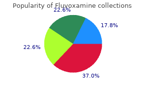
Generic fluvoxamine 100mg mastercard
Exaggeration of this physiologic jaundice raises the risk of bilirubin-induced neuropathy anxiety chat rooms buy fluvoxamine now. Serum bilirubin concentrations reach 10 mg/dL in about 16% of newborns, and exceed 15 mg/dL in 5% [165]. Jaundice of the newborn results from an increased bilirubin load and a lower capacity of the liver to dispose of bilirubin. Exaggeration of these factors and/or superimposition of additional mechanisms may result in a pathologic level of hyperbilirubinemia. Important risk factors for severe hyperbilirubinemia in babies born at 35 weeks or more gestation include lower gestational age, exclusive breast-feeding (particularly in the presence of excessive weight loss), clinical jaundice noted in the first 24 h, presence of hemolytic diseases. In addition, East Asian ethnicity and neonatal jaundice in a previous sibling were risk factors for severe hyperbilirubinemia [167]. Bilirubin production, as measured by carbon monoxide production, is increased in the newborn period [168]. The excess bilirubin is derived from erythroid and nonerythroid sources and from shortened erythrocyte half-life [169]. Rh incompatibility between mother and fetus used to be a common cause of severe neonatal jaundice before treatment of the mother with anti-Rh immunoglobulins became available [170]. Maturation of canalicular excretion processes may take longer than does the maturation of uptake and conjugation. Therefore, in the late newborn period, canalicular excretion becomes rate limiting in hepatic bilirubin disposition. In cases in which the bilirubin load is higher than normal, conjugated bilirubin accumulates in serum [182]. Because of the lack of an established intestinal microbiota in the newborn, there is reduced bacterial degradation of bilirubin, resulting in an increased absorption of unconjugated bilirubin [183]. A greater fraction of unconjugated bilirubin is absorbed from the intestine in infants fed maternal milk. Common causes of overproduction of bilirubin include hematologic conditions associated with hemolysis, such as hereditary spherocytosis, and toxic or idiosyncratic drug reactions. Ineffective erythropoiesis that occurs in thalassemia and vitamin B12 deficiency, and a rare group of disorders called dyserythropoietic anemias, result in unconjugated hyperbilirubinemia of various degrees [184]. In patients with hemolytic jaundice but normal liver function, a small amount of conjugated bilirubin produced in the liver may appear in the circulation [172,185], but the unconjugated:conjugated bilirubin ratio remains normal. In sickle cell anemia, a combination of hemolysis and liver abnormalities may lead to the accumulation of both unconjugated and conjugated bilirubin. Crigler and Najjar [186] described this rare, recessively inherited syndrome in 1952 in six infants from three unrelated families. Five of the six infants in the initial report died of bilirubin-induced encephalopathy within 15 months. The sixth patient remained free of brain damage until the age of 15 years, when kernicterus developed and death followed in 6 months [187]. Another related patient remained without brain damage until 18 years of age, but then developed bilirubin encephalopathy and died at the age of 24 years [188]. The original family described by Crigler and Najjar had a high degree of consanguinity. Several members of this family had other recessively inherited disorders, such as Morquio syndrome, homocystinuria, metachromatic leukodystrophy, and bird-headed dwarfism. In subsequently studied families, the other recessively inherited disorders were not observed. However, in many patients, residual neurologic abnormalities, a sequela to a previous episode of bilirubin encephalopathy, may be detected. However, many survivors develop kernicterus around puberty or in early adult life [188]. Use of phototherapy usually prolongs survival without encephalopathy to adolescence, and liver transplantation results in long-term survival (see later). Because of a relatively high concentration of unconjugated bilirubin in bile, pigment stones are common. The concentration of serum bilirubin increases during intercurrent illnesses and decreases on exposure to the sun or bright artificial light [189]. The bile is paler than normal [190] and contains only small amounts of unconjugated bilirubin [191]. There is no evidence of hemolysis [190], and bilirubin is produced at a normal rate [192]. Historically, liver histology has been reported as normal, except that, in several patients, bilirubin plugs were found to be deposited in bile canaliculi and bile ducts [186,191], probably resulting from biliary excretion of unconjugated bilirubin or its photoisomers. There was no evidence of portal hypertension or significant association with gallstones. The transplant recipients with liver fibrosis were older than those without, suggesting an accrual of fibrosis risk with age. In addition, many of these patients have reduction of the level of glucuronidation of phenolic substrates [196]. Such genetic lesions may consist of point mutations, deletions, or insertions within the coding region. The genetic lesions may result in mutation of a single critical amino acid or deletion of segments of the enzyme. A slash separating two mutations indicates that the patient carried two different mutations on the two alleles. The same strategy can be applied to perform prenatal diagnosis using chorionic villus samples as the starting material [216]. The Gunn rat is the only natural mutant animal model that develops bilirubin encephalopathy spontaneously [218]. Gunn rats have a single guanosine base deletion in the common region exon 4, which results in a frameshift, leading to a premature termination codon. These mice have higher serum bilirubin levels than do the Gunn rats, requiring therapeutic intervention to prevent mortality. New experimental therapies based on liver cell transplantation and gene therapy are currently being developed. Phototherapy is the most commonly used medical therapy for severe unconjugated hyperbilirubinemia [191]. In general, recommendation for phototherapy in neonates is based on age-related serum bilirubin concentrations. Beyond the age of 3 or 4 years, thickening of skin, pigmentation, and lower surface area in relation to body mass reduce the effect of phototherapy [191,225]. During neurologic emergencies, serum bilirubin concentration can be acutely reduced by plasmapheresis [191]. If serum total bilirubin concentrations exceed these levels by 5 mg/dL (85 M), exchange transfusion should be combined with intensive phototherapy because after removal of albumin-bound bilirubin from blood, bilirubin is mobilized from tissue stores to the plasma. Attempts to remove plasma bilirubin by affinity chromatography on albumin-conjugated agarose gel columns were hindered in patients because of the removal of formed elements of blood [226]. Although this procedure is associated with some risk and commits the patient to prolonged immunosuppression, it has been curative in several cases and has dramatically improved the outlook for these patients. Non-iron metalloporphyrins are strong inhibitors of microsomal heme oxygenase activity [19].
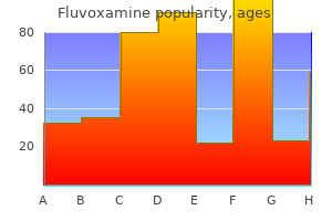
Best fluvoxamine 50 mg
The size of the aerosol particles has indeed a major influence on their behavioral properties anxiety symptoms rash trusted 100mg fluvoxamine, and the aerosol particle radius or diameter is a key property used to characterize aerosols. These larger droplets are primarily deposited on the surface beneath the nebulizer by gravitational settling and inertial impaction [57]. Moreover, it has been shown to work in environments highly saturated with humidity. Whereas endoscopic microcatheters are able to spray aqueous solutions [58], they cannot reliably aerosolize polymer-based formulations with higher viscosity. Aerosolizers based on microperforated membranes did not function reliably with complex solutions or in environments saturated with humidity [59]. Electrostatic precipitation devices have been certified for clearing the visual field from surgical smoke during laparoscopy [60] and the same technology can be used for precipitating therapeutic aerosols. After three procedures and concomitant chemotherapy, response or stable disease was achieved in approximately half of cases [63]. Pressurized intraperitoneal aerosol chemotherapy consists of six key breakthrough qualities: 1. Adequacy of drug distribution throughout the entire peritoneal cavity If anticancer drugs cannot reach all the cells within a tumor, their effectiveness is compromised. At the beginning, therapy intervals are 6 weeks; in the case of objective tumor regression this can be prolonged to 3 or even 6 months. Decrease in the outflow of drug from the tumor by capillary flow Advanced peritoneal cancer patients generally suffer gastrointestinal symptoms that deteriorate until death. Global quality of life improved and disease-related symptoms were stabilized for several months in the majority of patients. This increases the pharmacokinetic advantage of regional delivery and limits toxicity. These clinical data confirmed previous preclinical studies demonstrating that intraperitoneal administration of nano- and microsized formulations of paclitaxel resulted in superior antitumor activity against mouse ovarian cancer xenografts compared to intravenous administration [80]. Two dose-finding studies are currently being conducted to determine the optimal dosage of oxaliplatin [67,68]. Results of one of these studies have been presented and confirmed the arbitrary dosage of 92 mg/m2, significant toxicity having been observed at a dosage of 140 mg/m2 [67]. Some patients have experienced major or even complete histological response allowing a secondary complete resection. Comprehensive clinical reports on the use of these substances have been published [76] and reviewed [77]. This technology, first developed by Marie Dutreix from Institut Curie in Paris, might be interesting for potentializing the effect of radiotherapy and/or chemotherapy on peritoneal metastasis. A combination with systemic administration of angiogenesis inhibitors has been reported to be safe [92]. Healthy cells acquire a competitive advantage over cancer cells, which will continue dividing with 210 10. In silico modeling Knowledge of the peritoneal tissue, cancerrelated modifications of this tissue, pharmacodynamics and pharmacokinetics characteristics, and available clinical experience highlight current limitations of intraperitoneal chemotherapy. Drug uptake and distribution are indeed dependent on the substance to be administered. These multiple, interdependent parameters can barely be tested in bench experiments and simulations might facilitate and speed up this research considerably. In silico modeling offers the possibility to test different protocols or drug formulations for intraperitoneal delivery, might provide unique insights into the effect of modifying factors on peritoneal drug uptake, and will limit the need for in vivo experiments (reviewed in Ref. To be most effect, such simulations must be supported by bench data, for example, high-resolution optical imaging of tumor tissue from animal models. Such in vivo imaging of vascular perfusion will visualize the uptake of therapeutic agents, as well as their spatiotemporal distribution within tumors [93]. Following demonstrations of efficacy in the abdominal cavity, new intrathoracic applications are under development, paving the way for new treatments for mesothelioma [71], another rare form of cancer that commonly develops in the lining of the lungs. Epithelial-mesenchymal transition in human cancer: comprehensive reprogramming of metabolism, epigenetics, and differentiation. Hypoxia, cytokines and stromal recruitment: parallels between pathophysiology of encapsulating peritoneal sclerosis, endometriosis and peritoneal metastasis. Mesenchymal conversion of mesoo thelial cells is a key event in the pathophysiology of the peritoneum during peritoneal dialysis. Evolving treatment strategies and outcomes in advanced gastric cancer with peritoneal metastasis. Mucinous adenocarcinomas with intra-abdominal dissemination: a review of current therapy. Intraoperative incident dark field imaging of the human peritoneal microcirculation. Interstitial fluid pressure predicts survival in patients with cervix cancer independent of clinical prognostic factors and tumor oxygen measurements. Tumor interstitial fluid pressure may regulate angiogenicn factors in osteosarcoma. Description of a novel approach for intraperitoneal drug delivery and the related device. Rationale supporting the use of vasoconstrictors for intraperitoneal chemotherapy with platinum derivatives. Pleural macrophages differentially alter pleural mesothelial cell glycosaminoglycan production. High intra-abdominal pressure enhances the penetration and antitumor effect of intraperitoneal cisplatin on experimental peritoneal carcinomatosis. Effects of intra-abdominal pressure on pharmacokinetics andx tissue distribution of doxorubicin after intraperitoneal administration. High pressure enhances the effect of hyperthermia in intraperitoneal chemotherapy with oxaliplatin: an experimental study. Laparoscopic hyperthermic chemotherapy for palliation of debilitating malignant ascites. Pressurized Intra Peritoneal Aerosol Chemotherapy in patients suffering from peritoneal carcinomatosis of pancreatic adenocarcinoma. A phase I, single-arm, openlabel, dose escalation study of intraperitoneal cisplatin and doxorubicin in patients with recurrent ovarian cancer and peritoneal carcinomatosis. Alyami M, H bner M, Grass F, Bakrin N, Villeneuve L, u Laplace N, Passot G, Glehen O, Kepenekian V. Pressurised intraperitoneal aerosol chemotherapy: rationale, evidence, and potential indications. Van De Sande L, Graversen M, Hubner M, Pocard M, Reymond M, Vaira M, Cosyns S, Willaert W, Ceelen W. Electrostatic precipitation is a novel way of maintaining visual field clarity during laparoscopic surgery: a prospective double-blind randomized controlled pilot study. Pressurized intraperitoneal aerosol chemotherapy with oxaliplatin in colorectal peritoneal metastasis. Complete cytoreductive surgery plus intraperitoneal chemohyperthermia with oxaliplatin for peritoneal carcinomatosis of colorectal origin. Effects of carrier on disposition and antitumor activity of intraperitoneal paclitaxel. High penetration of paclitaxel in abdominal wall of rabbits after hyperthermic intraperitoneal administration of nab-paclitaxel compared to standard paclitaxel formulation. Therapeutic approach of human peritoneal carcinomatosis with Dbait in combination with capnoperitoneum: proof of concept. Pressurized intraluminal aerosol chemotherapy with Dbait in the distal esophagus of swine. Intraperitoneal chemotherapy of peritoneal carcinomatosis using pressurized aerosol as an alternative to liquid solution: first evidence for efficacy. Bevacizumab increases the risk of gastrointestinal perforation in cancer patients: a meta-analysis with a focus on different subgroups.
Buy fluvoxamine 50mg cheap
Unfortunately anxiety 5 steps purchase fluvoxamine 100mg without a prescription, because tests of liver function often remain relatively normal despite severe vascular disease in the liver, patients have difficulty gaining access to the transplant list. Testing the stool for occult blood is useless because most patients swallow blood from bouts of epistaxis, which turns the test positive. We also thank all of our colleagues who shared their published and unpublished work and commented on sections of this chapter. The recent availability of molecular diagnosis offers promise of presymptomatic and prenatal diagnosis. The Foundation also sponsors national conferences for patient education and support, and biennial international research symposia. Epistaxis as an indication of impaired nutrition, and of degeneration of the vascular system. On a family form of recurring epistaxis, associated with multiple telangiectases of the skin and mucous membranes. Multiple hereditary developmental angiomata (telangiectases) of the skin and mucous membranes associated with recurring haemorrhages. Multiple hereditary telangiectases causing hemorrhage (Hereditary Hemorrhagic Telangiectasia). Mutations in the activin receptor-like kinase 1 gene in hereditary haemorrhagic telangiectasia type 2. Diagnostic criteria for hereditary hemorrhagic telangiectasia (Rendu-Osler-Weber syndrome). In 1999, a mortality analysis of patients in one county in Denmark showed that patients younger than age 60 years died at twice the rate of the population average [16]. Improved survival outcomes in cancer patients with hereditary hemorrhagic telangiectasia. Value of capillary microscopy in the diagnosis of hereditary hemorrhagic telangiectasia. Molecular classification of patients with unexplained hamartomatous and hyperplastic polyposis. Neurovascular phenotypes in hereditary haemorrhagic telangiectasia patients according to age. Pulmonary arteriovenous malformations, hereditary hemorrhagic telangiectasia, and brain abscess. Spinal arteriovenous fistulae in patients with hereditary hemorrhagic telangiectasia: a case report and systematic review of the literature. Life-threatening pulmonary hemorrhage with pulmonary arteriovenous malformations and hereditary hemorrhagic telangiectasia. Hereditary haemorrhagic telangiectasia (Osler-Weber-Rendu syndrome): a view from the 21st century. Hereditary haemorrhagic telangiectasia: a population-based study of prevalence and mortality in Danish patients. The prevalence and manifestations of hereditary hemorrhagic telangiectasia in the Afro-Caribbean population of the Netherlands Antilles: a family screening. Complications and mortality in hereditary hemorrhagic telangiectasia: a population-based study. International guidelines for the diagnosis and management of hereditary haemorrhagic telangiectasia. Angiography of hepatic vascular malformations associated with hereditary hemorrhagic telangiectasia. Natural history and outcome of hepatic vascular malformations in a large cohort of patients with hereditary hemorrhagic telangiectasia. Age-related clinical profile of hereditary hemorrhagic telangiectasia in an epidemiologically recruited population. Large aneurysms of the ascending aorta and major coronary arteries in a patient with hereditary hemorrhagic telangiectasia. A disease locus for hereditary haemorrhagic telangiectasia maps to chromosome 9q33-34. Endoglin is a component of the transforming growth factor-beta receptor system in human endothelial cells. Mutant endoglin in hereditary hemorrhagic telangiectasia type 1 is transiently expressed intracellularly and is not a dominant negative. Hereditary hemorrhagic telangiectasia: an overview of diagnosis, management, and pathogenesis. Arterial endothelium-specific activin receptor-like kinase 1 expression suggests its [54] [55] [56] [57] [58] [59] [60] [61] [62] [63] [64] [65] [66] role in arterialization and vascular remodeling. The activin receptor-like kinase 1 gene: genomic structure and mutations in hereditary hemorrhagic telangiectasia type 2. Generalized juvenile polyposis with pulmonary arteriovenous malformations and hypertrophic osteoarthropathy. Hereditary hemorrhagic telangiectasia associated with multiple pulmonary arteriovenous malformations and juvenile polyposis. Germline mutations of the gene encoding bone morphogenetic protein receptor 1A in juvenile polyposis. Genotype-phenotype correlation in hereditary hemorrhagic telangiectasia: mutations and manifestations. Potential role of modifier genes influencing transforming growth factor- beta1 levels in the development of vascular defects in endoglin heterozygous mice with hereditary hemorrhagic telangiectasia. Functional analysis of endoglin mutations from hereditary hemorrhagic telangiectasia type 1 patients reveals different mechanisms for endoglin loss of function. Defective fluid shear stress mechanotransduction mediates hereditary hemorrhagic telangiectasia. Epistatic interactions between modifier genes confer strain-specific redundancy for Tgfb1 in developmental angiogenesis. Genetic modifiers interact with maternal determinants in vascular development of Tgfb1(-/-) mice. Clinical heterogeneity in hereditary haemorrhagic telangiectasia: are pulmonary arteriovenous malformations more common in families linked to endoglin Genotype-phenotype relationship for localization and age distribution of telangiectases in hereditary hemorrhagic telangiectasia. Clinical and molecular genetic features of pulmonary hypertension in patients with hereditary hemorrhagic telangiectasia. Activin receptor-like kinase 1 modulates transforming growth factor-b1 signaling in the regulation of angiogenesis. Real-time imaging of de novo arteriovenous malformation in a mouse model of hereditary hemorrhagic telangiectasia. Endothelial depletion of Acvrl1 in mice leads to arteriovenous malformations associated with reduced endoglin expression. Persistent infiltration and pro-inflammatory differentiation of monocytes cause unresolved inflammation in brain arteriovenous malformation. Mouse model for hereditary hemorrhagic telangiectasia has a generalized vascular abnormality. Endoglin is dispensable for angiogenesis, but required for endocardial cushion formation in the midgestation mouse embryo. Novel brain arteriovenous malformation mouse models for type 1 hereditary hemorrhagic telangiectasia. Endoglin expression in early development is associated with vasculogenesis and angiogenesis. Endoglin mediates vascular maturation by promoting vascular smooth muscle cell migration and spreading. Pulsatile shear and Gja5 modulate arterial identity and remodeling events during flow-driven arteriogenesis.
Diseases
- Culler Jones syndrome
- Multicentric reticulohistiocytosis
- Ganser syndrome
- Pterygium colli mental retardation digital anomalies
- Chromosome 8, trisomy 8p
- Craniosynostosis Fontaine type
- Gamma aminobutyric acid transaminase deficiency
- Ivemark syndrome
Cheap fluvoxamine 100 mg fast delivery
Significant issues remain regarding the utility of screening in non-Caucasian couples and implementation in clinical practices [518 anxiety symptoms neck tension cheap 100mg fluvoxamine amex,519]. Achievement of these goals requires a multifaceted management plan delivered by a team of health-care professionals. For this reason, a great deal of effort is expended to retard progression of pulmonary damage. At the present time, therapy is targeted at mucus clearance and control of infection (Table 12. Exercise has been advocated on the basis of patient well-being but may also promote more efficient sputum expectoration [531,532]. A significant fraction of patients have a reactive airway component to their disease that can be improved by -adrenergic bronchodilators [533]. Improved methods of mucus removal can aid in the clearance of secretions from the airways. Lung transplantation is the last resort in selected patients with extensive irreversible damage [544]. Combinatorial treatment is likely to be extended to an even larger set of variants that permit residual function and respond well in clinical trials [551,596]. New correctors of higher efficacy for individuals carrying one copy of F508del show considerable promise [548,549]. Administration of aminoglycoside-derived compounds to nasal epithelia has been shown to correct the ion transport defect in patients carrying a nonsense variant [601]. Alternatively, it may be possible to improve the hydration of epithelial secretions by affecting the movement of ions other than chloride [609]. Other vectors that have been considered include sendai virus and feline immunodeficiency virus [566,567]. The challenging issues presented by gene therapy has led to renewed efforts to target the molecular defect. Novel approaches have also been devised to control viral and bacterial infection and the inflammatory response they elicit. Bacterial genes that regulate formation of biofilms are particularly attractive [619,620]. It appears that long-term alternate-day with anti-inflammatory steroids such as prednisone may improve lung function but treatment side-effects remain problematic for this medication [625,626]. Ibuprofen given over a 4-year period reduced the rate of pulmonary function decline with few adverse reactions [626]. These are achieved by providing exogenous pancreatic enzymes, supplementing fat-soluble vitamins (A,D,E, and K), and providing sufficient calories to compensate for less efficient absorption and increased energy requirements. Efficacy of pancreatic enzyme replacement can be limited by gastric acid inactivation, therefore, current preparations of pancreatic enzymes have an enteric coating that dissolves at pH levels that exist in the small intestine. Although relatively safe, some patients taking high-dose pancreatic supplements have had colonic strictures [636,637]. Dietary manipulation to reduce fat intake had been in vogue in the past, but a normal diet with moderate amounts of fat is recommended [638]. Correlation between the degree of fatty acid deficiency and severity of lung disease has been documented [639,640]. The most important factor may be attention to the dietary preferences of the patient to ensure that adequate calories are consumed [645]. For some patients, enteral supplementation is used to provide adequate caloric intake [414]. As outlined for lung disease, treatment is aimed at several phases of pathogenesis. The goals are prevention of mucus plugging of bile ducts and reduction of hepatoxicity caused by bile acids. Choleretic agents can be used to reduce bile viscosity and prevent mucus plugging. Ursodeoxycholic acid is used to alter biliary bile and composition to minimize cell damage. For patients with advanced disease, liver transplantation is the only therapeutic option. Treating obstruction in the pig and ferret model is challenging and limits full utilization of these models. Intestinal obstruction and perforation are a frequent life-limiting complication in the mice, but significant lung disease is not readily apparent [652]. These strain differences may be a consequence of variation in alternative pathways of chloride transport that are more active in mice than in humans [655]. Murine models can also be used to test the contribution of genetic modifiers to disease variation. Cystic-fibrosis-like disease unrelated to the cystic fibrosis transmembrane conductance regulator. Cystic fibrosis mutations in North American populations of French ancestry: analysis of Quebec French-Canadian and Louisiana Acadian families. Early diagnosis of cystic fibrosis through neonatal screening prevents severe malnutrition and improves long-term growth. Efficacy of statewide neonatal screening for cystic fibrosis by assay of trypsinogen concentrations. Epidemiology and survival analysis of cystic fibrosis in an area of intense neonatal screening over 30 years. Comparison of the Clinical Manifestations of cystic fibrosis in black and white patients. The prevalence and clinical characteristics of cystic fibrosis in South Asian Canadian immigrants. Late diagnosis defines a unique population of long-term survivors of cystic fibrosis. Congenital bilateral absence of the vas deferens - a primarily genital form of cystic fibrosis. Mutations in the cystic fibrosis gene in patients with congenital absence of the vas deferens. A novel mutation in the cystic fibrosis gene in patients with pulmonary disease but normal sweat chloride concentrations. Poor recovery from cystic fibrosis pulmonary exacerbations is associated with poor long-term outcomes. Longitudinal analysis of pulmonary function decline in patients with cystic fibrosis. Improved respiratory prognosis in patients with cystic fibrosis with normal fat absorption. Genotype-phenotype correlation in cystic fibrosis patients compound heterozygous for the A455E mutation. Molecular identification of bacteria in bronchoalveolar lavage fluid from children with cystic fibrosis. Cystic fibrosis gene mutation in two sisters with mild disease and normal sweat electrolyte levels. Clinical and genetic comparisons of patients with cystic fibrosis, with or without meconium ileus. Similarity of the tracheobronchial mucous glands and epithelium in infants with and without cystic fibrosis. Quantitative evaluation of the development of tracheal submucosal glands in infants with cystic fibrosis and control infants.
Discount fluvoxamine 50mg on-line
Cold temperature can be made use of during compounding and mixing which can be performed in cold room and cold tank conditions [4] anxiety symptoms generalized anxiety disorder buy 100 mg fluvoxamine overnight delivery. Most of these can be eliminated by using proper process, equipment, and analytical setup. Simulation program of cytotoxic compounding robot for monoclonal antibodies and anti-infectious sterile drug preparation shows development of a specific simulation program for validation of a cytotoxic compounding robot used in preparation of sterile monoclonal antibodies [11]. Microbial surfaces during processing might pose a source of microbial contamination. This may not necessarily be aseptic (unless aseptic formulation utilizing presterilized components/ingredients are to be used), but there should be more stringent control of microorganisms. The material of construction for cabinets and counters should be preferably made up of stainless steel and must fit snugly to walls to avoid catch areas where dirt can accumulate [4]. Since extremely high standards of purity must be met by sterile products, it is essential to follow the prescribed specifications. Parameters Measurement of particle and microbial limit Grade classification Measurement of particles Grade of area cleanliness European standards As per cubic meter A, B, C, and D Required at 5 m in addition to 0. In the case of aseptic air systems, static pressure in the innermost room for a series of rooms must have a higher pressure than the adjacent room leading toward the sterile room. Critical manufacturing areas must have sufficient positive air pressure and should meet predetermined values as per regulatory criteria [6]. The major variable in control of aseptic processing occurs not only due to sterilization process, cleanroom, or filtration processes but may be due to human-borne contamination [14]. The equipment used in production must be routinely calibrated and maintained, and should be inert. The production area must have a quality control unit which is responsible for any discrepancy, records review, etc. Recommended cleaning frequency of manufacturing sites and amenities used is provided in Table 8. There is a difference in the requirements of critical areas and controlled areas for aseptic processing operations. Controlled areas including personnel changing rooms and nonsterile manufacturing areas. Entrances and exits should be provided with airlocks and air supplied should be filtered and conditioned [6]. Personnel working in compounding of sterile preparations should ensure that all necessary precautions are taken to ensure the desired quality of product. The personnel in charge should perform antiseptic hand cleansing along with disinfection of nonsterile compounding surfaces. They should manipulate sterile products aseptically, and sterilize high-risk level compounding sterile preparations [17]. Products that are not capable of maintaining efficacy under stress conditions of terminal sterilization must be manufactured by aseptic processing. The ointment base may be sterilized by dry heat or filtration at a temperature high enough to ensure fluidity. After freezing the material, surrounding pressure is reduced, which allows frozen water to sublimate directly into a gas phase [18]. In the case of aseptic filling, different product-related factors need to be taken into account like type of container, type of product, filling speed, container size, closure system, and fill volume. In the case of the filling process, factors to consider are filling lines and duration of fill [19]. In the case of materials involving sterilization, selection of an appropriate sterilization technique is important. Sterilization is basically a process that eliminates or effectively kills all microorganisms like viruses, bacteria, and spore forms except prions from a surface, food, equipment, medication, or biological culture medium [20]. Sterilization finds its major application in medical and surgical industry, where different materials like surgical gloves need to be sterile since they come in direct contact with the bloodstream [21]. A key step in manufacturing of Product recipe preparation is carried out in the formulation area, which typically includes areas for weighing, mixing, and formulating product. The basic characteristics of a sterile dosage form include safety (free of adverse effects), sterility (freedom from microbiological contamination), and pyrogen-free (the dosage form is free from pyrogens, endotoxins, and contamination). Sterility also ensures that the dosage form is free from visible particle contamination and is chemically, physically, and microbiologically stable. Most importantly, sterile dosage forms should be isotonic with biological fluids [4]. Depending upon product monograph, regulatory guidelines, and pharmacopeia referred to (Table 8. Sterilization of pharmaceutical dosage forms 4 Types of sterilization methods Methods used for sterilization differs depending upon the material to be sterilized- for example, a formulation containing a thermolabile drug cannot be sterilized by dry heat sterilization. Thus, the choice of sterilization method is based upon the properties of the dosage form subjected to sterilization. Effectiveness of different sterilization methods depends upon several factors like type and number of microorganisms, number and size of cracks on the instrument which may be present during sterilization of formulation, etc. It is commonly utilized as a depyrogenation step, which causes inactivation of bacterial endotoxins as well as destruction of microorganisms [23]. With dry heat ovens, the primary method of heat transfer is forced convection, in which fluid is forced to flow by fan or other mechanical means [24]. Dry heat coagulates proteins in any organism, causing free radical damage, drying of cells, and depending upon temperature-time combination, it can incinerate microbial cells. This method of sterilization, which requires a prolonged exposure time and a temperature higher than for moist heat sterilization, is more appropriate for heat-stable, nonaqueous materials which cannot be steam sterilized due to undesirable effects [22]. Sterilization by dry heat is relatively inefficient compared to steam since it requires longer exposure times. Healthcare materials sterilized by dry heat sterilization include acetals, metal instruments, polymers, teflons, etc. This method of sterilization is not useful for aqueous liquids since it results in degradation [25]. Dry-heat batch sterilization is based on the principle of convective heat transfer to heat the load while dry-heat tunnel sterilization found major application in sterilization and depyrogenation processing of glass [28]. Currently, processes used in dry heat sterilization include dry-heat oven sterilization and dry-heat tunnel sterilization. A baffle closes, which results in increase in temperature of oven to reach the operating level (which is the heat-up phase). Once the practical temperature of oven reaches the set temperature, the exposure stage starts. Once the timed exposure phase ends, the heating elements switch off and cooling begins [19, 28]. In dry-heat tunnel sterilization, energy flows from a container at higher temperature to the one at lower temperature without the aid any intervening medium [29]. Infrared tunnels transmit heat by exposing material surface to radiation by direct rays. This is achieved when infrared heaters which are placed in the roof of the tunnel direct rays onto the surface of the material to be sterilized. This heat then diffuses through the material by conduction, radiation, and turbulent airflow [28]. Typically used dry heat sterilizers are the cox rapid heat transfer sterilizer and Wayne dry heat sterilizer. Validation of dry heat process occurs using microorganisms with high resistance (known as biological indicators) like Bacillus subtilis var. A biological indicator for dry heat involves a carrier or dried suspension consisting of a known amount of spores which have been demonstrated to be resistant to dry heat sterilization. On this basis, moist heat sterilization provides a heat-up period which is at minimum 12 times faster than the typical dry heat sterilization process [30]. If appropriately applied, this method can be used to inactivate prions which cannot be treated by other methods [31].
Purchase 50 mg fluvoxamine free shipping
Type 1 and Type 2 T-helper cells Cell Type 1 and Type 2 T-helper cell (Th1 and Th2 cell) are cells of the T-cell lineage that protect against intracellular bacteria and protozoa (Th1) and extracellular parasites (Th2) via stimulation of B-cell maturation and activation of other immune cells anxiety symptoms palpitations buy fluvoxamine 50 mg with amex. Psoriatic skin lesions contain increased numbers of differentiated T-helper 17 (Th17) cells. The differentiation of naive T cells toward the Th17 phenotype is promoted by a number of cytokines. In psoriasis, keratinocytes, which surround a wound, produce and release high levels of psoriasin and calgranulins in the wound exudate. Interleukin-23 and interleukin-17: importance in pathogenesis and therapy of psoriasis. There are several clinical forms and a number of rare syndromes that include ichthyosis as one of their symptoms. Ichthyosis vulgaris is the mildest form of hereditary nonsyndromic ichthyosis, characterized by xerosis (abnormal skin dryness), eczema, pruritus, and atopic manifestations. X-linked ichthyosis is the second most common ichthyosis, with a prevalence of 1:2000 to 1:6000. Recessive X-linked ichthyosis is clinically characterized by widespread, dark brown, polygonal scales and generalized dryness (Takeichi and Akiyama, 2016). Babies with harlequin ichthyosis are born with very thick hard skin in form rhomb-shaped plates divided by deep cracks (fissures). Diseases of the skin and subcutaneous tissue Key cellular contributors and processes Corneodesmosomes Anatomic structure Corneodesmosomes are specialized cell-cell adhesion structures that interconnect corneocytes, the major cell type in the stratum corneum. In the skin, lamellar granules are secreted into the extracellular space between the epidermal layers and contain molecules, lipids, and proteins required for maintaining the lipid barrier and layered structure of the epidermis. Lipid skin barrier Process the lipid skin barrier is a layer of lipids, ceramides, and fatty acids produced by keratinocytes in the stratum corneum layer of epidermis. The lipid matrix prevents excessive water loss through the epidermis and forms a physical barrier against harmful agents. Stratum corneum Anatomic structure Stratum corneum is the outer layer of the epidermis composed by dead corneocytes filled with keratin (skin cells) submerged in an intercellular matrix composed of lipids and fatty acids. Stratum corneum serves as a tough protective barrier for the inner layers of live cells. Stratum granulosum Anatomic structure Stratum granulosum (granular layer) is one of the intermediate layers of the epidermis localized between stratum corneum and stratum spinosum (although in thick skin, there is an additional layer just underneath stratum corneum called stratum lucidum). Granular layer keratinocytes contain dense lipid-rich granules called keratohyalin granules, which participate in the formation of hydrophobic barrier in the skin. Keratohyalin granules are important in the formation of the protective skin barrier and the terminal differentiation of the epidermis. Filaggrin metabolites work as natural moisturizing factors, which are able to absorb a large amount of water, maintaining the necessary level of humidification of the tissue and the appropriate pH. Outcome effects Filaggrin is one of the main contributors to epidermal barrier integrity, and accordingly, filaggrin defects leads to the epidermal barrier dysfunctions. Signaling Profilaggrin is cleaved by several enzymes when it is converted to the functional filaggrin monomer protein. Filaggrin monomers can bind to keratin 1 or keratin 10 and to bundles of keratin intermediate filaments in the lower stratum corneum. Thus keratinocytes are flattened, and scales are formed on the lower stratum corneum. In patients with X-linked ichthyosis, the remodeling dynamics of corneodesmosome (an adhesion structure between living keratinocytes and the enucleated stratum corneum cells) is delayed. The formation of the peripheral, honeycomb pattern of corneodesmosome distribution is affected because corneodesmosome does not degrade properly. Outcome effects Extra cholesterol sulfate in the stratum corneum destabilizes permeability barrier homeostasis by modifying the organization of the lamellar lipids and the ability of those lipids to inhibit corneodesmosome proteolysis. Reduced generation of cholesterol from cholesterol sulfate (reduced by approximately 50% in the disease) also contributes to the barrier abnormality (Kitajima, 2015). Ca2+, if present in sufficient quantities, could stabilize the highly anionic sulfate groups (resulting from persistent cholesterol sulfate) in the extracellular lipids and form Ca2+ bridges. Formation of Ca2+ bridges between corneodesmosomes takes part in the delayed degradation of corneodesmosome observed in X-linked ichthyosis skin. Corneodesmosomes mature from desmosomes and cannot reassemble once detached by degradation. Corneodesmosome degradation is needed for the formation of more widespread lipid multilayers and wider intercellular space in the stratum corneum in order for the water-barrier function to be effective (Elias et al. Outcome effects the massive hyperkeratosis (stratum corneum thickening) that occurs in the patients with harlequin ichthyosis could be a compensatory response to a deficient lipid barrier. Role of cholesterol sulfate in epidermal structure and function: lessons from X-linked ichthyosis. A systematic review of clinical trials of treatments for the congenital ichthyoses, excluding ichthyosis vulgaris. Implications of normal and disordered remodeling dynamics of corneodesmosomes in stratum corneum. Diseases of the musculoskeletal system There is a broad spectrum of musculoskeletal disorders. They can be classified as arthritis (osteoarthritis and other types), connective tissue disorders (rheumatoid arthritis, systemic lupus erythematosus, and others), skeletal muscle diseases (polymyositis and others), bone diseases (osteopetrosis, osteoporosis, and others), and musculoskeletal neoplasias. Pathologies of skeletal muscle and connective tissue are usually closely related to the immune and nervous systems. Together, they involve cell differentiation pathways, and therefore they deserve several separate chapters. Osteoarthritis is the most common form of arthritis that causes joint pain, stiffness, and results in a breakdown of joint cartilage and underlying bone. Osteoporosis is the most common cause of bone fractures among the elderly, especially in women because of decreased bone density after menopause that results from lower levels of estrogen expression. On the contrary, osteopetrosis makes bones abnormally dense and prone to fracture. Nevertheless, it is a worthy example of a complex syndrome-like disease, which can be caused by mutations in several genes, and therefore it illustrates the multifactorial nature of bone-related diseases. This condition is characterized by the breakdown of cartilage, the tough but flexible tissue that covers the ends of the bones at the joints and allows smooth joint movements. Abnormalities may initiate intraarticular stresses that lead to joint damage in articular cartilage, subchondral bone, ligaments, menisci, periarticular muscles, peripheral nerves, or the synovium. The result is the breakdown of cartilage and bone leading to symptoms of pain, stiffness, and functional disability (Ferri and Ferri, 2018). The excessive mechanical loading of cartilage is a proven trigger of osteoarthritis. The mechanical stress initiates the release of proinflammatory cytokines, which alter the normal function of chondrocytes and lead to cartilage damage: Pathway 1. Inflammation, mechanical, and oxidative stress and other factors induce the apoptosis of chondrocytes: Pathway 2. Diseases of the musculoskeletal system Cartilage ossification and chondrocyte hypertrophy characteristic of osteoarthritis are its other main pathogenic features although their cause and underlying mechanisms are still not fully understood: Pathway 3. Diseases of the musculoskeletal system 537 Key cellular contributors and processes Apoptosis Process Apoptosis is a highly regulated chain of events leading to cell destruction that occurs in multicellular organisms. Chondrocyte Cell Chondrocytes are the only cell type present in healthy cartilage tissue. Chondroptosis Process the term chondroptosis refers to the process of nontypical programmed death (apoptosis) in chondrocytes with specific features such as cytoplasmic vacuolization without nuclear fragmentation. Chondroptosis is a highly regulated process required for cartilage degradation during skeleton development. Endochondral ossification Process Endochondral ossification is the process of bone development in which growing cartilage is gradually replaced by bone tissue. Necrosis Process Necrosis is a premature death of living cells by autolysis caused by disease, trauma, or insufficient blood supply to the organ or tissue. Chondrocytes dye through a combination of apoptosis, necrosis, autophagy, and chondroptosis. Adult chondrocytes seldom divide and therefore display almost no cellular turnover. Necrotic cell death (necrosis) occurs under conditions of extreme tissue damage, such as ischemia or trauma, when apoptosis fails to initiate.
Order fluvoxamine 50mg with amex
Dysfunction in cell-cell adhesion protein complexes also may cause instances of autosomal recessive deafness anxiety related to fluvoxamine 100 mg fast delivery. Loss-of-function mutations in several genes coding myosins and cell adhesion proteins are responsible for the development of the rare congenital disorder known as Usher syndrome. Some genes, encoding proteins implicated in synaptogenesis, may be mutated and exhibit diminished functions in the congenital hearing loss. Outcome effects Due to dysfunctions of these proteins, the glutamatergic synapse between cochlear hair cells and peripheral axon of spiral ganglion neuron neurotransmission is impaired resulting in hearing loss (Charizopoulou et al. Signaling the neurotransmitter glutamate needs to be loaded into synaptic vesicles before it is released into the synaptic cleft. Mutations in other genes that play a role in vesicle exocytosis in cochlear hair cells have been associated with hearing loss. The absence or loss of function of one of the components of the complex results in a delay in synaptic maturation. Perilymph has an ionic composition similar to extracellular fluid found elsewhere in the body. Hearing depends on the high K+ concentration in endolymph that bathes the apical membranes of sensory hair cells. K+ ions exit from hair cells, transfer between endolymph and perilymph, and are recycled by Deiter cells, fibrocytes, and marginal cells of the stria vascularis. Dysfunctions in the proteins involved in mechanoelectrical transduction and K+ recycling cause hearing loss. Outcome effects Dysfunctional proteins of mechanoelectrical transducer channel and K+ channels impair the K+ circulation in endolymph of the inner ear and the transduction of sound waves into neuronal signals normally produced by action potential generation in hair cell membrane. Novel myosin mutations for hereditary hearing loss revealed by targeted genomic capture and massively parallel sequencing. Gipc3 mutations associated with audiogenic seizures and sensorineural hearing loss in mouse and human. Usher I syndrome: unravelling the mechanisms that underlie the cohesion of the growing hair bundle in inner ear sensory cells. Molecular and physiological bases of the K+ circulation in the mammalian inner ear. Association of cadherin23 single nucleotide polymorphism with age-related hearing impairment in Han Chinese. Functional significance of channels and transporters expressed in the inner ear and kidney. Apoptosis in acquired and genetic hearing impairment: the programmed death of the hair cell. Molecular basis of human Usher syndrome: deciphering the meshes of the Usher protein network provides insights into the pathomechanisms of the Usher disease. Otoferlin, defective in a human deafness form, is essential for exocytosis at the auditory ribbon synapse. Contribution of genetic factors to noiseinduced hearing loss: a human studies review. Stereocilin connects outer hair cell stereocilia to one another and to the tectorial membrane. Genetic variations in protocadherin 15 and their interactions with noise exposure associated with noise-induced hearing loss in Chinese population. Otitis media is the rapid onset of signs and symptoms of inflammation in the middle ear. The most common etiologic factor is a viral upper respiratory tract infection, which causes inflammation and dysfunction of the eustachian tube leading to the transient aspiration of nasopharyngeal secretions into the middle ear. Bacterial colonization from the nasopharynx in conjunction with eustachian tube dysfunction also leads to the infection. The epithelial cells of the middle ear contain several defense mechanisms including (1) the presence of mucous glycoproteins and surfactants, which trap infectious agents; (2) the ability to secrete defense molecules such as the defensins or interferons; and (3) increased antibody production through the adaptive immune response. Diseases of the ear molecules and cytokines by epithelial cells, which in turn is needed for activation of cells of immune system: Pathway 1. Pathogens also stimulate extra mucus production in the middle ear, however, which further complicate the reduction of inflammation characteristic of otitis media. Middle ear Anatomic structure Middle ear is the internal part of the ear that conducts sound from the outer to the inner ear. Human disease pathways Insufficient activation of immune response in the middle ear epithelium cells in otitis media 319 leading to the expression of proinflammatory proteins and the stimulation of immune responses. Middle ear epithelial cells also produce lysozyme, an antimicrobial molecule of innate immunity that degrades the peptidoglycans found in bacterial cell walls. The viscous mucus of the middle ear is a heterogeneous mixture of secreted polypeptides, mainly mucins. The rise of mucin production is a vital defense response against invading microbes (also see Asthma). Excess mucin production, however, results in a conductive hearing loss observed in otitis media. Outcome effects Abnormally generous amount of viscous mucus in the middle ear prevents active mucociliary clearance in otitis media. Pneumolysin, endotoxin, and lipopolysaccharides are typical trigger signals produced by S. Understanding the aetiology and resolution of chronic otitis media from animal and human studies. Altered Toll- and Nod-like receptor expression in human middle ear mucosa from patients with chronic middle ear disease. Role of Toll-like receptor 4 in innate immune responses in a mouse model of acute otitis media. Synergistic effect of interleukin 1 alpha on nontypeable Haemophilus influenzae-induced up-regulation of human beta-defensin 2 in middle ear epithelial cells. Lysozyme M deficiency leads to an increased susceptibility to Streptococcus pneumoniae-induced otitis media. Diseases of the circulatory system Diseases of the circulatory system include various pathologies of the heart and blood vessels. Together, these are diseases with the highest frequency in the general human population. High blood cholesterol, high blood pressure, inflammation, tobacco use, and physical inactivity are well-known risk factors for cardiovascular diseases. While a genetic predisposition is unequivocally important in the pathogenesis of diseases of the circulatory system, environmental factors have a particularly high impact, and they can be prevented. In atherosclerosis the plaque accumulates inside the artery and may narrow arteries resulting in stroke, coronary or peripheral artery disease, and other serious conditions. High blood pressure is a principal risk factor for many serious conditions including stroke and heart attack. High blood pressure is the result of a complex interplay between lifestyle, hereditary, and environmental factors. According to generalized data, about one-fourth of the human population suffers from arterial hypertension, and half of these people may not know about their condition. Most cases of high blood pressure are classified as primary (essential) high blood pressure and a minority as secondary high blood pressure. Pulmonary hypertension refers to increased blood pressure within the arteries of the lungs. It develops due to severe vasoconstriction of pulmonary arterioles and leads to clinical manifestations such as shortness of breath, chest pain, and fast heartbeat. Diseases of the heart muscle, known as cardiomyopathies, are wellknown causes of heart failure. Hypertrophic cardiomyopathy is one of the most common inherited conditions worldwide. In hypertrophic cardiomyopathy a portion of the heart muscle (in the ventricles and intraventricular septum) becomes thickened and hypertrophic. That results in narrowing of the ventricles, increased heart muscle stiffness, and decreased ability of the heart to pump blood effectively.
