

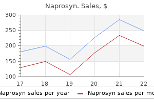
Cheap naprosyn online
Pediatric palliative care is being increasingly integrated into treatment protocols and therapies hemophilic arthritis definition order naprosyn overnight. Precautions New medical technologies have complicated treatment decisions for parents. Newborns are often subjected to stressful and painful tests and procedures, even when their condition or illness is incompatible with life. Overwhelmed parents are often unaware of the long-term consequences of the medical decisions that they are asked to make, especially when they are dealing with multiple specialists. In addition, language and cultural barriers can exacerbate communication problems between parents, physicians, and palliative care providers. It can be very difficult for young children to understand why they are undergoing painful procedures for diagnosis and treatment and many parents want to protect their child from a serious prognosis. However, studies have found that even very young children can understand the seriousness of their disease and that open communication between children and their parents and care providers can help alleviate their fears and maintain hope. Infants and children are often unable to communicate symptoms and specific sources of discomfort and pain. A major component of pediatric palliative care is the identification and treatment of such symptoms. Palliative caregivers must work closely with pharmacists to develop syrups, dissolvable capsules, or skin patches. It is helpful for children to take a medication before leaving the hospital to ensure that they can swallow it. Parents are often reluctant to administer pain medications, either because they fear that the child will become addicted to narcotics or because they are waiting for the child to cry or complain of severe pain. Prescriptions for painkillers should be filled immediately, so that the medication is available in the middle of the night. Many families prefer to have their child die at home and many pediatric palliative care programs include end-of-life planning. Studies have found that the overall quality of care is generally better in the home than in a hospital or hospice. The first pediatric palliative home care program in the United States was established in Connecticut in 1974. Since the 1980s a variety of homeand hospice-based pediatric palliative care programs have been established across the United States and around the world. Chronic-An illness or condition of long duration, frequent recurrence, or slow progression. Hospice-A facility or program that provides for the physical and emotional needs of the terminally ill in a caring environment. Opiate-A drug containing or derived from opium- such as codeine, morphine, and heroin-that alleviates pain and induces sleep. Palliative treatment-Treatments-such as chemotherapy, radiation therapy, or surgery-that ease the symptoms of a disease without curing it. Respite care-Temporary care of a patient to provide parents or other caregivers with a period of physical, mental, and emotional rest. Even when prescribed, parents may withhold medication because of fear of side effects or addiction. Some physicians do not believe it is medically necessary and many hospitals claim they cannot afford it. Palliative care is poorly reimbursed by Medicaid and it has been more difficult to demonstrate cost savings from reduced emergency care and hospital stays with pediatric palliative care than with adult programs. Preparation Palliative care for children usually begins with the diagnosis of a chronic or life-threatening illness. It can appear to skip beats, beat rapidly, beat irregularly, or thump in the chest. Although palpitations are very common and often harmless, they can be frightening to a person, who is usually unaware of his or her heartbeat. Palpitations that are caused by certain types of abnormal heart rhythms (arrhythmias) can be serious, and even fatal if left untreated. Recognizable arrhythmias are present in a small number of patients who have palpitations. Immediate medical attention should be sought for palpitations that feel like a very fast series of heartbeats, last more than two or three minutes, and are unrelated to strenuous physical activity, obvious fright, or anger. Medical attention should also be sought if palpitations are accompanied by chest pain, dizziness, shortness of breath, or an overall feeling of weakness. Most people have experienced a skipped or missed heartbeat, which is really an early beat and not a skipped beat at all. After a premature heartbeat, the heart rests for an instant then beats with extra force, making a person feel as if the heart has skipped a beat. After chest pain, palpitations are the most common reason that people are referred for cardiology evaluation. Causes and symptoms Palpitations can be caused by anxiety, arrhythmias, caffeine, certain medications, cocaine and amphetamines, emotional stress, overeating, panic, somatization, and vigorous exercise. But, anxiety, dizziness, shortness of breath, and chest pain may be signs of more severe arrhythmias. In this procedure, the patient wears a small, portable tape recorder that is attached to a belt or shoulder strap and connected to electrode disks on his or her chest. Some medical centers are now using event recorders that the patient can carry for weeks or months. When palpitations occur, the patient presses a button on the device, which captures the information about the palpitations for physician evaluation. Some arrhythmias are harmless, while others, such as ventricular tachycardia, ventricular fibrillation, and ventricular standstill, can be fatal. Homeopathic remedies such as Lachesis, Digitalis, and Aconite (Aconitum nnapellus) may be used to control palpitations but should be taken only when prescribed by a homeopathic physician. Mind/body medicine such as meditation and yoga can help the person relax, eliminating or reducing palpitations caused by anxiety or stress. Reducing or eliminating tea, cola, coffee, and chocolate, and consuming adequate amounts of the mineralscalcium, magnesium, and potassium can help reduce or eliminate palpitations. Prognosis Most palpitations are harmless, but some can be a sign of heart trouble, which could be fatal if left untreated. If the cause of the palpitations is determined to be an arrhythmia, medical or surgical treatment may be indicated, although surgery is rarely needed. Alternatives Alternative treatments for palpitations should be used only as a complement to traditional medicine. Alternative treatments include: aromatherapy, Chinese herbs, herbal therapies, homeopathic medicine, exercise, mind/body medicine, and diet and nutrition. In aromatherapy, adding citrus oils to bath water may help with minor palpitations. Some Chinese herbs can also help, but others can worsen arrhythmias, so a qualified herbalist should be consulted. Herbal therapies such as hawthorn (Crataegus laevigata) and motherwort (Leonurus cardiaca) can help with palpitations. Exercise can also help, but a treadmill stress test performed by a physician should be considered first to make sure that exercise is safe. European Society of Cardiology, the European Heart House, 2035 Route des Colles, B. Among these individuals, the best candidates for pancreas transplantation are typically: Pancreas transplantation Definition Pancreas transplantation is a surgical procedure in which a diseased pancreas is replaced with a healthy pancreas that has been obtained from an immunologically compatible cadavear or living donor. Patients with type 1 diabetes have experienced partial or complete damage to the insulinproducing beta cells of the pancreas. Consequently, they are unable to generate sufficient insulin to control blood glucose levels. Long-term uncontrolled high blood glucose levels can cause damage to every system of the body, so type 1 patients must inject insulin to do the work of the beta cells. Pancreas transplantation allows the body to once again make and secrete its own 3804 Pancreas-only transplants account for about 10% of the 1200 pancreas transplants performed each year in the United States. There are about 100 medical centers in the United States that perform pancreas transplantations. Description Once a donor pancreas is located and tissue typing deems it compatible, the patient is contacted and prepared for surgery. In a successful transplant, the pancreas begins producing insulin, bringing the regulation of glucose back under control.
Purchase naprosyn overnight delivery
Description Physicians recognize a number of different forms of psoriatic arthritis arthritis yahoo buy 500mg naprosyn free shipping. In some patients, the arthritic symptoms will affect the small joints at the ends of the fingers and toes. In others, symptoms will affect joints on one side of the body but not on the other. In addition, there are patients whose larger joints on both sides of the body simultaneously become affected, as in rheumatoid arthritis. In many patients, symptoms of psoriasis precede the arthritis symptoms; a clue to possible joint disease is pitting and other changes in the fingernails. Both the skin and joint symptoms will come and go; there is no clear relationship between the severity of the psoriasis symptoms and arthritis pain at any given time. Recent surveys suggest that between one in five people and one in two people with psoriasis may also have some arthritis symptoms. Psoriatic arthritis mutilans-A severe form of psoriatic arthritis that destroys the joints of the fingers and toes and causes the bones to fuse, leaving patients with gnarled and club-like hands and feet. Rheumatoid arthritis-A systemic disease that primarily affects the joints, causing inflammation, changes in structure, and loss of function. Rheumatoid factor-A series of antibodies that signal the presence of rheumatoid arthritis. People with psoriatic arthritis are more likely than others to have close relatives with the disease, but they are just as likely to have relatives with psoriasis but no joint disease. Researchers believe genes increasing the susceptibility to developing psoriasis may be located on chromosome 6p and chromosome 17, but the specific genetic abnormality has not been identified. Symptoms of psoriatic arthritis include dry, scaly, silver patches of skin combined with joint pain and destructive changes in the feet, hands, knees, and spine. Antimalaria drugs and systemic corticosteroids should be avoided because they can cause dermatitis or exacerbate psoriasis when they are discontinued. Several treatments are useful for both the skin lesions and the joint inflammation of psoriatic arthritis. Etretinate, a vitamin A derivative; methotrexate, a potent suppressor of the immune system; and ultraviolet light therapy have all been successfully used to treat psoriatic arthritis. Alternatives Food allergies/intolerances are believed to play a role in most autoimmune disorders, including psoriatic arthritis. Constitutional homeopathy can work deeply and effectively with this condition, if the proper prescription is given. Acupuncture, Chinese herbal medicine, and Western herbal medicine can all be useful in managing the symptoms of psoriatic arthritis. Alternative treatments recommended for psoriasis and rheumatoid arthritis may also be helpful in treating psoriatic arthritis. Diagnosis Skin and nail changes characteristic of psoriasis with accompanying arthritic symptoms are the hallmarks of psoriatic arthritis. A blood test for rheumatoid factor, antibodies that suggest the presence of rheumatoid arthritis, is negative in nearly all patients with psoriatic arthritis. X rays may show characteristic damage to the larger joints on either side of the body, as well as fusion of the joints at the ends of the fingers and toes. Treatment Treatment for psoriatic arthritis is meant to control the skin lesions of psoriasis and the joint inflammation of arthritis. Nonsteroidal anti-inflammatory drugs, gold salts, and sulfasalazine are standard arthritis treatments, but have no effect on psoriasis. For many, the joint and other arthritis symptoms are much milder than those experienced in rheumatoid arthritis. One in five people with psoriatic arthritis, however, face potentially crippling joint 4213 Psychoanalysis disease. In some cases, the course of the arthritis can be far more mutilating than in rheumatoid arthritis. Demographics As of 2014, there are 35 training institutes for psychoanalysts in the United States approved by the American Psychoanalytic Association and between 75 and 100 independent training institutes. It is difficult to obtain precise statistics on the number of patients who undergo psychoanalysis in an average year; however, because of the time and expense involved in this form of mental health treatment, considerably fewer people consult psychoanalysts than other mental health professionals. Freud expanded upon this practice by introducing a method that he called free association, in which a patient is encouraged to speak in a nonnarrative or rambling manner under the expectation that he or she will eventually reveal or uncover the unconscious heart of the problem. This sort of undirected self-exploration became one of the signature tenets of psychoanalysis. Continuing his research into the mind and the unconscious, Freud published the Interpretation of Dreams in 1900. In this work he outlined his ideas about the construction of the mind and human personality. This book was followed by the major publications of the Freudian canon: the Psychopathology of Everyday Life in 1904, and A Case of Hysteria as well as Three Essays on the Theory of Sexuality in 1905. Psychoanalysis is classified as an insightoriented rather than a supportive form of therapy, because it is based on the notion that people are better able to make changes in their lives when they have improved their understanding of themselves through identifying their assumptions about life and the early life experiences that gave rise to them. In the words of the American Psychoanalytic Association, 'Psychoanalytic treatment gives patients the opportunity to examine these assumptions, understand their origins in their lives, modify them if necessary, and make better choices for themselves. Psychoanalysts maintain that they can successfully treat phobias, anxiety attacks, obsessions and compulsions, mood disorders, repetitive patterns of failure in relationships and employment, unresolved grief, and a more general feeling of alienation or estrangement from others. Although Freud delivered his talks in German, they were translated into English within weeks and made his name a household word among English-speaking physicians and psychologists. Psychoanalysis had emerged as a significant intellectual achievement on a par with the work of Albert Einstein in physics and in many ways comparable to the modernist movement in the visual arts. Psychoanalysis was in its prime by the 1920s; after the shock and disillusionment following World War I, Western intellectuals made undergoing psychoanalysis a fashionable form of treatment. Major concepts Freud believed that human personality is constructed of three parts: the id, the ego, and the superego. The id, according to this schema, is comprised largely of such instinctual drives as desires for food or sexual pleasure. These drives are essentially unconscious, yielding satisfaction when they are fulfilled and frustration or anxiety when they are thwarted. The superego acts in many ways like the ego, as a moderator of behavior-but whereas the ego moderates urges based on social constraints, the superego operates as an arbiter of right and wrong. Having constructed this framework of human personality, Freud used it to demonstrate the ways in which instinctual drives inevitably run aground on strictly social codes (upheld by the ego) and notions of morality (upheld by the superego). The resultant conflicts, according to psychoanalytic theory, lie at the heart of most anxiety disorders and other neurotic problems. He considered dreams to have two parts: the manifest content, which is the narrative that one is able to remember upon waking; and the latent content, which is the underlying and largely symbolic message of the dream. Because Freud believed dreams to represent unfulfilled longings of the id, psychoanalysis deals heavily with dream interpretation. Psychoanalytic theory suggests that conditions like blindness, paralysis, and severe headaches can result from unfulfilled longings that the patient is unable to confront on a conscious level. Because of this inability, the patient develops some acceptable symptom, such as headaches, for which he or she can then seek medical attention. Freud identified several defense mechanisms, such as repression, displacement, denial, rationalization, projection, and identification. Each has its own peculiar dynamic, but all work to distance a person from a conflict that is too difficult to confront realistically. These conflicts, according to psychoanalytic theory, originate during one of the four developmental stages Freud identified. These stages, and the infantile sexuality he identified as occurring within them, are some of the most controversial aspects of psychoanalytic theory. Freud suggested that adult neuroses result from and can be traced back to frustrated sexual drives during these stages. Freud defined the stages as the oral stage, birth to one year; the anal stage, one to three years; the phallic stage, three to five years; and latency, five years to the beginning of puberty. In each of the major stages, the infant has sexual longings which, because of social mores, are left largely unfulfilled and lead to the formation of neuroses. Freud hypothesized that children in the phallic stage of development form the Oedipus complex, easily the most renowned and controversial theoretical construction of the Freudian canon. The Oedipus complex refers to the notion that a child begins associating his genitals with sexual pleasure during the phallic stage and becomes erotically attracted to the parent of the opposite sex while at the same time developing an intense jealousy of the same-sex parent.
Buy genuine naprosyn on-line
If an open prostatectomy is needed rheumatoid arthritis inflammation diet order naprosyn pills in toronto, an incision may be made the lower abdomen (C) or the perineal area (D). The glandular cells produce a milk-like fluid that mixes with seminal fluid and sperm to produce semen. The medically preferred anesthesia for open prostatectomy is a spinal or epidural nerve block. General anesthesia may be used if the patient has an anatomic or medical contraindication for regional anesthesia. With the use of open prostatectomy, the prostate is accessed through an incision that allows manual manipulation by the surgeon. The patient lies on his back on the operating table and is prepared and draped for this procedure. Following the cystoscopy, the patient is changed to a Trendelenburg position (feet higher than head). The abdominal muscle (rectus abdominis) is separated, and a retractor is placed at the incision site to widen the surgical field. Further maneuvering is essential to clearly locate the veins (dorsal vein complex) and the bladder neck. Once the structures have been identified and the blood supply controlled, an incision is made deep into the level of the tumor. Scissors are used to dissect the prostatic tissue (prostatic capsule) from the underlying tissue of the prostatic tumor. The wound is closed after complete removal of the tumor and hemostasis (stoppage of bleeding) occurs. Additionally, open prostatectomy is indicated for males with: acute urinary distention bladder outlet obstructions pathological changes in the bladder, ureters, or kidneys due to prostate obstruction recurrent blood in urine of prostate origin recurrent or persistent urinary tract infections Prostatectomy gland, and previous pelvic surgery that may obstruct access to the prostate gland. This procedure is less common than it used to be due to difficulties with circumventing the nerves and to inadequate access of the lymph nodes. This approach is ideal for prostate cancer surgery, which also allows the surgeon access to remove the surrounding lymph nodes. The suprapubic approach utilizes an incision of the lower anterior (front) bladder wall. The primary advantage over the retropubic approach is that the suprapubic route allows for direct visualization of the bladder neck and bladder mucosa. The major disadvantage is that visualization of the top part of the tumor is degraded. Additionally, with the suprapubic approach, hemostasis (stoppage of bleeding during surgery) may be more difficult due to poor visualization after removal of the tumor. Using a scalpel, a lower midline incision is made from the umbilicus to the pubic area. Using electrocautery (a special tool that produces heat at the tip, useful for hemostasis or tissue excision) and scissors, dissection proceeds until the prostatic tumor is identified and removed. After maintaining hemostasis and arterial blood supply to the prostate, the incisions to the bladder and abdominal wall are closed. A resectoscope (or resection cystoscope)-which is an instrument with a wide-angle telescope and an electrically activated wire loop-is moved up the urethra to the prostate. Laser prostate surgery Prostatectomy is used to reduce or eliminate severe urinary symptoms caused by an enlarged prostate. The laser beam is used to shrink tissue (in a process called ablation) or remove excess tissue (enucleation) that is blocking the urethra and preventing urine flow. It is especially useful when the enlarged prostate causes problems with urination. The method vaporizes enlarged prostate tissue by the creation of an ionized plasma corona. This procedure is considered the least intrusive of all the prostatectomy techniques currently available. The patient normally remains in the hospital for approximately two days after the surgery. Additionally, preoperative patients should have lower urinary tract studies, including urinary flow rate and post void residual urine in the bladder. Prostatectomy procedures are performed by a urological surgeon who typically completes one year of general surgery training and four to five years of urology training. This allows for an even higher degree of precision and for smaller incisions, resulting in less pain, bleeding, and risk of infection, as well as faster healing times and shorter hospital stays. A risk factor is the presence of normally functioning testicles; research indicates that castration can minimize prostatic enlargement. It appears that the glandular tissues that multiply abnormally use male hormones produced in the testicles differently than do normal tissues. Antispasmodics (bladder relaxant) may be given either by mouth or rectally if bladder spasms occur. After the catheter is removed, rectal antispasmodics are stopped to allow the bladder to return to normal. Beginning to walk as soon as possible after surgery will help normal bowel function to return. It also promotes more effective breathing, improves circulation, prevents stiffness of joints, and relieves pressure. After being discharged from the hospital, men should follow these recommendations for four to six weeks: Do not perform any heavy lifting or strenuous exercises. Avoid constipation by increasing roughage in the diet; take milk of magnesia or some other over the counter laxative if necessary. Do not take aspirin, Motrin, Advil, Nuprin, or nonsteroidal analgesics for at least six weeks after surgery because they may cause bleeding. Open prostatectomy is a major surgical operation requiring an inpatient hospital stay of four to seven days. Blood transfusions are generally not required due to improvements in surgical technique. Immediately after the operation, the surgeon must closely monitor urinary output and fluid status. On the first day after surgery, most patients are given a clear liquid diet and asked to sit up four times. On the second postoperative day, the urethral catheter is removed if the urine does not contain blood. On the third postoperative day, the pelvic drain is removed if drainage is less than 75 milliliters (mL) in 24 hours. Laparoscopic radical prostatectomy may allow the patient to go home the day after surgery. Other recommendations include: Laparoscopic radical prostatectomy is usually associated with very positive results because it involves lower blood loss and transfusion rates than other prostatectomy procedures. In addition, it involves shorter hospital stays, better pain control, reduced catheterization time, and faster return to everyday activities when compared to other methods. The overall rate of morbidity and mortality is extremely low for patients of open prostatectomy. Laparoscopic radical prostatectomy is considered the safest of the prostatectomy procedures. Prostatectomy Risks For transurethral resection of the prostate, patients with heart problems are not considered candidates for the procedure because heavy bleeding risks are possible. For open prostatectomy, patients normally will not have the adverse effects of bleeding. Hematuria (blood in the urine) is typically resolved within two days after surgery. The patient should begin a regular diet and moderate increases in activity soon after surgery.
Purchase genuine naprosyn online
Since muscle pain and weakness are common and usually temporary arthritis pain suddenly worse 500mg naprosyn for sale, it is important to determine if the patient also has risk factors for rhabdomyolysis. Creatinine-A protein produced by muscle and filtered out by the kidneys; high creatinine levels can indicate kidney failure. Electrolytes-Ions such as potassium, calcium, and sodium that are dissolved in bodily fluids such as blood and which regulate or affect most metabolic processes. Heme-The iron-containing portion of the myoglobin molecule that provides oxygen to muscle tissue. Hemodialysis-The process of removing blood from a patient, purifying it by dialysis, and returning it to the body, usually because of kidney failure. Diuretics and bicarbonate may be given to prevent kidney damage if urine output is sufficient. Patients with milder rhabdomyolysis may return to normal activities within a few weeks, although fatigue and muscle pain may linger. With adequate support, the prognosis in children is often good, although recurrent episodes may indicate underlying metabolic or muscle structural defects. In large-scale natural disasters, such as earthquakes, the ability of emergency medical teams to provide hydration and dialysis improves survival, and many patients recover completely. Drinking fluids dilutes the urine and helps flush out of the kidneys any myoglobin released by muscles. It is also essential to pay immediate attention to signs of heat exhaustion in hot, humid conditions. High school and college athletes should be educated about the signs of dehydration and heat-related injuries. Patients at risk for exercise-induced or hyperthermia-induced rhabdomyolysis should avoid overly strenuous exercise and ensure adequate hydration and body cooling when exercising. Patients should contact their physician if they experience moderate-to-severe muscle aches when first taking a statin. They should stop the drug immediately if signs and symptoms of rhabdomyolysis develop. Patients should also discontinue the use of recreational or prescription drugs or alcohol that contributed to the development of their rhabdomyolysis. Patients with metabolic disorders may be able to prevent rhabdomyolysis by dietary modifications. Families with inherited deficiencies in muscle enzymes and metabolism should seek genetic counseling. Description Throat infection with a member of the Group A streptococcus (strep) bacteria is a common problem among school-aged children. However, when such a throat infection occurs without symptoms, or when a course of medication is not taken for the full 10 days, there is a 3% chance of that person developing rheumatic fever. Children between the ages of 5 and 15 are most susceptible to strep throat, and therefore most susceptible to rheumatic fever. Other risk factors include poverty, overcrowding (as in military camps), and lack of access to good medical care. Just as strep throat occurs most frequently in fall, winter, and early spring, so does rheumatic fever. This could be related to the above theory, in that these families may have cell antigens which more closely resemble streptococcal antigens than do members of other families. The joints (especially those of the ankles, knees, elbows, and wrists) become red, hot, swollen, shiny, and extraordinarily painful. Unlike many other forms of arthritis, the arthritis may not occur symmetrically (affecting a particular joint on both the right and left sides, simultaneously). The joints become so tender that even the touch of bedsheets or clothing is terribly painful. The patient begins experiencing a change in coordination, often first noted by changes in handwriting. A rash called erythema marginatum develops (especially in those patients who will develop heart problems from their illness), composed of pink splotches, which may eventually spread into each other. These are called subcutaneous nodules; they are hard to the touch, but not painful. These nodules most commonly occur over the knee and elbow joint, as well as over the spine. Pancarditis is an inflammation that affects all aspects of the heart, including the lining of the heart (endocardium), the sac containing the heart (pericardium), and the heart muscle itself (myocardium). This may result in blood which either leaks back in the wrong direction, or has a difficult time passing a stiff, poorly moving valve. Either way, damage to a valve can result in the heart having to work very hard in order to move the 4401 Rheumatic fever A magnified image of cardiac muscle damaged by chronic myocarditis caused by rheumatic fever. One theory, less supported by research evidence, suggests that the bacteria produce some kind of poisonous chemical (toxin). This toxin is sent into circulation throughout the bloodstream, thus affecting other systems of the body. The body produces immune cells (called antibodies), which are specifically designed to recognize and destroy invading agents; in this case, streptococcal bacteria. The antibodies are able to recognize the bacteria because the bacteria contain special markers called antigens. The heart may not be able to 'work around' the damaged valve, which may result in a consistently inadequate amount of blood entering the circulation. Chorea-Involuntary movements in which the arms or legs may jerk or flail uncontrollably. Pancarditis-Inflammation of the lining of the heart, the sac around the heart, and the muscle of the heart. In either case, it must also be proved that the individual has had a previous infection with streptococcus. Tests are also performed to provide evidence of recent infection with group A streptococcal bacteria. These bacteria can then be specially processed, and examined under a microscope, to identify streptococcal bacteria. Other tests can be performed to see if the patient is producing specific antibodies; that are only made in response to a recent strep infection. Arthritis quickly improves when the patient is given a preparation containing aspirin, or some other anti-inflammatory agent (ibuprofen). Mild carditis will also improve with such anti-inflammatory agents, although more severe cases of carditis will require steroid medications. A number of medications are available to treat the involuntary movements of chorea, including diazepam for mild cases, and haloperidol for more severe cases. This can mean a small daily dose of penicillin by mouth, or an injection every three weeks. Some practitioners keep patients on this regimen for five years, or until they reach 18 years of age (whichever comes first). The normally thin and delicate synovium becomes thick and stiff, with numerous infoldings on the surface. The cartilage may be attacked and destroyed, and the bone, articular capsule, and ligaments may begin to wear away. Inflammation-A localized, normally protective, immune response elicited by injury or destruction of tissues and characterized by pain, heat, redness, swelling, or loss of function. Synovial membrane; synovium-The membrane that lines the inside of the articular capsule of a joint and produces lubricating synovial fluid. X chromosome, which may explain why women are more susceptible, since they have two X chromosomes and men have only one.
Order naprosyn with a visa
According to the American Association of Endodontists arthritis pain tylenol or advil buy 500 mg naprosyn otc, more than 15 million root canal treatments are performed every year in North America as of 2014, with a 97% success rate. If a tooth becomes diseased or injured, bacteria may build up inside the pulp, spreading infection from the natural crown of the tooth to the root tips in the jawbone. Pus accumulating at the ends of the roots can form a painful abscess that can damage the bone supporting the teeth. It can also result in prolonged sensitivity to heat or cold, swelling, and tenderness in the surrounding gums, facial swelling, or discoloration of the tooth. In some cases, however, the pulp may die so gradually that there is little noticeable pain. A thin sheet of rubber called a rubber dam is placed in the mouth and around the base of the tooth to isolate the tooth and help to keep the operative field dry. The dentist removes any tooth decay and makes an opening through the natural crown of the tooth into the pulp chamber. Creating this access also relieves the pressure inside the tooth and can dramatically ease pain. The dentist determines the length of the root canals, usually with a series of x rays. Small wire-like files are then used to clean the entire canal space of diseased pulp tissue and bacteria. Apicoectomy-Also called root resectioning, in which the root tip of a tooth is accessed in the bone and a small amount is shaved away. Endodontic-Pertaining to the inside structures of the tooth, including the dental pulp and tooth root, and the periapical tissue surrounding the root. Endodontist-A dentist who specializes in the diagnosis and treatment of disorders affecting the inside structures of teeth. Pulpitis-Inflammation of the pulp of a tooth involving the blood vessels and nerves. Root canal-The space within a tooth that runs from the pulp chamber to the tip of the root. Smear layer-A layer of organic and inorganic material produced on teeth by dental instrumentation that may also contain bacteria and their by-products. Once the canals are completely clean, they are filled with gutta percha and a sealer cement to prevent bacteria from entering the tooth in the future. A metal post may be placed in the pulp chamber for added structural support and better retention of the crown restoration. The tooth is protected by a temporary filling or crown until a permanent restoration may be made. This restoration is usually a gold or porcelain crown, although it may be a gold inlay, or an amalgam or composite filling (paste fillings that harden). In theory, the beam of intense light from the erbium laser that the dentist uses melts the debris (also called the smear layer) inside the tooth, cleansing it completely. It is possible, however, for a laser beam to miss some of the infection within the tooth, particularly if the root canal itself is unusually shaped or has a number of small crevices. In addition, the use of lasers in root canal treatment has been reported to occasionally damage the tooth. Root canal treatment Diagnosis/Preparation Signs that a root canal treatment is necessary include severe pain while chewing, prolonged sensitivity to heat or cold, or a darkening of the tooth. Swelling and tenderness of the gums or pimples appearing on the gums are also common symptoms. The dentist will take an x ray of the tooth to determine if there is any sign of infection in the surrounding bone. Aftercare Once a root canal treatment is performed, the recipient must have a crown placed over the tooth to protect it. However, replacing an extracted tooth with a fixed bridge, a removable partial denture, or an implant to maintain the space and restore the chewing function is typically even more expensive. During the time when antibiotics are being used, care should be taken to avoid using the tooth to chew food. The tooth has been structurally weakened and may break, or there is a possibility of the interior of the tooth becoming reinfected. If the tooth feels sensitive following the procedure, a standard over-the-counter pain medication such as ibuprofen or naproxen may be taken. The canals are also slightly enlarged and shaped to receive an inert (non-reactive) filling material called gutta percha. However, the tooth is not filled and permanently sealed until it is completely free of active infection. The dentist may place a temporary seal, or leave the tooth open to drain, and prescribe an antibiotic to counter any spread of infection from the tooth. If infection and inflammation recur and an x ray indicates a repeat treatment is feasible, the old filling material is removed and the canals are thoroughly cleaned out. The dentist will try to identify and correct problems with the first root canal treatment before filling and sealing the tooth a second time. In cases where an x ray indicates that another root canal treatment cannot correct the problem, endodontic surgery may be performed. In a procedure called an apicoectomy, or root resectioning, the root end of the tooth is accessed in the bone, and a small amount is shaved away. The area is cleaned of diseased tissue and a filling is placed to reseal the canal. However, because it does not contain an internal nerve, it no longer has sensitivity to hot, cold, or sweets. Because these are signs of dental decay, the root canal recipient must receive regular dental check-ups with periodic x rays to avoid further disease in the tooth. However, with routine wear, the filling or crown may eventually need to be replaced. Morbidity and mortality rates About 5% of patients will experience persistent pain after root canal therapy. In some cases, despite proper root canal treatment and endodontic surgery, the tooth dies and must be extracted. After restoration or extraction, the two main goals are to allow normal chewing and to maintain proper alignment and spacing between teeth. A fixed bridge, a removable partial denture or an implant will accomplish both goals. It is marked by redness (erythema) of the face, flushing of the skin, and the presence of hard pimples (papules) or pus-filled pimples (pustules), and small visible spider-like veins called telangiectasias. In later stages of the disease, the face may swell and the nose may take on a bulb-like appearance called rhinophyma. Telangiectasia may appear around the borders of the eyelid, the eyelids may be chronically inflamed, and small lumps called chalazions may develop. The cornea of the eye, the transparent covering over the lens, can also be affected, and in some cases vision will be affected. The consensus among many experts is that multiple factors may lead to an overreaction of the facial blood vessels, which triggers flushing. Over time, persistent episodes of redness and flushing leave the face continually inflamed. Certain genetic factors may also come into play, although these have not been fully described. It may be more common in people of Celtic background, although this is an area of disagreement among experts. Certain antibiotics are useful in the treatment of rosacea, leading some researchers to suspect a bacterium or other infectious agent may be the cause. One of the newest suspects is a bacterium called Helicobacter pylori, which has been implicated in causing many cases of stomach ulcers but the evidence here is mixed. Other investigators have observed that a particular parasite, the mite Demodex folliculorum, can be found in areas of the skin affected by rosacea. The mite can also be detected, however, in the skin of people who do not have the disease. Description Rosacea produces redness and flushing of the skin, as well as pustules and papules. Areas of the face, including the nose, cheeks, forehead, and chin, are the primary sites, but some people experience symptoms on their necks, backs, scalp, arms, and legs. The similarity in appearance of rosacea to acne led people in the past to erroneously call the disease acne rosacea or adult acne.
TFd (Transfer Factor). Naprosyn.
- What other names is Transfer Factor known by?
- What is Transfer Factor?
- How does Transfer Factor work?
- Lung cancer.
- Chronic fatigue syndrome.
- Are there safety concerns?
Source: http://www.rxlist.com/script/main/art.asp?articlekey=96972
Purchase 500 mg naprosyn mastercard
The kit could be useful in the event of a bioterrorist attack as well as in countries without advanced microbiology laboratories arthritis pain scale generic 250mg naprosyn with amex. Treatment As soon as plague is suspected, the patient should be isolated, and local and state departments notified. Alternatives include gentamicin, chloramphenicol, tetracycline, or trimethoprim/ sulfamethoxazole. The presence of plague bacteria in a blood smear is a grave sign and indicates septicemic plague. Septicemic plague has a mortality rate of 40% in treated cases and 100% in untreated cases. Buboes-Smooth, oval, reddened, and very painful swellings in the armpits, groin, or neck that occur as a result of infection with the plague. Endemic-A disease that occurs naturally in a geographic area or population group. Pandemic-A disease that occurs throughout a regional group, the population of a country, or the world. Septicemia-The medical term for blood poisoning, in which bacteria have invaded the bloodstream and circulates throughout the body. Pregnant women should not be vaccinated unless the need for protection is greater than the risk to the unborn child. The inadequacy of the vaccines available as of 2014 explains why it is important to protect against rodents, fleas, and people with plague. A team of researchers in the United Kingdom reported in the summer of 2004 that an injected subunit vaccine is likely to offer the best protection against both bubonic and pneumonic forms of plague. All plague patients should be isolated for 48 hours after antibiotic treatment begins. Pneumonic plague patients should be completely isolated until sputum cultures show no sign of infection. Anyone working in a rodent-infested area should wear insect repellent on skin and clothing. Plague vaccines have been used with varying effectiveness since the late nineteenth century. Experts believe that vaccination lowers the chance of infection and the severity of the disease. However, the effectiveness of the vaccine against pneumonic plague is not clearly known. Moreover, its unpleasant side effects make it a poor choice unless there is a substantial long-term risk of infection. Jones, Abby, Catharine Bosio, Angela Duffy, Andrew Goodyear, Martin Schriefer, and Steven Dow. Also, the sample must be collected properly: drawn into a chilled syringe and collection tube, placed on ice, and sent to the performing laboratory immediately. Patients with primary hyperaldosteronism (caused by an adrenal tumor that overproduces aldosterone) will have an increased aldosterone level with decreased renin activity. Conversely, patients with secondary hyperaldosteronism (caused by certain types of kidney disease) will have increased levels of renin. Renin stimulation test the renin stimulation test is performed to help diagnose and distinguish the two forms of hyperaldosteronism. In cases of primary hyperaldosteronism, the blood volume is greatly expanded, and a change in position or reduced salt intake does not result in decreased kidney blood flow or decreased blood sodium. However, in secondary hyperaldosteronism, blood sodium levels decrease with a lowered salt intake, and when the patient is standing upright, the kidney blood flow decreases as well. Captopril test the captopril test is a screening test for hypertension of kidney origin (renovascular hypertension). Precautions Patients taking diuretics, antihypertensives, vasodilators, oral contraceptives, and licorice should discontinue use of these substances for two to four weeks before the test. It should be noted that renin is increased in pregnancy and in diets with reduced salt intake. Also, since renin is affected by body position, as well as by diurnal (daily) variation, blood samples should be drawn in the morning, and the position of the patient (sitting or lying down) should be noted. Description When the kidneys release the enzyme renin in response to certain conditions (high blood potassium, low blood sodium, decreased blood volume), it is the first step in what is called the renin-angiotensinaldosterone cycle. Primary aldosteronism, the symptoms of which include hypertension and low blood potassium (hypokalemia), is considered 'low-renin aldosteronism. Symptoms include hypertension, impaired kidney function, thirst and muscle weakness. Risks for this test are minimal, but may include slight bleeding from the puncture site, fainting or feeling lightheaded after venipuncture, or hematoma (blood accumulating under the puncture site). Flores Plasmapheresis the need for a more invasive radiographic evaluation such as renal arteriography. Definition Plasmapheresis is a blood purification procedure used to treat several autoimmune diseases. It is recommended that the patient be fasting (nothing to eat or drink) from midnight the day of the test. In many autoimmune diseases, the chief weapons of attack are antibodies, proteins that circulate in the bloodstream until they meet and bind with the target tissue. Once bound, they impair the functions of the target, and signal other immune components to respond as well. Plasmapheresis is used to remove antibodies from the bloodstream, thereby preventing them from attacking their targets. Neurologic diseases comprise 90% of the diseases that could profit from plasmapheresis. Decreased renin levels may indicate increased blood volume due to a high-sodium diet, salt-retaining steroids, primary aldosteronism, licorice ingestion syndrome, or essential hypertension with low renin levels. Penicillin is an example of a substance that causes severe allergic reactions for some people. Antibody-Chemicals produced by the body to defend it against bacteria, viruses, or other cells foreign to the body (antigens). It is a watery fluid that carries red cells, white cells, and platelets throughout the body. Because of concerns over viral infection and allergic reaction, fresh plasma is not routinely used. Instead, the most common substitute is saline solution with sterilized human albumin protein. During the course of a single session, two to three liters of plasma is removed and replaced. Plasmapheresis requires insertion of a venous catheter, either in a limb or central vein. Central veins allow higher flow rates and are more convenient for repeat procedures, but are more often the site of complications, especially bacterial infection. While most of the anticlotting agent is removed from the blood during treatment, some is returned to the patient. Three procedures are available: Preparation Good nutrition and plenty of rest make the procedure less stressful. Aftercare the patient may experience dizziness, nausea, numbness, tingling, or lightheadedness during or after the procedure. These effects usually pass quickly, allowing the patient to return to normal activities the same day. Results Plasmapheresis is an effective temporary treatment for: 'Discontinuous flow centrifugation. Approximately 300 mL of blood is removed at a time and centrifuged to separate plasma from blood cells.
Syndromes
- Monoclonal gammopathy of unknown significance (MGUS)
- Stomach pain
- Backache, which occurs with routine activities
- Laparoscopy
- Over-the-counter pain relievers or prescription pain medications to control pain (neuralgia)
- The ends of your intestines that are sewn together comes apart (anastomotic leak -- this may be life-threatening)
- Developmental milestones record - 2 years
Cheap 500mg naprosyn overnight delivery
Range of motion is determined using a goniometer-an instrument that measures the largest angle through which a joint can move rheumatoid arthritis yahoo order naprosyn online. The therapist determines whether restricted motion is due to tight muscles or tight ligaments and tendons. Physical therapy also teaches patients to use assistive and adaptive devices such as crutches, wheelchairs, and prostheses. For range-of-motion stretches the muscles are often first warmed with heat to improve effectiveness and reduce pain. Tight ligaments or tendons require gentle stretching, whereas the joint can be stretched more vigorously if tight muscles are causing poor range of motion. An affected joint must be moved beyond the point of pain, but should not cause residual pain after the movement is stopped. There are three types of range-of-motion exercises: Physical therapy strength coordination and balance posture motor function muscle performance respiration active exercise for patients who can move their limbs and exercise a muscle or joint without assistance active-assistive exercise for patients who need some help moving their limbs and exercising muscles or for whom moving joints is painful passive exercise in which the therapist moves the limbs Based on these evaluations, physical therapists develop treatment plans, including purpose, strategy, and anticipated outcomes. Physical therapists may treat a wide range of conditions or specialize in certain areas. They often consult or collaborate with physicians, nurses, dentists, occupational therapists, speech-language pathologists, audiologists, educators, and/or social workers. Working under the direct supervision of a physical therapist, physical therapy assistants often implement treatment plans and record patient responses. About 60% of physical therapists work in hospitals or in the offices of other healthcare practitioners, especially physicians. Physical therapy also can take place in the following locations: There are a variety of other physical therapy exercises: There are many muscle-strengthening exercises, all of which progressively increase resistance. Movement against gravity is used for very weak muscles and the resistance is gradually increased using stretchy bands or weights. Rehabilitation from a stroke or brain damage often requires coordination exercises that involve specific tasks that work multiple joints and muscles, such as picking up an object. Once a patient can balance while standing, ambulation exercises begin with walking using parallel bars and progressing to a walker, crutches or a cane, possibly wearing a brace or assistive belt to prevent falls. Once a patient can walk on a level surface, ambulation exercises involve stepping over curbs or climbing stairs. This may include teaching family members and caregivers how to correctly support the patient. Transfer training-moving safely and independently from bed to chair, chair to toilet, or chair to standing-is a critical component of physical therapy. It is often required for patients who have had a hip fracture, amputation, or stroke. Transfer training techniques depend on whether the patient can do the following: Physical therapy uses a variety of techniques to reduce swelling and relieve pain, including the following: bear weight on one or both legs balance well can use assistive devices hot and cold packs paraffin baths electrical stimulation ultrasound massage, including deep-tissue massage traction Pediatric physical therapists evaluate children in the context of their daily routines and activities. Evaluation may include the following: Paralysis is another factor that determines what the patient can do. Tilt tables are used for patients who have had strict bed rest for several weeks or have had a spinal cord injury, since they can become dizzy when standing up. Tilt tables retrain blood vessels to narrow and widen appropriately with changes in posture. The patient lies face-up on a padded table with a footboard and is held in place with a safety belt as the table is slowly tilted. To decrease lower back pain and restore mobility, physical therapy involves: mobility analyzing gait (walking and running) sensory and neuromotor development muscle and joint function strength and endurance posture and balance cardiopulmonary status oral motor skills use of assistive technologies Physical therapy for children can include the following: manual therapies, including spinal manipulation, to improve the mobility of joints and soft tissues specific strengthening and/or flexibility exercises training for sitting, sleeping, bending, lifting, and performing chores education about back care Physical therapy for patients with diabetes includes: testing sensation in the feet teaching patients how to protect feet that have lost sensation recommending footwear or assistive devices adapting shoes or orthotic devices for walking decreasing cramping during walking caring for skin ulcers and sores supervising exercise programs movement and mobility posture, positioning, and lifting strengthening motor learning coordination and balance cardiopulmonary endurance developmental activities adapting daily routines and activities fitting and use of assistive technology orthotics and prosthetics burn and wound care safety, health, and prevention programs Pediatric physical therapy uses many of the same evaluation and therapeutic techniques as adult physical therapy, but it often includes toys and pediatric therapy gyms with balls, benches, swings, and slides. Pediatric physical therapy may also include: Physical therapists design exercise programs to prevent knee or other injuries, beginning with an evaluation of body traits that predispose a patient to injury. Physical therapy started soon after breast cancer surgery, including massage and shoulder exercises, can reduce or prevent the common complication of lymphedema. Physical therapists treat patients with a variety of conditions and diseases, including the following: Demographics the use of physical therapy to treat patients of all ages-from newborns to the elderly-is becoming increasingly widespread. As of 2014, there was over 33 million noninstitutionalized adults in the United States with some physical function limitation. Between 2010 and 2018 employment of physical therapists was expected to have grown by 30%, much more than the average for all occupations. There are several reasons for the growth of physical therapy: Physical therapy all types of injuries, including sprains, strains, fractures, and head injuries back and neck pain knee pain shoulder pain repetitive stress and overuse injuries poor posture arthritis heart disease stroke diabetes osteoporosis lymphedema Common applications of physical therapy are: retraining muscles and adjusting to the use of artificial joints strengthening leg muscles following a hip fracture assessing and fitting walking aids such as canes and walkers strengthening arm muscles for using walking aids treating pain from tendinitis/bursitis and arthritis to avoid the use of prescription pain medications in patients at risk for heart disease rehabilitating stroke victims for walking safely, with or without a walking aid the increasingly population of elderly people in the United States is especially vulnerable to chronic and debilitating conditions that require physical therapy. Advances in physical therapy have led to treatments for many disabling conditions that were previously untreatable. The federally mandated Individuals with Disabilities Education Act guarantees student access to physical therapy. A growing number of employers are using physical therapists to evaluate worksites, develop exercise programs, and teach safe work habits to reduce injuries. Benefits Physical therapy can help patients gain and maintain mobility, independence, and good quality of life. It can help prevent and manage medical conditions and motivate patients to improve on their own. Physical therapy can prevent loss of mobility by designing exercise programs based on individual characteristics. Physical therapy and medical management have been found to be as effective as knee surgery for relieving stiffness and pain from moderate to severe osteoarthritis. Pediatric physical therapy is commonly used for children with the following problems: birth defects such as spina bifida genetic disorders prenatal drug or alcohol exposure cerebral palsy orthopedic disabilities and limb deficiencies developmental delays heart and lung conditions muscle diseases head injuries acute trauma Preparation Patient attitude and cooperation are key to successful physical therapy. Patients must be active participants in their treatment, aware of the short-term and long-term goals of their therapy, and able to communicate with their therapists. Aftercare Physical therapy often requires patients to follow a specially designed exercise program. Stroke-A sudden diminishing or loss of consciousness, sensation, or voluntary movement from a rupture or obstruction of a blood vessel in the brain. Tilt table-Also called a tiltboard, an apparatus for rotating a person from horizontal to an oblique or vertical position. Traction-Pulling force exerted on a skeletal structure by a special device or piece of equipment. Margaret Alic, PhD Physiology Definition Physiology is the branch of biology that studies how living things function, including their growth, development, and nutrient use. Physiology also encompasses the physical and biological functioning of various organ systems. The Physiological Society, an international organization that promotes physiology, describes this branch of biology as 'the study of how molecules, cells and organs interact to form a whole being,' describing it as 'essential to the development of new treatments for disease. The study of human physiology is a core competency necessary for anyone from a personal trainer to a physician who wishes to enter a career in the health sciences. Physiology examines how the major organ systems of the body-circulatory, nervous, musculoskeletal, respiratory, gastrointestinal, integumentary, urinary, reproductive, endocrine, and immune systems-work together. Although everyone who works in an allied health field needs a background in physiology, only a few people, mostly research scientists, consider themselves physiologists. Some patients who have chronic diseases such as chronic heart and lung diseases are familiar with exercise physiologists. Exercise physiology is a subspecialty of kinesiology, which explores how physical activity affects human mental and physical health. Exercise physiology is the study of the short-term response to exercise and the adaption of the body to regular exercise. Besides working with medical patients and their care team, exercise physiologists often work with athletes as trainers or conditioning experts. Hippocrates believed it essential to understand the relationship between structure and function of various organs in the body. However, because of religious and legal prohibitions about dissecting human cadavers, much of the understanding of how the human body worked was extrapolated from the dissection of pigs, monkeys, dogs and other animals. Although illegal and semi-legal dissections were quietly carried out, it was not until 1752 that England allowed the legal dissection of criminals hanged for murder. The supply of bodies available for dissection, however, remained quite limited until the mid-1800s, when enough finally became available to provide a needed view inside the human body, and a clearer view of its inner workings. After that, knowledge of physiology increased rapidly, as did technical innovations that allowed scientists to study the body in new ways. Today, however, academic researchers do much of the basic research in the laboratory, and after thorough testing and necessary approvals, medical and allied health professionals then apply their research findings in clinical settings.

Order 250mg naprosyn
Pylorus-The area that controls food passage from the stomach to the first part of the small intestine (duodenum) rheumatoid arthritis wrist radiology order generic naprosyn on-line. Small intestine-The part of the intestines that consists of the duodenum, jejunum, and ileum. Demographics Pediatric surgeons provide treatment for young patients-newborns up through late adolescence. Description Pediatric surgery is performed by a pediatric surgeon who has had five years of general surgery training along with further specialized instruction and experience, and is certified in pediatric general surgery or in a specific pediatric specialty. Pediatric surgery is the surgical branch that uses operative techniques to correct certain pediatric conditions. There are different specialties within the field that include: pediatric general surgery pediatric otolaryngology (ear, nose, and throat) pediatric ophthalmology (eye) pediatric urology (urogenital system) pediatric orthopedic (bone) surgery pediatric neurological (brain and spinal cord) surgery pediatric plastic (reconstructive and cosmetic) surgery the American Academy of Pediatrics has established specific guidelines for referral to subspecialists. The pediatric patient has special considerations that differentiate him or her, both physically and psychologically, from an adult. A neonate (newborn) poses a great challenge in surgical treatment, since the tiny structures and immature organ systems may not cope with disease-induced stress and the physical demands of a major operative procedure. A newborn infant may still be developing key bodily functions, or may have special requirements. Key areas of concern in the newborn include: overview (with symptoms) of the more common pediatric conditions that require surgery typically performed by a pediatric surgeon. Alimentary tract obstruction Obstruction of the alimentary tract (tubes of digestion extending from the mouth to the anus) is characterized by four cardinal symptoms: cardiovascular (heart) system thermoregulation (temperature requirements of 73 F [22. This is a congenital deformity that occurs the pediatric surgeon is trained to treat the entire spectrum of surgical illnesses. Symptoms include severe respiratory distress (the neonate cannot breathe) and excessive 3871 Pediatric surgery salivation. Other clinical signs include cyanosis (bluish discoloration of the skin due to oxygen deprivation), choking, and coughing. It is a life-threatening illness characterized by abdominal distention, bilious vomiting, lethargy, fever, and occult (not obvious) or gross (clearly seen) rectal bleeding. Additionally, affected patients may exhibit signs of hypothermia (temperature less than 96. Abdominal wall defects Omphalocele is a defect that involves protrusion of abdominal contents into an external sac within the base of the umbilical cord. About 75% of omphalocele patients have serious genetic deformities involving these body systems: cardiovascular (heart), musculoskeletal (muscle and bones), genitourinary (genital and bladder systems), and central nervous (brain and spinal cord). The overall survival rate for infants with omphalocele varies and depends on defect size, other associated genetic abnormalities, and age of newborn. Gastroschisis is a defect in the abdominal wall to the side (lateral) of the umbilicus. The bowel protrudes to the outside of the abdomen during intrauterine life (while the embryo is developing inside the uterus). The amniotic fluid has an irritating effect on the exposed bowel, and causes infection of the bowels. Some pediatric surgeons and obstetricians recommend cesarean section (early elective delivery) to spare bowel trauma. The newborn patients typically require surgery, tube feedings for three to four weeks, and hospitalization for several weeks. Pyloric atresia is a condition that occurs when the pyloric valve, located between the stomach and duodenum, fails to open. Food cannot pass out of the stomach, resulting in vomiting clear gastric juice at attempted feedings. Other areas of the colon (duodenum, jejunum, ileum) can be obstructed during development, with symptoms present at birth. Most of these disorders share the four cardinal symptoms of alimentary obstruction. Intussusception is the most common cause of intestinal obstruction in patients who are three months to one year of age. The cause of intussusception is not known, and it is more common in males who are well nourished and apparently healthy. The symptoms include a sudden onset of abdominal pain characterized by episodic screaming and drawing up of the legs. In 60% of patients, vomiting and blood in the stool are common findings (either bright red or occult [hidden]). Typically, the bowel movements look like currant jelly, consisting of mucus and blood mixed together. Currant jelly stool is the most common clinical observation for patients with intussusception. During physical examination, patients will exhibit abdominal distention, and in 65% of cases there is a sausage-shaped mass that can be felt in the upper right portion of the abdomen toward the mid-abdomen. Anorectal anomalies There are many different types of anorectal anomalies common to male and female neonates, as well as deformities that are gender-specific since involvement of genitalia can occur. The surgery for these cases is complicated, and must be performed by an experienced pediatric surgeon. Through this defect, the abdominal contents enter the lung cavity and interfere with normal lung development. Usually the infants are fullterm, and the defect occurs on the left side in the majority-88%-of patients. The diverticulum is an outgrowth of intestine that is located in a portion of the intestines called the ileum. Symptoms of obstruction are more often observed in infants, and bleeding is more common in patients after age four. Intestinal polyps Juvenile polyps are usually present between the ages of 4 and 14 years, and tend to be inflammatory. The most common symptom of intestinal polyps is rectal bleeding, which is commonly due to a solitary polyp (80% of cases). Diagnosis can be done by proctosigmoidoscopy, which allows visualization of 85% of polyps. Acute appendicitis Acute appendicitis is a relatively common surgical emergency that is misdiagnosed in 28% of patients due to a broad spectrum of symptoms that can confuse the clinician. The classic clinical symptom of acute appendicitis is the onset of pain in the middle region of the abdomen that is followed by anorexia (loss of appetite), nausea, and vomiting. The pain is persistent and radiates to the right lower abdomen, becoming more intense and localized. The physical and abdominal examinations must be carefully and accurately performed. Patients with acute appendicitis usually have an increased white blood cell (cells that fight infection) count. If the appendix is perforated (ruptured), complications can occur as a result of kidney (renal) failure, seizures due to fever, and gram-negative sepsis (an infection that enters the bloodstream and interferes with life-saving chemical reactions). Patients who are very young, or those who were misdiagnosed and incurred long delays in treatment, are susceptible to death. Inflammatory bowel diseases Some cases (approximately 25%) of inflammatory bowel disease are found in persons younger than 20 years of age. The diagnosis of inflammatory bowel disease is usually based on presenting clinical symptoms, laboratory analysis results, endoscopic appearance, and radiologic findings. Pyloric stenosis is a common hereditary condition that affects males more than females, and occurs in one to three per 1,000 births. The typical symptoms include a progressive, often projectile, vomiting after attempted feedings. The gastric vomitus (bloody in 80% of patients) usually begins during the second and third week of life, and increases in force and frequency.
Order naprosyn australia
Other causes of abnormal findings include viral arthritis medication starting with m buy naprosyn visa, fungal, or parasitic infections, and tuberculosis. Risks Risks for this procedure include respiratory distress on the side of the biopsy, as well as bleeding, possible shoulder pain, pneumothorax (immediate) or pneumonia (delayed). Flores Preparation Preparations for this procedure vary, depending on the type of procedure requested. Pleural biopsy can be performed in several ways: percutaneous needle biopsy (described above), by thoracoscopy (insertion of a visual device called a laparoscope into the pleural space for inspection), or by open pleural biopsy, which requires general anesthesia. Pleural effusion Definition Pleural effusion occurs when too much fluid collects in the pleural space (the space between the two layers of the pleura). Aftercare Potential complications of this procedure include bleeding or injury to the lung, or a condition called pneumothorax, in which air enters the pleural cavity (the space between the two layers of pleura lining the lungs and the chest wall). Because of these possibilities, the patient is to report any shortness of breath, and to note any signs of bleeding, decreased blood pressure, or increased pulse rate. Description There are two thin membranes in the chest, one (the visceral pleura) lining the lungs, and the other (the parietal pleura) covering the inside of the chest wall. Normally, small blood vessels in the pleural linings produce a small amount of fluid that lubricates the opposed pleural membranes so that they can glide smoothly against one another during breathing movements. When either too much fluid forms or something prevents its removal, the result is an excess of pleural fluid-an effusion. The most common causes are disease of the heart or lungs, and inflammation or infection of the pleura. Pleural effusion itself is not a disease as much as a result of many different diseases. For this reason, there is no 'typical' patient in terms of age, sex, or other characteristics. Instead, anyone who develops one of the many conditions that can produce an effusion may be affected. Abnormal results Abnormal findings include tumors called neoplasms (any new or abnormal growth) that can be either benign or malignant. Pleural tumors are divided into two classifications: primary (mesothelioma), or metastatic (arising from cancer sites elsewhere in the body). These tumors are often associated with an accumulation of fluid between the pleural layers called a pleural effusion, which itself may be caused by pneumonia, heart failure, cancer, or blood clot in the lungs (pulmonary embolism). Transudates A transudate is a clear fluid, similar to blood serum, that forms not because the pleural surfaces themselves are diseased, but because the forces that normally produce and remove pleural fluid at the same rate are out of balance. When the heart fails, pressure in the small blood vessels that remove pleural fluid is increased and fluid 'backs up' in the pleural space, forming an effusion. Or, if too little protein is present in the blood, the vessels are less able to hold the fluid part of blood within them and it leaks out into the pleural space. Exudates An exudate-which often is a cloudy fluid, containing cells and much protein-results from disease of the pleura itself. Among the most common are infections such as bacterial pneumonia and tuberculosis; blood clots in the lungs; and connective tissue diseases, such as rheumatoid arthritis. Cancer and disease in organs such as the pancreas also may give rise to an exudative pleural effusion. Special types of pleural effusion Some of the pleural disorders that produce an exudate also cause bleeding into the pleural space. If the effusion contains half or more of the number of red blood cells present in the blood itself, it is called hemothorax. When a pleural effusion has a milky appearance and contains a large amount of fat, it is called chylothorax. Lymph fluid that drains from tissues throughout the body into small lymph vessels finally collects in a large duct (the thoracic duct) running through the chest to empty into a major vein. When this fluid, or chyle, leaks out of the duct into the pleural space, chylothorax is the result. When only one side is affected it usually is the right (because patients usually lie on their right side). This is seen in some forms of kidney disease; when patients have bowel disease and absorb too little of what they eat; and when an excessive amount of fluid is given intravenously. About 5% of patients with a chronic scarring disease of the liver called cirrhosis develop pleural effusion. Causes of exudative pleural effusions Pleural effusion A wide range of conditions may be the cause of an exudative pleural effusion: Causes and symptoms Causes of transudative pleural effusion Among the most important specific causes of a transudative pleural effusion are: Pleural tumors account for up to 40% of one-sided pleural effusions. They may arise in the pleura itself (mesothelioma), or from other sites, notably the lung. Pneumonia affects about three million persons each year, and four of every 10 patients will develop pleural effusion. If effective treatment is not provided, an extensive effusion can form that is very difficult to treat. Patients with any of a wide range of infections by a virus, fungus, or parasite that involve the lungs may have pleural effusion. Up to half of all patients who develop blood clots in their lungs (pulmonary embolism) will have pleural effusion, and this sometimes is the only sign of embolism. Patients with disease of the liver or pancreas may have an exudative effusion, and the same is true for any patient who undergoes extensive abdominal surgery. Injury to the chest may produce pleural effusion in the form of either hemothorax or chylothorax. This causes pleural effusions in about 40% of patients and is often present on both sides of the chest. Fluid filling the pleural space makes it hard for the lungs to fully expand, causing the patient to take many breaths so as to get enough oxygen. This is more likely when the effusion results from recent abdominal surgery, cancer, or tuberculosis. Tapping on the chest will show that the usual crisp sounds have become dull, and on listening with a stethoscope the normal breath sounds are muted. If the pleura is inflamed, there may be a scratchy sound called a 'pleural friction rub. Dyspepsia-A vague feeling of being too full and having heartburn, bloating, and nausea. Exudate-The type of pleural effusion that results from inflammation or other disease of the pleura itself. The pleura is divided into two areas separated by fluid-the visceral pleura, which covers the lungs, and the parietal pleura, which lines the chest wall and covers the diaphragm. Pleural space-The potential area between the visceral and parietal layers of the pleurae. Pneumonia-An acute inflammation of the lungs, usually caused by bacterial infection. Sclerosis-The process by which an irritating material is placed in the pleural space in order to inflame the pleural membranes and cause them to stick together, eliminating the pleural space and recurrent effusions. Thoracentesis-Placing a needle, tube, or catheter in the pleural space to remove the fluid of pleural effusion. Transudate-The type of pleural effusion seen with heart failure or other disorders of the circulation. Diagnosis When pleural effusion is suspected, the best way to confirm it is to take chest x-rays, both straight-on and from the side. The fluid itself can be seen at the bottom of the lung or lungs, hiding the normal lung structure. An ultrasound scan may disclose a small effusion that caused no abnormal findings during chest examination. In order to learn what has caused the effusion, a needle or catheter is often used to obtain a fluid sample, which is examined for cells and its chemical makeup. This procedure, called a thoracentesis, is the way to determine whether an effusion is a transudate or exudate, giving a clue as to the underlying cause. In some cases-for instance when cancer or bacterial infection is present-the specific cause can be determined and the correct treatment planned.
