

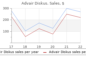
Buy generic advair diskus 500 mcg line
Transition planning helps the student identify career interests and select courses of study that will help him or her attain postsecondary or vocational goals asthma definition zen buy genuine advair diskus line. This coordination of services may include preparation for postsecondary education, vocational training, job shadowing or community-based apprenticeships, and/or training in independent living skills. Tier 1 specifies high-quality, scientifically based instruction for all students that is provided by qualified personnel to ensure that academic or behavioral difficulties are not due to inadequate instruction. All students are screened on a periodic basis to establish an academic and behavioral baseline and to identify struggling learners who need additional support. Students identified as being "at risk" through universal screenings and/or results on state- or districtwide tests receive supplemental instruction during the school day in the regular education classroom. At the end of this period, students who show significant progress are returned to the general education curriculum. On the basis of levels of performance and rates of progress, Tier 2 specifies provision of increasingly intensive instruction matched to the needs of the struggling learner. These interventions are provided in small group settings in addition to instruction in the general education setting. Students who do not show adequate progress are considered for more intensive interventions as part of Tier 3. The data collected during Tiers 1, 2, and 3 are included, along with the results of psychoeducational evaluation, and are used to make the eligibility decision. Curriculum-based assessment utilizes local norms gathered from universal screening within a single school district. As such, the cutoff for inclusion in the "at risk" group may differ significantly from one district to another. Those programs include public school districts, institutions of higher education, and other state and local education agencies. To qualify under Section 504, disability is defined broadly as a physical or mental impairment that substantially limits one or more major life activities. When a student is determined to be eligible for services under Section 504, the school must eliminate barriers to his or her access to full participation in school activities, including the general education curriculum. Examples of accommodations may include testing in a quiet room, preferential seating, audio textbooks, modified homework assignments, or a sign language interpreter. A 10-chapter English Learner Tool Kit8 followed that provided state and local educational agencies with guidelines to ensure that students who are English language learners receive the high-quality services they need to be college and career ready. The following are among these guidelines: Schools must identify, in a timely manner, students who are English learners in need of language assistance services by using valid and reliable assessment tools and communicate these results to parents or legal guardians. Schools must offer students who are English learners services and programs until they are proficient in English and can participate meaningfully in educational programs without English learner supports. Schools must provide appropriate special education services to students who are English learners with disabilities who are found to be eligible for special education and related services. Gifted and Talented Students Children who are gifted comprise 5% to 20% of the general school-age population, depending on how "gifted" is defined, or depending on the criteria being used to identify students who are gifted. Rather, the majority of programs and services that gifted and talented students receive are determined by state laws and policies and funded at the state and local level. State laws that define 492 American Academy of Pediatrics Developmental and Behavioral Pediatrics gifted and talented programming and teacher training requirements, along with available funding for gifted education, vary widely. Most states and school districts use the following definition:10 "Children and youth with outstanding talent perform or show the potential for performing at remarkably high levels of accomplishment when compared with others of their age, experience, or environment. These children and youth exhibit high capability in intellectual, creative, and/or artistic areas, possess an unusual leadership capacity, or excel in specific academic fields. Outstanding talents are present in children and youth from all cultural groups, across all economic strata, and in all areas of human endeavor (p. The primary pediatric health care professional also may consult with district school psychologists about the identification process and programming unique to a given school. Educational Advocacy Navigating the special education process is often overwhelming and confusing for families. Because school is central to the lives of youth with developmental-behavioral disorders, the primary pediatric health care professional who is knowledgeable about psychoeducational evaluation, special education, and related services will best be able to advocate for supports. The behaviors must result in clinically significant impairment in academic, social, or occupational functioning, and criteria cannot be met for antisocial personality disorder. To meet criteria for the diagnosis, there must be 3 behavioral outbursts involving damage, destruction of property, and/or physical assault within a 12-month period. An individual with intermittent explosive disorder must have a chronological age (or equivalent developmental level) of at least 6 years. The recurrent outbursts are not premeditated, are grossly out of proportion to any trigger, are associated with significant impairment in functioning, and are not better explained by another mental health or medical disorder. Epidemiology of Disruptive Behavior Disorders It may not be possible to determine the prevalence of disruptive behaviors because many do not cause sufficient impairment to warrant a medical diagnosis. However, noncompliant and oppositional behaviors comprise some of the most common concerns among parents and are the most frequently reported behavior problems seen by primary care pediatricians. Parents often place demands on their children, such as cleaning up their toys, and some of these demands may seem aversive to the child. The child may respond to this aversive event by displaying a coercive response, such as yelling or having a tantrum. The child is likely to continue to yell or have a tantrum when future demands are placed because the child has learned that he will get his own way by doing so. Another response the parent could make to the yelling or tantrum is to try to comfort the child and explain the reason for the request. If any of these maladaptive parent-child interactions continue over time, a persistent and worsening pattern of noncompliance and defiance may develop. Important Points to Consider When Taking the History Although children may present to the primary pediatric health care professional with any number of disruptive behaviors, the most common referral symptoms for disruptive behavior disorders are fighting, stealing, lying, cruelty, fire-setting, substance abuse, and sexual misconduct. At different ages, children display different types of disruptive behaviors, with property and status offenses more prevalent at older ages. For example, in a child who displays aggression, it is important to determine what type of aggression the child shows, such as verbal, physical, etc, and to whom the aggression is directed, such as parents, other children, animals, etc. It is also important to ask parents, teachers, and other adult caregivers how disruptive these behaviors are. Has your child, in the past 3 months, been spiteful or vindictive, or blamed others for his or her own mistakes How often has your child been angry and resentful or deliberately annoying to others Areas to assess include attention, level of activity and impulsiveness, social interactions, and communication skills. Other factors to consider when evaluating a child with disruptive behaviors include anxiety, mood disorders, cognitive and/or learning problems, substance abuse, and history of physical or sexual abuse. Supplements to Clinical Interview Standardized questionnaires can aid the primary pediatric health care professional in evaluating disruptive behavior. These include information about oppositional and disruptive behaviors, and thus can be useful. Finally, the Modified Overt Aggression Scale20 is another useful supplement to the clinical interview in evaluating disruptive behavior. Outside of research purposes or a medical history and physical examination indicating abnormal neurological status, neuroimaging is not recommended. In addition, if sexual abuse or unprotected sexual activity is present, testing for sexually transmitted infections may be warranted. Risk Factors for the Development of Disruptive Behavior Disorders Biological Genetic Antenatal and perinatal complications Brain injury, brain disease Male sex Environmental toxins, such as lead Individual Cognitive impairment Difficult temperament Aggressiveness Hyperactivity, impulsivity Attention problems Language impairment Reading problems Family Single-parent household or family divorce Domestic violence Lack of permanent family Parental substance abuse or antisocial behavior Child maltreatment or neglect Parent-child conflict Excessive parental control Lack of parental supervision Maternal depression or anxiety Social/School Low socioeconomic status Rejection by peers Association with deviant peers History of being bullied Neighborhood violence Disorganized or dysfunctional school Intense exposure to violence via media One interesting study evaluated the effect of poverty on the prevalence of oppositional and conduct disorders. In this study, 9- to 13-year-old rural children, of whom onequarter were Native American and the remaining were predominantly white, were given annual psychiatric examinations for 8 years. Halfway through the study, a casino opened up on the reservation, which gave every Native American family an income supplement, moving 14% of study families out of poverty. Reducing poverty among Native American families resulted in a reduction of oppositional and conduct problems in their children. However, neuroimaging studies have found the frontal 501 Chapter 21: Disruptive Behavior Disorders lobe to be associated with violence and aggression.
Diseases
- Chronic granulomatous disease
- Sclerocornea, syndactyly, ambiguous genitalia
- Osteopathia condensans disseminata with osteopoikilosis
- Non-small cell lung cancer
- Compartment syndrome
- Faciocardiomelic dysplasia lethal
Purchase advair diskus online from canada
Intravascular thrombosis Various mechanisms leading to capillary thrombosis have been demonstrated in patients with non-traumatic osteonecrosis asthma symptoms in 12 month old safe advair diskus 250 mcg. Usually this takes the form of a linear tangential fracture close to the articular surface, possibly due to shearing stress. However, until very late the articular cartilage retains its thickness and viability. The articular cartilage is obviously intact and the subchondral bone is well vascularized. The necrotic segment (B) has a texture similar to that of normal bone, but it may develop fine cracks. In the later stages the necrotic bone breaks up and finally the joint surface is destroyed. Local tenderness may be present and, if a superficial bone is affected, there may be some swelling. Imaging X-ray the early signs of ischaemia are confined to the bone marrow and cannot be detected by plain X-ray examination. X-ray changes, when they appear (seldom before 3 months after the onset of ischaemia), are due to (1) reactive new bone formation at the boundary of the ischaemic area, and (2) trabecular failure in the necrotic segment. Occasionally the necrotic portion separates from the parent bone as a discrete fragment. However, it is now recognized that in the case of the femoral head and the medial femoral condyle such necrotic fragments may have resulted from small osteoarticular fractures which only later failed to unite and lost their blood supply. This represents an undisplaced subarticular fracture in the early necrotic segment. At this stage the femoral head may still be spherical and (unlike osteoarthritis) the articular space is still well-defined. Radioscintigraphy Radionuclide scanning with 99mTc sulphur colloid, which is taken up in myeloid tissue, may reveal an avascular segment. More often, however, the picture is dominated by increased activity, reflecting hyperaemia and new bone formation in the area around the infarct. However, it does show the area of bone destruction very clearly and it may be useful in planning surgery. A cannula introduced into the metaphysis enables measurements to be taken (1) at rest and (2) after rapid injection of saline. Venous stasis can also be demonstrated by venography after injection of radio-opaque medium into the bone. Similar findings have been recorded in osteoarthritis, but the change is not nearly as marked as in osteonecrosis. All these areas are located beneath convex articular surfaces; osteonecrosis is seldom seen beneath a concave articular surface. Stage 2 is the reparative stage where the femoral head contour is still normal but there are early signs of reactive change in the subchondral area. There are clear-cut X-ray signs of osteonecrosis with evidence of structural damage and distortion of the bone outline. Two general observations can be made: (1) the size of the ischaemic segment is determined at a very early stage and it rarely increases after that; (2) small lesions which do not involve the maximally loaded zone of the articular surface tend not to collapse, whereas large lesions extending under the maximally loaded articular surface break down in over 60% of cases. There may be a record of high-dosage corticosteroid administration; for example, after renal transplantation. Alcohol abuse is often difficult to determine because patients tend to hide the information. Unfortunately, the tests are expensive and there is understandable resistance to adopting this approach in routine management. Prevention Where risk factors for osteonecrosis are recognized, preventive steps can be taken especially in the management of corticosteroid medication and alcohol abuse. It is important also to be aware of the cumulative effect of even moderate doses of corticosteroids in patients with a history of alcohol abuse. Decompression procedures for divers and compressed-air workers should be rigorously applied. Only general principles will be discussed here; the treatment of osteonecrosis in specific sites is dealt with in the appropriate chapters on regional orthopaedics. Lesions in heavily loaded joints have a poor prognosis and will probably end in structural failure if left untreated. Simple measures to reduce loading of weight-bearing joints may help, though their value has not been proven. Some lesions heal spontaneously and with minimal deformity; this is seen especially in areas which are not heavily loaded: the non-weight-bearing joints, the superomedial part of the femoral head and the nonweight-bearing surfaces of the femoral condyles and talus. There is low-level evidence that drug therapy (such as bisphosphonates, statins and low molecular weight Once there is structural damage and distortion of the articular surface, conservative operations are inappropriate. Three options are available: (1) non-operative management, concentrating on pain control, modification of daily activities and, where appropriate, splintage of the joint; (2) arthrodesis of the joint. A cumulative dose of 2000 mg of prednisone equivalent administered over several years. It is important to bear in mind that multiple causative agents have an additive effect; thus, osteonecrosis has been encountered after comparatively short courses and low doses of corticosteroids (totals of 800 mg or less), but in these cases an additive factor can almost always be identified. In recent years it has spread more widely in Europe but it is rarely encountered south of the equator. Sickle-cell disease is most likely in homozygous offspring (those with HbS genes from both mother and father), but it may also occur in heterozygous children with HbS/C haemoglobinopathy and HbS/ thalassaemia. Inheritance of one HbS gene and one normal -globin gene confers the (heterozygous) sickle-cell trait; HbS concentration is low and sickling occurs only under conditions of hypoxia. In the established disorder, the main clinical features are due to a combination of chronic haemolytic anaemia and a tendency to clumping of the sickle-shaped cells which results in diminished capillary flow and recurrent episodes of intracapillary thrombosis. Secondary changes such as trabecular coarsening, infarctions of the marrow, periostitis and osteonecrosis are common. Complications include hyperuricaemia (due to increased red cell turnover) and an increased susceptibility to bacterial infection. Clinical features Children during the first two years of life may present with swelling of the hands and feet. In older children a typical feature is recurrent episodes of severe pain, sometimes associated with fever. Other bone changes are due to a combination of marrow hyperplasia and medullary infarctions. Instead of having an X in the Hb beta chain, the replacement of this residue with Y results in Hb precipitation at low oxygen tensions. In deoxygenated blood there is increased aggregation of the haemoglobin molecules and distortion of the red cells, which become somewhat sickle-shaped. At first this is reversible and the cells reacquire their normal shape when the blood is oxygenated. Eventually, however, the red cell membrane becomes damaged and the cells are permanently deformed. The sickle-cell trait, which originated in West and Central Africa centuries ago, is an example of natural selection for survival in areas where malaria was endemic. Anaesthesia carries definite risks; failure to maintain adequate oxygenation may recipitate vascular occlusion in the central nervous system, lungs or kidneys. Prophylactic antibiotics are advisable as the risk of postoperative infection is high. Under increased air pressure the blood and other tissues (especially fat) become supersaturated with nitrogen; if decompression is too rapid, the gas is released as bubbles, which cause local tissue damage, generalized embolic phenomena and intracapillary coagulation. Prolonged compression may also cause swelling of marrow fat cells and decreased intramedullary blood flow, possibly due to oxygen toxicity. In the most acute cases there can be circulatory and respiratory collapse, severe neurological changes, coma and death. Management the aim is prevention; the incidence of osteonecrosis is proportional to the working pressure, the length of exposure, the rate of decompression and the number of exposures. Strict enforcement of suitable working schedules has reduced the risks considerably. Trabecular coarsening and thickening of the cortices may be mistaken for signs of infection.
Buy advair diskus 500mcg
Patients develop pain asthma definition yay buy cheap advair diskus line, swelling and tenderness over the sternoclavicular region and X-rays show hyperostosis of the medial ends of the clavicles, the adjacent sternum, the anterior ends of the upper ribs and the soft tissues in between. Biopsy is of little help; the histological changes are non-specific and microorganisms have not been identified. A peculiarity which links this condition with the next is an association with pustular lesions on the palms and soles (palmoplantar pustulosis) and pustular psoriasis. X-rays these show the characteristic features of osteoarthritis; the changes are often bilateral, even though only one side may be hurting. Treatment the initial treatment is non-surgical with activity modification analgesics or steroid injections. If this is ineffectual, pain may be relieved by excision of the lateral end of the clavicle. Trimming of the bony roughness, or excision of the outer end of the clavicle, may also be needed during subacromial decompression for rotator cuff impingement. Arthroscopic repair of Bankart lesions produces results comparable to those obtained by open surgery. Subsequent modifications and the introduction of glenoid resurfacing broadened the indications to include other disease processes, including end-stage glenohumeral osteoarthritis and rheumatoid arthritis. At the same time a biopsy can be taken which may assist in the diagnosis of synovial disorders such as rheumatoid arthritis or pigmented villonodular synovitis. There has been a transition over the last 20 years from its use in diagnosis to that of repair and reconstructive procedures. The relative merits of total shoulder arthroplasty and hemiarthroplasty are not clear. Glenoid resurfacing is contraindicated if inadequate bone stock or irreparable rotator cuff tears (or both) are present. On the other hand, individual studies have reported less consistent pain relief with isolated humeral head replacement. With isolated humeral head replacement, the glenoid can undergo progressive erosion over time, often leading to deteriorating results. A number of techniques have been reported with extra-articular arthrodesis, intra-articular arthrodesis and a combination of both. Extra-articular arthrodesis is primarily a historic procedure that was used before the antibiotic era to treat tuberculosis. A variety of methods of internal fixation for intra-articular arthrodesis have been described. It is generally agreed that internal fixation is desirable because it maintains the position of the arthrodesis and can decrease the length of time spent in plaster immobilization. The optimal position is 30 degrees of flexion, 30 degrees of abduction and 30 degrees of internal rotation. Complications the commonest, in order of frequency, are loosening of the components, glenohumeral instability, rotator cuff failure, periprosthetic fracture, infection and implant failure. Glenoid fixation remains a challenge; lucent lines around the glenoid component are very common but are not always symptomatic. Arthroplasty for fractures, avascular necrosis or proximal humeral tumours gives good pain relief and shoulder movement, although power is always diminished. Where there is more extensive joint destruction and disruption of the soft tissues (for example, in rheumatoid arthritis), pain relief is still excellent but the range of movement is only moderately improved. The greater the integrity of the surrounding soft tissues (and especially the rotator cuff), the more stable the new joint will be, and thus the better the outcome of the operation. In severe cuff failure, reverse geometry arthroplasty has been used with reasonable success in the short term in elderly patients, although further research is needed to assess longevity and continued functional improvement. Outcome Despite the restriction of glenohumeral movement, postoperative function is surprisingly good; and of course the joint is free of pain! Complications include non-union, infection, malposition often with too much internal rotation, prominence of the internal fixation and fracture of the humerus. Stability the shallow glenohumeral articulation has little inherent stability because the glenoid surface area is only one-quarter that of the humeral articular surface. The extent to which the socket is deepened by the labrum may seem trivial, but it must be significant because labral tears are associated with dislocation. The muscles provide kinetic stability: during abduction the rotator cuff muscles draw the head of the humerus firmly into its socket while the deltoid elevates the arm. Imagine what would happen if the deltoid muscle acted alone in abducting the shoulder. Because of the relatively unstable fulcrum, the deltoid would simply shrug the arm upwards at the side of the body. In reality, the rotator cuff muscles, particularly the supraspinatus, draw the head of the humerus firmly into the socket and slightly downwards, thus allowing the deltoid to act as a true abductor. The first 30 degrees of abduction occurs almost entirely at the glenohumeral joint with slight movement of the clavicle at the sternoclavicular joint. From 30 to 90 degrees of abduction the scapula gradually comes into play, with about one-third of the movement coming from the scapula rotating on the thorax. As the arm rises above shoulder height, it rolls into external rotation so that the greater tuberosity clears the projecting acromion. The sternoclavicular joint participates in movements close to the trunk (for example, shrugging or bracing the shoulders); the acromioclavicular joint moves in the last 60 degrees of abduction. Rotator cuff the rotator cuff is a sheet of conjoint tendons closely applied over the top of the shoulder capsule and inserting into the greater tuberosity of the humerus. It is made up of subscapularis in front, supraspinatus above and infraspinatus and teres minor behind. Separating the tendons from the arch, and allowing them to glide, is the subacromial bursa. Of the four cuff tendons, the supraspinatus is the most exposed; it runs over the top 381 the elbow 14 Loss of function may be significant with elbow pathology because the role of the elbow is to position the hand in space to manipulate the environment and bring objects towards the individual. Pain from the dorsal aspect of the elbow is likely to be due to olecranon bursitis or rarely triceps tendinopathy; anterior elbow pain is likely to be due to distal biceps tendinopathy. Pain in childhood or adolescence requires further investigation to rule out osteochondritides. Remember that pain in the elbow is sometimes referred pain from the cervical spine. Stiffness, which means loss of range of movement, may develop slowly and will not be noticed until it is relatively marked. It can be very disabling, making it hard for the patient to reach their mouth (loss of flexion) or perform simple toileting (loss of supination). A sudden symmetrical loss of range suggests an increase of fluid within the joint which may be synovial fluid (effusion), blood or pus. A localized soft-tissue lump on the point of the olecranon is likely to be an olecranon bursitis. Note that the carrying angle cannot be assessed reliably if there is a flexion contracture of the elbow. Varus and valgus deformities (cubitus varus and cubitus valgus) are usually the result of trauma around the elbow. A varus deformity can be demonstrated by asking the patient to abduct the shoulders to 90 degrees with the palms facing forwards: the arm takes on the appearance of a rifle butt (gunstock deformity). Look for scars around the elbow, in particular posterior to the medial epicondyle that may be related to previous ulnar nerve surgery.
Buy advair diskus us
They may avoid situations or insist on the presence of a companion due to perceived difficulty escaping or being unable to find help in the event of incapacitating or embarrassing symptoms asthma treatment kerala discount advair diskus online master card. These children or adolescents usually find it difficult to control their worries and have concerns related to their competence or quality of their performance, and whether or not others are evaluating them. They exhibit at least 3 symptoms that include feeling on edge, fatigue, difficulty concentrating, irritability, muscle tension, and sleep disturbance. Functional impairment is often secondary to the associated symptoms and the considerable time spent worrying. Obsessions and compulsions may be related to themes of contamination, symmetry, and fear of harm to oneself and others. Children aged 6 years or younger may not experience the traumatic event themselves but may witness it. Symptoms include: (1) intrusion symptoms, such as re-experiencing recurrent and distressing memories and dreams, dissociative reactions, intense psychological distress, and/or marked physiological reactions; (2) persistent avoidance of associated stimuli; (3) negative alterations in cognition and mood associated with the traumatic event or feelings of detachment; and (4) marked alterations in arousal and reactivity associated with the traumatic event. Anxiety Screening Tools Structured and semistructured diagnostic interviews, self-report rating scales, and clinician-rated instruments are the most common methods for identifying and measuring anxiety in the pediatric population. Ideally, information should be obtained from multiple informants, including parents and teachers. Younger children should be screened with parent report measures or interviewed with the use of visual aids, such as a feeling or mood thermometer. If the screening measures are positive for anxiety symptoms, the primary pediatric health care professional should determine which anxiety disorder might be present, the severity, and the degree of functional impairment. Specific phobias and dissociative disorders have also been known to occur in response to a traumatic event. Finally, any neurological damage that may have occurred due to the traumatic event should be assessed. Treatment Due to the shortage of mental/behavioral health professionals in almost all communities, primary pediatric health care professionals often need to become involved with the treatment and ongoing symptom reassessment of children with anxiety disorders. In follow-up studies of children who received treatment for their anxiety disorders, there was a decreased risk of developing another mental health disorder after 3 to 4 years. Measures overall anxiety levels and academic stress, test anxiety, peer and family conflicts, and drug problems. Screening Tools for the Identification of Obsessive-Compulsive Disorder Approximate Time to Complete May take up to 120 minutes to administer and score. Screening Tools for the Identification of Traumatic Experiences and Symptoms of Posttraumatic Stress Disorder No. Cognitive behavioral therapy works by examining the relationship between cognitions, behaviors, and feelings. Individual programs, such as Coping Cat, have been shown to be effective in treating children with anxiety disorders by reducing symptoms and functional impairment,53 and these improvements have been shown to be sustained up to 1 year later. The quality of the parent-child relationship and parenting behaviors have been associated with anxiety symptoms in children. Accommodation predicts symptom severity, degree of child impairment, family dysfunction and distress, and poor treatment outcomes. The greatest safety evidence otherwise is for fluvoxamine, which had the highest rate of clinical response and tolerance in a comparison meta-analysis of 16 clinical trials. In addition, fluoxetine, fluvoxamine, and paroxetine were better tolerated than sertraline and venlafaxine. When remission is achieved, treatment should be continued for at least 6 to 12 months before consideration of discontinuing the medication. Gastrointestinal symptoms tend to be self-limited and are rarely severe enough to warrant discontinuation. More significant adverse reactions may include disinhibition, agitation, mania, or psychosis. There is an increased risk during the first 9 days of treatment or if the starting doses are higher than usual, confirming the need for close follow-up during the titration period. Serotonin syndrome is a triad of mental status changes, autonomic hyperreactivity, and neuromuscular abnormalities and symptoms that can range from mild to life-threatening. Emergency treatment of anxiety symptoms with benzodiazepines has been effective for adults due to their rapid onset, although randomized controlled trials using alprazolam were not found to be any more effective than placebo in reducing anxiety symptoms in children. These include clomipramine and fluvoxamine in children 10 years of age or older, fluoxetine, and sertraline. It is also important not to convey a sense of blame to parents for accommodations they are providing but instead empower them by explaining how a change in their behavior can help their child. It is critical not to "take sides" with one or the other parent but encourage them to support each other in changing practices. They are associated with negative academic, social, and health outcomes, including mood disorders in adulthood, substance abuse, early pregnancy and parenthood, and increased suicide risk. Although the diagnosis of mood disorders in the pediatric population is based primarily on adult criteria,2 symptoms of mood disorders in children and adolescents may differ from those of adults. Importantly, irritability may be a more pronounced symptom than depressed mood or mania; also, what may appear to be boredom can instead be lack of motivation or fatigue. Having a negative or maladaptive coping style,85 a sense of hopelessness,86 and low self-esteem are considered personality risk factors. Medical risk factors include low birth weight, a history of concussion87 or traumatic brain injury,88 chronic medical illness, and prior diagnoses of anxiety and conduct disorders. Comorbidity is common in mood disorders, and studies have shown that 50% to 90% of youth with depression will have a comorbid diagnosis. In autopsy studies, 50% to 60% of adolescent suicide victims had a depressive disorder at the time of death. There is evidence that stress induces modifications in the hypothalamic-pituitary-adrenal axis, and the overproduction of stress hormones has been found to cause reduced neurogenesis in the hippocampus, which has been associated with adolescent and adult depression. However, no specific gene has been identified, and bipolar disease is considered genetically complex. Diagnostic changes include the addition of new diagnoses: disruptive mood dysregulation disorder and persistent depressive disorder (which replaces dysthymia and chronic major depressive disorder). Additional changes include the addition of the specifier "with mixed features" (ie, features of both mania and depression) to both depressive and bipolar disorders, and the removal of bereavement as an exclusion for depression. The common feature of all these disorders is the presence of sad, empty, or irritable mood accompanied by functional impairment. In children, depression may present differently depending on the development of the child. However, retrospective analyses of adult patients with bipolar disorder suggested that symptoms began much earlier than previously believed, and analyses of several longitudinal research samples suggested that children can present with symptoms of mania. Mania manifests as at least 3 of the following (4 if mood is only irritable): inflated self-esteem or grandiosity, decreased need for sleep (getting by with little or no need for sleep, as opposed to insomnia, which is difficulty falling or staying asleep), increased talking or pressured speech, flight of ideas, racing thoughts, distractibility, an increase in goal-directed activity or psychomotor agitation, and excessive involvement in activities with a potential for significant consequences. These manic episodes may have been preceded by and may be followed by periods of hypomania or major depression, although it is not necessary for diagnosis. Juvenile mania often presents as labile and erratic changes in mood and energy levels with irritability and belligerence being more common than euphoria. In young children, excessive silliness, hypersexuality, decreased need for sleep, and daredevil and reckless acts are also seen. Hypomania is defined as a distinct period of abnormally and persistently increased activity or energy, lasting at least 4 consecutive days and persisting most of the day, nearly every day. Several screening instruments are available for depression (as detailed in Table 22. Among these, the Beck Depression Inventory and the Patient Health Questionnaire for Adolescents reported the highest sensitivities (approximately 89%) and specificities (approximately 75%). Differential Diagnoses the main differential diagnoses of mood disorders are other mood disorders, as depression is often the first symptom of pediatric bipolar disorder, and youth with depressive disorders often experience manic episodes. As such, it is important to determine that the mood episode symptoms and behavior represent a significant departure from baseline functioning. Also, although youth with disruptive behavior disorders may defy bedtime rules, they do not typically have a decreased need for sleep. Grandiosity, hypersexuality, racing thoughts, and flight of ideas are also distinct characteristics of bipolar disorders. Children with generalized anxiety typically have more chronic symptoms, such as chronic irritability, restlessness, and impaired concentration. Scale and subscale scores report on emotional problems, functional problems, negative mood, physical symptoms, negative self-esteem, interpersonal problems, and ineffectiveness. The first two items are used to screen for depression, and if positive, the entire scale can then be used to assess symptom severity; question 9 screens for suicidal ideation.
Methyl-Synephrine HCl (Bitter Orange). Advair Diskus.
- How does Bitter Orange work?
- What other names is Bitter Orange known by?
- Dosing considerations for Bitter Orange.
- Are there any interactions with medications?
- Weight loss, nasal congestion, intestinal gas, cancer, stomach and intestinal upset, intestinal ulcers, regulating cholesterol, diabetes, chronic fatigue syndrome (CFS), liver and gallbladder problems, stimulation of the heart and circulation, eye swelling, colds, headaches, nerve and muscle pain, bruises, stimulating appetite, mild sleep problems (insomnia), and other conditions.
- Are there safety concerns?
Source: http://www.rxlist.com/script/main/art.asp?articlekey=96937
Purchase 500mcg advair diskus overnight delivery
Although there has historically been concern that stimulants may worsen tic behaviors asthma va disability rating buy advair diskus 500 mcg, tics wax and wane over time; increased frequency of tics may be due to the natural history of the disorder rather than to stimulant use. Finally, much concern has been raised regarding the possible cardiac side effects of stimulant use. Clinicians are urged to document pertinent negative findings in the medical record prior to beginning treatment with a stimulant. Alpha-2 Agonists Alpha-2 agonists, available as clonidine and guanfacine, have also demonstrated efficacy in the treatment of hyperactivity and inattention. Clonidine was initially developed in the 1960s as a treatment for hypertension and acts on the alpha-2 receptors 2A, 2B, and 2C in the prefrontal cortex. Guanfacine was developed in the late 20th century and more specifically binds to alpha-2A receptors (therefore exerting less effect on blood pressure). Alpha-2 receptors in the prefrontal cortex have been implicated in the maintenance of attention and focus. It is theorized that these medications enhance the transmission of dopamine and norepinephrine in this region, albeit through different mechanisms than with stimulants. In clinical practice, however, off-label use of immediate-release clonidine and guanfacine is common. Although alpha-2 agonists are not approved for other psychiatric disorders, they are sometimes used for the treatment of oppositional behavior, aggression, and delayed sleep onset. They may be used as monotherapy in individuals who have not been able to tolerate any stimulant. Unlike stimulants, alpha-2 agonists demonstrate coverage throughout the day and can be helpful particularly for individuals who struggle with attention problems in the afternoon or evening (after a stimulant has worn off). For short-acting alpha-2 agonists (typically given twice daily), clinicians should start with an evening dose (0. The medication dose can be increased in a stepwise fashion every 3 to 7 days to the maximum tolerated dose. Short-acting formulations typically come in tablets and can be divided in half for finer dose manipulation. Clinicians should start an initial evening dose (guanfacine extended-release 1 mg or clonidine extendedrelease 0. As with stimulants, dosing of alpha-2 agonists is generally limited by the side effects rather than directed by weight-based targets (although average and maximum doses can be seen in Table 23. The most common adverse effects of alpha-2 agonists include fatigue, sedation, and somnolence, with higher rates being reported with clonidine than with guanfacine. These effects are dose dependent and may decrease over time as an individual becomes accustomed to the medication; however, if these side effects do not improve within the first week, they are unlikely to resolve. For those taking a shortacting formulation, sedation may decrease if patients are changed to a long-acting medication (especially if it can be given in the evening). Anticholinergic effects, such as dry eyes, dry mouth, and constipation, can also be seen. However, sudden cessation of alpha-2 agonists, especially when individuals are taking higher doses, can precipitate a withdrawal syndrome, which includes increased blood pressure, headache, tremor, restlessness, and nausea. It is recommended that when stopping the medication, clinicians gradually taper the dose, with adjustments made every 3 to 7 days. It is theorized that atomoxetine may selectively inhibit mechanisms of norepinephrine reuptake in the synaptic clefts of the prefrontal cortex, resulting in similar neurochemical changes seen in stimulant use. As with to alpha-2 agonists, medication efficacy is consistent over the course of the day without the waxing and waning effects seen in stimulant therapy. Finally, unlike stimulants, atomoxetine has a low potential for abuse or misuse, making it an attractive option for clinicians or individuals who are uncomfortable with using controlled substances. In addition, it may take 2 to 4 weeks for atomoxetine to develop its therapeutic effect. However, restarting the medication requires a retitration to the target dose, and the effects may take 2 to 4 weeks to be seen; medication "holidays" are discouraged. Common side effects of atomoxetine include difficulty with sleep, decreased appetite, somnolence, irritability, gastrointestinal symptoms, and sexual dysfunction. There may be mild deleterious effects on adult height, although these changes appear to be reversible. There is no indication for obtaining serial liver function enzymes for monitoring purposes. Parents and individuals should be cautioned about the signs and symptoms of acute hepatitis and instructed to seek appropriate medical care if necessary. Vital signs, especially heart rate and blood pressure, as well as growth parameters, should be measured at each visit. Informal teacher reports or comments written on progress reports and report cards can also be valuable sources of information. Individuals may also provide self-reported symptom severity, although research suggests that teenagers and young adults may underreport impairments when compared with teacher and parent feedback. This should include a careful evaluation of the psychosocial history, including a history of substance use. Initial diagnostic indications for use included schizophrenia and bipolar disorder. Currently, aggression is the more common target of antipsychotic therapy in the pediatric population. Meta-analyses suggest that the classification of antipsychotics into "first" and "second" generation is a meaningless distinction, as each antipsychotic has an individual efficacy and rate of adverse events. Aripiprazole has been shown in several studies to be beneficial for nonsuicidal selfinjury; however, most of this research concerns adolescents and adults with borderline personality disorder or anxiety disorders who engage in practices such as cutting, carving, and burning. Sudden discontinuation of medication can result in symptoms of withdrawal, including agitation, activation, insomnia, psychosis, and dyskinesia. Although rates of individual side effects may differ from drug to drug, patients taking any antipsychotic should be closely monitored for the following potential adverse effects. Therefore, all patients prescribed antipsychotics should have blood glucose and lipid profiles monitored closely. In a joint statement issued by the American Diabetes Association, the American Psychiatric Association, and the American Association of Clinical Endocrinologists, an expert panel recommended the following:45 1. If possible, baseline screening prior to starting a medication (and referrals as appropriate), including a. A personal and family history of diabetes, dyslipidemia, hypertension, and cardiovascular disease b. Personal and family history, waist circumference, blood pressure, and fasting blood glucose annually 5. The D2 binding of antipsychotics can result in increased prolactin levels within 1 to 9 days. Hyperprolactinemia, defined as >20 ng/mL for adult men and >25 ng/mL for adult women, can result in issues such as sexual problems and gynecomastia. Adverse drug events such as gynecomastia have gained attention due to the increased frequency of lawsuits in which claimants allege the drug manufacturers did not provide sufficient warning about gynecomastia. Should symptoms of hyperprolactinemia develop, the clinician may choose to decrease or discontinue the antipsychotic or try a different antipsychotic. Clinicians should be careful to document discussions regarding the potential risk of hyperprolactinemia (as well as other adverse effects) when considering antipsychotic treatment. Symptoms include muscle rigidity, fever, altered consciousness, and autonomic dysfunction; these symptoms typically occur within 2 weeks of initiation of therapy or dose adjustments. Response to psychological or medication treatment should be monitored over time through the use of symptom rating scales administered on a periodic basis. Benzodiazepines, such as clonazepam, lorazepam, and alprazolam, have not been extensively studied in children, and thus have a limited role for medication treatment of pediatric anxiety. Common adverse effects include agitation, disinhibition, and impaired memory and learning. Clinicians should consider the risk of abuse, addiction, and diversion when prescribing a benzodiazepine. If symptomatic improvement is not evident after 6 to 8 weeks, then the dose should be increased again for another trial until either the maximum dose is reached or side effects become intolerable. These are usually transient but can be mitigated by reducing the dose or slowing the rate of upward titration. Psychiatric effects include disinhibition, agitation, mania or hypomania, and worsened anxiety.
Order 250 mcg advair diskus free shipping
Arthrodesis is widely considered to be the best option for dealing with painful instability in the radiocarpal joint asthmatic bronchitis relief buy advair diskus with american express. Bone grafts are not necessarily added but can be taken from the ulnar head if it is excised. In this group, ulnar head replacement rather than ulnar head excision should be considered if the patient has high demands from their upper limb. Furthermore the dominant wrist should, if possible, be fused in slight extension to provide reliable power grip, while the non-dominant wrist is fused in some flexion (or replaced) so as to provide the posture needed for perineal care. Partial excision of the radial styloid Osteoarthritis following a scaphoid fracture may be limited to that part of the joint. This can be done by open or arthroscopic means and at the same time a partial wrist denervation may be performed. Since these usually present as distinct syndromes, they are considered separately. The appearance may be normal but there is often swelling over the back of the wrist, and movements are limited and painful. The outcome of these procedures is similar (about 60% grip strength, 60% movement). Proximal row carpectomy is easier to perform and risks fewer complications; four-corner fusion may give a more stable grip in torsion. The radiocarpal and intercarpal joints are decorticated, bone graft is impacted and a plate is fixed to the third metacarpal and the distal radius. Long-term survivorship studies have yet to show whether replacement arthroplasty will hold up in patients with higher demands. Degenerative changes are seen in primary or secondary osteoarthritis (possibly following marked and long-standing instability of the joint). On examination, the joint is swollen and in advanced cases is held in an adducted position, with prominence of the subluxed metacarpal base. Stressing the joint by the examiner pushing the metacarpal head into adduction against the side of the index metacarpal, and by the examiner extending the thumb metacarpal backwards parallel to the index metacarpal, are the most sensitive tests. Imaging X-rays show narrowing and then lateral subluxation of the trapeziometacarpal joint. Treatment Asymptomatic radiological disease is common, affecting 60% of women over 60. Most symptomatic patients can be treated by reassurance that it will settle, anti-inflammatory preparations, local corticosteroid injections and temporary splintage. Attempts have been made to prevent postoperative collapse of the joint and proximal migration of the metacarpal by rerouting a slip of flexor carpi radialis or abductor pollicis longus tendon. Replacement arthroplasty Replacement arthroplasty using a silicone spacer has a high complication rate and the results are unpredictable. Metal-onpolyethene implants and pyrocarbon implants are also available but they are experimental and long-term durability is uncertain. There is no evidence that implants are better than trapeziectomy, except that the earlier recovery is quicker with an implant. Arthrodesis Arthrodesis of the trapeziometacarpal joint relieves pain, but the restriction of movement and high failure rate are distinct drawbacks. If the metacarpophalangeal joint has been secondarily damaged by hyperextension, then either a sesamoid arthrodesis (which restores flexion but preserves movement) or fusion (at 25 degrees for stable pinch) is indicated. Excisional arthroplasty Excision of the trapezium gives pain relief and return of function, though thumb pinch is always weak. Treatment may be by (c) excision of trapezium, (d) arthrodesis, (e,f) silastic replacement or (g) total replacement. The patient points to the front of the scaphoid tubercle as the source of the pain (whereas in carpometacarpal arthritis the patient points to the back of the thumb base). Normal X-rays do not show the disease; a lateral in 25 degrees supination is needed. Excision of the distal pole of the scaphoid is easier but can cause midcarpal collapse unless only the smallest amount is removed. Trapeziectomy with undercutting of the trapezoid is straightforward, especially if there is concomitant trapeziometacarpal arthritis. Pyrocarbon interposition arthroplasty is an experimental option, but whether this has any immediate or long-term advantage over simple excision is unknown. Occasionally, surgical excision is undertaken with excellent results unless the immediately adjacent ulnar nerve is damaged. Tenosynovitis can be caused by unaccustomed overuse but sometimes it occurs spontaneously. Early treatment, including rest, anti-inflammatory medication and injection of corticosteroids, may break this vicious circle. The first dorsal compartment (abductor pollicis longus and extensor pollicis brevis) and the second dorsal compartment (extensor carpi radialis brevis) are most commonly affected. The differential diagnosis includes arthritis at the base of the thumb, scaphoid non-union and the intersection syndrome (see below). Resistant cases need an operation, which consists of slitting the thickened tendon sheath. Sometimes there is duplication of tendons and even of the sheath, in which case both sheaths need to be divided. Care should be taken to prevent injury to the dorsal sensory branches of the radial nerve, which may cause intractable dysaesthesia. It may be initiated by overuse but it also occurs spontaneously, particularly in middle-aged women, and sometimes during pregnancy. There is usually an associated tenosynovitis within the second extensor compartment containing extensors carpi radialis longus and brevis. There may be a history of unaccustomed activity such as pruning roses or wringing out clothes. Sometimes there is a visible swelling over the radial styloid and the tendon sheath feels thick and hard. In a positive test this is acutely painful; repeating the movement with the thumb left free is relatively painless. Treatment Treatment involves rest, splintage, steroid injection and, in resistant cases, surgical widening of the second compartment and exploration of the intersection. Patients present with pain and crepitus on the dorsum of the wrist; flexing and extending the fingers produces a fine, palpable crepitus over the common extensor compartment. This sheath can give way after a sudden supination stress (usually in tennis or rugby). Surgical repair is usually required and involves either a direct reattachment or a reconstruction using a patch of retinaculum. Epidemiological studies suggest that these conditions are no more common among keyboard operators than in the general population. Treatment of these conditions is the same as for the other types of tenosynovitis. On examination there is a discrete tender point over the back of the midcarpus and the pain is reproduced by full passive wrist extension. Treatment should be initially with a steroid injection; if that fails, then arthroscopic excision may succeed. It arises from leakage of synovial fluid from a joint or tendon sheath and contains a glairy, viscous fluid. Although it can appear anywhere around the carpus, it usually develops on the dorsal surface of the scapholunate ligament.
Buy genuine advair diskus line
These movements are then repeated but carried out against resistance asthmatic bronchitis duration buy advair diskus with mastercard, to test for muscle power. The wrist X-ray should be taken in a standard position of mid-pronation with the elbow at 90 degrees; often both wrists must be X-rayed for comparison. Moving the wrist under image intensification is useful to investigate some cases of carpal instability. The embryonic arm buds appear about 4 weeks after fertilization and from then on the limbs develop progressively in three axes, from proximal to distal, radial to ulnar and dorsal to palmar. By 6 weeks the digital rays begin to appear and then develop in concert with the general mesenchymal differentiation that gives rise to the primitive skeleton and muscles. Growth and apoptosis (genetically programmed cell death) result in modelling of the limbs and the formation of joints and separate digits. The process is more or less complete by the end of the eighth week after fertilization, at which time primary ossification centres begin to appear in the long bones. Ossification centres in the epiphyses and carpal bones do not emerge until after birth, so X-rays in the neonatal period must be interpreted with this in mind. Malformations may occur during embryonic development because of defective formation or incomplete separation of mesenchymal components, the former accounting for partial or complete absence of a part and the latter for coalitions between adjacent elements. Congenital limb anomalies can be sporadic or syndromic (associated with other abnormalities). The overall incidence of congenital upper-limb anomalies is estimated to be about 1 in 600 live births, and many are not severe enough to require operative treatment. Malformations may be caused by heritable genetic mutations, or intrauterine damage. The thickness of the cuts may be too large to detect injury to thin structures such as the lunotriquetral ligament, scapholunate ligament or triangular fibrocartilage but modern scanners have ever-increasing sensitivity and specificity. Defects in the triangular fibrocartilage, scapholunate ligaments or lunotriquetral ligaments can be identified by arthrography. Radionuclide scan A localized area of increased activity may reveal an osteoid osteoma, an occult scaphoid fracture or early osteoarthritis. Fluoroscopy Fluoroscopic examination may be needed to demonstrate some patterns of carpal instability. Ligament tears, articular cartilage damage, osteoarthritis, occult ganglia, synovitis and triangular fibrocartilage lesions can be recognized and in some cases treated. These groups are then divided into four subgroups of abnormal axis formation/ differentiation: (1) proximal to distal axis, (2) radial to ulnar axis, (3) dorsal to ventral axis, and (4) unspecified axis, and there are a number of further subcategories in each. Investigations It may be useful to obtain radiographs of the contralateral normal limb for comparison. Remember that many congenital wrist and hand anomalies are part of a larger syndrome. For example, skeletal, cardiac, haematological, gastrointestinal, renal or craniofacial anomalies are commonly associated with radial dysplasia, and these should be sought. Children with radial dysplasia (and/or thumb hypoplasia) or thumb duplication should also be tested for Fanconi anaemia. Genetic counselling should be made available for inherited or unusual conditions, and it may be helpful in reaching a diagnosis. There may be issues of maternal guilt, parental anger and resentment, as well as unrealistic expectations about the outcome and possibilities of surgery. It is important to gain the confidence of the family at the initial consultation; remember that these children are likely to be longterm patients. They must be given a diagnosis, an indication of prognosis, reassurance about the future and a longterm plan of treatment, including a schedule of surgery, which may require staged operations over a number of years. It follows that in some conditions the indication for surgery is clear, for example release of ring constrictions causing limb ischaemia in a neonate. In others, such as treatment of adolescent camptodactyly, decision making may be more difficult. The hand must be considered in the context of the whole upper limb as well as other systemic conditions. For example, syndactyly between digits of unequal length will lead to deviation of the longer toward the shorter digit, and for this reason they should be treated early before deformity becomes established. One exception is ring constriction syndrome, in which the fingertips may be tight and painful with poor soft-tissue cover. Although as a general principle we try to preserve digital length, these are improved by minimal shortening to provide pain-free, durable soft-tissue cover and a more pleasing appearance. For those who are old enough to talk to you, some key questions can help to initiate conversations: for example, who is your best friend at school, what is your favourite subject at school, which sports team do you support Psychological factors should always be considered; teasing usually starts around the age of 6, and the early involvement of clinical psychologists may be helpful. The clinic waiting room provides a useful opportunity for families to share experiences, as do patient support groups. White coats, crowded clinic rooms, and other intimidating hospital paraphernalia should be avoided. Toys encourage children to play in an unconstrained manner, which allows close observation of their hand function (it is impossible to make young children demonstrate hand movements). Absence of skin creases at joints indicates lack of movement, either due to lack of motors, or stiffness (tight soft tissues, abnormal or absent joint). Proximal forearm Children adapt surprisingly well to congenital limb absence, particularly as they still have sensibility and proprioception at the distal end of the abbreviated limb, the lack of which limits prosthetic use. Some may learn to use prosthetics, both for function and cosmesis, and uptake is likely to increase with improved technology. The results of non-vascularized transfer of a toe phalanx into the existing skin envelope are disappointing, and this technique is falling out of favour. Treatment Mild radial dysplasia is treated from birth by gentle stretching and splintage, best done by the parents. More serious cases can be treated by distraction, prior to radialization with a tension-free soft-tissue correction. Prolonged pin fixation and splintage is still required to avoid recurrence of the deformity. Attention must be paid to the elbow; if the joint is stiff, the radially deviated wrist can actually be advantageous, as the child can then get the hand to his or her mouth (for eating) and the perineum (for toilet care). Surgical correction of the wrist in these cases can result in a functional disaster. This condition, also known as phocomelia, may affect more than one limb and is sometimes associated with craniofacial deformities. When there is no basal joint or the thumb is absent, pollicization of the index finger can be performed (as long as the index finger is not also hypoplastic, when it will not make a good thumb). Ulnar longitudinal deficiency this is approximately half as common as radial dysplasia (1 in 100 000), and about half of these patients have associated musculoskeletal problems. Children may present with ulnar deviation of one or both wrists, due to partial or complete absence of the ulna; in addition, some of the carpal bones may be absent and the ulnar rays of the hand may be missing. With growth the radius elongates disproportionately and becomes bowed; ultimately the radial head may dislocate. The diagnosis may be difficult when there are absent ulnar digital rays in the hand, and the ulna is present. In such cases, there may also be syndactyly between the radial digits, all of which can be in the plane of the palm, providing only side-to-side pinch (and not opposition) between the thumb and index fingers. If wrist deformity and radial bowing are progressive and severe, surgery may be advisable and consists of excision of any tethering ulnar anlage and osteotomy of the radius. If the radial head has dislocated and elbow movement is restricted, the radial head can be excised; if the forearm is unstable, the distal radius can be fused to the proximal ulna to make a one-bone forearm. If the distal ulna is affected in these conditions, growth at the distal physis may be retarded; the distal ulna tapers and is short.
Advair diskus 250mcg line
On Antibiotic treatment follows the same guidelines as presented for acute haematogenous osteomyelitis asthma symptoms 86 buy advair diskus with american express. The initial choice of antibiotics is based on judgement of the most likely pathogens. Neonates and infants up to the age of 6 months should be protected against staphylococcus and Gram-negative streptococci with one of the penicillinase-resistant penicillins. If the initial examination shows Gram-negative organisms a third-generation cephalosporin is added. More appropriate drugs can be substituted after full microbiological investigation. If articular cartilage has been preserved, gentle and gradually increasing active movements are encouraged. If articular cartilage has been destroyed, the aim is to keep the joint immobile while ankylosis is awaited. Splintage in the optimum position is therefore continuously maintained, usually by plaster, until ankylosis is sound. If the condition is suspected, the patient should be questioned about possible contacts during the previous days or weeks and they should be examined for other signs of genitourinary infection. Joint aspiration may reveal a high white blood cell count and typical Gram-negative organisms, but bacteriological investigations are often disappointing. Samples should also be taken from the various mucosal surfaces and tests should be performed for other sexually transmitted infections. Patients will usually respond quite quickly to a third-generation cephalosporin given intravenously or intramuscularly. However, bear in mind that many patients with gonococcal infection also have chlamydial infection, which is resistant to cephalosporins; both are sensitive to quinolone antibiotics such as ciprofloxacin and ofloxacin. If the organism is found to be sensitive to penicillin (and the patient is not allergic), treatment with ampicillin or amoxicillin and clavulanic acid is also effective. Complications Infants under 6 months of age have the highest incidence of complications, most of which affect the hip. The most obvious risk factors are a delay in diagnosis and treatment (more than 4 days) and concomitant osteomyelitis of the proximal femur. Subluxation and dislocation of the hip, or instability of the knee should be prevented by appropriate posturing or splintage. Damage to the cartilaginous physis or the epiphysis in the growing child is the most serious complication. Sequelae include retarded growth, partial or complete destruction of the epiphysis, deformity of the joint, epiphyseal osteonecrosis, acetabular dysplasia and pseudarthrosis of the hip. Articular cartilage erosion (chondrolysis) is seen in older patients and this may result in restricted movement or complete ankylosis of the joint. The usual organisms are Staphylococcus aureus and Streptococcus; however, opportunistic infection by unusual organisms is not uncommon. The patient may present with an acutely painful, inflamed joint and marked systemic features of bacteraemia or septicaemia. In some cases the infection is confined to a single, unusual site such as the sacroiliac joint; in others several joints may be affected simultaneously. Opportunistic infection by unusual organisms may produce a more indolent clinical picture. Patients with staphylococcal and streptococcal infections usually respond well to antibiotic treatment and joint drainage; opportunistic infections may be more difficult to control. Even in affluent communities the incidence of sexually transmitted diseases has increased (probably related to the increased use of non-barrier contraception) and with it the risk of gonococcal and syphilitic bone and joint diseases and their sequelae. Lyme disease, which also originates with a spirochaetal infection, is better regarded as due to a systemic autoimmune response and is discussed in Chapter 3. The ones who survive manifest pathological changes similar to those described above, though with modified clinical appearances and a contracted timescale. The organism can also cross the placental barrier and enter the fetal blood stream directly during the latter half of pregnancy, giving rise to congenital syphilis. In acquired syphilis a primary ulcerous lesion, or chancre, appears at the site of inoculation about a month after initial infection. This usually heals without treatment but, a month or more after that, the disease enters a secondary phase characterized by the appearance of a maculopapular rash and bone and joint changes due to periostitis, osteitis and osteochondritis. After a variable length of time, this phase is followed by a latent period which may continue for many years. In congenital syphilis, the primary infection may be so severe that the fetus is either stillborn or the infant Clinical features of acquired syphilis Early features the patient usually presents with pain, swelling and tenderness of the bones, especially those with little soft-tissue covering, such as the frontal bones of the skull, the anterior surface of the tibia, the sternum and the ribs. X-rays may show typical features of periostitis and thickening of the cortex in these bones, as well as others that are not necessarily symptomatic. Late features the typical late feature, which may appear only after many years, is the syphilitic gumma, a dense granulomatous lesion associated with local bone resorption and adjacent areas of sclerosis. X-rays may show thick periosteal new bone formation at other sites, especially the tibia. The baby is sick and irritable and examination may show skin lesions, hepatosplenomegaly and anaemia. Several sites may be involved, often symmetrically, with slight swelling and tenderness at the ends or along the shafts of the tubular bones. Late congenital syphilis Bone lesions in older children and adolescents resemble those of acquired syphilis and some features occurring 10 or 15 years after birth may be manifestations of tertiary disease, the result of gumma formation and endarteritis. Gummata appear either as discrete, punched-out radiolucent areas in the medulla or as more extensive destructive lesions in the cortex. In areas where the disease is endemic, the typical skin lesions and an associated lymphadenopathy are quickly recognized. Treatment Treatment with benzylpenicillin, preferably given by intramuscular injection, is effective. For those who are hypersensitive to penicillin, erythromycin is a satisfactory alternative. The initial lesion is a small split in the skin (a cut, thorn-scratch, insect bite or other minor abrasion), which is then contaminated with all kinds of dirt or stagnant water. The most likely infecting organisms are Fusiformis fusiformis and Borrelia vincentii (both common in faeces). This results in an indolent ulcer which defies most forms of topical treatment (and certainly traditional remedies native to those parts of the world) Treatment Early lesions will usually respond to intramuscular injections of benzylpenicillin given weekly for 3 or 4 doses. Late lesions will require high-dosage intravenous penicillin for a week or 10 days, but some forms of tertiary syphilis will not respond at all. Yaws 52 Yaws is a non-venereal spirochaetal infection caused by Treponema pertenue. Early cases of tropical ulcer may respond to benzylpenicillin or erythromycin given daily for a week. Ulcers should be cleansed every day and kept covered with moist or non-adherent dressings. Late cases of ulceration will require painstaking cleansing and de-sloughing together with broadspectrum antibiotics effective against the causative anaerobic Gram-negative organisms as well as secondary infecting microbes cultured from swab samples. Soft-tissue and bone destruction may be severe enough to require extensive debridement and skin-grafting.
Order generic advair diskus from india
If the femoral head asthma triad definition order generic advair diskus line, neck and trochanters can be left, it is possible to fit a tilting-table prosthesis in which the upper femur sits flexed; if, however, a good prosthetic service is available, a disarticulation and moulding of the torso is preferable. Transfemoral amputations A longer stump offers the patient better control of the prosthesis and it is usual to leave at least 12 cm below the stump for the knee mechanism. However, recent gait studies suggest some latitude is present as long as the amputated femur is at least 57% of the length of the contralateral femur. Amputation through the knee is used at times but is often associated with poorer functional and psychological outcomes to above-knee amputees. Fitting a modern knee mechanism is troublesome and the sitting position reveals the knees to be grossly unequal in level. The main indication for this procedure is in children because the lower femoral physis is preserved, effectively permitting a stump length equivalent to an above-knee amputation to be reached when the child is mature. Transtibial (below-knee) amputations Healthy below-knee stumps can be fitted with excellent prostheses allowing good function and nearly normal gait. It gives excellent function in children, and shares the same advantage as a through-knee amputation in that the distal physis is preserved. Because the stump is designed to be end-bearing, the scar is brought away from the end by cutting a long posterior flap. The flap must contain not only the skin of the heel but the fibrofatty heel pad so as to provide a good surface for weight-bearing. The bones are divided just above the malleoli to provide a broad area of cancellous bone, to which the flap should stick firmly; otherwise the soft tissues tend to wobble about. Partial foot amputation the problem here is that the tendo Achillis tends to pull the foot into equinus; this can be prevented by splintage, tenotomy or tendon transfers. The foot may be amputated at any convenient level; for example, through the mid-tarsal joints (Chopart), through the tarsometatarsal joints (Lisfranc), through the metatarsal bones or through the metatarsophalangeal joints. It is best to disregard the classic descriptions and to leave as long a foot as possible provided it is plantigrade and that an adequate flap of plantar skin can be obtained. The only prosthesis needed is a specially moulded slipper worn inside a normal shoe. In the foot Where feasible, it is better to amputate through the base of the proximal phalanx than through the metatarsophalangeal joint. With diabetic gangrene, septic arthritis of the joint is not uncommon; the entire ray (toe plus metatarsal bone) should be amputated. The patient accepts and uses a prosthesis much better if it is fitted soon after operation; delay is unjustifiable now that modular components are available and only the socket need be made individually. The absence of sensory feedback limits their use, as the right amount of pressure for each object cannot be properly judged. In the lower limb, weight can be transmitted through the ischial tuberosity, patellar tendon, upper tibia or soft tissues. Combinations are permissible; recent developments in silicon and gel materials provide improved comfort in total-contact self-suspending sockets. Nerve A cut nerve always forms a neuroma and occasionally this is painful and tender. Alternatively, the epineural sleeve of the nerve stump is freed from nerve fascicles for 5 mm and then sealed with a synthetic tissue adhesive or buried within muscle or bone away from pressure points. Both features are prevalent in amputees to a varying extent, and they appear to have greater significance in those who also have features of depressive symptoms. The patient should be warned of the possibility; eventually the feeling recedes or disappears but, in some, long-term medication may be needed. A common deformity is fixed flexion and fixed abduction at the hip in above-knee stumps (because the adductors and hamstring muscles have been divided). Fixed flexion at the knee makes it difficult to walk properly and should also be prevented. If there has been infection, however, the spur may be large and painful and it may be necessary to excise the end of the bone with the spur. If the bone is transmitting little weight, it becomes osteoporotic and liable to fracture. Breakdown of skin flaps this may be due to ischaemia, suturing under excess tension or (in belowknee amputations) an unduly long tibia pressing against the flap. Gas gangrene Clostridia and spores from the perineum may infect a high above-knee amputation (or reamputation), especially if performed through ischaemic tissue. Ulceration is usually due to poor circulation, and reamputation at a higher level is then necessary. If, however, the circulation is satisfactory and the skin around an ulcer is healthy, it may be sufficient to excise 2. Blood supply Poor circulation gives a cold, blue stump that is liable to ulcerate. This problem chiefly arises with below-knee amputations and often reamputation is necessary. Careful counselling, meeting others with amputations and involvement in organized activities for amputees (the Paralympics being the ultimate example) will help. Stainless steel Because of its relative plasticity, stainless steel can be cold-worked. This is a process in which the metal is reshaped or resized, usually at room temperature, which increases its hardness and strength. The tensile plasticity (ductility) of stainless steel makes it possible to bend plates to required shapes during an operation without seriously disturbing their strength. Chromium is added to cobalt for passivation; an adherent oxide layer formed by the chromium provides corrosion resistance, as it does in stainless steel. Other elements are sometimes added, such as tungsten and molybdenum, to improve strength and machining ability. These alloys have a long track record of biocompatibility in human tissue and have also, through forging and cold-working, high strength. They usually contain aluminium and vanadium in low concentrations for strength; passivation (and thus corrosion resistance) is obtained by creating a titanium oxide layer. The elastic modulus of the metal is close to that of bone and this reduces the stress concentrations that can occur when stainless steel or cobalt chromium alloys are used. A disadvantage of titanium alloy is notch sensitivity; this is when a scratch or sharp angle created in the metal, either at manufacture or during insertion of the implant, can significantly reduce its fatigue life. Corrosion Corrosion is inevitable unless the implanted metal is treated, for example by passivation, which creates a protective passive layer; this is usually an oxide layer formed from chemical treatment. With passivated metal alloys used in orthopaedic surgery, corrosion is rarely a problem except when damage to the passive layer occurs; it may be initiated by abrasive damage or minute surface cracks due to fatigue failure. Even in the absence of these faults, failure can occur through crevice corrosion (where the process is heightened by low oxygen concentrations in crevices. The products of corrosion, metal ions and debris, cause a local inflammatory response that accelerates loosening. Recently there has been a renewed interest in wear of the trunnion (the join of the neck and modular head) in hip arthroplasty. Other factors are also involved: infection may delay union and lead to eventual implant fracture (e). In the early days of implant surgery, when highly corrodible metals were used, the same thing happened in the body. However, the passive alloys now used for implants tend to resist this phenomenon (titanium being particularly resistant to chemical attack), although the application of loads at the interface between different materials (such as the trunnion in total hip arthroplasty) may potentiate this process. Metal wear particles may provoke a lymphocyte-dominated vasculitis-associated reaction locally and their presence has also been demonstrated in lymph nodes and other organs far distant from the implant; the significance of this finding is uncertain. Wear of articular cartilage is offset partly by an ability to repair, although this capacity diminishes with age; this mechanism is obviously not possessed by prostheses. Titanium alloys have been shown to be less susceptible to the development of infection when exposed to the same inoculums of bacteria (as compared to stainless steel), but the mechanism of this difference is uncertain.
