

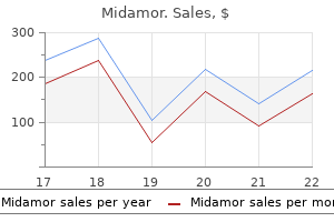
Buy line midamor
The -osmotic terms refer to the concentration of the solutions pulse pressure 47 45mg midamor visa, and the -tonic terms refer to the tendency of cells to swell or shrink. For example, it is possible to prepare a solution of glycerol and a solution of mannitol that are isosmotic to the cytoplasm of the cell. Because the solutions are isosmotic, they have the same concentration of solutes and water as the cytoplasm. However, glycerol can diffuse across the plasma membrane, whereas mannitol cannot. When glycerol diffuses into the cell, the solute concentration of the cytoplasm increases, and its water concentration decreases. In contrast, mannitol cannot enter the cell, and the isosmotic mannitol solution is also isotonic. Facilitated Diffusion Because of their chemical structure or size, many essential molecules, such as amino acids and glucose, cannot enter or exit the cell by diffusing directly through the plasma membrane. Instead, these molecules and ions cross the membrane by mediated transport, a membrane transport process by which membrane transport proteins mediate, or assist, the movement of large, water-soluble molecules or electrically charged molecules or ions across the plasma membrane. Facilitated diffusion is a mediated transport process that moves substances into or out of cells from a higher to a lower concentration (figure 3. Facilitated diffusion does not require metabolic energy to transport substances across the plasma membrane. The rate at which molecules or ions are transported is directly proportional to their concentration gradient up to the point of saturation, when all the carrier proteins or channels are occupied. Predict 5 the transport of glucose into and out of most cells, such as muscle cells and adipocytes, occurs by facilitated diffusion. Once glucose enters a cell, it is rapidly converted to another molecule, such as glucose-6phosphate or glycogen. Carrier molecule 1 Glucose Concentration gradient 2 1 the carrier protein binds with a molecule, such as glucose, on the outside of the plasma membrane. However, because it involves the bulk movement of material into the cell, vesicular transport does not demonstrate the degree of specificity or saturation that other forms of active membrane transport exhibit. Active transport can also move substances from higher Na+ to lower concentrations. The tendency for the ions to move back into the cell (down their concentration gradient) provides the energy necessary to move a different ion or some other molecule into the cell. For example, glucose moves from the lumen of the intestine into epithelial cells by secondary active transport (figure 3. The movement of Na+ down its concentration gradient provides the energy to move glucose molecules into the cell against their concentration gradient. Thus, glucose can accumulate at concentrations higher inside the cell than outside. Because the movement of glucose molecules against their concentration gradient results from the formation of a concentration gradient of Na+ by an active-transport mechanism, the process is called secondary active transport. The concentration gradient for Na+ provides energy required to move glucose against its concentration gradient. A portion of the plasma membrane wraps around a particle or droplet in the extracellular fluid. The portion of the plasma membrane then fuses, so that the particle or droplet is surrounded by a membrane. That portion of the membrane then "pinches off," so that the enclosed particle or droplet is within the cytoplasm of the cell, and the plasma membrane is left intact. In phagocytosis (fag-o -to -sl sis), which means "cell-eating," solid particles are ingested and phagocytic vesicles are formed (figure 3. White blood cells and some other cell types phagocytize bacteria, cell debris, and foreign particles. Phagocytosis is therefore important in eliminating harmful substances from the body. Pinocytosis often forms vesicles near the tips of deep invaginations of the plasma membrane. Pinocytotic vesicles form on the internal side of a capillary, are transported across the cell, and open by exocytosis outside the capillary. For example, cells that phagocytize bacteria and necrotic tissue do not phagocytize healthy cells. The plasma membrane may contain specific receptor molecules that recognize certain substances and allow them to be transported into the cell by phagocytosis or pinocytosis. This is called receptor-mediated endocytosis, and the receptor sites combine only with certain molecules (figure 3. Cholesterol and growth factors are examples of molecules that can be taken into a cell by receptor-mediated endocytosis. As a result of inadequate cholesterol uptake, cholesterol synthesis within these cells is not regulated, and too much cholesterol is produced. The excess cholesterol accumulates in blood vessels, resulting in atherosclerosis. Some cells release material through a vesicular transport mechanism called exocytosis (ekso -to -s l sis). These secretory vesicles then move to the plasma membrane, where the vesicle membrane fuses with the plasma membrane and the vesicle contents are expelled from the cell (figure 3. The secretion of digestive enzymes by the pancreas and the secretion of mucus by the salivary glands are examples of exocytosis. As the concentration of a solution increases, what happens to its osmotic pressure and to the tendency for water to move into the solution Compare isosmotic, hyperosmotic, and hyposmotic solutions with isotonic, hypertonic, and hypotonic solutions. Contrast facilitated diffusion and active transport in relation to energy expenditure and direction of movement with respect to the concentration gradient. Cytoplasm, the cellular material outside the nucleus but inside the plasma membrane, is about half cytosol and half organelles. The cytosol is a colloid, a viscous solution containing dissolved ions and molecules as well as suspended molecules, especially proteins. Many of these proteins are enzymes that catalyze the breakdown of molecules for energy or the synthesis of sugars, fatty acids, nucleotides, amino acids, and other molecules. Other proteins in the cytosol make up the cytoskeleton and cytoplasmic inclusions. Cytoskeleton Just as our skeleton supports many of the structures of our bodies, the cytoskeleton supports the cell and holds the nucleus and other organelles in place. In addition, some components of the cytoskeleton are responsible for changes in cell shape and the movement of cell organelles. The cytoskeleton consists of three groups of proteins: microtubules, actin filaments, and intermediate filaments (figure 3. The microtubules are about 25 nanometers (nm) in diameter, with walls about 5 nm thick. Nucleus Plasma membrane Mitochondrion Tubulin subunits Intermediate filament (b) Endoplasmic reticulum 5 nm 25 nm Ribosomes Microtubules are composed of tubulin protein subunits. Protein subunits 10 nm Actin subunits 8 nm Actin filaments (microfilaments) are composed of actin subunits and are about 8 nm in diameter. They help provide support and structure to the cytoplasm of the cell, much like an internal scaffolding. Microtubules are involved in cell division and in the transport of intracellular materials. Microtubules also form essential components of certain cell organelles, such as centrioles, spindle fibers, cilia, and flagella.
Generic midamor 45mg fast delivery
But in our bodies we tend to pay little attention to the movable joints until disease or damage makes movement very difficult arrhythmia chapter 1 buy genuine midamor online. Explain the structure of a fibrous joint, list the three types, and give an example of each type. Illustrate the structure of a synovial joint and explain the roles of the components of a synovial joint. Classify synovial joints based on the shape of the bones in the joint and give an example of each type. Frontal and parietal Occipital and parietal the two parietal bones Parietal and temporal Radius and ulna Styloid process and hyoid bone Styloid process and mandible Tibia and fibula Tooth and alveolar process None None None Slight Slight Slight Slight Slight Slight Joints, or articulations, are commonly named according to the bones or portions of bones that join together; for example, the temporomandibular joint is between the temporal bone and the mandible. Some joints are given the Greek or Latin equivalent of the common name, such as cubital (kbi-tl; cubit, elbow or forearm) joint for the elbow joint. Joints are classified structurally as fibrous, cartilaginous, or synovial, according to the major connective tissue type that binds the bones together and whether a fluid-filled joint capsule is present. Joints can also be classified in functional categories according to their degree of motion as synarthroses (nonmovable joints), amphiarthroses (slightly movable joints), or diarthroses (freely movable joints). In general, fibrous and cartilaginous joints have little or no movement, while synovial joints have considerable movement. Because this functional classification is somewhat limited, our discussions are based on the more precise structural classification scheme. Cartilaginous Joints Synchondroses Epiphyseal plate Sternocostal Diaphysis and epiphysis of a long bone Anterior cartilaginous part of first rib; between rib and sternum Sphenoid and occipital Bodies of adjacent vertebrae Manubrium and body of sternum the two hipbones Xiphoid process and body of sternum None Slight Sphenooccipital Symphyses Intervertebral Manubriosternal Symphysis pubis Xiphisternal None Slight None None except during childbirth None Fibrous Joints Fibrous joints are the articulating surfaces of two bones united by fibrous connective tissue. Joints in this group are further subdivided on the basis of structure as sutures, syndesmoses, or gomphoses (table 8. Sutures Sutures (soochoorz) are seams found only between the bones of the skull (figure 8. Few sutures are smooth, and the opposing bones often interdigitate (have interlocking, fingerlike processes). The tissue between the bones is dense regular collagenous connective tissue, and the periosteum on the inner and outer surfaces of the adjacent bones continues over the joint. The two layers of periosteum plus the dense fibrous connective tissue in between form a sutural ligament. In a newborn, some of the sutures have a membranous area called a fontanel (font-nel; little fountain, so named because the membrane can be seen to move with the pulse; soft spot). The fontanels make the skull flexible during the birth process and allow for growth of the head after birth (figure 8. The margins of bones within sutures are sites of continuous intramembranous bone growth, and many sutures eventually become ossified. For example, ossification of the suture between the two frontal bones occurs shortly after birth, so that they usually form a single frontal bone in the adult skull. A synostosis results when two bones grow together across a joint to form a single bone. Some movement may occur at syndesmoses because the ligaments are flexible; this occurs in the radioulnar syndesmosis, for example, which binds the radius and ulna together (figure 8. Frontal bones (not yet fused into a single bone) Frontal (anterior) fontanel Gomphoses Gomphoses (gom-fsz) are specialized joints consisting of pegs that fit into sockets and are held in place by fine bundles of regular collagenous connective tissue. The only gomphoses in the human body are the joints between the teeth and the sockets (alveoli) of the mandible and maxillae (figure 8. This small amount of movement also allows teeth to be gradually realigned by braces. Neglect of the teeth can result in gingivitis, an inflammation of the gingiva that is often caused by bacterial infection. Left untreated, gingivitis can spread to the tooth socket, resulting in periodontal disease, the leading cause of tooth loss in the United States. In periodontal disease, plaque and bacteria accumulate, resulting in inflammation that gradually destroys the periodontal ligaments and the bone. Proper brushing, flossing, and professional cleaning to remove plaque can usually prevent gingivitis and periodontal disease. Joints containing hyaline cartilage are called synchondroses; joints containing fibrocartilage are called symphyses. Synchondroses A synchondrosis (sinkon-drsis) consists of two bones joined by hyaline cartilage where little or no movement occurs (figure 8. As a result, even though the costochondral joints (between the ribs and the costal cartilages) are retained, most costal cartilages no longer qualify as synchondroses. Symphyses Oblique cord Radius Interosseous membrane Ulna Radioulnar syndesmosis A symphysis (simfi-sis) consists of fibrocartilage uniting two bones. Examples of symphyses include the junction between the manubrium and the body of the sternum (figure 8. Some of these joints are slightly movable because of the somewhat flexible nature of fibrocartilage. This is especially important for the intervertebral disks because the disk also acts as a shock absorber between the vertebrae. Name the two types of cartilaginous joints, tell the type of cartilage present, and give an example of each. In the case of epiphyseal plates, the synchondrosis is converted to a synostosis as bone replaces the existing cartilage (see "Bone Growth" in chapter 6). Other synchondroses are converted to synovial joints, whereas others persist throughout life. An example of a synchondrosis joint in an adult is the sternocostal synchondrosis between the first rib and the sternum by way of the first costal cartilage (figure 8. All the costal cartilages begin as synchondroses but, because movement occurs between them and the sternum, all but the first usually develop uring pregnancy, certain hormones, such as estrogen, progesterone, and relaxin, act on the connective tissue of joints, particularly the symphysis pubis, making them more stretchable and allowing the joints to loosen. After delivery, the connective tissue of the symphysis pubis returns to its original condition. However, the enlarged pelvic opening may not return completely to its original size, and the woman may have slightly wider hips after the birth of the child. The same hormones may act on the connective tissue of other joints in the body, such as the arches of the feet, causing them to relax, which may result in fallen arches. Increased mobility of the hip can result in congenital (appearing at birth) partial or complete dislocation of the hip. Fortunately, if detected early the condition can be corrected using a specialized harness or traction to realign the joint. Most joints that unite the bones of the appendicular skeleton are synovial joints, reflecting the far greater mobility of the appendicular skeleton compared with the axial skeleton. The articular surfaces of bones within synovial joints are covered with a thin layer of hyaline cartilage called articular cartilage. In some synovial joints, a flat pad of fibrocartilage, called an articular disk, or meniscus (m-niskus; pl. A meniscus is a type of articular disk that only partially spans the synovial cavity such that there is an opening in the center. Joints with menisci include the knee and wrist, while the disks in the temporomandibular, sternoclavicular, and acromioclavicular joints span the entire cavity. Fat pads help protect the articular cartilage by acting as a cushion around the joint. The membrane produces synovial fluid, a viscous lubricating film that covers the surfaces of a joint. Synovial fluid is a complex mixture of polysaccharides, proteins, lipids, and cells derived from serum (blood fluid) filtrate and secretions from the synovial cells. The major polysaccharide, hyaluronic acid, provides much of the slippery consistency and lubricating qualities of synovial fluid. Predict 3 What would happen if a synovial membrane covered the articular cartilage The articular surfaces of the bones that meet at a synovial joint are enclosed within a joint cavity filled with synovial fluid (figure 8. The joint cavity is surrounded by a joint capsule that helps hold the bones together while still allowing for movement. The joint capsule consists of two layers: an outer fibrous capsule and an inner synovial membrane. It consists of dense irregular connective tissue and is continuous with the fibrous layer of the periosteum that covers the bones united at the joint. Portions of the fibrous capsule may thicken, and the collagen fibers may become regularly arranged to form ligaments.
Discount midamor 45 mg with visa
Paliperidone should be dosed once daily in the morning due to the kinetics of its sustained-release dosage formulation arrhythmia lying down discount midamor 45 mg online. The patient should be counseled that a "ghost shell" of the insoluble tablet may be present in stool and can cause complications if the patient has any gastrointestinal narrowing or obstruction. Both risperidone and paliperidone are available in long-acting injectable dosage forms. The long-acting injectable formulation of risperidone (Risperdal Consta) is available as 12. The risperidone is suspended in glycolic acid-lactate copolymer microspheres that are slowly hydrolyzed, resulting in a delayed absorption of approximately three weeks. The dose of paliperidone palmitate needs to be reduced to 156 mg day 1 followed by 117 mg day 8 (both administered in the deltoid) in patients with mild renal impairment (creatinine clearance 50 mL/min to < 80 mL/min). Both injections should still be administered in the deltoid and followed by a monthly maintenance dose of 78 mg given in either the deltoid or gluteal muscle. Paliperidone palmitate should not be administered in patients with severe renal impairment (creatinine clearance below 50 mL/min). In patients who express this decreased activity enzymatic phenotype, the half-lives of risperidone and 9-hydroxyrisperidone will be approximately 17 hours and 30 hours, respectively. Paliperidone is primarily excreted unchanged (57%) in the urine and does not undergo hepatic metabolism. This would in theory suggest antidepressant actions, but there is little evidence to support this. Food will increase the absorption of ziprasidone by two-fold; therefore, it must also be administered with a meal of at least 500 calories. Ziprasidone has a lower propensity to cause weight gain, dyslipidemia, increased fasting triglycerides, or insulin resistance. No cases of torsades de pointes were observed in the 4,571 ziprasidone-treated patients. Clinically, this necessitates caution and careful consideration of the risks and benefits when contemplating the initiation of, or continued treatment with, ziprasidone. Asenapine (Saphris) the chemical structure of asenapine closely resembles that of the tetracyclic antidepressant mirtazapine. The bioavailability of asenapine is severely limited by extensive first-pass metabolism. The time of onset is not as rapid as might be expected due to compartmentalization in the oral mucosa. Upon administration, the compound is rapidly dissolved in the saliva and transported into the mucosal membranes until a pseudoequilibrium is reached. The limited surface area available for absorption requires split-day dosing for doses greater than 5 mg even though asenapine has a 24-hour half-life. The rate-limiting step in the absorption of asenapine is the transfer from the mucosal membranes into systemic circulation. However, it is differentiated by its potent alpha1 blockade, which results in high rates of orthostasis and sedation. This agent has low incidence of dyslipidemias but carries a moderate risk for weight gain. The addition of a potent inhibitor of either enzyme has the potential to increase the serum concentration of iloperidone; therefore, it is recommended to decrease the dose of iloperidone by 50% if concomitant administration is deemed necessary. Quetiapine (Seroquel) Quetiapine is a dibenzothiazepine derivative structurally related to clozapine. The high H1 and 1 blockade results in the high 216 Schizophrenia and Related Disorders incidence of sedation, dizziness, and orthostasis observed with quetiapine. The active metabolite of quetiapine, norquetiapine, possesses some differing pharmacological activity than the parent compound. The mean terminal half-life of quetiapine is approximately six hours, allowing it to reach steady-state concentrations within two days of administration. Quetiapine has no clinically significant impact on the pharmacokinetics of other medications. However, quetiapine may have additive sedative and hypotensive effects when combined with similar agents or alcohol. After one week of administration, olanzapine will reach steady-state concentrations and have an elimination half-life of 30 hours. Only 60% of the parent compound will reach systemic circulation following oral administration. Should smoking cessation occur, the resultant reversal in enzymatic induction will cause an increase in olanzapine serum levels. Therefore, changes in patient smoking status should be consistently monitored, and adjustments to the olanzapine dose should be made accordingly. This interaction should be kept in mind when patients are being admitted and discharged from smoke-free facilities. In addition to the oral capsule formulation, olanzapine is also available as an orally disintegrating tablet, a short-acting injectable, and a long-acting injectable formulation. Due to concerns about excessive orthostatic hypotension, the short-acting injectable should be dosed at least two hours after the initial 10 mg Im dose or four hours after a second 10 mg Im dose. The patient should be assessed for orthostatic hypotension and subsequent doses be withheld if significant orthostasis or syncope is observed. The turbidity in the bloodstream leads to the rapid dissolution of olanzapine pamoate. The injection must be given in a facility with readily available access to emergency response services, and the patient must be continuously observed for three hours post-injection by a healthcare professional for symptoms consistent with olanzapine overdose. The high administrative burden of coordinating the post-injection monitoring limits its use in routine clinical practice. The partial D2 agonism may also cause nausea and vomiting early in treatment and be somewhat activating in certain patients. In this subset of patients, morning dosing will help avoid disrupting sleep architecture. Aripiprazole is also differentiated by its lack of m1 and H1 activity and low risk of inducing weight gain, fasting triglyceride elevations, or insulin resistance. However, this does not mean it cannot induce weight gain in select individuals, particularly in the child and adolescent population. The 75-hour elimination half-life of aripiprazole may be of particular benefit in patients who have difficulty with medication adherence. If concurrent administration is clinically indicated, the dose of aripiprazole should be doubled. Aripiprazole is unlikely to exert any clinically relevant pharmacokinetic effect on other agents metabolized through cytochrome P450. An orally disintegrating tablet, short-acting Im injection, liquid, and long-acting injection are all available. The short-acting Im injection has a bioavailability of 100% versus the 87% of the oral tablet. The tablet, orally disintegrating tablet, and liquid formulation can be substituted at a 1:1 ratio. Mild renal impairment (CrCl 50 mL/min and < 80mL/min): Initiate with 156 mg Im on day 1, then 117 mg Im day 8. Like that of aripiprazole and ziprasidone, the metabolic profile of lurasidone is relatively clean, with low risk for weight gain or dyslipidemias. The absorption of lurasidone, like ziprasidone, is greatly increased by administration in a fed versus fasted state. This effect was seen independently of the dietary fat content of the meal, and drug exposure did not increase any further when the size of the meal was greater than 350 calories. Lurasidone has an elimination half-life of approximately 18 hours and will reach steady-state concentration at a given dose within seven days of consistent administration.
Purchase midamor visa
As the outer cells are damaged heart attack reasons buy cheap midamor 45mg on-line, they are replaced by cells from deeper layers; thus, a continuous barrier of epithelial cells is maintained in the tissue. Stratified squamous epithelium is found in areas of the body where abrasion can occur, such as the skin, mouth, throat, esophagus, anus, and vagina. For example, simple squamous epithelium forms blood and lymphatic capillaries, alveoli (air sacs) of the lungs, and parts of the kidney tubules. They have greater cytoplasmic volume relative to surface area than seen with squamous cells. When transitional epithelium is stretched, the folded regions of the plasma membrane can unfold. Transitional epithelium is specialized to expand in tissues such as the urinary bladder. Cell Connections Cells have structures that hold them to one another or to the basement membrane. These structures do three things: (1) mechanically bind the cells together, (2) help form a permeability barrier, and (3) provide a mechanism for intercellular communication. Epithelial cells have cell surface glycoproteins, which attach to other glycoproteins located on adjacent cells and in the basement membrane. Among the glycoprotein connections between cells, there are some relatively strong adhesive structures called desmosomes (dezm-smz). Desmosomes consist of adhesive glycoproteins that bind cells together and intracellular proteins attached to intermediate filaments that extend into the cytoplasm of the cells (figure 4. Many desmosomes are found in epithelial tissues that are subjected to stress, such as the stratified squamous epithelium of the skin. Hemidesmosomes, similar to one-half of a desmosome, attach epithelial cells to the basement membrane. Tight junctions are formed by proteins in the plasma membranes of adjacent cells that join one another to make a very tight seal. Near the free surface of simple epithelial cells, the tight junctions form a ring that completely surrounds each cell and binds adjacent cells together to prevent the passage of materials between cells. For example, in the stomach and the urinary bladder, chemicals cannot pass between cells. Thus, water and other substances must pass through the epithelial cells, which can actively regulate what is absorbed or secreted. Tight junctions are found in areas where a layer of simple epithelium forms a permeability barrier. For example, water can diffuse through epithelial cells, and active transport, symport, and facilitated diffusion move most nutrients through the epithelial cells of the small intestine. It is located between the plasma membranes of adjacent cells and acts as a weak glue that holds cells together. A gap junction is a small, specialized contact region between cells containing protein channels that aid intercellular communication by allowing ions and small molecules to pass from one cell to another (figure 4. In epithelium, the function of gap junctions is not entirely clear; gap junctions between ciliated epithelial cells may coordinate the movements of cilia. In cardiac and smooth muscle tissues, gap junctions are important in coordinating important functions. Because ions can pass through the gap junctions from one cell to the next, electrical signals can pass from cell to cell to coordinate the contraction of cardiac and smooth muscle cells. Thus, electrical signals that originate in one cell of the heart can spread from cell to cell and cause the entire heart to contract. In the heart, the gap junctions between cardiac muscle cells are found in specialized cell-to-cell connections called intercalated disks (see chapter 20). Intercalated disks contain both gap junctions and desmosomes that help hold adjacent cells in close contact. Predict 3 If a simple epithelium has well-developed tight junctions, explain how NaCl can move from one side of the epithelial layer to the other, what type of epithelium it is likely to be, and how the movement of NaCl causes water to move in the same direction. What is the function of each of the following characteristics of an epithelial free surface: is smooth, has cilia, has microvilli, is folded Name the possible ways by which epithelial cells are bound to one another and to the basement membrane. Glands are composed primarily of epithelium, with a supporting network of connective tissue. If the gland maintains an open contact with the epithelium from which it developed, a duct is present. Alternatively, some glands become separated from the epithelium of their origin and have no ducts; these are called endocrine (end-krin) glands. The cellular products of endocrine glands, which are called hormones (hrmnz), are secreted into the bloodstream and carried throughout the body. Most exocrine glands are composed of many cells and are called multicellular glands, but some exocrine glands are composed of a single cell and are called unicellular glands (figure 4. Multicellular exocrine glands can be classified according to the structure of their ducts and secretory regions (figure 4. If there are multiple secretory regions that branch off the duct, then the gland is called branched. For both simple and compound glands, the shape of the secretory regions further defines the gland. Cell shed into the duct (c) Holocrine gland Entire cells are shed by the gland and become part of the secretion. Merocrine secretion involves the release of secretory products by exocytosis (figure 4. Merocrine secretion is used by water-producing sweat glands and the exocrine portion of the pancreas. Apocrine (ap-krin) secretion involves the release of secretory products as pinchedoff fragments of the gland cells (figure 4. The milkproducing mammary glands release milk by a combination of apocrine and mostly merocrine secretion. Products accumulate in the cytoplasm of each epithelial cell, the cell ruptures and dies, and the entire cell becomes part of the secretion. How are multicellular exocrine glands classified on the basis of their duct system Describe the three main components of the extracellular matrix of connective tissue. Give an example of each type of connective tissue, describe its characteristic functions, and state its location in the body. Connective tissue is a diverse primary tissue type that makes up part of every organ in the body. Connective tissue differs from the other three tissue types in that it consists of cells separated from each other by abundant extracellular matrix. Functions of Connective Tissue Connective tissue performs the following major functions: 1. Sheets of connective tissue form capsules around organs, such as the liver and kidneys. For example, connective tissues separate muscles, arteries, veins, and nerves from one another. Strong cables, or bands, of connective tissue called tendons attach muscles to bone, whereas connective tissue bands called ligaments hold bones together. Bones of the skeletal system provide rigid support for the body, and the semirigid cartilage supports structures such as the nose, ears, and joint surfaces. Adipose tissue (fat) stores high-energy molecules, and bones store minerals, such as calcium and phosphate. Adipose tissue cushions and protects the tissue it surrounds and provides an insulating layer beneath the skin that helps conserve heat. Blood transports the gases, nutrients, enzymes, hormones, and cells of the immune system throughout the body. Cells of the immune system and blood protect against toxins and tissue injury, as well as against microorganisms. Undifferentiated mesenchymal cells are a type of adult stem cell that persist in connective tissue. They have the potential to form multiple cell types, such as fibroblasts or smooth muscle cells, in response to injury.
Cheap midamor 45mg overnight delivery
Cyanide poisoning by inhalation or absorption through the skin can also occur in certain manufacturing processes arrhythmia medicine cheap midamor 45 mg online, and cyanide gas was used to kill people during the Holocaust. Deliberate suicide by ingesting cyanide is rare but was made famous by suicide capsules in spy movies. Cyanide has only rarely been used as a weapon, although in 1982 seven people died after taking Tylenol that someone had laced with cyanide, which led to the widespread current use of tamper-proof capsules and packaging. Define gene, and explain how genes determine the structures and functions of cells. Learn to Predict From page 25 Answer the saliva catalyzed a decomposition reaction, breaking down the starch to a different material. Chapter 2 teaches us that enzymes are protein catalysts that speed up chemical reactions by lowering the activation energy. Activation energy is the minimum energy that the reactants must have to start the chemical reaction. Therefore, the digestive enzymes in the saliva lowered the activation energy needed to break the bonds in the starch molecules. To understand the reactions occurring in the test tube, you will need to ask yourself the following questions: (1) What was in the saliva that changed the starch To answer these questions, let us determine what important information was provided in the question. First, we know that starch, which is a polysaccharide, is the primary material in the test tube and that iodine stains starch a blue color. Finally, we are told that after 30 minutes the blue color disappeared, indicating that starch was no longer present in the test tube. Much of the structure and function of healthy or diseased organisms can be understood at the chemical level. A hydrogen bond is the weak attraction between a positively charged hydrogen and negatively charged oxygen or other polar molecule. Hydrogen bonds are important in determining properties of water and the three-dimensional structure of large molecules. An element is the simplest type of matter having unique chemical and physical properties. An atom is the smallest particle of an element that has the chemical characteristics of that element. Protons are positively charged, electrons are negatively charged, and neutrons have no charge. Protons and neutrons are in the nucleus; electrons are located around the nucleus and can be represented by an electron cloud. The atomic mass of an element is the average mass of its naturally occurring isotopes weighted according to their abundance. The molar mass of a substance is the mass of 1 mole of the substance expressed in grams. A synthesis reaction is the chemical combination of two or more substances to form a new or larger substance. A decomposition reaction is the chemical breakdown of a larger substance to two or more different and smaller substances. Reversible Reactions Reversible reactions produce an equilibrium condition in which the amount of reactants relative to the amount of products remains constant. Oxidation-Reduction Reactions Oxidation-reduction reactions involve the complete or partial transfer of electrons between atoms. An atom that loses 1 or more electrons becomes positively charged and is called a cation. An anion is an atom that becomes negatively charged after accepting 1 or more electrons. An ionic bond results from the attraction of the oppositely charged cation and anion to each other. A polar covalent bond results when the sharing of electrons is unequal and can produce a polar molecule that is electrically asymmetric. Potential energy is stored energy, and kinetic energy is energy resulting from the movement of an object. Chemical reactions in which the products contain more potential energy than the reactants require the input of energy. Chemical reactions in which the products have less potential energy than the reactants release energy. Heat energy Heat energy is energy that flows between objects that are at different temperatures. Heat energy is released in chemical reactions and is responsible for body temperature. A molecule is two or more atoms chemically combined to form a structure that behaves as an independent unit. The kinds and numbers of atoms (or ions) in a molecule or compound can be represented by a formula consisting of the symbols of the atoms (or ions) plus subscripts denoting the number of each type of atom (or ion). Activation energy is the minimum energy that the reactants must have to start a chemical reaction. Enzymes are specialized protein catalysts that lower the activation energy for chemical reactions. Enzymes speed up chemical reactions but are not consumed or altered in the process. Increased temperature and concentration of reactants can increase the rate of chemical reactions. Water is a polar molecule composed of one atom of oxygen and two atoms of hydrogen. Because water molecules form hydrogen bonds with each other, water is good at stabilizing body temperature, protecting against friction and trauma, making chemical reactions possible, directly participating in chemical reactions. A mixture is a combination of two or more substances physically blended together, but not chemically combined. A solution is any liquid, gas, or solid in which the substances are uniformly distributed, with no clear boundary between the substances. A suspension is a mixture containing materials that separate from each other unless they are continually, physically blended together. A colloid is a mixture in which a dispersed (solutelike) substance is distributed throughout a dispersing (solventlike) substance. Disaccharide molecules are formed by dehydration reactions between two monosaccharides. A polysaccharide is composed of many monosaccharides bound together to form a long chain. Fatty acids can be saturated (having only single covalent bonds between carbon atoms) or unsaturated (having one or more double covalent bonds between carbon atoms). Phospholipids are lipids in which a fatty acid is replaced by a phosphatecontaining molecule. Other lipids include fat-soluble vitamins, prostaglandins, thromboxanes, and leukotrienes. The building blocks of a protein are amino acids, which are joined by peptide bonds. The number, kind, and arrangement of amino acids determine the primary structure of a protein. Hydrogen bonds between amino acids determine secondary structure, and hydrogen bonds between amino acids and water determine tertiary structure. Enzymes are protein catalysts that speed up chemical reactions by lowering their activation energy. Cofactors are ions or organic molecules, such as vitamins, that are required for some enzymes to function. A buffer is a solution of a conjugate acid-base pair that resists changes in pH when acids or bases are added to the solution. The basic unit of nucleic acids is the nucleotide, which is a monosaccharide with an attached phosphate and a nitrogenous base.
Dog-Tree (American Dogwood). Midamor.
- Are there safety concerns?
- What is American Dogwood?
- How does American Dogwood work?
- Headaches, fatigue, weakness, fever, chronic diarrhea, loss of appetite, malaria, treating boils and wounds, and other conditions.
- Dosing considerations for American Dogwood.
Source: http://www.rxlist.com/script/main/art.asp?articlekey=96525
Discount 45 mg midamor with amex
Granulation tissue being replaced with new connective tissue 4 Approximately 1 month after the injury arrhythmia sounds midamor 45mg without a prescription, the wound has completely closed, the scab has been sloughed, and the granulation tissue is being replaced by new connective tissue. The clot contains the threadlike protein fibrin (fbrin), which binds the edges of the wound together. The surface of the clot dries to form a scab, which seals the wound and helps prevent infection. An inflammatory response induces 134 vasodilation and takes more blood cells and other substances to the area. Fibrin and blood cells move into the wounded tissues because of the increased vascular permeability. Phagocytic white blood cells called neutrophils (nootr-filz) then move into the tissue to help fight the infection (figure 4. Neutrophils are killed in this process and can accumulate as a mixture of dead cells and fluid called pus (ps). Fibroblasts from surrounding connective tissue migrate into the clot and produce collagen and other extracellular matrix components. Capillaries grow from blood vessels at the edge of the wound and revascularize the area, and fibrin in the clot is broken down and removed. Granulation tissue, a delicate, granular-appearing connective tissue that consists of fibroblasts, collagen, and capillaries, replaces the clot. A large amount of granulation tissue is converted to a scar, which consists of dense irregular collagenous connective tissue. Later, the scar becomes white as collagen accumulates and the vascular channels are compressed. Repair by secondary union proceeds in a similar fashion, but with some differences. Because the wound edges are far apart, the clot may not close the gap completely, and it takes the epithelial cells much longer to regenerate and cover the wound. Also, the increased tissue damage means that both the degree of inflammation and the risk of infection are greater and there is more cell debris for the phagocytes to remove. Much more granulation tissue forms, and the contraction of fibroblasts in the granulation tissue leads to wound contracture, resulting in disfiguring and debilitating scars. Thus, it is advisable to suture a large wound, so that it can heal by primary rather than secondary union. Healing is faster, with a lowered risk of infection and a reduced degree of scarring. Compare labile, stable, and permanent cells according to their ability to regenerate. The rates of healing and scarring in the elderly are very different from those in the very young, and major changes in skin structure develop. All these changes result in the differences among young, middle-aged, and older people. At the tissue level, age-related changes affect cells and the extracellular materials they produce. Collagen fibers become more irregular in structure, even though their number may increase. As a consequence, connective tissues with abundant collagen, such as tendons and ligaments, become less flexible and more fragile. Reduced flexibility and elasticity of connective tissue are responsible for increased wrinkling of the skin, as well as the increased tendency for bones to break in older people. Arterial walls also become less elastic due to changes in the structure of elastic and collagen fibers. Atherosclerosis results as plaques form in the walls of blood vessels, which contain collagen fibers, lipids, and calcium deposits (see chapter 21). Basal cell and squamous cell carcinomas are types of skin cancer derived from epithelial tissue. Adenocarcinomas (ad-n-kar-sinmaz) are types of carcinomas derived from glandular epithelium. A sarcoma (sarkm) is a relatively rare type of cancer derived from mesodermal tissue (muscle and connective tissue). For example, an osteosarcoma (ost-sar-km) is cancer of bone, and a chondrosarcoma (kondr-sar-km) is cancer of cartilage. Identifying the tissue of origin is useful for the diagnosis and treatment of cancer. Since tumor cells have altered shapes compared with their morphology in tissues (see Clinical Genetics, "Genetic Changes in Cancer Cells," in chapter 3), molecular markers are commonly used to identify the type of tumor. For example, specific types of carcinomas express keratin filaments that are characteristic of different types of epithelial tissue. Other intermediate filaments are diagnostic of sarcomas and other types of cancers. Advances in nucleic acid technologies have opened the door for even more extensive gene expression profiling of cancers. In the future, it is likely that distinct molecular profiles of cancer will allow a more definitive diagnosis and prognosis, which may lead to targeted therapies tailored for individual patients. Age-related changes-for example, reduced visual acuity and reduced smell, taste, and touch sensations-are well documented. With advanced age, the number of neurons and muscle cells decreases substantially. Injuries in the very young heal more rapidly and more completely than in older people. A similar fracture in an adult heals more slowly, and a scar, seen in x-rays of the bone, is likely to persist throughout life. It is increasingly evident that many of the cell losses and functional declines of aging can be slowed by physical and mental exercise. Staying active, both physically and mentally, is often a good prescription for better health. Describe the age-related changes in tissues with abundant collagen and elastic fibers. Learn to Predict From page 103 Answer enteropathy reduced his ability to absorb nutrients and water, so we can conclude that the cell parts affected by the disease are the microvilli. Finally, the question asks us to explain why Matt suffers from bouts of diarrhea after eating gluten. We know that gluten damages the intestinal lining by decreasing the number of villi and microvilli. Chapter 3 showed us that water moves by osmosis to areas of higher solute concentration. Since the solutes are not being absorbed, the solute concentration remains high in the intestines, and water absorption decreases. As a result, the nutrients and water accumulate in the intestines, resulting in the watery feces of diarrhea. The question tells us that gluten enteropathy affects the intestinal lining, reducing its ability to absorb nutrients and water. It also reminds us that nutrient and water absorption occurs at the cellular level via several different transport processes. In chapter 4 we learned that epithelial tissue covers body surfaces, including the lining of the intestines. Further reading showed that the intestinal lining is composed of simple columnar epithelial tissue. As stated in the question, the intestinal lining is organized into fingerlike projections called villi, which are covered by the simple columnar epithelium. In chapter 3, we learned that microvilli are extensions of the plasma membrane that increase the surface area for absorption. Tissues are collections of similar cells and the extracellular substances surrounding them. The four primary tissue types are epithelial, connective, muscle, and nervous tissues. Summary Functions of Epithelial Tissues Epithelial tissues protect underlying structures, act as barriers, permit some substances to pass through epithelial layers, secrete substances, and absorb substances.
Effective 45 mg midamor
Cartilage has no blood vessels or nerves hypertension 2 generic 45mg midamor with mastercard, except those of the perichondrium; it therefore heals very slowly after an injury because the cells and nutrients necessary for tissue repair cannot reach the damaged area easily. Hyaline (h-lin) cartilage has large amounts of both collagen fibers and proteoglycans (table 4. Collagen fibers are evenly dispersed throughout the ground substance, and hyaline cartilage in joints has a very smooth surface. Specimens appear to have a glassy, translucent matrix when viewed through a microscope. Hyaline cartilage is found where strong support and some flexibility are needed, such as in the rib cage and within the trachea and bronchi (see chapter 23). It also covers the surfaces of bones that move smoothly against each other in joints. Hyaline cartilage forms most of the skeleton before it is replaced by bone in the embryo, and it is involved in growth that increases the length of bones (see chapter 6). Compared with hyaline cartilage, fibrocartilage has much thicker bundles of collagen fibers dispersed through its matrix. It is found in areas of the body where a great deal of pressure is applied to joints, such as in the knee, in the jaw, and between the vertebrae. Some joints, such as the knee, have both hyaline and fibrocartilage connective tissue. In these joints, pads of fibrocartilage help absorb shocks and prevent bone-to-bone abrasion. Fibrocartilage injuries of the knee joint (meniscus tears) are common sports-related injuries. Elastic cartilage has numerous elastic fibers in addition to collagen and proteoglycans dispersed throughout its matrix (table 4. It is found in areas that have rigid but elastic properties, such as the external ears. Predict 6 One of several changes caused by rheumatoid arthritis in joints is the replacement of hyaline cartilage with dense irregular collagenous connective tissue. Bone Bone is a hard connective tissue that consists of living cells and mineralized matrix. The organic portion consists of protein fibers, primarily collagen, and other organic molecules. The mineral, or inorganic, portion consists of specialized crystals called hydroxyapatite (h-droksap-tt), which contains calcium and phosphate. Osteocytes (ost-stz), or bone cells, are located within holes in the matrix, which are called lacunae and are similar to the lacunae of cartilage. Spongy bone has spaces between trabeculae (tr-bek-l; beams), or plates, of bone and therefore resembles a sponge (table 4. Compact bone is more solid, with almost no space between many thin layers, or lamellae (l-mel; sing. Fluid Connective Tissue Blood Blood is unusual among the connective tissues because the matrix between the cells is liquid (table 4. The cells of most other connective tissues are more or less stationary within a relatively rigid matrix, but blood cells move freely within a fluid matrix. The matrix of blood is also unusual in that most of it is produced by cells contained in other tissues, rather than by blood cells. There are three types of cellular structures: red blood cells, white blood cells, and cell fragments called platelets. White blood cells sometimes leave the bloodstream and wander through other tissues. Hemopoietic tissue produces red and white blood cells and platelets; it is described in detail in chapter 19. Discuss the three types of muscle tissue by describing their general structures, locations in the body, and functions. The main characteristic of muscle tissue is that it contracts, or shortens, with a force and therefore is responsible for movement. Many people with Marfan syndrome have limbs, fingers, and toes that are disproportionately long in relation to the rest of the body. Connective tissues are weakened; as a consequence, the heart valves, which are composed largely of connective tissue, do not function normally, resulting in heart murmurs (abnormal heart sounds). Poor vision is common because the lenses of the eyes, which are normally held in place by elastic fibers, are positioned abnormally. The lungs are prone to collapse, and dilation of large arteries, such as the aorta, can occur. There is no cure for the condition, but treatments can reduce the danger of the symptoms. It has been speculated that President Lincoln may have had Marfan syndrome, but some geneticists now think it more likely that he had a rare inherited form of endocrine cancer that includes physical features of Marfan syndrome. M arfan syndrome is an autosomal dominant disorder that affects approximately 1 in 5000 people. The gene for Marfan syndrome codes for a protein called fibrillin-1, which is necessary for the normal structure of the elastic fibers of connective tissue. Children of a person with Marfan syndrome have a 50% chance of inheriting the disorder because it is an autosomal dominant trait (see chapter 29). However, about 25% of the cases of Marfan syndrome occur in children whose parents do not have the disorder. In these cases, a mutation of the gene contractile proteins, which are described in chapter 9. Muscles contract to move the entire body, to pump blood through the heart and blood vessels, and to decrease the size of hollow organs, such as the stomach and urinary bladder. The three types of muscle tissue-skeletal, cardiac, and smooth muscle-are grouped according to both structure and function (table 4. As the name implies, skeletal muscle attaches to the skeleton and enables the body to move. Skeletal muscle is under voluntary (conscious) control because a person can purposefully cause skeletal muscle contraction to achieve specific body movements. However, the nervous system can cause skeletal muscles to contract without conscious involvement, as occurs during reflex movements and the maintenance of muscle tone. Skeletal muscle cells are long, cylindrical cells, each containing many nuclei located at the periphery of the cell. Skeletal muscle cells are striated (strt-ed), or banded, because of the arrangement of contractile proteins within the cells (see chapter 9). Cardiac muscle is the muscle of the heart; it is responsible for pumping blood (table 4. They are often branched and connected to one another by intercalated (in-terk-l-ted; inserted between) disks, which contain specialized gap junctions and are important in coordinating cardiac muscle cell contractions (see chapter 20). Smooth muscle forms the walls of hollow organs (except the heart); it is also found in the skin and eyes (table 4. Smooth muscle is responsible for a number of functions, such as moving food through the digestive tract and emptying the urinary bladder. Smooth muscle cells are tapered at each end, have a single nucleus, and are not striated. Describe the structural and functional roles of neurons and glia in the nervous tissue. Nervous tissue is found in the brain, spinal cord, and nerves and is characterized by the ability to conduct electrical signals called action potentials. Nervous tissue consists of neurons, which are responsible for its conductive ability, and support cells called glia. Just as an electrical wiring system transports electricity throughout a house, neurons transport electrical signals throughout the body. Axons can be much longer than dendrites, and they have a constant diameter along their entire length. Pseudounipolar neurons have only a single, short process that extends from the cell body and then divides into two branches, which extend to the periphery and to the central nervous system (table 4.
Purchase 45 mg midamor mastercard
In severe medial knee injuries blood pressure medication vision buy cheapest midamor, the anterior cruciate ligament is also damaged (figure 8B). Tearing of the medial collateral ligament, medial meniscus, and anterior cruciate ligament is often referred to as "the unhappy triad of injuries. For each of the following joints, name the bones of the joint, the specific parts of the bones that form the joint, the type of joint, and the possible movement(s) at the joint: temporomandibular, shoulder, elbow, hip, knee, and ankle. Explain the differences in stability and movement between the shoulder and the hip joints. List the common knee injuries, and tell which part of the knee is most often damaged in each type. Describe a sprain, and identify which portions of the ankle joint are most commonly damaged when it is sprained. A sprained ankle results when the ligaments of the ankle are torn partially or completely. The calcaneofibular ligament tears most often (figure 8C), followed in frequency by the anterior talofibular ligament. A fibular fracture can occur with severe inversion because the talus can slide against the lateral malleolus and break it (see chapter 7). Explain the most effective preventive measures against the effects of aging on the joints. Those that occur in synovial joints have the greatest impact and often present major problems for elderly people. Biomedical engineers are designing synthetic replacement materials to accomplish these objectives. Prosthetic joints are usually composed of metal, such as stainless steel, titanium alloys, or cobalt-chrome alloys, in combination with modern plastics, such as high-density polyethylene, silastic, or elastomer. The bone of the articular area is removed on one side (a procedure called hemireplacement) or on both sides (total replacement) of the joint, and the artificial articular areas are glued to the bone with a synthetic adhesive, such as methylmethacrylate. The smooth metal surface rubbing against the smooth plastic surface provides low-friction contact, with a range of movement that depends on the design. The success of joint replacement depends on the joint being replaced, the age and condition of the patient, the state of the technology, and the definition of success. The technology is improving constantly, so current reports do not adequately reflect the effect of the most recent improvements. The major reason for the failure of prosthetic joints is loosening of the artificial joint from the bone to which it is attached. Osteoarthritis is the leading disease requiring joint replacement and accounts for two-thirds of the patients. The major design objectives for joint prostheses (artificial replacements) include developing stable articulations, low friction, solid fixation to the bone, and normal range age-related modifications that cross-link proteins together. These changes are most prevalent in long-lived proteins, such as collagen, which is abundant in fibrous connective tissue. Tissue repair slows as cell proliferation rates decline and the rate of new blood vessel development decreases. When a person is young, the production of new, resilient matrix compensates for the wear. However, as a person ages, the rate of replacement declines and the matrix becomes more rigid, thus increasing its rate of wear. The production rate of lubricating synovial fluid also declines with age, further contributing to the wear of the articular cartilage. Furthermore, the ligaments and tendons surrounding a joint shorten and become less flexible with age, resulting in decreased range of motion. The most effective preventive measure against the effects of aging on the joints is to strengthen the bones and muscles and maintain flexibility. This can be accomplished through a combination of regular physical activity, stretching, and a healthy diet. List the age-related factors that contribute to cartilage wear in synovial joints. Describe the age-related factors that cause loss of flexibility and loss of range of motion in synovial joints. Learn to Predict From page 242 Answer knee joint by stretching diagonally from the femur at the back of the joint to the tibia in the front, which normally prevents forward or anterior movement of the tibia from underneath the femur. In this test, the patient lies on his or her back with the hips flexed at a 45-degree angle, knees bent at a 90-degree angle, and feet flat on the examining table. To answer this question, we first need to review the functions of the major knee ligaments that stabilize the knee joint: the cruciate and collateral ligaments. Joints are classified structurally according to the type of connective tissue that binds them together and whether fluid is present between the bones. They consist of the following: Articular cartilage on the ends of bones that provides a smooth surface for articulation. A joint cavity is surrounded by a joint capsule of fibrous connective tissue, which holds the bones together while permitting flexibility. Bursae are extensions of synovial joints that protect skin, tendons, or bone from structures that could rub against them. Synovial joints are classified according to the shape of the adjoining articular surfaces: plane (two flat surfaces), saddle (two saddleshaped surfaces), hinge (concave and convex surfaces), pivot (cylindrical projection inside a ring), ball-and-socket (rounded surface into a socket), and ellipsoid (ellipsoid concave and convex surfaces). Fibrous joints, in which bones are connected by fibrous tissue with no joint cavity, are capable of little or no movement. Sutures involve interdigitating bones held together by dense fibrous connective tissue. Gomphoses are joints in which pegs fit into sockets and are held in place by periodontal ligaments (teeth in the jaws). Synchondroses are immovable joints in which bones are joined by hyaline cartilage. Angular movements include flexion and extension, plantar flexion and dorsiflexion, and abduction and adduction. Special movements include elevation and depression, protraction and retraction, excursion, opposition and reposition, and inversion and eversion. The temporomandibular joint is a complex hinge and gliding joint between the temporal and mandibular bones. It is capable of elevation and depression, protraction and retraction, and lateral and medial excursion. The shoulder joint is a ball-and-socket joint between the head of the humerus and the glenoid cavity of the scapula that permits a wide range of motion. The shoulder joint is capable of flexion and extension, abduction and adduction, rotation, and circumduction. The elbow joint is a compound hinge joint between the humerus, the ulna, and the radius. The hip joint is a ball-and-socket joint between the head of the femur and the acetabulum of the hipbone. It is greatly strengthened by ligaments and is capable of a wide range of motion, including flexion, extension, abduction, adduction, rotation, and circumduction. The knee joint is a complex ellipsoid joint between the femur and the tibia that is supported by many ligaments. The ankle joint is a special hinge joint of the tibia, the fibula, and the talus that allows dorsiflexion and plantar flexion and inversion and eversion. With age, the connective tissue of the joints becomes less flexible and less elastic. The resulting joint rigidity increases the rate of wear in the articulating surfaces.
